Chiropractic Student Perspectives on The Use of Virtual Radiography Simulation An Observational Study Reporting on Chiropractic Students' ...
←
→
Page content transcription
If your browser does not render page correctly, please read the page content below
Chiropractic Student Perspectives on The Use of Virtual
Radiography Simulation An Observational Study Reporting on
Chiropractic Students’ Perception on The Use of Virtual
Radiography as A Clinical Learning Tool
Madeleine Shanahan
University of Canberra
Tom Molyneux
RMIT University - Bundoora East Campus: RMIT University Bundoora Campus
Dein Vindigni ( dein.vindigni@rmit.edu.au )
RMIT University Bundoora Campus
Research article
Keywords: Chiropractors, Radiography, Simulation
Posted Date: October 9th, 2020
DOI: https://doi.org/10.21203/rs.3.rs-87469/v1
License: This work is licensed under a Creative Commons Attribution 4.0 International License. Read Full License
Version of Record: A version of this preprint was published at BMC Medical Education on July 28th, 2021. See the published
version at https://doi.org/10.1186/s12909-021-02827-0.
Page 1/13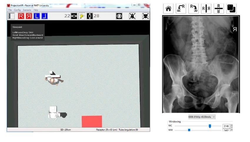
Abstract
Background Virtual radiography provides students with an opportunity to practise their clinical skills in patient positioning and
evaluating radiographic images. The purpose of this pilot study was to introduce Projection VR™, a software radiography
simulation program, into a student chiropractic program and evaluate its potential application as a teaching and learning tool.

Methods Undergraduate chiropractic students, enrolled in a radiographic course (unit within the chiropractic program), were
invited to attend a scheduled laboratory where they were introduced to, and undertook purposefully designed activities using the
radiography simulation. At the end of this activity, students were asked to complete an online survey to describe their perceptions
of the educational value of the software program. Descriptive and inferential statistics were used to evaluate outcomes.

Results Responses were received from 44 out of the 47 students who attended the scheduled laboratory (response rate 92%).
Overall students were positive about this simulation identifying that it was easy to use (95%) and that they could control the
equipment as needed (95%). The main reported benefits included students being enabled to repeat tasks until they were satisfied
with the results (98%) and being able to quickly assess images and determine if changes needed to be made (98%). Participants
reported improvement in their understanding of the effect of exposure factors on patient radiation dose (93%) as well as their
technical image evaluation (84%) and problem-solving skills (80%).

Conclusions The results of this study suggest that virtual radiography is a valuable complementary resource in providing
chiropractic students with radiographic knowledge and skills.

Background
Radiography has been integrated into chiropractic teaching programs since 1910 when B.J. Palmer purchased an x-ray unit for
the Palmer School of Chiropractic in Davenport, Iowa [1]. It has been used as a diagnostic tool in the biomechanical evaluation
of the spine and pelvis and to help identify contraindications to manual therapy.

It continues to be taught as a core course in all chiropractic programs in Australia and most chiropractic programs throughout
the world [2–4].

Maintaining the quality of imaging whilst minimising the radiation dose to patients is a priority which is highlighted by the
Australian Radiation Protection and Nuclear Safety Agency [5] and one which is emphasised in clinical radiographic training and
radiology courses (MEDS2144 Introduction to Diagnostic Imaging, MEDS2143 Advanced Diagnostic Imaging and REHA2203
Chiropractic 6 Theory) at RMIT University [6].

The opportunity to participate in simulated radiography is complementary to rigorous training in the theoretical and clinical
aspects of plain film diagnostic imaging.

In recent years, computer-based simulation radiography has been introduced in undergraduate radiography programs with
promising results [7, 8] .However, its usefulness in achieving clinical learning outcomes in pre-clinical undergraduate chiropractic
programs has not been previously evaluated.

This study aimed to introduce a software radiography simulation tool called Projection VR™ into a chiropractic, pre-clinical
undergraduate setting and gather student feedback about its application as a clinical learning tool and strategy.

Methods
The Human Research Ethics Committee of the Royal Melbourne Institute of Technology (RMIT) University (BSEHAPP 06-15)
approved the project, including its design and recruitment methods.

Participants:

                                                             Page 2/13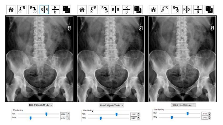
Chiropractic students in the second semester of year four of the program in 2016 were recruited as part of their traditional
practical sessions in radiography. Informed consent was obtained in the first test item of an online survey, with respondents able
to exit at this point if they so preferred.

Procedure:

Participating chiropractic students were scheduled to attend one laboratory session which used the computer-based virtual
radiography simulation software Projection VR™. Projection VR™ simulation in this university setting could be adequately
delivered via Windows 8 or 7 (64 or 32 bit) with a graphics processor of at least DirectX and Shader Model 3.0 or 4.0 hardware
support and 512 megabytes or more of dedicated video RAM. The standard computer laboratory equipment at the university met
or exceeded these requirements.

A worksheet on simulated radiography of the lumbar spine was developed for chiropractic students using Projection VR™. No
training was conducted prior to using the simulated radiography system as students learned to use the technology while they
undertook the activity under the guidance of an experienced lecturer in radiography. The detailed worksheet allowed the students
to use the technology as they undertook the laboratory activity. Each student used the simulation individually.

There were three key areas of focus for this activity. Firstly, for each student to simulate patient positioning and technical set up
in preparation for taking the Anterior to Posterior lumbopelvic image and to generate an AP lumbopelvic (APLP) image (Figure 1).
Secondly, having produced an unrotated APLP, students were then asked to rotate their patient so that the patient’s right posterior
side was closer to the image receptor (Figure 2). Before generating the image, students were asked what distinguishing
anatomical features they expected to see on the image (Figure 2). This strategy was used to support active and engage critical
thinking as students consciously paused and reflected before they undertook their next action [9]. Thirdly, the effect of exposure
factor selection on the digital image as well as patient dose was investigated. This was tested using two methods, namely
application of the 15% rule and the effect of decreasing and increasing milliamp seconds (mAs) on digital images. Increasing
kilo volt peak (kVp) with a concomitant decrease in mAs is expressed as the 15% kVp rule. The 15% rule states that a 15%
increase in kVp is the equivalent of doubling the exposure received at the image receptor [10, 11]. To maintain exposure at the
image receptor, the mAs is halved. The increase in kVp, when applying the 15% rule, is variable and dependent on the original
kVp. For example, at 60 kVp, the calculated change in kVp is be 9 kVp whereas at 80 kVp, the required change is 12 kVp. Studies
examining application of the 15% kVp rule demonstrate a considerable reduction in patient dose (22% - 60%) without adversely
affecting image quality [12, 13]. This is an important finding as increasing kVp reduces subject contrast and could therefore
potentially negatively impact image quality [10, 11] . Before generating the image acquired using the 15% rule, students were
asked what change if any they may expect to see on the image (Figure 3) and what impact, if any, applying the 15% rule would
have on patient dose. Technical data available in Projection VR™ relating to radiation dose for the two images generated is
provided in Figure 4. Entrance surface dose (ESD) measurements were compared for the two exposure techniques. The final
aspect of selection of exposure factors on digital images was undertaken by asking students what difference they would expect
to see on the radiographic image and on patient dose if they were to half or double the mAs. Generated images are provided in
Figure 5.

Throughout the session, students were asked to predict the outcome of each change in patient position or exposure factors
before they generated and saw images or technical data. This method was used to encourage students to think critically in
applying their decision-making skills in a clinical setting and has been found to reinforce learning by other authors [9, 14, 15] .

Evaluation of virtual radiography

Students who were enrolled in the radiographic positioning course in the chiropractic program were invited to participate
anonymously in an online Qualtrics survey during the session. The survey comprised a mix of quantitative and qualitative items
including Likert scales and open-ended questions. This assessed the ease with which students used Projection VR™ and the
extent to which they developed program skills. Survey data were then entered for analysis.

Data Analysis:

                                                              Page 3/13
The survey data were entered into SPSS 21.0® and descriptive and inferential statistics were used for analysis. Cross
tabulations were performed using age group and gender and self-reported confidence using computer technology to determine if
relationships existed. Differences between groups were examined using chi-square analysis and Fisher's exact test.

Analysis of survey data was descriptive only as this research involved an exploratory snap-shot process - that is, the survey was
only administered once [16] .

Results
Responses were received from 44 out of 47 participants in the practical sessions. The gender and age characteristics of the
responding students are presented in Table 1.

                                                          Table 1
                                             Demographic characteristics of survey
                                                    respondents (n = 44)
                                             Characteristic                    Number (%)

                                             Gender             Female         30 (68)

                                                                Male           14 (32)

                                             Age (years)        18–21          22 (50)

                                                                22–25          11 (25)

                                                                26–29          4 (9)

                                                                30 +           7 (16)

The majority of students were female (68%) (M = 14, F = 30). All age groups were represented in this sample and included both
high school leavers and mature aged students.

Technology

Given that Projection VR™ involves computer-based simulation, participants were asked about their confidence in using this
technology as part of their learning strategy. Eighty-two% (n = 36) of responding participants described themselves as confident
or moderately confident in the use of computer technology. Only 18% (eight students) reported that they had previous experience
using computer-based simulation. No relationship was shown to exist (p > .05) between gender or age and students’ perception
of their confidence in the use of computer technology.

Students’ perceptions of ease of use of Projection VR™ are summarised in Table 2.

                                                           Table 2
                                   Student perceptions ease of use of Projection VR™ (n = 44)
 Question                                             Strongly         Agree      Neither agree   Dis-    Strongly      Total
                                                      agree                       nor disagree    agree   disagree      Number

 Projection VR™ is easy to use                        8                34         2               0       0             44

 I could control the equipment as I needed when       15               27         2               0       0             44
 using Projection VR™

 I liked using Projection VR™                         17               22         4               1       0             44

 Technical problems made using Projection VR™         4                6          11              14      9             44
 difficult

 The Projection VR™ laboratory worksheet was          7                27         7               0       1             44
 easy to follow

                                                              Page 4/13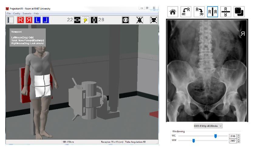
Overall, students were positive regarding ease of use of the simulation software and their ability to control the equipment during
the learning activity. Some students commented that the simulation could be improved by making it easier to use, with one
student specifying “some of the keys made it hard to set up accurately”.

Chiropractic students’ perceptions of use of Projection VR™ as a learning tool for radiography is displayed in Table 3.

                                                             Table 3
                         Student perceptions on Projection VR™ as a learning tool for radiography (n = 44)
 Question                                                    Strongly    Agree     Neither agree     Dis-     Strongly    Total
                                                             agree                 nor disagree      agree    disagree    Number
 Using Projection VR™

 Encouraged me to think more about radiographic              17          22        3                 2        0           44
 procedures

 Had a positive effect on my ability to set up a             15          17        8                 3        1           44
 radiographic examination

 Allowed me to quickly see images and understand if          25          18        1                 0        0           44
 changes needed to be made

 Had a positive effect on my ability to evaluate             17          20        7                 0        0           44
 radiographic images

 Helped me learn as I was able to repeat activities until    21          22        1                 0        0           44
 I was satisfied with the results

 Encouraged me to solve problems                             12          23        8                 1        0           44

 Had a positive effect on my confidence level in setting     11          23        8                 2        0           44
 up radiographic examinations

 Had a positive effect on my confidence level in             9           26        7                 2        0           44
 evaluating radiographic images

 Encouraged me to think more about evaluating                20          20        4                 0        0           44
 radiographic images

 Helped me understand the effect of changing                 19          22        2                 1        0           44
 radiographic exposure factors on patient dose

 Projection VR is a valuable part of the laboratory          19          19        5                 1        0           44

 Projection VR complements other learning activities in      19          21        2                 2        0           44
 the laboratory

 Projection VR should continue to be used in the             21          19        3                 1        0           44
 laboratory

Students generally perceived Projection VR™ as a worthwhile educational tool that quickly generated radiographic images and
enabled them to refine the process until they were confident in its application. Student comments described benefits of using the
simulation including “Easy and quick visualisation of an x-ray image”, “It was a good simulation to be able to see what we're
actually imaging” and “you can visualise anything you want to, at whatever angle you want to. Very helpful especially for visual
learners like myself”.

Students also reported that using the program encouraged them to think more about radiographic technique and it facilitated
their problem-solving skills. Student comments included “made me think about what I was doing”, “helped me to think wisely on
how the image should be produced” and “enabled me to produce images and see where I needed to correct myself to get a better
image”.

In addition, students identified that the simulation activity enhanced their understanding of technical factor selection and
radiation dose. For example, students stated that using the simulation “helped me to understand the exposure”, “helped me
understand the technical side, not just positioning” and it “gave us extra information on patient dose and levels of radiation”.
                                                             Page 5/13
Students also perceived the use of this simulation program as enhancing their learning opportunities. Their comments included
“it combines the theory and practical context taught during Chiropractic Theory 6 whilst introducing a new way of expanding our
skills and knowledge through technology” and “regular practice sessions with the computers would be great to assist the
physical learning”. Students also recognised that this simulation provided a safe learning environment as it did not require the
use of radiation “being able to redo and correct any mistakes without worry”.
Students did report limitations of the simulation including that, as movement of x-ray tube was controlled by computer keys,
“some buttons are difficult to find” and “some of the keys made it hard to set up accurately”. It was also noted that as you are
unable to palpate the virtual patient “more bony landmarks on patient” were needed.
Discussion
The aim of this study was to explore chiropractic students’ perceptions of the Projection VR TM to assist in developing their
radiographic skills and confidence in a laboratory setting. This study suggested that the simulation did improve students’
learning experience.
The Projection VR™ was previously incorporated into the Medical Imaging program in the School of Health and Biomedical
Sciences (SHBS) at RMIT University with most technical complications being resolved by the time the simulation program was
trialled with chiropractic students. In general, the program supported students’ skill development and enhanced confidence
levels. Moreover, gender and age have been reported as possible barriers to using computer technology. In this study, differences
in confidence levels were not associated with gender and age.
Another potential application of this program is remote access by students. The advantages of such access include the flexible
delivery of learning and teaching, overcoming geographical barriers in terms of travel as well as students being able to acquire
skills and knowledge at their own pace [17].
Students in this study identified that remote access to this simulation would be a beneficial change e.g. “simulation of practicals
into computers makes practice easier and more accessible”, “make it readily available to practise at home” and “use of it at
home via RMIT website, lists of views required for exam so we can practice”.
Given that there is variety in the reported levels of confidence, computer skills and abilities among students, the option of their
being able to progress through simulation activities at their own pace is likely to facilitate the learning experience.
Confidence and skill development
In general, the introduction of Projection VR™ increased students’ confidence in patient positioning procedures and their ability to
evaluate radiographic images. It has been reported that enhancing students’ clinical radiographic skills as they make the
transition from their pre-clinical undergraduate education to clinical practice may help to alleviate the stress associated with this
transition [18].
Having acquired the skills to confidently set up radiographic procedures and evaluate images, students have reported being able
to better focus their energies on refining their communication and patient-interaction skills [19].
Students also described that participating in the Projection VR™ simulation positively influenced their ability to problem solve.
These findings are consistent with other published reports that highlight the value of students critically reflecting on their
perceived strengths and weaknesses as a step to solving future clinical challenges and contributing to a range of other important
clinical and professional standards [20, 21].
Ninety-three percent of students identified that the simulation activity enhanced their understanding of the effect of changing
radiographic exposure factors on patient dose. Chiropractors who perform radiography have a responsibility to select exposure
parameters which minimise patient dose when producing clinically diagnostic images. Key parameters that a chiropractor
controls and can manipulate for radiographic examinations include tube voltage (kVp), tube current and time (mAs) and source
Page 6/13to image distance (SID). If SID is traditionally fixed at 150 cm for chiropractic planar imaging [22], then student chiropractors
should develop a good understanding of 15% rule as a radiation dose reduction strategy. Projection VR™ simulation provides
similar percent dose reduction measurements to direct dosimetry measurements when assessing application of the 15% rule
[23]. Student comments also highlighted the value of using this simulation to better understand technical factor selection for
planar radiography “It helped me learn more about the technical side of medical imaging, such as kVp and dosages” and “gave
us extra information on patient dose”. Projection VR™ is a useful educational tool to support student learning focussed on
exposure parameters and dose reduction technique in planar radiography. Potential applications of the simulation program
within the chiropractic curriculum may include:
a) assist in providing a blended learning approach to teaching radiographic positioning that includes the theoretical basis of
radiography in chiropractic practice, face-to-face practicals and virtual radiography to complement and reinforce these more
traditional approaches to teaching and learning;
b) help students to better understand how positioning influences radiation exposure (including factors such as skin dose and
absorbed dose);
c) provide a cost-effective and efficient mechanism to 'practice' positioning;
d) assist in demonstrating how positioning influences radiographic anatomy;
e) currently none of the four chiropractic programs in Australia incorporate virtual radiography in their curricula and the
preliminary findings of this study demonstrate the potential to incorporate virtual chiropractic radiography into their curricula as
part of an effective blended learning approach to learning and teaching.
Study limitations
This study explored the students’ perceptions of virtual radiography in the undergraduate pre-clinical radiography course as part
of a university chiropractic program.
Previous authors have noted that student performance may be influenced by a variety of factors [7, 19] and this interplay of
factors may confound the ability to independently evaluate the role of specific potential enablers such as simulated teaching.
This is particularly true when an innovative teaching tool or strategy is introduced into a new learning environment [19].
Documenting the student usage patterns of innovative teaching programs such as simulated radiography may also provide
valuable information about how to best achieve flexibly delivered, clinical educational programs such as this in the future.
Conclusion
The Projection VR™ software was adapted for use in an undergraduate university chiropractic radiography program.
The introduction of this software was associated with multiple benefits including an increase in skills and confidence in
students’ ability to effectively prepare their patients for radiographic positioning and quality imaging while minimising their
exposure to irradiation dose.
The software also promoted their problem-solving skills in preparation for their transition to professional practice.
List Of Abbreviations
RMIT Royal Melbourne Institute of Technology
SHBS School of Health and Biomedical Sciences
Declarations
Page 7/13Ethics Approval and Consent to Participate
The project, including its design and recruitment methods was approved by the Human Research Ethics Committee of the Royal
Melbourne Institute of Technology (RMIT) University (BSEHAPP 06-15). Informed consent was implied by completion and
submission of the anonymous survey.
Consent for Publication
All authors have provided their written consent for this manuscript to be published
Availability of data and materials
The datasets used and/or analysed during the current study are available from the corresponding author on reasonable request.
Competing Interests
All authors declare they have no competing interests
Funding
This study was unfunded
Authors Contribution
Author MS originally developed, delivered and evaluated a virtual radiography program for use by radiography students and
collaborated with TM and DV to adapt the radiography teaching tool for chiropractic students. Authors TM and DV helped in
constructing variables and questions of particular relevance for chiropractic educators and students. Authors MS, TM and DV
each assisted in analysing and interpreting the patient data, writing and editing the manuscript. All authors read and approved
the final manuscript.
Acknowledgements
Thanks to Dr Joe Messina and Mr Bruno Treglia for supervising the students in the radiography lab and to all the chiropractic
students who responded to our survey. Also, to Associate Professor Cliff Da Costa for his assistance in the statistical analysis
and Dr Andisheh Bastani for her help in formatting the manuscript.
Authors’ Information
MS School of Health Sciences, University of Canberra. madeleine.shanahan@canberra.edu.au and Adjunct Associate Professor,
School of Health and Biomedical Sciences, RMIT University.
TM Discipline of Chiropractic, School of Health and Biomedical Sciences, RMIT University. tom.molymeux@rmit.edu.au
DV Discipline of Chiropractic, School of Health and Biomedical Sciences, RMIT University. dein.vindigni@rmit.edu.au
References
1. Peterson D,Wiese G. Chiropractic: An illustrated history. 1st ed ed. Mosby Incorporated. 1995.
2. The Council on Chiropractic Education. CCE Accreditation Standards 2018 In: Council on Chiropractic Education Australasia.
http://www.cce-usa.org/uploads/1/0/6/5/106500339/2018_cce_accreditation_standards.pdf. 24 June 2020.
3. Council on Chiropractic Education Australasia. Chiropractic Accreditation and Competency Standards 2017 In: Council on
Chiropractic Education Australasia. 2017.
https://www.ccea.com.au/files/1015/0450/1916/CCEA_Accreditation_and_Competency_Standards_2017.pdf. 24 June
2020.
Page 8/134. European Council on Chiropractic Education. Accreditation Procedures and Standards. 2018. http://www.cce-
europe.com/downloads.html. 24 June 2020.
5. Australian Radiation Protection and Nuclear Safety Agency. Codes and standards applicable to sources.
https://www.arpansa.gov.au/regulation-and-licensing/licensing/information-for-licence-holders/licence-
conditions/applicable-codes-and-standards. 24 June 2020.
6. RMIT University. BP280 - Bachelor of Health Science/Bachelor of Applied Science (Chiropractic).
http://www1.rmit.edu.au/programs/structure/bp280hsddausbu. 24 June 2020.
7. Cosson P,Willis R. Comparison of student radiographers’ performance in a real x-ray room after training with a screen based
computer simulator. 2012. http://www.shaderware.com/distrib/etc/WhitePaper-
ComparisonOfStudentRadiographersPerformanceInaRealXrayRoomAfterTrainingWithAScreenBasedComputerSimulator.pdf.
24 June 2020.
8. Shanahan MJR. Student perspective on using a virtual radiography simulation. 2016. 22: 3. p. 217-222.
9. Rodgers CJTcr. Defining reflection: Another look at John Dewey and reflective thinking. 2002. 104: 4. p. 842-866.
10. Bushong SC. Radiologic science for technologists-E-book: physics, biology, and protection. 10th ed. Elsevier Health
Sciences. 2013.
11. Carlton RR,Adler AM. Principles of Radiographic Imaging. 5th ed. Cengage Learning. 2012.
12. Allen E, Hogg P, Ma WK, Szczepura KJR. Fact or fiction: an analysis of the 10 kVp ‘rule’in computed radiography. 2013. 19: 3.
p. 223-227.
13. Lança L, Franco L, Ahmed A, Harderwijk M, Marti C, Nasir S, et al. 10 kVp rule–an anthropomorphic pelvis phantom imaging
study using a CR system: impact on image quality and effective dose using AEC and manual mode. 2014. 20: 4. p. 333-338.
14. Jonassen DH. Computers as mindtools for schools: Engaging critical thinking. 2nd ed. Prentice hall. 2000.
15. Jonassen DH. Meaningful learning with technology. 3rd edition ed. Upper Saddle River, N.J: Pearson/Merrill Prentice Hall.
2008.
16. Lambert VA,Lambert CEJPRIJoNR. Qualitative descriptive research: An acceptable design. 2012. 16: 4. p. 255-256.
17. MacDonald-Hill J,Warren-Forward HJR. Feasibility study into the use of online instrumentation courses for medical radiation
scientists. 2015. 21: 3. p. 282-287.
18. Mason SLJRT. Radiography student perceptions of clinical stressors. 2006. 77: 6. p. 437-450.
19. Bridge P, Crowe S, Gibson G, Ellemor N, Hargrave C, Carmichael MJR. A virtual radiation therapy workflow training
simulation. 2016. 22: 1. p. e59-e63.
20. Kuiper RA,Pesut DJ. Promoting cognitive and metacognitive reflective reasoning skills in nursing practice: self‐regulated
learning theory. J Journal of advanced nursing. 2004. 45: 4. p. 381-391.
21. Mann K, Gordon J, MacLeod A. Reflection and reflective practice in health professions education: a systematic review. J
Advances in health sciences education. 2009. 14: 4. p. 595-621.
22. Yochum TR,Rowe LJ. Essentials of skeletal radiology. 2nd ed. Lippincott Williams & Wilkins. 1987.
23. Shanahan MJR. A pilot study investigating two dose reduction techniques for AP lumbar spine radiography using direct
dosimetry and Projection VR. 2017. 23: 3. p. 222-228.
Figures
Page 9/13Figure 1
Technical set up for Anterior to Posterior lumbopelvic (left) and resultant image generated (right) using Projection VR™
Figure 2
Page 10/13Technical set up for rotated (Right anterior oblique position) Anterior to Posterior lumbopelvic (left) and resultant image
generated (right) using Projection VR™
Figure 3
Images generated using Projection VR™ with 15% rule applied. Image on right 81 kVp, 40 mAs and image on left 93 kVp, 20 mAs.
Page 11/13Figure 4
Technical data display available when using Projection VR™ allowing for comparison of Entrance Surface Dose (ESD) with 15%
rule applied. Image on right 81 kVp, 40 mAs and image on left 93 kVp, 20 mAs.
Page 12/13Figure 5
Images generated using Projection VR™ at 81 kVp and varying mAs 20 mAs (left), 40 mAs (centre) and 80 mAs (right)
Page 13/13You can also read