Ciliate mitoribosome illuminates evolutionary steps of mitochondrial translation - eLife
←
→
Page content transcription
If your browser does not render page correctly, please read the page content below
RESEARCH ARTICLE

                                      Ciliate mitoribosome illuminates
                                      evolutionary steps of mitochondrial
                                      translation
                                      Victor Tobiasson1,2, Alexey Amunts1,2*
                                      1
                                       Science for Life Laboratory, Department of Biochemistry and Biophysics,
                                      Stockholm University, Solna, Sweden; 2Department of Medical Biochemistry and
                                      Biophysics, Karolinska Institutet, Solna, Sweden

                                      Abstract To understand the steps involved in the evolution of translation, we used Tetrahymena
                                      thermophila, a ciliate with high coding capacity of the mitochondrial genome, as the model
                                      organism and characterized its mitochondrial ribosome (mitoribosome) using cryo-EM. The
                                      structure of the mitoribosome reveals an assembly of 94-ribosomal proteins and four-rRNAs with
                                      an additional protein mass of ~700 kDa on the small subunit, while the large subunit lacks 5S rRNA.
                                      The structure also shows that the small subunit head is constrained, tRNA binding sites are formed
                                      by mitochondria-specific protein elements, conserved protein bS1 is excluded, and bacterial RNA
                                      polymerase binding site is blocked. We provide evidence for anintrinsic protein targeting system
                                      through visualization of mitochondria-specific mL105 by the exit tunnel that would facilitate the
                                      recruitment of a nascent polypeptide. Functional protein uS3m is encoded by three complementary
                                      genes from the nucleus and mitochondrion, establishing a link between genetic drift and
                                      mitochondrial translation. Finally, we reannotated nine open reading frames in the mitochondrial
                                      genome that code for mitoribosomal proteins.

*For correspondence:
amunts@scilifelab.se
                                      Introduction
Competing interests: The              Mitoribosomes are composed of a catalytic rRNA core, encoded in the mitochondrial genome, and
authors declare that no
                                      an outer shell of mitoribosomal proteins. During evolution, genetic information has been transferred
competing interests exist.
                                      from mitochondria to the nucleus independently in different species, the current mitochondrial
Funding: See page 11                  genomes are highly diverse (Janouškovec et al., 2017). Previous structural studies have reported
Received: 25 May 2020                 atomic models of mitoribosomes from eukaryotic supergroups such as Holozoa, Holomycota
Accepted: 08 June 2020                (Brown et al., 2014; Amunts et al., 2014; Greber et al., 2014; Amunts et al., 2015; Greber et al.,
Published: 18 June 2020               2015; Desai et al., 2017), and Discoba (Ramrath et al., 2018), previously Excavata (Adl et al.,
                                      2019). These mitoribosomes translate only a few mRNAs, of which all but one code for hydrophobic
Reviewing editor: Cynthia
                                      membrane subunits of the oxygenic phosphorylation complexes. A preliminary evolutionary analysis
Wolberger, Johns Hopkins
University School of Medicine,
                                      of some of those structures showed that mitoribosomes have evolved and gained new functions
United States                         through a combination of destabilizing changes in mitochondrial DNA coding for rRNA
                                      (Petrov et al., 2019) and neutral evolution (Gray et al., 2010; Lukeš et al., 2011). However, the cur-
   Copyright Tobiasson and
                                      rent insight is limited because the analyzed systems reflect a relatively narrow sampling of mitochon-
Amunts. This article is distributed
                                      drial genome diversity, and it remains unclear whether other dispersal evolutionary strategies exist.
under the terms of the Creative
Commons Attribution License,          Recently, single-cell RNA sequencing unraveled unexpected diversity in mitochondrial genomes,
which permits unrestricted use        implying that mitochondria-encoded soluble proteins are widespread (Keeling and McCutcheon,
and redistribution provided that      2017; Wideman et al., 2020). Therefore, to generate data to understand the evolution and function
the original author and source are    of mitochondrial translation, new evidence representing a larger variation of species is needed
credited.                             (Lukeš et al., 2018).

Tobiasson and Amunts. eLife 2020;9:e59264. DOI: https://doi.org/10.7554/eLife.59264                                                   1 of 15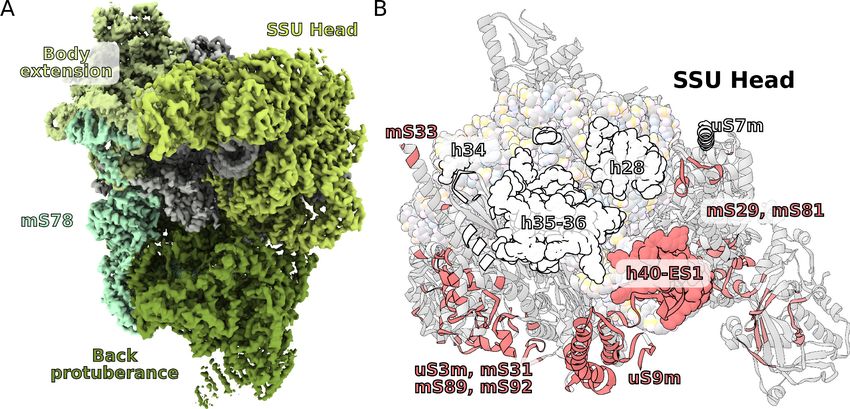
Research article Structural Biology and Molecular Biophysics
To this end, we searched for a model organism using two main criteria: 1) evolutionary distance
from the previously characterized supergroups and 2) coding capacity of the mitochondrial genome
beyond the conventional hydrophobic membrane proteins. The search singled out Tetrahymena
thermophila, a ciliate protist from the phylum Alveolata. The T. thermophila mitochondrial genome
differs substantially from those previously characterized. It encodes 43 proteins, half of which are sol-
uble, varying in length from 59 to 1344 amino acids. In addition, 22 have unknown functions and are
annotated as Ymf genes (Brunk et al., 2003). A further rationale for investigating T. thermophila is
that it is an established and accessible laboratory instrument for genetics (Nanney and Simon,
1999), as well as a source of fundamental discoveries in biology. These include induction of cell-divi-
sion synchrony (Scherbaum and Zeuthen, 1954), discoveries of lysosomes (Elliott and Bak, 1964),
peroxisomes (Baudhuin et al., 1965), linear mitochondrial DNA (Suyama and Miura, 1968), dynein
(Gibbons and Rowe, 1965), catalytic RNA (Kruger et al., 1982), and the molecular characterization
of telomeres (Blackburn and Gall, 1978). In addition, alveolates are infectious agents
commonly involved in meat or pet trade (Chambouvet et al., 2020).
Results and discussion
New proteins and conserved rRNA
To characterize the T. thermophila mitoribosome, we determined its structure using cryo-EM at
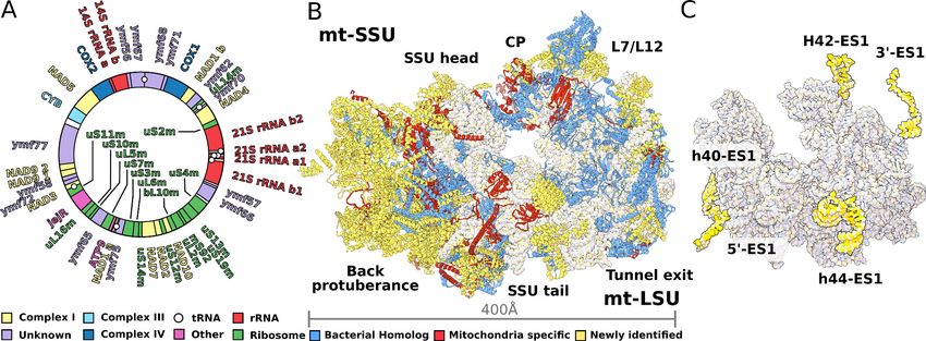
3.30–3.67 Å (Figure 1, Figure 1—figure supplement 1, Supplementary file 1). The resulting 4.0-
MDa model consists of 92 different proteins, including two bL12m dimers. Of these, 46 proteins are
mitochondria-specific, while 27 are newly identified including 9 Ymf-encoded with previously unas-
signed functions (Figure 1A, Figure 1—figure supplements 2 and 3, Supplementary file 1,
Video 1). The protein nomenclature is consistent with the previous structures, whereas additional
proteins are named to avoid overlap. Most of the newly identified proteins are associated with the
small subunit and distributed across the head and the two regions that we named as the back
Figure 1. Structure of T. thermophila mitoribosome and newly identified proteins in the mitochondrial DNA. (A) Schematic representation of the
mitochondrial genome of T. thermophila with newly identified proteins labeled inside the circle. (B) The overall structure of the mitoribosome showing
mitochondria specific and newly identified proteins. (C) Mitoribosomal rRNA showing expansion segments (relative to E. coli) in yellow.
The online version of this article includes the following figure supplement(s) for figure 1:
Figure supplement 1. Electron microscopy data processing workflow.
Figure supplement 2. Positions of newly annotated proteins on the mitoribosome.
Figure supplement 3. Comparison of evolutionary conservation for mitoribosomal proteins.
Figure supplement 4. Secondary structure diagram of the T. thermophila LSU rRNA.
Figure supplement 5. Secondary structure diagram of the T. thermophila SSU rRNA.
Tobiasson and Amunts. eLife 2020;9:e59264. DOI: https://doi.org/10.7554/eLife.59264 2 of 15
Research article                                                                       Structural Biology and Molecular Biophysics

                                                                                       protuberance and the body extension. These
                                                                                       include SelR, a methionine-R-sulfoxide reductase
                                                                                       (Kryukov et al., 2002) in the body extension,
                                                                                       mS81 and an apo-frataxin protein (Castro et al.,
                                                                                       2019) in the back protuberance, mS86. In the
                                                                                       large mitoribosomal subunit (LSU), we identified
                                                                                       a protein binding two iron-sulfur clusters via
                                                                                       CDGSH motifs (Sengupta et al., 2018), mL107.
                                                                                       Proteins in additional locations include mS93
                                                                                       bound to h44-ES1 in the small mitoribosomal
                                  Video 1. Structure of the ciliate mitoribosome.      subunit (SSU) tail, which we identified as the
                                  https://elifesciences.org/articles/59264#video1      MTERF family of proteins. The MTERF family
                                                                                       consists of four members featuring a 30-amino
                                                                                       acid motif, containing leucine zipper-like hep-
                                                                                       tads, reported to be involved in mitochondrial
                                  transcription as well as DNA replication (Roberti et al., 2009).
                                     The rRNAs in both mitoribosomal subunits are split into two fragments (Figure 1A, Figure 1—fig-
                                  ure supplements 4 and 5). However, the overall rRNA structure is conserved (Figure 1C), and
                                  expansion segments (ES) constitute only 3%, while 7% of the rRNA has been reduced. Some of those
                                  rRNA deletions are not structurally restabilized by proteins. For example, h56-59 of domain III is
                                  reduced to a single flexible h56 with no binding protein observed (Figure 2). The conservation of
                                  rRNA and the apparent absence of stabilizing protein elements imply that drivers other than rRNA
                                  reduction impact the mitoribosomal evolution.

                                  The mRNA channel is defined by split protein assembly uS3m encoded
                                  in different genomes
                                  A fundamental stage in translation is the binding of mRNA to a dedicated channel, positioning the
                                  start codon of the open reading frame in the P-site to match the proper anticodon. In all the previ-
                                  ously studied translation systems, including from organelles, the mRNA channel entrance is formed
                                  mainly by the conserved proteins uS3 and uS5.
                                      In T. thermophila, we report that the assembly corresponding to uS3m is composed of three sep-
                                  arate proteins encoded in the nucleic and mitochondrial genomes. The first protein, which was previ-
                                  ously annotated as ‘Ribosomal protein S3’, partially corresponds to the N-terminal stabilizing
                                  domain (NTD) of the bacterial uS3 (Figure 3, Figure 3—figure supplement 1). We renamed it to
                                  mS92 as it constitutes the smallest fragment of uS3. The topology of the first ~50 amino acids is con-
                                  served; however, the similarity is broken by residues Tyr51-Tyr52-Tyr53 (Alexandrov et al., 2012)
                                  causing the mitochondrial structure to deviate from the original helical kink. The new conformation
                                  is then stabilized by the second protein mS31, with its short helix, as well as Lys57 via negatively
                                  charged rRNA phosphate. In the mitochondrial genome, we found the third protein encoded in the
                                  previously unknown Ymf64, which mainly corresponds to the functional C-terminal domain (CTD) of
                                  uS3 and forms a part of the NTD (Figure 3). We named it uS3m. The full NTD is then formed by two
                                  antiparallel b-strands from mS92 that are complemented by two b-strands from mS31 and an addi-
                                  tional single b-strand from uS3m (Figure 3). The resulting b-sheet, therefore, consists of three differ-
                                  ent proteins assembled to make a bacterial-like uS3 NTD. In regard to the CTD, in Holozoa, it is
                                  completely deleted from the mitoribosome resulting in an expanded channel entrance
                                  (Brown et al., 2014), whereas in T. thermophila its genomic sequence is found to be split from the
                                  NTD. Assuming all ribosomes originally had intact uS3, the genomic splitting of uS3m reported here
                                  might represent a possible structural intermediate in the evolution of the mitoribosome toward the
                                  loss of the CTD observed in Holozoa. A similar splitting of the mitoribosomal protein uL2m has been
                                  recently reported in plants (Waltz et al., 2020).
                                      The involvement of two genomes to produce a functional and stable protein further indicates an
                                  evolutionary link between genetic drift and mitochondrial translation. Our experimental finding
                                  therefore illustrates the requirement of tight mito-nuclear coevolution to maintain mitochondrial
                                  activities (Shtolz and Mishmar, 2019; Mishmar, 2020).

Tobiasson and Amunts. eLife 2020;9:e59264. DOI: https://doi.org/10.7554/eLife.59264                                                  3 of 15
Research article                                                                                Structural Biology and Molecular Biophysics

Figure 2. Evolution of rRNA domain III reduction. Comparison of H48-60 rRNA between E. coli and mitochondria of T. thermophila and H. sapiens.
Left, LSU model with rRNA is shown in gray and domain III is highlighted, conserved proteins are in blue, shared mitoribosomal proteins are in red,
and specific mitoribosomal proteins are in yellow. Right, schematic representation of the rRNA region subjected to reduction. In H. sapiens, the rRNA
deletions are structurally restabilized by proteins mL37 and mL65. In T. thermophila, the rRNA reduction is less severe, and flexible elements such as
H56 have no binding protein partners, suggesting an intermediate stage between E. coli and H. sapiens.

Tobiasson and Amunts. eLife 2020;9:e59264. DOI: https://doi.org/10.7554/eLife.59264                                                              4 of 15
Research article Structural Biology and Molecular Biophysics
Figure 3. Functional uS3m consists of three separate proteins encoded in the nuclear and mitochondrial genomes. Comparison of uS3 between E. coli
ribosome and T. thermophila mitoribosome. The topology diagram of uS3 shows the organization of its domains that are replaced by three different
proteins in T. thermophila. Mitoribosomal mS31 (nuclear encoded), mS92 (nuclear encoded), and one strand from uS3m (mitochondria encoded)
collectively correspond to uS3-NTD, whereas mitoribosomal uS3m corresponds to uS3-CTD.
The online version of this article includes the following figure supplement(s) for figure 3:
Figure supplement 1. Local resolution and density for the uS3m module.
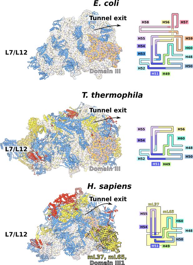
The expanded SSU head is connected to the body by an extensive
protein network
The SSU head plays a key role during the process of translation elongation in promoting tRNA trans-
location. Flexible interactions with the body allow uncoupling of the head–body movement that is
necessary for the sequential conformational changes during the translation cycle (Ratje et al., 2010).
The T. thermophila SSU head has seven extra proteins (mS29, 31, 33, 35, 75, 81, 89), while the
bacterial homologs are extended by ~40%. In addition, two sites enriched with mitochondria specific
proteins are identified on the SSU solvent-facing side. The first is the back protuberance that con-
sists of eleven proteins (mS45, 47, 78-CTD, 83–88, 91, 92) facing away from the LSU binding inter-
face; the second is the body extension that consists of eight proteins (mS23, 26, 37, 76, 77, 78-NTD,
79, uS11-NTD) protruding from the tRNA E-site (Figure 1B, Figure 4A, Video 1). The two moieties
Tobiasson and Amunts. eLife 2020;9:e59264. DOI: https://doi.org/10.7554/eLife.59264 5 of 15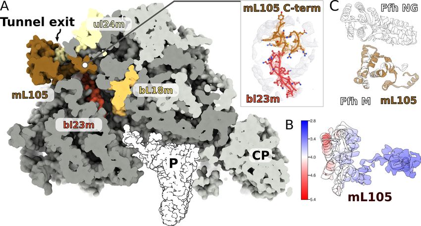
Research article                                                                                   Structural Biology and Molecular Biophysics

Figure 4. Unique functional features provide a distinct architecture of the mitoribosomal subunits. (A) The overall structure of the SSU is substantially
affected by the back protuberance and body extension that are bound to the head and interconnected through mS78. (B) The contact area between
the SSU head and body is illustrated on the head. Mitochondria specific contacts are shown in red, and conserved contacts are shown in white.
The online version of this article includes the following figure supplement(s) for figure 4:
Figure supplement 1. SSU head interactions with body extension.

                                      appear to be interconnected with each other through mS78, which is the largest protein in our struc-
                                      ture with 1509 modeled amino acids (Figure 4A, Supplementary file 2). The protein mS78 is
                                      divided into three domains: 1) NTD forming an extensive helical network with mS23, which builds
                                      the periphery of the body extension; 2) a central helical repeat linking the two new sites; and 3) CTD
                                      consisting of 34 helices capping the back protuberance.
                                         The structures of the back protuberance and body extension indicate altered functions of the
                                      SSU. While the back protuberance coincides with putative RNA polymerase binding sites
                                      (Demo et al., 2017; Kohler et al., 2017), the body extension excludes bS1, which is considered to
                                      be one of the most ancient and conserved ribosomal proteins with functions involved in unfolding
                                      mRNAs for active translation (Qu et al., 2012). In addition, both protrusions interact with the head
                                      (Figure 4A). The back protuberance and the head are connected by nine proteins (mS31, 45, 47, 85,
                                      87, 88, 89, 92, and uS9m) and h40-ES1. The body extension and the head are connected by four
                                      proteins (mS23, 26, 29, and 37). Additionally, mitochondria-specific extensions of uS9m, mS23, 29,
                                      and 31 form multiple interactions linking the head to the body (Figure 4—figure supplement 1). As
                                      a result, the buried surface area between the SSU head and body is almost doubled compared to
                                      that of bacteria: ~40,000 Å2 vs ~21,000 Å2 (Figure 4B).
                                         Consistently with the altered structure of the head, the tRNA binding sites have also been remod-
                                      eled (Figure 5). The C-terminal extension of uS13m presents a positively charged surface replacing
                                      the A-site finger. A newly identified protein mL102 makes further interactions with the A-site tRNA
                                      acceptor stem by extending its C-terminus downward from between the CP and 7/12 stalk. At the
                                      P-site, mL40 extends with its positively charged N-terminal helix toward the tRNA elbow. Taken
                                      together, the increased surface area between the head and body and the positioning of mitochon-
                                      drial protein elements facing the A- and P-sites suggest altered interactions with the ligands.

Tobiasson and Amunts. eLife 2020;9:e59264. DOI: https://doi.org/10.7554/eLife.59264                                                                  6 of 15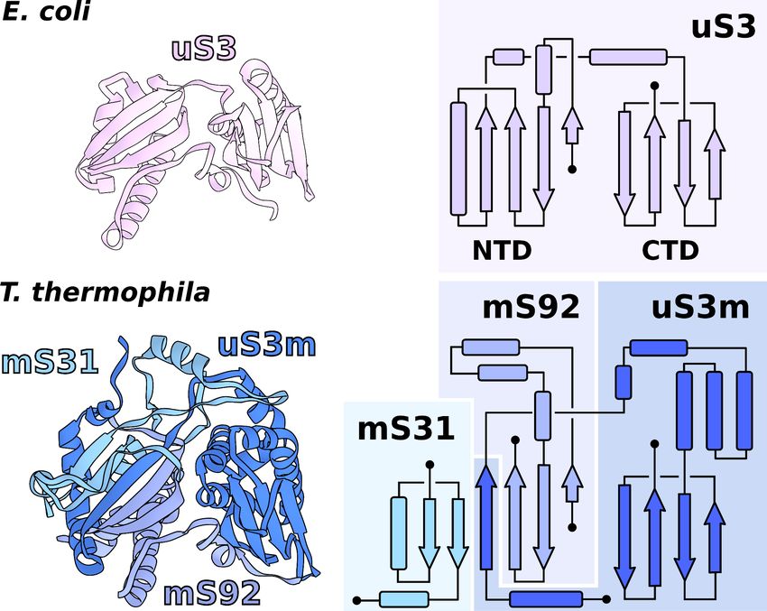
Research article                                                                               Structural Biology and Molecular Biophysics

Figure 5. Specific protein elements interacting with tRNA binding sites. The conventional tRNA binding sites are indicated in white based on the
canonical L-shape of tRNAs (PDBID: 5MDZ). Related proteins of the LSU and SSU are shown in blue/purple and white, respectively. Mitochondria
specific elements encasing the tRNA binding sites are shown in red.

                                    Minimal central protuberance lacking 5S rRNA is a potential
                                    evolutionary intermediate
                                    The central protuberance (CP) is a ubiquitous element of the LSU that forms bridges with the SSU
                                    head. Unlike cytosolic ribosomes that contain 5S rRNA as a core component of the CP, in mitochon-
                                    dria, the 5S rRNA has been replaced by other RNAs or protein. Analysis of the previously reported
                                    mitoribosomal structures concluded that the replacing elements, such as a Holozoan tRNA encoded
                                    between the two rRNA genes in the mitochondrial genome, could be incorporated into the CP to
                                    restabilize its core (Petrov et al., 2019).
                                       In the T. thermophila mitoribosome, although the 5S is missing, no RNA or any substantial protein
                                    replacement is found (Figure 6, Video 1). This is despite a tRNA being encoded between rRNA
                                    genes, as in Holozoa (Figure 1A). The positions of the 5S C- and D-loops are partially occupied by a
                                    small protein loop of bL21m and an N-terminal extension of bL27m, respectively. Yet, the overall
                                    architecture of the CP remains intact, and the peripheral proteins are arranged similarly to
                                    those of other mitoribosomes. Also on the rRNA level, only a minor deviation from bacteria is found,
                                    which is in the 15-nucleotide long tip of H84 interacting with uL5m. The protein uL5m is traditionally
                                    associated with the 5S rRNA and is heavily reduced in our structure. Intriguingly, uL5m is the first
                                    reported mitoribosomal homolog that appears to be reduced. No other proteins are seen stabilizing
                                    the CP and the density suggests it is flexible.
                                       Therefore, the T. thermophila mitoribosome has a minimal and the most flexible known CP, indi-
                                    cating a less significant role of LSU in the stabilization of the enlarged SSU head. These structural
                                    changes might reflect an intermediate evolutionary step after the loss of 5S rRNA and before
                                    the acquisition of a compensating structural replacement.

                                    Native L7/L12 stalk with four bL12m copies
                                    The L7/L12 stalk mediates interactions with translational factors and is organized in three structural
                                    regions: the rRNA base (H42-44 rRNA), a protein linker (a8 of uL10, dimers of bL12 NTDs), and a
                                    mobile factor-recruiting domain (bL12 CTDs). Due to its high flexibility, the stalk has not been well

Tobiasson and Amunts. eLife 2020;9:e59264. DOI: https://doi.org/10.7554/eLife.59264                                                            7 of 15
Research article Structural Biology and Molecular Biophysics
Figure 6. Specific features of the LSU. The LSU central protuberance lacks 5S rRNA, and the L7/L12 stalk consists
of only two dimers. Superposition of E. coli 5S rRNA reveals no substantial protein replacement, apart from minor
elements shown in red. The model of the native L7/L12 stalk reveals unusual conformation due to the presence of
rRNA expansion H42-ES1 and mL104.
The online version of this article includes the following figure supplement(s) for figure 6:
Figure supplement 1. Structure of the L7/L12 stalk.
resolved in the previous cryo-EM reconstructions (Brown et al., 2014; Amunts et al., 2014;
Greber et al., 2014; Amunts et al., 2015; Greber et al., 2015; Desai et al., 2017). However, a
computational analysis of the mitoribosomal stalk predicted six bL12m copies arranged in three
dimers (Davydov et al., 2013).
Our data shows a well-resolved linker domain allowing for modeling of the native uL10m and
bL12m dimers (Figure 6, Video 1). In contrast to the prediction, only four bL12m copies arranged in
two dimers fit the density. For uL10m we found a matching sequence encoded in the mitochondrial
Ymf74 sequence. Superimposition with the bacterial stalk revealed that the linker domain a8 of
uL10m is straight and rigid, lacking the representative kinks that define its bL12-binding capacity in
other ribosomes (Liljas and Sanyal, 2018; Figure 6—figure supplement 1). The structural basis for
the stalk rigidity originates in a 25-nucleotide expansion H42-ES1 in the rRNA base. It is bound by
the 90 kDa helical repeat protein mL104. This forms a stabilizing interface for the proximal bL12m
dimer (Figure 4—figure supplement 1). As a result, the protrusion of the stalk is more distinct, plac-
ing the mobile factor-recruiting domains of bL12 CTDs further away from the A-site (Figure 1B, Fig-
ure 6). Such an arrangement provides a mechanical constraint on the number of bL12m copies
within functional distance to test aa-tRNAs to a codon in the A-site and rationalizing the presence of
only two bL12m dimers.
Intrinsic protein visualized in the tunnel suggests putative targeting
Previous structural studies have focused on mitoribosomes that almost exclusively synthesized trans-
membrane proteins of the oxidative phosphorylation chain. Therefore, a translation-independent
Tobiasson and Amunts. eLife 2020;9:e59264. DOI: https://doi.org/10.7554/eLife.59264 8 of 15Research article Structural Biology and Molecular Biophysics
membrane targeting was indicated (Pfeffer et al., 2015; Englmeier et al., 2017). In many other line-
ages of eukaryotes, multiple soluble proteins are encoded in mitochondria, however, whether a sep-
arate targeting system similar to the cytosolic translation apparatus exists is not known.
Since in the cytosol, ribosomes engage a universally conserved targeting system called the signal
recognition particle (SRP), we carefully inspected the region on the mitoribosome that is equivalent
to the SRP accessory proteins’ binding sites. We observed a globular density near the tunnel exit, in
the vicinity of H7, extending ~40 Å from the mitoribosome surface (Figure 7A). The local resolution
of 3.2–5.0 Å in this region allowed modeling 150 residues arranged in eight helices, which belong to
a nuclear-encoded protein that we named mL105 (Figure 7B, Figure 7—figure supplement 1). We
found that this protein is annotated as ‘signal peptide-binding domain protein fragment’. The topol-
ogy and sequence of mL105 suggest homology to the M-domain of the bacterial SRP binding pro-
tein Ffh in its ribosome-bound form, while it lacks the GTP binding NG-domain (Figure 7C). The
C-terminal helix of mL105 inserts into the tunnel and forms interactions with the hairpin loop of
bL23m (Figure 7A). Moreover, it would inevitably contact the nascent chain. A similar functional
insertion of Ffh was suggested based on the low resolution reconstruction of the reconstituted cyto-
solic system (Jomaa et al., 2016) and confirmed by cross-linking data (Denks et al., 2017). We
therefore report an intrinsic feature of the ciliate mitoribosome that provides the first evidence for a
putative protein targeting in mitochondria.
Conclusions
Although the requirement for proteins synthesized in mitochondria is vital, the underlying mecha-
nisms were previously only partially explored in the isolated species. The current structural analysis
Figure 7. A signal peptide-binding domain protein is bound to the LSU. (A) Slicing through the LSU shows the tunnel path and the targeting protein
mL105 bound at the tunnel exit. The protein mL105 is positioned in a way that would affect the nascent polypeptide path. Inset displays interactions
between mL105 CTD and bL23m close to the tunnel exit. (B) Model and density for mL105 colored by local resolution. (C) Superposition of the
ribosome-bound Ffh M-domain (PDBID: 5GAF) with mL105 shows structural similarity.
The online version of this article includes the following figure supplement(s) for figure 7:
Figure supplement 1. Local resolution density for mL105.
Tobiasson and Amunts. eLife 2020;9:e59264. DOI: https://doi.org/10.7554/eLife.59264 9 of 15Research article Structural Biology and Molecular Biophysics
of the ciliate mitoribosome, which is evolutionarily distant from the previously characterized, shows
that mitochondria have evolved independent features related to all functional aspects of translation.
The data revealed extra proteins on the SSU that might affect the conformational landscape of the
head movement, identified protein-rich tRNA binding sites, and reported prime evidence for a pro-
tein targeting system through a signal peptide-binding domain protein found at the LSU tunnel exit.
From the evolutionary perspective, a surprising feature is that despite the multiple additional pro-
teins that form a distinct overall structure, the rRNA is generally conserved. This suggest evolution-
ary drivers other than rRNA for mitoribosomal diversity. In addition, our finding that rRNA deletions
are not structurally restabilized by proteins imply that the ciliate mitoribosome represents an evolu-
tionary intermediate between other eukaryotic supergroups, providing a reference point for further
investigation of the development of translation. Finally, we report a functional protein composed of
three separate proteins encoded in the nuclear and mitochondrial genomes, providing experimental
link between genetic drift and mitochondrial translation.
Together, the distinct T. thermophila mitoribosome structural model illustrates the functional
diversity of mitochondrial translation and provides a framework for examination of its evolution.
Materials and methods
Strains and growth conditions
T. thermophila SB210 cells (from ATCC) were cultured axenically in 1% (w/v) yeast extract, 2% prote-
ose peptone supplemented with 3% (v/v) glycerol at 36˚C with 80 rpm shaking. Upon reaching mid
log phase (~8105 cells/ml), the cells were harvested by centrifugation for 10 min at 1300 g. All the
subsequent steps were performed at 4˚C. The pellets were resuspended in a minimal volume of
homogenization buffer (20 mM HEPES KOH pH 7.5, 350 mM mannitol, 5 mM EDTA). Lysis was
achieved by homogenization in a Dounce homogenizer with 30 strokes of a tightly fitting plunger.
Debris were removed by two centrifugations at 800 g for 10 min. Mitochondria were then pelleted
by centrifugation at 7000 g for 20 min. The pellets were resuspended in minimal volume and loaded
onto a discontinuous sucrose gradient (20 mM HEPES KOH pH 7.5, 1 mM EDTA with 15, 23, 32%,
and 60% sucrose) and centrifuged at 90,000 g for 1 hr. Mitochondria were collected between the
60% and 32% sucrose layers and flash frozen in aliquots of 2 mL in liquid nitrogen.
Purification of mitoribosomes
All the subsequent steps were carried out on ice. About 100 mg of mitochondrial protein was
thawed and solubilized by adding 5 volumes of Triton buffer (25 mM HEPES KOH pH 7.5, 20 mM
KCl, 25 mM MgOAc, 1.7% Triton X-100 and ROCHE complete EDTA-free protease inhibitor tablets),
followed by a 15 min incubation on a rolling mill for 15 min. Mitochondrial membranes were
removed by centrifugation at 30,000 g for 20 min. The released mitochondrial proteins were trans-
ferred onto 1 M sucrose cushion (25 mM Hepes KOH pH 7.5, 20 mM KCl, 15 mM MgOAc, 1% Triton
X-100, and 1 M sucrose), and mitochondrial ribosomes pelleted at 235,000 g for 4 hr. The pellet was
resuspended in a minimal amount of buffer (25 mM HEPES KOH pH 7.5, 20 mM KCl, 15 mM
MgOAc). Aggregates were removed by centrifugation for 5 min at 14,000 g. The clarified superna-
tant was transferred to linear 15–30% (w/v) sucrose gradients. Gradients were centrifuged at 90,000
g for 16 hr and fractionated into 400 mL fractions. Fractions with an enriched absorbance at 260 nm
were pooled and centrifuged at 235,000 g for 60 min to pellet mitoribosomes. Final pellets were
resuspended in the same buffer without sucrose.
Cryo-EM and model building
Quantifoil R2/2 300 mesh copper grids were coated with a ~3 nm continuous carbon support pro-
duced in house and glow discharged for 30 s at 25 mA. About 3 mL of the purified ribosomal sample
at OD260 3.0 was applied and incubated for 30 s at 100% humidity at 4˚C before blotting for 3 s and
plunge frozen into liquid ethane using an FEI Vitrobot MkIV. The cryo-EM data were collected using
a Titan Krios microscope operated at a voltage of 300 kV and equipped with a K2 Summit detector
(Gatan). Data were collected at a nominal magnification of 130,000x corresponding to a pixel size of
1.07 Å2/px, and a fluence of 30 electrons divided into 20 dose fractions at a flux of 5 e-/Å2s and a
Tobiasson and Amunts. eLife 2020;9:e59264. DOI: https://doi.org/10.7554/eLife.59264 10 of 15Research article Structural Biology and Molecular Biophysics
final defocus range from 0.5 to 3.0 mm. A total of 5511 micrographs were selected after manual
screening based on CTF fitting and overall qualitative appearance.
Initial motion correction was carried out using motioncor2 version 2.1 (Zheng et al., 2017), fol-
lowed by CTF estimation by Gctf version 1.06 (Zhang, 2016). All further processing was done with
RELION 2.7 and 3.0 (Zivanov et al., 2018). The workflow of the data processing is shown in Fig-
ure 1—figure supplement 1. A total of 450,250 particles were picked using Gaussian blobs and
extracted with a box of 550 pixels. The box was binned to 128 pixels for the following classification
steps. After several rounds of 2D classification, 133,650 particles were left for the 3D classification.
After 3D classification, three classes with good quality were selected for the final reconstruction. In
total, 99,380 particles were merged for 3D refinement. The final resolution of the 3D auto-refine-
ment after post-processing was 3.67 Å. Application of masks for the LSU and SSU during refinement
further improved the resolution of these regions to 3.38 Å and 3.61 Å, respectively. We also applied
local masks to the L7/L12 stalk, CP, SSU head, back protuberance, which resulted in improved qual-
ity of local maps with resolutions ranging between 3.30 Å and 3.61 Å. All the resolutions were esti-
mated with the gold-standard Fourier shell correlation 0.143 criterion with high-resolution noise
substitution. All the local resolution maps were calculated using RELION 3.0 (Zivanov et al., 2018).
Model building was done in Coot 0.8.9.2 (Emsley et al., 2010). Initially, models of the mitoribo-
some from S. cerevisiae (PDB ID: 5MRC) and H. sapiens (PDB ID: 3J9M) were fitted to the map and
served as protein backbone and rRNA references. Most of the proteins were built de novo using a
combination of bulky side chain patterns, fold identification by PDBeFold (Krissinel and Henrick,
2004), mass-spec data, and assigning putative primary sequence followed by BLAST (Altschul et al.,
1990) searches. The model was initially built and refined against a composite map consisting of all
six masked regions and finally refined and validated using the consensus map. All models were
refined iteratively using PHENIX (Liebschner et al., 2019) realspace refinement and validated using
MolProbity (Williams et al., 2018). The data collection, model refinement and validation statistics
are presented in Supplementary file 1. All figures were generated using either Chimera
(Pettersen et al., 2004) or ChimeraX (Goddard et al., 2018) with annotations and vector
editing was done using Inkscape.
Acknowledgements
The authors thank the SciLifeLab cryo-EM and mass spectrometry facilities, and G von Heijne for his
comments on the manuscript. This work was supported by the Swedish Foundation for Strategic
Research (FFL15:0325), Ragnar Söderberg Foundation (M44/16), Swedish Research Council
(NT_2015–04107), Cancerfonden (2017/1041), European Research Council (ERC-2018-StG-805230),
Knut and Alice Wallenberg Foundation (2018.0080), EMBO Young Investigator Program. The cryo-
EM facility is funded by the Knut and Alice Wallenberg, Family Erling Persson, and Kempe
foundations.
Additional information
Funding
Funder Grant reference number Author
Ragnar Söderbergs stiftelse M44/16 Alexey Amunts
Cancerfonden 2017/1041 Alexey Amunts
H2020 European Research ERC-2018-StG- 805230 Alexey Amunts
Council
Knut och Alice Wallenbergs 2018.0080 Alexey Amunts
Stiftelse
European Molecular Biology EMBO Young Investigator Alexey Amunts
Organization Program
Swedish Foundation for Stra- FFL15:0325 Alexey Amunts
tegic Research
Swedish Research Council NT_2015–04107 Alexey Amunts
Tobiasson and Amunts. eLife 2020;9:e59264. DOI: https://doi.org/10.7554/eLife.59264 11 of 15Research article Structural Biology and Molecular Biophysics
The funders had no role in study design, data collection and interpretation, or the
decision to submit the work for publication.
Author contributions
Victor Tobiasson, Alexey Amunts, Conceptualization, Data curation, Formal analysis, Validation,
Investigation, Visualization, Methodology, Writing - original draft, Writing - review and editing
Author ORCIDs
Victor Tobiasson https://orcid.org/0000-0001-8920-017X
Alexey Amunts https://orcid.org/0000-0002-5302-1740
Decision letter and Author response
Decision letter https://doi.org/10.7554/eLife.59264.sa1
Author response https://doi.org/10.7554/eLife.59264.sa2
Additional files
Supplementary files
. Supplementary file 1. Cryo-EM data collection, refinement and validation statistics.
. Supplementary file 2. Summary of the mitoribosomal proteins.
. Transparent reporting form
Data availability
The electron density maps have been deposited into EMDB, with accession codes EMD-11032
(monosome), EMD-11033 (LSU), EMD-11034 (SSU), EMD-11035 (CP), EMD-11036 (L7/L12 stalk),
EMD-11037 (head), EMD-11038 (back protuberance). The model has been deposited in the PDB,
with accession code 6Z1P.
The following datasets were generated:
Database and
Author(s) Year Dataset title Dataset URL Identifier
Tobiasson V, 2020 monosome https://www.ebi.ac.uk/ Electron Microscopy
Amunts A pdbe/entry/emdb/EMD- Data Bank, EMD-110
11032 32
Tobiasson V, 2020 LSU https://www.ebi.ac.uk/ Electron Microscopy
Amunts A pdbe/entry/emdb/EMD- Data Bank, EMD-110
11033 33
Tobiasson V, 2020 SSU https://www.ebi.ac.uk/ Electron Microscopy
Amunts A pdbe/entry/emdb/EMD- Data Bank, EMD-110
11034 34
Tobiasson V, 2020 CP https://www.ebi.ac.uk/ Electron Microscopy
Amunts A pdbe/entry/emdb/EMD- Data Bank, EMD-110
11035 35
Tobiasson V, 2020 L7/L12 stalk https://www.ebi.ac.uk/ Electron Microscopy
Amunts A pdbe/entry/emdb/EMD- Data Bank, EMD-110
11036 36
Tobiasson V, 2020 head https://www.ebi.ac.uk/ Electron Microscopy
Amunts A pdbe/entry/emdb/EMD- Data Bank, EMD-110
11037 37
Tobiasson V, 2020 back protuberance https://www.ebi.ac.uk/ Electron Microscopy
Amunts A pdbe/entry/emdb/EMD- Data Bank, EMD-110
11038 38
Tobiasson V, 2020 Model https://www.rcsb.org/ RCSB Protein Data
Amunts A structure/6Z1P Bank, 6Z1P
Tobiasson and Amunts. eLife 2020;9:e59264. DOI: https://doi.org/10.7554/eLife.59264 12 of 15Research article Structural Biology and Molecular Biophysics
References
Adl SM, Bass D, Lane CE, Lukeš J, Schoch CL, Smirnov A, Agatha S, Berney C, Brown MW, Burki F, Cárdenas P,
Čepička I, Chistyakova L, Del Campo J, Dunthorn M, Edvardsen B, Eglit Y, Guillou L, Hampl V, Heiss AA, et al.
2019. Revisions to the classification, nomenclature, and diversity of eukaryotes. The Journal of Eukaryotic
Microbiology 66:4–119. DOI: https://doi.org/10.1111/jeu.12691, PMID: 30257078
Alexandrov AI, Polyanskaya AB, Serpionov GV, Ter-Avanesyan MD, Kushnirov VV. 2012. The effects of amino
acid composition of glutamine-rich domains on amyloid formation and fragmentation. PLOS ONE 7:e46458.
DOI: https://doi.org/10.1371/journal.pone.0046458, PMID: 23071575
Altschul SF, Gish W, Miller W, Myers EW, Lipman DJ. 1990. Basic local alignment search tool. Journal of
Molecular Biology 215:403–410. DOI: https://doi.org/10.1016/S0022-2836(05)80360-2, PMID: 2231712
Amunts A, Brown A, Bai XC, Llácer JL, Hussain T, Emsley P, Long F, Murshudov G, Scheres SHW, Ramakrishnan
V. 2014. Structure of the yeast mitochondrial large ribosomal subunit. Science 343:1485–1489. DOI: https://
doi.org/10.1126/science.1249410, PMID: 24675956
Amunts A, Brown A, Toots J, Scheres SHW, Ramakrishnan V. 2015. Ribosome. The structure of the human
mitochondrial ribosome. Science 348:95–98. DOI: https://doi.org/10.1126/science.aaa1193, PMID: 25838379
Baudhuin P, Mueller M, Poole B, de Duve C. 1965. Non-mitochondrial oxidizing particles (MICROBODIES) IN rat
liver and kidney and in Tetrahymena pyriformis. Biochemical and Biophysical Research Communications 20:53–
59. DOI: https://doi.org/10.1016/0006-291X(65)90949-6, PMID: 14341941
Blackburn EH, Gall JG. 1978. A tandemly repeated sequence at the termini of the extrachromosomal ribosomal
RNA genes in Tetrahymena. Journal of Molecular Biology 120:33–53. DOI: https://doi.org/10.1016/0022-2836
(78)90294-2, PMID: 642006
Brown A, Amunts A, Bai XC, Sugimoto Y, Edwards PC, Murshudov G, Scheres SHW, Ramakrishnan V. 2014.
Structure of the large ribosomal subunit from human mitochondria. Science 346:718–722. DOI: https://doi.org/
10.1126/science.1258026, PMID: 25278503
Brunk CF, Lee LC, Tran AB, Li J. 2003. Complete sequence of the mitochondrial genome of Tetrahymena
thermophila and comparative methods for identifying highly divergent genes. Nucleic Acids Research 31:1673–
1682. DOI: https://doi.org/10.1093/nar/gkg270, PMID: 12626709
Castro IH, Pignataro MF, Sewell KE, Espeche LD, Herrera MG, Noguera ME, Dain L, Nadra AD, Aran M, Smal C,
Gallo M, Santos J. 2019. Frataxin structure and function. Sub-Cellular Biochemistry 93:393–438. DOI: https://
doi.org/10.1007/978-3-030-28151-9_13, PMID: 31939159
Chambouvet A, Smilansky V, Jirků M, Isidoro-Ayza M, Itoı̈z S, Derelle E, Monier A, Gower DJ, Wilkinson M,
Yabsley MJ, Lukeš J, Richards TA. 2020. Diverse alveolate infections of tadpoles, a new threat to frogs? PLOS
Pathogens 16:e1008107. DOI: https://doi.org/10.1371/journal.ppat.1008107, PMID: 32053700
Davydov II, Wohlgemuth I, Artamonova II, Urlaub H, Tonevitsky AG, Rodnina MV. 2013. Evolution of the protein
stoichiometry in the L12 stalk of bacterial and organellar ribosomes. Nature Communications 4:1387.
DOI: https://doi.org/10.1038/ncomms2373, PMID: 23340427
Demo G, Rasouly A, Vasilyev N, Svetlov V, Loveland AB, Diaz-Avalos R, Grigorieff N, Nudler E, Korostelev AA.
2017. Structure of RNA polymerase bound to ribosomal 30S subunit. eLife 6:e28560. DOI: https://doi.org/10.
7554/eLife.28560, PMID: 29027901
Denks K, Sliwinski N, Erichsen V, Borodkina B, Origi A, Koch HG. 2017. The signal recognition particle contacts
uL23 and scans substrate translation inside the ribosomal tunnel. Nature Microbiology 2:16265. DOI: https://
doi.org/10.1038/nmicrobiol.2016.265, PMID: 28134917
Desai N, Brown A, Amunts A, Ramakrishnan V. 2017. The structure of the yeast mitochondrial ribosome. Science
355:528–531. DOI: https://doi.org/10.1126/science.aal2415, PMID: 28154081
Elliott AM, Bak IJ. 1964. The contractile vacuole and related structures in Tetrahymena pyriformis. The Journal of
Protozoology 11:250–261. DOI: https://doi.org/10.1111/j.1550-7408.1964.tb01752.x, PMID: 14179751
Emsley P, Lohkamp B, Scott WG, Cowtan K. 2010. Features and development of coot. Acta Crystallographica.
Section D, Biological Crystallography 66:486–501. DOI: https://doi.org/10.1107/S0907444910007493,
PMID: 20383002
Englmeier R, Pfeffer S, Förster F. 2017. Structure of the human mitochondrial ribosome studied in situ by
cryoelectron tomography. Structure 25:1574–1581. DOI: https://doi.org/10.1016/j.str.2017.07.011, PMID: 2
8867615
Gibbons IR, Rowe AJ. 1965. Dynein: a protein with adenosine triphosphatase activity from cilia. Science 149:
424–426. DOI: https://doi.org/10.1126/science.149.3682.424, PMID: 17809406
Goddard TD, Huang CC, Meng EC, Pettersen EF, Couch GS, Morris JH, Ferrin TE. 2018. UCSF ChimeraX:
meeting modern challenges in visualization and analysis. Protein Science 27:14–25. DOI: https://doi.org/10.
1002/pro.3235, PMID: 28710774
Gray MW, Lukes J, Archibald JM, Keeling PJ, Doolittle WF. 2010. Irremediable complexity? Science 330:920–
921. DOI: https://doi.org/10.1126/science.1198594
Greber BJ, Boehringer D, Leibundgut M, Bieri P, Leitner A, Schmitz N, Aebersold R, Ban N. 2014. The complete
structure of the large subunit of the mammalian mitochondrial ribosome. Nature 515:283–286. DOI: https://
doi.org/10.1038/nature13895, PMID: 25271403
Greber BJ, Bieri P, Leibundgut M, Leitner A, Aebersold R, Boehringer D, Ban N. 2015. Ribosome. The complete
structure of the 55S mammalian mitochondrial ribosome. Science 348:303–308. DOI: https://doi.org/10.1126/
science.aaa3872, PMID: 25837512
Tobiasson and Amunts. eLife 2020;9:e59264. DOI: https://doi.org/10.7554/eLife.59264 13 of 15Research article Structural Biology and Molecular Biophysics
Janouškovec J, Tikhonenkov DV, Burki F, Howe AT, Rohwer FL, Mylnikov AP, Keeling PJ. 2017. A new lineage of
eukaryotes illuminates early mitochondrial genome reduction. Current Biology 27:3717–3724. DOI: https://doi.
org/10.1016/j.cub.2017.10.051, PMID: 29174886
Jomaa A, Boehringer D, Leibundgut M, Ban N. 2016. Structures of the E. coli translating ribosome with SRP and
its receptor and with the translocon. Nature Communications 7:10471. DOI: https://doi.org/10.1038/
ncomms10471, PMID: 26804923
Keeling PJ, McCutcheon JP. 2017. Endosymbiosis: the feeling is not mutual. Journal of Theoretical Biology 434:
75–79. DOI: https://doi.org/10.1016/j.jtbi.2017.06.008, PMID: 28624393
Kohler R, Mooney RA, Mills DJ, Landick R, Cramer P. 2017. Architecture of a transcribing-translating
expressome. Science 356:194–197. DOI: https://doi.org/10.1126/science.aal3059, PMID: 28408604
Krissinel E, Henrick K. 2004. Secondary-structure matching (SSM), a new tool for fast protein structure alignment
in three dimensions. Acta Crystallographica Section D Biological Crystallography 60:2256–2268. DOI: https://
doi.org/10.1107/S0907444904026460, PMID: 15572779
Kruger K, Grabowski PJ, Zaug AJ, Sands J, Gottschling DE, Cech TR. 1982. Self-splicing RNA: autoexcision and
autocyclization of the ribosomal RNA intervening sequence of Tetrahymena. Cell 31:147–157. DOI: https://doi.
org/10.1016/0092-8674(82)90414-7, PMID: 6297745
Kryukov GV, Kumar RA, Koc A, Sun Z, Gladyshev VN. 2002. Selenoprotein R is a zinc-containing stereo-specific
methionine sulfoxide reductase. PNAS 99:4245–4250. DOI: https://doi.org/10.1073/pnas.072603099, PMID: 11
929995
Liebschner D, Afonine PV, Baker ML, Bunkóczi G, Chen VB, Croll TI, Hintze B, Hung L-W, Jain S, McCoy AJ,
Moriarty NW, Oeffner RD, Poon BK, Prisant MG, Read RJ, Richardson JS, Richardson DC, Sammito MD,
Sobolev OV, Stockwell DH, et al. 2019. Macromolecular structure determination using X-rays, neutrons and
electrons: recent developments in Phenix . Acta Crystallographica Section D Structural Biology 75:861–877.
DOI: https://doi.org/10.1107/S2059798319011471
Liljas A, Sanyal S. 2018. The enigmatic ribosomal stalk. Quarterly Reviews of Biophysics 51:e12. DOI: https://doi.
org/10.1017/S0033583518000100, PMID: 30912488
Lukeš J, Archibald JM, Keeling PJ, Doolittle WF, Gray MW. 2011. How a neutral evolutionary ratchet can build
cellular complexity. IUBMB Life 63:528–537. DOI: https://doi.org/10.1002/iub.489, PMID: 21698757
Lukeš J, Wheeler R, Jirsová D, David V, Archibald JM. 2018. Massive mitochondrial DNA content in diplonemid
and kinetoplastid protists. IUBMB Life 70:1267–1274. DOI: https://doi.org/10.1002/iub.1894, PMID: 30291814
Mishmar D. 2020. mtDNA in the crossroads of evolution and disease. Nature Reviews Molecular Cell Biology 21:
181. DOI: https://doi.org/10.1038/s41580-020-0213-4, PMID: 31969697
Nanney DL, Simon EM. 1999. Chapter 1 laboratory and evolutionary history of Tetrahymena thermophila.
Methods in Cell Biology 63:3–25. DOI: https://doi.org/10.1016/S0091-679X(08)61527-7
Petrov AS, Wood EC, Bernier CR, Norris AM, Brown A, Amunts A. 2019. Structural patching fosters divergence
of mitochondrial ribosomes. Molecular Biology and Evolution 36:207–219. DOI: https://doi.org/10.1093/
molbev/msy221, PMID: 30517740
Pettersen EF, Goddard TD, Huang CC, Couch GS, Greenblatt DM, Meng EC, Ferrin TE. 2004. UCSF chimera–a
visualization system for exploratory research and analysis. Journal of Computational Chemistry 25:1605–1612.
DOI: https://doi.org/10.1002/jcc.20084, PMID: 15264254
Pfeffer S, Woellhaf MW, Herrmann JM, Förster F. 2015. Organization of the mitochondrial translation machinery
studied in situ by cryoelectron tomography. Nature Communications 6:6019. DOI: https://doi.org/10.1038/
ncomms7019, PMID: 25609543
Qu X, Lancaster L, Noller HF, Bustamante C, Tinoco I. 2012. Ribosomal protein S1 unwinds double-stranded RNA
in multiple steps. PNAS 109:14458–14463. DOI: https://doi.org/10.1073/pnas.1208950109
Ramrath DJF, Niemann M, Leibundgut M, Bieri P, Prange C, Horn EK, Leitner A, Boehringer D, Schneider A, Ban
N. 2018. Evolutionary shift toward protein-based architecture in trypanosomal mitochondrial ribosomes.
Science 362:eaau7735. DOI: https://doi.org/10.1126/science.aau7735, PMID: 30213880
Ratje AH, Loerke J, Mikolajka A, Brünner M, Hildebrand PW, Starosta AL, Dönhöfer A, Connell SR, Fucini P,
Mielke T, Whitford PC, Onuchic JN, Yu Y, Sanbonmatsu KY, Hartmann RK, Penczek PA, Wilson DN, Spahn CM.
2010. Head swivel on the ribosome facilitates translocation by means of intra-subunit tRNA hybrid sites. Nature
468:713–716. DOI: https://doi.org/10.1038/nature09547, PMID: 21124459
Roberti M, Polosa PL, Bruni F, Manzari C, Deceglie S, Gadaleta MN, Cantatore P. 2009. The MTERF family
proteins: mitochondrial transcription regulators and beyond. Biochimica Et Biophysica Acta (BBA) -
Bioenergetics 1787:303–311. DOI: https://doi.org/10.1016/j.bbabio.2009.01.013
Scherbaum O, Zeuthen E. 1954. Induction of synchronous cell division in mass cultures of Tetrahymena piriformis.
Experimental Cell Research 6:221–227. DOI: https://doi.org/10.1016/0014-4827(54)90164-0, PMID: 13142000
Sengupta S, Nechushtai R, Jennings PA, Onuchic JN, Padilla PA, Azad RK, Mittler R. 2018. Phylogenetic analysis
of the CDGSH iron-sulfur binding domain reveals its ancient origin. Scientific Reports 8:6. DOI: https://doi.org/
10.1038/s41598-018-23305-6
Shtolz N, Mishmar D. 2019. The mitochondrial Genome–on Selective Constraints and Signatures at the
Organism, Cell, and Single Mitochondrion Levels. Frontiers in Ecology and Evolution 7:342. DOI: https://doi.
org/10.3389/fevo.2019.00342
Suyama Y, Miura K. 1968. Size and structural variations of mitochondrial DNA. PNAS 60:235–242. DOI: https://
doi.org/10.1073/pnas.60.1.235, PMID: 16591633
Waltz F, Soufari H, Bochler A, Giegé P, Hashem Y. 2020. Cryo-EM structure of the RNA-rich plant mitochondrial
ribosome. Nature Plants 6:377–383. DOI: https://doi.org/10.1038/s41477-020-0631-5, PMID: 32251374
Tobiasson and Amunts. eLife 2020;9:e59264. DOI: https://doi.org/10.7554/eLife.59264 14 of 15Research article Structural Biology and Molecular Biophysics
Wideman JG, Monier A, Rodrı́guez-Martı́nez R, Leonard G, Cook E, Poirier C, Maguire F, Milner DS, Irwin NAT,
Moore K, Santoro AE, Keeling PJ, Worden AZ, Richards TA. 2020. Unexpected mitochondrial genome diversity
revealed by targeted single-cell genomics of heterotrophic flagellated protists. Nature Microbiology 5:154–
165. DOI: https://doi.org/10.1038/s41564-019-0605-4, PMID: 31768028
Williams CJ, Headd JJ, Moriarty NW, Prisant MG, Videau LL, Deis LN, Verma V, Keedy DA, Hintze BJ, Chen VB,
Jain S, Lewis SM, Arendall WB, Snoeyink J, Adams PD, Lovell SC, Richardson JS, Richardson DC. 2018.
MolProbity: more and better reference data for improved all-atom structure validation. Protein Science 27:293–
315. DOI: https://doi.org/10.1002/pro.3330, PMID: 29067766
Zhang K. 2016. Gctf: real-time CTF determination and correction. Journal of Structural Biology 193:1–12.
DOI: https://doi.org/10.1016/j.jsb.2015.11.003, PMID: 26592709
Zheng SQ, Palovcak E, Armache JP, Verba KA, Cheng Y, Agard DA. 2017. MotionCor2: anisotropic correction of
beam-induced motion for improved cryo-electron microscopy. Nature Methods 14:331–332. DOI: https://doi.
org/10.1038/nmeth.4193, PMID: 28250466
Zivanov J, Nakane T, Forsberg BO, Kimanius D, Hagen WJ, Lindahl E, Scheres SH. 2018. New tools for
automated high-resolution cryo-EM structure determination in RELION-3. eLife 7:e42166. DOI: https://doi.org/
10.7554/eLife.42166, PMID: 30412051
Tobiasson and Amunts. eLife 2020;9:e59264. DOI: https://doi.org/10.7554/eLife.59264 15 of 15You can also read