Clinical and Gait Parameters Related to Pelvic Retraction in Patients with Spastic Hemiplegia - MDPI
←
→
Page content transcription
If your browser does not render page correctly, please read the page content below
Journal of
           Clinical Medicine

Article
Clinical and Gait Parameters Related to Pelvic
Retraction in Patients with Spastic Hemiplegia
Kun-Bo Park 1 , Hoon Park 2 , Byoung Kyu Park 3 , Sharkawy Wagih Abdel-Baki 1 and
Hyun Woo Kim 1, *
 1    Division of Pediatric Orthopaedic Surgery, Severance Children’s Hospital,
      Yonsei University College of Medicine, 50-1 Yonsei-ro, Seodaemun-gu, Seoul 03722, Korea;
      pedoskbp@yuhs.ac (K.-B.P.); Sharkawy_ortho@hotmail.com (S.W.A.-B.)
 2    Department of Orthopaedic Surgery, Gangnam Severance Hospital, Seoul 06273, Korea; hoondeng@yuhs.ac
 3    Department of Orthopaedic Surgery, Inje University Haeundae Paik Hospital, Busan 48108, Korea;
      yspbk@naver.com
 *    Correspondence: pedhkim@yuhs.ac; Tel.: +82-2-2228-2180

 Received: 1 April 2019; Accepted: 13 May 2019; Published: 14 May 2019

 Abstract: Pelvic retraction during walking is a common finding seen in patients with spastic
 hemiplegia. However, potential factors related to this condition have not been comprehensively
 examined in a systemic manner in previous studies. The purpose of this study was to elucidate
 any clinical and gait parameters related to pelvic retraction in patients with hemiplegic cerebral
 palsy. A total of 212 independent ambulatory patients were enrolled in the study. Group I consisted
 of 113 patients who had persistent pelvic retraction, and Group II of 99 with a normal range of
 pelvic rotation throughout the gait cycle as evidenced by kinematic analysis. A multivariate logistic
 regression analysis using a clustering technique was performed, with use of eight gait factors and
 five clinical factors. Decreased ankle dorsiflexion, increased hip internal rotation, increased anterior
 pelvic tilt, the Winters classification type II, and asymmetrical posturing of the upper extremity during
 gait were found to be related to pelvic retraction. This is the only study including a broader array of
 assessment domains of both clinical and gait parameters with a considerably large and homogenous
 population with hemiplegia. Further studies will be needed to see whether the rectification of those
 parameters may improve abnormal gait and pelvic retraction in hemiplegia.

 Keywords: pelvic retraction; hemiplegia; cerebral palsy

1. Introduction
     During normal gait, the ipsilateral pelvis is internally rotated at the initial foot contact owing to
forward positioning of the foot, and then there is a continuous external rotation of the pelvis to an extent
of less than 5 degrees by the time of contralateral foot contact [1]. This normal pelvic movement
can be compromised in patients with underlying pathologic conditions. Pelvic retraction, excessive
external rotation of the pelvis during gait, is a common finding seen in patients with cerebral palsy [2].
It produces functional problems and cosmetic concerns due to an asymmetric gait.
     Pelvic retraction has been considered a consequence of primary neurological deficit per se, resulting
from a lesion of the central nervous system [3,4]. However, it may be secondary to muscle spasticity or
may be a coping response to the torsional deformity of the lower extremity. O’Sullivan et al. examined
average and maximum values of gait parameters and found that ankle plantarflexor tightness, increased
hip flexion and hip internal rotation were the significant features seen in patients with cerebral palsy
and pelvic retraction [2]. On the contrary, contractures of hip and knee flexors were reported to
be factors associated with pelvic retraction in another study [5]. Several surgical procedures have

J. Clin. Med. 2019, 8, 679; doi:10.3390/jcm8050679                                 www.mdpi.com/journal/jcm
J. Clin. Med. 2019, 8, 679                                                                           2 of 11

also been introduced to reduce pelvic retraction [6,7]; some improvements of pelvic retraction after
derotational osteotomy of the femur were observed, and an increased hip internal rotation associated
with an increased femoral anteversion was suggested to be the contributing factor for pelvic retraction
in hemiplegia [8,9].
      However, a review of the literature is difficult because heterogeneous groups of patients with
hemiplegia and diplegia were included in most studies. To identify any factors related to pelvic
retraction in cerebral palsy, the inclusion of a large patient cohort with homogeneous disease entity
is the first requirement as patients with spastic hemiplegia and diplegia show different gait patterns.
Furthermore, the definition of pelvic retraction was inconstant among studies and many authors simply
used the average or maximum value of pelvic rotation in order to examine the degrees of external
rotation of the pelvis and to determine pelvic retraction; however, those values cannot represent real
persistent pelvic retraction during the entire phases of gait and patients with exaggerated pelvic motion
may be misinterpreted as having pelvic retraction [10,11].
      For better prediction of treatment outcomes, not only the three-dimensional gait parameters but
also the clinical features related to abnormal gait pattern should be considered [8,12–15]. Nonetheless,
there have been more endeavours at the interpretation and biomechanical analysis of abnormal pelvic
motions with small subgroups of patients enrolled in each study [2,5,7,16]. Also, it is necessary to
investigate the effects of unilateral involvement of the entire limbs and the severity of clinical deficits
of the ipsilateral limbs on the development of pelvic retraction. The purpose of this present study
is to identify any clinical features and gait parameters that may be related to pelvic retraction in
patients with spastic hemiplegic cerebral palsy (SHCP). To the best of our knowledge, this is the only
study including a broader array of assessment domains of both clinical and gait parameters with
a considerably large and homogenous population with hemiplegia.

2. Experimental Section
     This was a retrospective study and was approved by our hospital’s institutional review board
(IRB No. 4-2009-0035). Written informed consent was obtained from all participants’ parents. The research
methods were carried out in accordance with the relevant institutional guidelines and regulations.

2.1. Subjects
     Three hundred and twelve independent ambulatory patients with SHCP (Gross Motor Function
Classification System, GMFCS I or II) who underwent three-dimensional instrumented gait analysis
(VICON 370 Motion Analysis System, Oxford Metrics, Oxford, England) were recruited. First,
we observed the pattern of pelvic rotation using each patient’s kinematic graphs, and the presence
of a pelvic retraction was defined as when the consistently increased external rotation of the pelvis
throughout the gait cycle (Figure 1a) was more than two standard deviations (SDs) from our normative
database (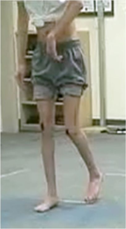
J. Clin. Med. 2019, 8, x FOR PEER REVIEW 3 of 12
J. Clin. Med. 2019, 8, x FOR PEER REVIEW 3 of 12
J. Clin. Med. 2019, 8, 679 3 of 11
(a) (b) (c)
Figure 1. Demonstration of three types of pelvic rotation which may be seen in a patient with left hemiplegia. (a)
Pelvic retraction: the average degree of external pelvic rotation on the left side (red line) is 15.64°, and the
consistently increased external rotation of the pelvis throughout the gait cycle is more than two standard deviations
from the norm. (b) Increased internal rotation of the pelvis compared to the contralateral right side (green line):
the average degree (a) of internal pelvic rotation on the left(b) side is 4.91° which is within normal(c) range. (c) Excessive
range of pelvic motion: the average degree of external pelvic rotation on the left side is 6.05°; however, increased
Figure
Figure 1. Demonstration
internal 1. Demonstration
rotation of three
at early stance of
types three
phase types of
of pelvic
followed bypelvic
rotation rotation
which
a sinusoidal which
may
type may
be seen
of pelvic be seen
in aare
rotation in anoted.
patient
patient withleft
with
Patients lefthemiplegia.
with an (a)
hemiplegia.
increased internal (a) Pelvic
rotation retraction:
of the pelvis the average degree
or an excessive of
range external pelvic
of pelviconmotionrotation
were on the
excluded left side
in this (red
study.line)
Pelvic retraction: the average degree of external pelvic rotation the left side (red line) is 15.64°, and the
is 15.64◦ , and the consistently increased external rotation of the pelvis throughout the gait cycle is more
consistently increased
than
externaldeviations
two standard
rotation of the pelvis throughout
from the norm.
the gaitrotation
cycle is more thancompared
two standard deviations
In total, 212 patients were enrolled in the (b)
studyIncreased
(Figureinternal
2). There wereof 84the pelvisfemales
(39.6%) andto 128
from the norm. (b)contralateral
the
(60.4%) Increased
males. internal
right
The average sideagerotation
(green
at theline):ofthe
time the
of pelvis
average
gait compared
degree
analysis eightto
of internal
was the and
pelvic
years contralateral
rotation on theright
eight months left side38(green line):
side
(range,
◦
the average months
degree ofwhich
is 4.91to internal
29 years). pelvic
is within rotation
One normal
hundred range.
and ontwenty-seven
the
(c) left side
Excessive is 4.91°
range which
of pelvic
patients is within
motion:
(59.9%) had normal
the average
right range.
degree
hemiplegia, of 85
and (c) Excessive
external pelvic rotation on the left side is 6.05 ◦ ; however, increased internal rotation at early stance
(40.1%) had left hemiplegia. The subjects were divided into two groups;
range of pelvic motion: the average degree of external pelvic rotation on the left side is 6.05°; however, increased Group I consisted of 113
phase(53.3%)
patients followed whoby ahadsinusoidal type of pelvic
pelvic retraction, androtation
Groupare noted. Patients
II comprised with an (46.7%)
99 patients increasedwho internal
did not
internal rotation at early stance phase followed by a sinusoidal type of pelvic rotation are noted. Patients with an
haverotation
pelvicof the pelvis or an excessive range of pelvic motion were excluded in this study.
retraction.
increased internal rotation of the pelvis or an excessive range of pelvic motion were excluded in this study.
In total, 212 patients were enrolled in the study (Figure 2). There were 84 (39.6%) females and 128
(60.4%) males. The average age at the time of gait analysis was eight years and eight months (range, 38
months to 29 years). One hundred and twenty-seven patients (59.9%) had right hemiplegia, and 85
(40.1%) had left hemiplegia. The subjects were divided into two groups; Group I consisted of 113
patients (53.3%) who had pelvic retraction, and Group II comprised 99 patients (46.7%) who did not
have pelvic retraction.
Figure2.2.Flow
Figure Flowdiagram
diagram of
of inclusion
inclusion and
and exclusion
exclusioncriteria
criteriaininthe
thestudy.
study.
2.2.
2.2.Clinical
Clinicaland
andGait
GaitParameters
Parameters
AAthorough
thoroughphysical
physicalexamination
examination waswas performed
performed in in all
all patients:
patients: modified
modifiedAshworth
Ashworthscale
scale[17],
[17],
passive range of motion in the hip, knee, and ankle joints; popliteal angle for hamstring
passive range of motion in the hip, knee, and ankle joints; popliteal angle for hamstring tightness, tightness,
Duncan-Elytest
Duncan-Ely testfor
forrectus
rectusfemoris
femoris spasticity
spasticity or
or tightness,
tightness, and
and ankle
ankle dorsiflexion
dorsiflexionangle
angleduring
duringthe the
Silfverskiöldtest
Silfverskiöld testfor
forthe
thedetection
detectionof ofgastrocnemius/Achilles
gastrocnemius/Achilles tendon
tendon tightness.
tightness.The
Thedegree
degreeofofankle
ankle
dorsiflexionwas
dorsiflexion wasmeasured
measuredwith withthe
theknee
kneeflexed
flexed and
and extended.
extended. Limb
Limb length
lengthdiscrepancy
discrepancywas
wasassessed
assessed
by measuring the distance from the anterior superior iliac spine to the medial
by measuring the distance from the anterior superior iliac spine to the medial malleolus malleolus in bothinsides
both
sides in all patients. A scanogram was also checked in 175 (82.5%) patients (93 patients in Group I
and 82 in Group II). Femoral anteversion was measured by the trochanteric palpation method [18]
and tibial torsion was measured using the bimalleolar axis in all patients [19]. One hundred and
seventy-two patients (81.1%) also underwent a computed tomographic (CT) scan to measure both
Figure 2.and
femoral anteversion Flow diagram
tibial torsion.of inclusion and exclusion criteria in the study.
2.2. Clinical and Gait Parameters
A thorough physical examination was performed in all patients: modified Ashworth scale [17],
passive range of motion in the hip, knee, and ankle joints; popliteal angle for hamstring tightness,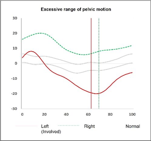
patients (81.1%) also underwent a computed tomographic (CT) scan to measure both femoral
anteversion and tibial torsion.
Gait analysis was performed using a VICON 370 Motion Analysis System (Oxford Metrics, Oxford,
England) with six infrared cameras and a Helen Hayes marker set. Data on ground–reaction forces
were gathered
J. Clin. Med.from
2019, 8, multiple
679 force platforms (Advanced Mechanical Technology, Watertown, 4 of 11 MA,
USA). All subjects were asked to walk barefoot at a self-selected speed along a 15-m walkway with
markers in place. We selected
Gait analysis values
was performed at athe
using VICON initial
370 contact, maximum,
Motion Analysis System minimum,
(Oxford Metrics,andOxford,
average values
England) with six infrared cameras and a Helen Hayes marker
for each kinematic parameter at each phase of gait. Several variables that have been set. Data on ground–reaction forces wereregarded as
gathered from multiple force platforms (Advanced Mechanical Technology,
clinically unimportant were excluded; furthermore, those variables have been known to be interrelated Watertown, MA, USA).
All subjects were asked to walk barefoot at a self-selected speed along a 15-m walkway with markers
with each other in a complex way, and may act as confounding factors in the interpretation of gait data
in place. We selected values at the initial contact, maximum, minimum, and average values for each
and in understanding
kinematic parameter abnormal
at each movement
phase of gait.[2,5,9,14,20–22].
Several variables that have been regarded as clinically
Two pediatric were
unimportant orthopaedic
excluded; surgeons
furthermore, reviewed all the
those variables videotaping
have been knownfor visual
to be gait analysis,
interrelated with three-
each other
dimensional gait in a complex
analysis way,dynamic
data, and may act as confoundingmeasurements
foot-pressure factors in the interpretation of gait data and
(pedobarographs), gross clinical
in understanding abnormal movement [2,5,9,14,20–22].
photos, and standing plain radiographs of the lower extremity in all patients. The presence of an
Two pediatric orthopaedic surgeons reviewed all the videotaping for visual gait analysis,
equinovarus foot deformity was defined as when inversion and plantar flexion of the hindfoot were
three-dimensional gait analysis data, dynamic foot-pressure measurements (pedobarographs), gross
present clinical
in standing
photos, position
and standing andplain
a significant
radiographspressure
of the lower exerted
extremity oninthe lateral The
all patients. forefoot and midfoot
presence
compared of an toequinovarus
the non-involved
foot deformity werewasconfirmed
defined as when on the dynamic
inversion pedobarographs
and plantar (Tekscan, South
flexion of the hindfoot
Boston, were
MA,present
USA). in standing position and a significant
The determination of kneepressure exerted on
recurvatum the lateral
gait and forefoot
the type and according
midfoot to the
compared to the non-involved were confirmed on the dynamic pedobarographs
classification by Winters et al. [23] were done using gait data and visual analysis. We referred (Tekscan, South Boston,
MA, USA). The determination of knee recurvatum gait and the type according to the classification
recurvatum gait as a clinical static variable as it is a descriptive type of qualitative classification. We
by Winters et al. [23] were done using gait data and visual analysis. We referred recurvatum gait as
modified the original
a clinical Winters
static variable as itclassification as follows;
is a descriptive type a patient
of qualitative with aWenormal
classification. range
modified of knee motion
the original
but having
Wintersincreased kneeasflexion
classification follows;at initial with
a patient contact andrange
a normal terminal
of kneestance
motion phase of gait
but having was categorized
increased
into type III (Figure 3). Consequently, patients with the original type III with increased 3).
knee flexion at initial contact and terminal stance phase of gait was categorized into type III (Figure knee flexion
Consequently, patients with the original type III with increased knee flexion
and type IV with increased hip flexion, hip internal rotation, and pelvic retraction were then re- and type IV with increased
hip flexion, hip internal rotation, and pelvic retraction were then re-classified as type IV and type V,
classified as type IV and type V, respectively. The presence of an asymmetrical posturing of the upper
respectively. The presence of an asymmetrical posturing of the upper extremity was defined as when
extremitythewas defined
patient has more as when
than 30the patient
degrees has more
of elbow flexionthan 30 degrees
contractures and of elbow
typical flexionbecomes
posturing contractures and
typical posturing
apparent when becomes apparent
the patient when 4)the
walks (Figure patient walks (Figure 4) [24,25].
[24,25].
Figure 3. The Winters classification (modified and reproduced) [23,26].
Figure 3. The Winters classification (modified and reproduced) [23,31].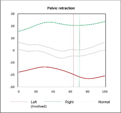
J. Clin. Med. 2019, 8, x FOR PEER REVIEW 5 of 12
J. Clin. Med. 2019, 8, 679 5 of 11
Figure 4. 4.
Figure AA hemiplegic
hemiplegicpatient
patientwith
withasymmetrical posturingof
asymmetrical posturing ofthe
theupper
upperextremity
extremity during
during walking.
walking.
2.3.2.3. Statistical
Statistical Analyses
Analyses
Statistical analyseswere
wereperformed
performed using
using IBM ® ®
Statistical analyses IBM®SPSS
SPSS® software
software version
version2323 (IBM
(IBMCorporation,
Corporation,
Armonk, NY, USA). The level of significance was set at p < 0.05. An independent
Armonk, NY, USA). The level of significance was set at p < 0.05. An independent t-test was t-test was used for for
used
initial comparison between the groups. Twenty-one gait parameters were selected
initial comparison between the groups. Twenty-one gait parameters were selected for the clustering for the clustering
technique analysis: range of pelvic motion in the sagittal plane, average anterior pelvic tilt, range
technique analysis: range of pelvic motion in the sagittal plane, average anterior pelvic tilt, range of
of pelvic motion in the coronal plane, average pelvic obliquity, minimum hip sagittal angle, average
pelvic motion in the coronal plane, average pelvic obliquity, minimum hip sagittal angle, average hip
hip sagittal angle, maximum hip coronal angle, average hip coronal angle, maximum hip transverse
sagittal angle, maximum hip coronal angle, average hip coronal angle, maximum hip transverse angle,
angle, average hip transverse angle, minimum knee sagittal angle, range of knee motion in sagittal
average hip transverse angle, minimum knee sagittal angle, range of knee motion in sagittal plane, knee
plane, knee sagittal angle at initial contact, average knee transverse angle, maximum ankle sagittal
sagittal angle at initial contact, average knee transverse angle, maximum ankle sagittal angle, minimum
angle, minimum ankle sagittal angle, average ankle sagittal angle, ankle sagittal angle at initial contact,
ankle sagittal
average angle,
ankle averageangle,
transverse ankle maximum
sagittal angle,
foot ankle sagittalangle,
progression angleandat initial
averagecontact, average ankle
foot progression
transverse angle,sixmaximum
angle. With foot progression
temporospatial parameters, angle,
a total and
of 27average foot progression
gait parameters angle.asWith
were clustered gait six
temporospatial
factors, with the consideration of the differences in patients’ ages and the correlation between the gait the
parameters, a total of 27 gait parameters were clustered as gait factors, with
consideration of the differences in patients’ ages and the correlation between the gait parameters
parameters [2,13,14,22,27].
[2,13,14,22,26].
Finally, eight gait factors (ankle dorsiflexion, temporospatial parameter (walking speed, stride
&Finally, eighttemporospatial
step length), gait factors (ankle dorsiflexion,
parameter (cadence,temporospatial parameter
stride & step time), internal(walking
rotationspeed,
of footstride
and &
stepankle,
length),
kneetemporospatial parameter
extension, pelvic obliquity (cadence, stride & step
and hip abduction, time),
anterior internal
pelvic rotation
tilt, hip internalofrotation)
foot andandankle,
knee extension,
five pelvic
clinical factors obliquity
(modified and hip
Winters abduction,Achilles
classification, anteriortendon
pelvictightness,
tilt, hip gastrocnemius
internal rotation) and five
tightness,
clinical factors (modified
pes equinovarus, Winters posturing
asymmetrical classification, Achilles
of the tendon tightness,
upper extremity) gastrocnemius
were included tightness, pes
for the multivariate
logistic regression
equinovarus, analysis.posturing of the upper extremity) were included for the multivariate
asymmetrical
logistic regression analysis.
3. Results
3. Results
3.1. Comparisons between the Groups (Group I vs Group II)
Patients (7.94
3.1. Comparisons ± 4.49
between the years)
Groupsin(Group
the group of pelvic
I vs Group II) retraction (Group I) were younger than those
(9.49 ± 5.06 years) in the group of normal range of pelvic rotation (Group II) (p = 0.0189). There were
noPatients (7.94
statistical ± 4.49 years)
differences between in the
thegroups
groupinofterms
pelvicof retraction
limb length(Group I) were
discrepancy, younger
femoral than those
anteversion,
(9.49
and± 5.06 years)
tibial in the
torsion. group of normal
Gastrocnemius and range
Achillesof pelvic
tendonrotation (Group
tightness were moreII) (p =frequent
0.0189). in
There
GroupwereII no
statistical
(Table 1). Pes equinovarus, asymmetrical posturing of the upper extremity, and severe types ofand
differences between the groups in terms of limb length discrepancy, femoral anteversion,
tibial torsion.Winters
modified Gastrocnemius andwere
classification Achilles tendon more
encountered tightness were more
frequently in Groupfrequent in Group
I. However, thereIIwas
(Table
no 1).
Pesdifference
equinovarus, asymmetrical posturing of the upper extremity, and severe types
in the existence of recurvatum gait between the groups. Modified Winters types IV and V of modified Winters
classification
were seen in were
smallencountered
numbers ofmore frequently
the total in Group affecting
study population, I. However, there was
36 (17.0%) and no difference
6 (2.8%) in the
patients,
existence of recurvatum gait between the groups. Modified Winters types IV and V were seen in small
respectively; and types II, IV, and V were more common in Group I, and type I was more common in
Group of
numbers II (Table 2). study population, affecting 36 (17.0%) and 6 (2.8%) patients, respectively; and
the total
types II, IV, and V were more common in Group I, and type I was more common in Group II (Table 2).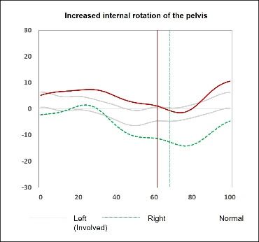
J. Clin. Med. 2019, 8, 679                                                                                                         6 of 11

                             Table 1. Comparisons of clinical parameters between the groups.

                                                                    Group I                       Group II                    *p
           Limb length discrepancy, mm                             6.8 ± 7.0                      6.7 ± 7.2                  0.881
    Limb length discrepancy (by scanogram), mm                   6.61 ± 4.67                     7.16 ± 6.30                 0.521
           Femoral anteversion, degrees                           28.8 ± 11.7                    26.6 ± 13.0                 0.182
      Femoral anteversion (by CT) (1) , degrees                 29.25 ± 11.88                  26.22 ± 12.43                 0.105
               Tibial torsion, degrees                           29.4 ± 12.1                      26.5 ± 9.4                 0.061
         Tibial torsion (by CT) (2) , degrees                   29.09 ± 10.54                  27.51 ± 10.00                 0.371
                Hip flexion, degrees                            120.78 ± 5.55                  120.43 ± 2.41                 0.565
           Hip external rotation, degrees                        45.37 ± 9.96                  48.12 ± 13.32                 0.159
           Hip internal rotation, degrees                        42.26 ± 9.79                  42.39 ± 12.88                 0.943
              Popliteal angle, degrees                          21.49 ± 13.85                  21.55 ± 12.33                 0.977
          Rectus femoris tightness, grade                   0(32), 1(24), 2(3), 3(0)       0(43), 1(16), 2(2), 3(1)          0.153
         Gastrocnemius tightness, degrees                       83.87 ± 17.15                   90.64 ± 8.34
J. Clin. Med. 2019, 8, 679 7 of 11
Table 4. Cont.
Group I Group II *p
Maximum knee flexion 41.55 ± 10.00 45.78 ± 9.20 0.002 *
Maximum ankle dorsiflexion 3.39 ± 13.48 10.46 ± 7.64J. Clin. Med. 2019, 8, 679 8 of 11
Table 6. Multivariate logistic regression analysis with gait and clinical factors.
95% Wald
Estimate Standard Error Pr > k2
Confidence Limits
Factor 1 (Ankle dorsiflexion) 0.382 0.350 0.192 0.757 0.006 *
Factor 2 (Walking speed, stride & step length) 0.689 0.236 0.434 1.094 0.114
Factor 3 (Cadence, stride & step time) 1.013 0.219 0.660 1.555 0.953
Factor 4 (Internal rotation of foot and ankle) 0.935 0.236 0.589 1.484 0.776
Factor 5 (Knee extension) 1.411 0.332 0.736 2.706 0.299
Factor 6 (Pelvic obliquity and hip abduction) 1.037 0.257 0.627 1.715 0.889
Factor 7 (Anterior pelvic tilt) 2.305 0.263 1.376 3.861 0.002 *
Factor 8 (Hip internal rotation) 1.540 0.217 1.006 2.358 0.047 *
5 vs 1 0.015 1.590J. Clin. Med. 2019, 8, 679 9 of 11
Decreased ankle dorsiflexion found in patients with Group I was due to tightness of the
gastrocnemius and Achilles tendon, and this was confirmed by comparing the physical examination
findings between the groups. Normal forward progression of the tibia over the supporting foot during
the stance phase of gait is prevented by tight calf muscles [2,5,7,30]. Pelvic retraction might occur
as a consequence of insufficient forward progression of the body during the stance phase, and the
lengthening of shortened ankle plantarflexors would improve pelvic retraction [6]. However, we could
not appreciate any other effects of tight gastrocnemius and Achilles tendon, as these clinical features
were not statistically significant in the multivariate logistic regression analysis.
On the contrary to the previous observations [2], our results showed that there is no relationship
between pelvic retraction and tightness/spasticity of the rectus femoris. Despite no difference in the
rectus tightness, patients in Group I had lower degrees of hip extension and had decreased maximum
knee flexion during gait compared to Group II. The rectus femoris tightness has been thought to be
secondary to longstanding inappropriate hip extension, and this diminished range of hip extension is
related to ankle equinus [2]. Decreased ankle dorsiflexion may cause reduced hip extension and may
also elicit decreased knee flexion. On the other hand, reduced hip and knee motions in the sagittal
plane have been known to be associated with an excessive range of pelvic motion [1]. It is our opinion
that there may be differences in terms of the degree of spasticity of the rectus femoris or the amount of
reduced range of motion, compared to the patients with excessive pelvic motions.
Some previous studies introduced modified Winters classifications [26,32] in order to allocate
subsets of patients that cannot be classified with the original system, and to make up the limitations in
classifying types I, II, and III which were determined only by abnormalities in the sagittal plane. We also
adapted a modified Winters classification in the study because 20% of our patients had increased knee
flexion at initial and terminal stance phasse of gait, as well as having a normal range of knee motion
in the sagittal plane; they were categorized into modified type III, and patients with original type III
with increased knee flexion were classified as modified type IV. The characteristic finding of modified
types III and IV was knee flexion compared to type II which has only ankle equinus. In our logistic
regression analysis, allocation into type II was found to be related to pelvic retraction. Increased
knee flexion is the characteristic finding in modified types III and IV; this increased knee flexion
may act as a compensation for forward progression of the tibia. We think that pelvic retraction may
decrease with forward progression of the knee. Patients with modified type V in our series showed
less pelvic retraction than patients with type I. However, as only six patients (2.8%) were modified
type V compared to 47 patients with type I (22.2%), it may be difficult to conclude that patients with
modified type V have less risk of pelvic retraction.
No previous studies mentioned an association between asymmetrical posturing of the upper
extremity during walking and pelvic retraction. Asymmetrical posturing was more frequent in the
group with pelvic retraction, and was also found to be a related factor to pelvic retraction in the logistic
regression analysis. Nevertheless, it is not certain that any surgeries on the upper extremity may
improve pelvic retraction as the asymmetrical posturing itself is related not only to the trunk balance
during gait but also to the severity of a primary neurologic impairment per se [33].
Our study has several limitations: we selected only five clinical factors, however those clinical
factors have traditionally been frequent in patients with hemiplegia and have also been considered
as significant factors related to the abnormal gait; scanogram and CT scan were not performed in
about 20% of our series as they refused radiological exposure. Nonetheless, the trochanteric palpation
method is a useful and reliable method to measure femoral anteversion in patients with cerebral
palsy [18], and the use of the bimalleolar axis for tibial torsion shows a high correlation with CT
measurement [19]. Furthermore, there were no significant differences in both physical examination
and radiographic evaluation between the groups; and as the clinically perceived pelvic retraction
may be a movement that may result from complex movements occurring in the sagittal, coronal,
or transverse plane, the clinician should be aware that there may be differences between the real gait
and three-dimensional gait analysis.J. Clin. Med. 2019, 8, 679 10 of 11
5. Conclusions
To the best of our knowledge, this is the only study including a broader array of assessment
domains of both clinical and gait parameters with a considerably large and homogenous population
with hemiplegia. We conclude that pelvic retraction is more likely in patients with decreased ankle
dorsiflexion, increased hip internal rotation, and increased anterior pelvic tilt. However, clinical
features such as Winters classification type II and asymmetrical posturing are also frequently seen
in patients with pelvic retraction. Further studies will be needed to see if the rectification of those
parameters may improve abnormal gait and pelvic retraction in spastic hemiplegia.
Author Contributions: Conceptualization, K.-B.P. and H.W.K.; Data curation, H.W.K.; Formal analysis, H.P.;
Funding acquisition, H.W.K.; Investigation, B.K.P. and S.W.A.-B.; Methodology, H.P. and B.K.P.; Project
administration, K.-B.P.; Resources, H.W.K.; Software, K.-B.P.; Supervision, H.W.K.; Validation, H.P. and S.W.A.-B.;
Visualization, H.W.K.; Writing–original draft, K.-B.P.; Writing–review & editing, K.-B.P. and H.W.K.
Funding: This research was funded by a faculty research grant of Yonsei University College of Medicine,
grant number 6-2010-0195. The APC was funded by Yonsei University College of Medicine.
Acknowledgments: The authors wish to thank Yoon Hae Kwak for her great help with initial data acquisition
and Hye-Jin Chi for her help with the statistical analysis.
Conflicts of Interest: The authors declare no conflict of interest. The funders had no role in the design of the
study; in the collection, analyses, or interpretation of data; in the writing of the manuscript, or in the decision to
publish the results.
References
1. Gage, J.R.; Schwartz, M.H. Normal gait. In The Identification and Treatment of Gait Problems in Cerebral Palsy,
2nd ed.; Gage, J.R., Schwartz, M.H., Koop, S.E., Novacheck, T.F., Eds.; Mac Keith: London, UK, 2009;
pp. 31–64.
2. O’Sullivan, R.; Walsh, M.; Jenkinson, A.; O’Brien, T. Factors associated with pelvic retraction during gait in
cerebral palsy. Gait Posture 2007, 25, 425–431. [CrossRef] [PubMed]
3. DeLuca, P.A.; Davis, R.B., 3rd; Ounpuu, S.; Rose, S.; Sirkin, R. Alterations in surgical decision making in
patients with cerebral palsy based on three-dimensional gait analysis. J. Pediatr. Orthop. 1997, 17, 608–614.
[CrossRef]
4. Aktas, S.; Aiona, M.D.; Orendurff, M. Evaluation of rotational gait abnormality in the patients cerebral palsy.
J. Pediatr. Orthop. 2000, 20, 217–220. [CrossRef] [PubMed]
5. De Morais Filho, M.C.; Kawamura, C.M.; Andrade, P.H.; Dos Santos, M.B.; Pickel, M.R.; Neto, R.B. Factors
associated with pelvic asymmetry in transverse plane during gait in patients with cerebral palsy. J. Pediatr.
Orthop. B 2009, 18, 320–324. [CrossRef]
6. Park, C.I.; Park, E.S.; Kim, H.W.; Rha, D.W. Soft tissue surgery for equinus deformity in spastic hemiplegic
cerebral palsy: Effects on kinematic and kinetic parameters. Yonsei Med. J. 2006, 47, 657–666. [CrossRef]
[PubMed]
7. Carty, C.P.; Walsh, H.P.; Gillett, J.G.; Phillips, T.; Edwards, J.M.; deLacy, M.; Boyd, R.N. The effect of
femoral derotation osteotomy on transverse plane hip and pelvic kinematics in children with cerebral palsy:
A systematic review and meta-analysis. Gait Posture 2014, 40, 333–340. [CrossRef]
8. Aminian, A.; Vankoski, S.J.; Dias, L.; Novak, R.A. Spastic hemiplegic cerebral palsy and the femoral derotation
osteotomy: Effect at the pelvis and hip in the transverse plane during gait. J. Pediatr. Orthop. 2003, 23, 314–320.
[CrossRef]
9. Saraph, V.; Zwick, E.B.; Zwick, G.; Dreier, M.; Steinwender, G.; Linhart, W. Effect of derotation osteotomy
of the femur on hip and pelvis rotations in hemiplegic and diplegic children. J. Pediatr. Orthop. B 2002,
11, 159–166. [PubMed]
10. Park, J.; Seeley, M.K.; Francom, D.; Reese, C.S.; Hopkins, J.T. Functional vs. traditional analysis in
biomechanical gait data: An alternative statistical approach. J. Hum. Kinet. 2017, 60, 39–49. [CrossRef]
11. Miller, F. Cerebral Palsy, 1st ed.; Springer: New York, NY, USA, 2005; pp. 335–340.
12. Cimolin, V.; Galli, M.; Tenore, N.; Albertini, G.; Crivellini, M. Gait strategy of uninvolved limb in children
with spastic hemiplegia. Eura Medicophys. 2007, 43, 303–310. [PubMed]J. Clin. Med. 2019, 8, 679 11 of 11
13. O’Malley, M.J.; Abel, M.F.; Damiano, D.L.; Vaughan, C.L. Fuzzy clustering of children with cerebral palsy
based on temporal-distance gait parameters. IEEE Trans. Rehabil. Eng. 1997, 5, 300–309. [CrossRef] [PubMed]
14. Carriero, A.; Zavatsky, A.; Stebbins, J.; Theologis, T.; Shefelbine, S.J. Determination of gait patterns in children
with spastic diplegic cerebral palsy using principal components. Gait Posture 2009, 29, 71–75. [CrossRef]
[PubMed]
15. Salazar-Torres, J.J.; McDowell, B.C.; Kerr, C.; Cosgrove, A.P. Pelvic kinematics and their relationship to gait
type in hemiplegic cerebral palsy. Gait Posture 2011, 33, 620–624. [CrossRef]
16. Niklasch, M.; Doderlein, L.; Klotz, M.C.; Braatz, F.; Wolf, S.I.; Dreher, T. Asymmetric pelvic and hip rotation
in children with bilateral cerebral palsy: Uni- or bilateral femoral derotation osteotomy? Gait Posture 2015,
41, 670–675. [CrossRef]
17. Ansari, N.N.; Naghdi, S.; Arab, T.K.; Jalaie, S. The interrater and intrarater reliability of the Modified
Ashworth Scale in the assessment of muscle spasticity: Limb and muscle group effect. NeuroRehabilitation
2008, 23, 231–237.
18. Davids, J.R.; Benfanti, P.; Blackhurst, D.W.; Allen, B.L. Assessment of femoral anteversion in children with
cerebral palsy: Accuracy of the trochanteric prominence angle test. J. Pediar. Orthop. 2002, 22, 173–178.
[CrossRef]
19. Sangeux, M.; Mahy, J.; Graham, H.K. Do physical examination and CT-scan measures of femoral neck.
A anteversion and tibial torsion relate to each other? Gait Posture 2014, 39, 12–16. [CrossRef]
20. Ounpuu, S.; DeLuca, P.; Davis, R.; Romness, M. Long-term effects of femoral derotation osteotomies:
An evaluation using three-dimensional gait analysis. J. Pediatr. Orthop. 2002, 22, 139–145. [CrossRef]
21. Graham, H.K.; Baker, R.; Dobson, F.; Morris, M.E. Multilevel orthopaedic surgery in group IV spastic
hemiplegia. J. Bone Joint Surg. Br. 2005, 87, 548–555. [CrossRef]
22. Carriero, A.; Zavatsky, A.; Stebbins, J.; Theologis, T.; Shefelbine, S.J. Correlation between lower limb bone
morphology and gait characteristics in children with spastic diplegic cerebral palsy. J. Pediatr. Orthop. 2009,
29, 73–79. [CrossRef] [PubMed]
23. Winters, T.F., Jr.; Gage, J.R.; Hicks, R. Gait patterns in spastic hemiplegia in children and young adults. J. Bone
Joint Surg. Am. 1987, 69, 437–441.
24. Graham, H.K.; Thomason, P.; Novacheck, T.F. Cerebral Palsy. In Lovell & Winter’s Pediatric Orthopaedics, 7th ed.;
Weinstein, S.L., Flynn, J.M., Eds.; Wolters Kluwer: Philadelphia, PA, USA, 2014; Volume 1, pp. 488–489.
25. Carlson, M.G.; Hearns, K.A.; Inkellis, E.; Leach, M.E. Early results of surgical intervention for elbow deformity
in cerebral palsy based on degree of contracture. J. Hand Surg. Am. 2012, 37, 1665–1671. [CrossRef]
26. Rodda, J.; Graham, H.K. Classification of gait patterns in spastic hemiplegia and spastic diplegia: A basis for
a management algorithm. Eur. J. Neurol. 2001, 8 (Suppl. 5), 98–108. [CrossRef]
27. Kay, R.M.; Rethlefsen, S.; Reed, M.; Do, K.P.; Skaggs, D.L.; Wren, T.A. Changes in pelvic rotation after soft
tissue and bony surgery in ambulatory children with cerebral palsy. J. Pediatr. Orthop. 2004, 24, 278–282.
[CrossRef]
28. Jiang, N.; Peng, L.; Al-Qwbani, M.; Xie, G.P.; Yang, Q.M.; Chai, Y.; Zhang, Q.; Yu, B. Femoral version,
neck-shaft angle, and acetabular anteversion in Chinese Han population: A retrospective analysis of
466 healthy adults. Medicine 2015, 94, e891. [CrossRef] [PubMed]
29. Kristiansen, L.P.; Gunderson, R.B.; Steen, H.; Reikeras, O. The normal development of tibial torsion.
Skeletal Radiol. 2001, 30, 519–522. [CrossRef]
30. Brunner, R.; Dreher, T.; Romkes, J.; Frigo, C. Effects of plantarflexion on pelvis and lower limb kinematics.
Gait Posture 2008, 28, 150–156. [CrossRef]
31. Rutz, E.; Passmore, E.; Baker, R.; Graham, H.K. Multilevel surgery improves gait in spastic hemiplegia but
does not resolve hip dysplasia. Clin. Orthop. Relat. Res. 2012, 470, 1294–1302. [CrossRef]
32. Riad, J.; Haglund-Akerlind, Y.; Miller, F. Classification of spastic hemiplegic cerebral palsy in children.
J. Pediatr. Orthop. 2007, 27, 758–764. [CrossRef]
33. Riad, J.; Coleman, S.; Miller, F. Arm posturing during walking in children with spastic hemiplegic cerebral
palsy. J. Pediatr. Orthop. 2007, 27, 137–141. [CrossRef]
© 2019 by the authors. Licensee MDPI, Basel, Switzerland. This article is an open access
article distributed under the terms and conditions of the Creative Commons Attribution
(CC BY) license (http://creativecommons.org/licenses/by/4.0/).You can also read