Clinical and Pathological Data of 17 Non-Epithelial Pancreatic Tumors in Cats - MDPI
←
→
Page content transcription
If your browser does not render page correctly, please read the page content below
veterinary
           sciences
Article
Clinical and Pathological Data of 17 Non-Epithelial
Pancreatic Tumors in Cats
Katrin Törner 1, *, Marlies Staudacher 2 , Katja Steiger 3     and Heike Aupperle-Lellbach 1
 1    LABOKLIN GmbH & Co. KG, 97688 Bad Kissingen, Germany; aupperle@laboklin.de
 2    Tierärztliche Klinik Dr. Staudacher, 52078 Aachen, Germany; m.staudacher@tgz-aachen.de
 3    Institute of Pathology, Technische Universität München, 81675 Munich, Germany; katja.steiger@tum.de
 *    Correspondence: toerner@laboklin.de

 Received: 26 March 2020; Accepted: 21 April 2020; Published: 27 April 2020

 Abstract: Tumors of mesenchymal origin are rarely reported in the pancreas. Therefore, this study
 characterized 17 feline non-epithelial pancreatic tumors, including clinical data, histopathology,
 and immunohistochemistry. Seventeen feline pancreatic tissue samples were investigated
 histopathologically and immunohistochemically. Selected pancreatic and inflammatory serum
 parameters, e.g., feline pancreatic lipase immunoreactivity (fPLI), 1,2-o-dilauryl-rac-glycero-3-glutaric
 acid-(60 -methylresorufin) ester (DGGR) lipase and serum amyloid A (SAA), were recorded,
 when available. The neoplasms were characterized as round (n = 13) or spindle (n = 4) cell
 tumors. Round cell tumors included 12 lymphomas and one mast cell tumor in ectopic splenic tissue
 within the pancreas. Lymphomas were of T-cell (n = 9) or B-cell (n = 3) origin. These cats showed
 leukocytosis (3/3) and increased fPLI (5/5), DGGR lipase (3/5) and SAA (4/5) values. Spindle cell tumors
 included two hemangiosarcomas, one pleomorphic sarcoma and one fibrosarcoma. The cat with
 pleomorphic sarcoma showed increased SAA value. Overall survival time was two weeks to seven
 months. These are the first descriptions of a pancreatic pleomorphic sarcoma and a mast cell tumor in
 accessory spleens within feline pancreas. Although rare, pancreatic tumors should be considered in
 cats presenting with clinical signs and clinical pathology changes of pancreatitis. Only histopathology
 can certainly distinguish solitary pancreatitis from a neoplasm with inflammation.

 Keywords: fPLI; DGGR lipase; histopathology; lymphoma; mast cell tumor; sarcoma

1. Introduction
      Pancreatic neoplasms are rare in cats, dogs [1–3], and humans [4]. Carcinoma is the most common
tumor type in cats and dogs, but adenomas, sarcomas, lymphomas, unclassified tumors, and metastatic
neoplasms have also been described [1–3,5]. Pancreatic neoplasms generally cause non-specific
constitutional and gastrointestinal signs such as lethargy, anorexia, weight loss, diarrhea, vomiting,
abdominal pain, or palpable abdominal masses [6–8].
      Lymphomas are common neoplasms in most domestic species. Gastrointestinal lymphoma is the
most common form of lymphoma in cats and is associated with clinical presentation such as weight
loss, vomiting, diarrhea anorexia, and lethargy [9]. However, pancreatic involvement has been reported
in only some cases and pancreas-specific blood parameters were not included in the studies [10–13].
While retroviral diseases (feline leukemia virus (FeLV) and feline immunodeficiency virus) are risk
factors, a high number of cats with alimentary lymphomas have been reported to be virus negative [14].
      As a mutant of FeLV, the feline sarcomavirus can cause sarcomas in cats [15], but literature
about feline pancreatic sarcomas is scant. There is one case report of a feline primary pancreatic
carcinosarcoma with metastases in uterus, omentum, and diaphragm [16]. Mesenchymal pancreatic
tumors in humans are reported to have a poor prognosis with a median overall survival time of

Vet. Sci. 2020, 7, 55; doi:10.3390/vetsci7020055                                  www.mdpi.com/journal/vetsci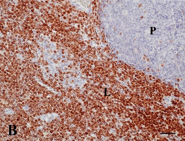
Vet. Sci. 2020, 7, 55                                                                                2 of 13

21 months [17,18]. Visceral hemangiosarcomas are a common neoplasm in dogs, especially German
Shepherds, but are uncommon in cats [19]. Hemangiosarcomas in dogs often occur in the spleen
and the right atrium [19]. In cats, they arise mostly in liver, intestine, lymph nodes, or spleen [20].
The prognosis is poor in both dogs and cats [19–21].
      The aim of this study is to characterize 17 cases of feline non-epithelial pancreatic tumors including
clinical data, histopathology, and immunohistochemistry.

2. Materials and Methods
      During the 2011–2018 period, 571 feline pancreatic tissue samples, submitted routinely to
LABOKLIN GmbH & Co. KG (Bad Kissingen, Germany), were investigated. From these, 540 showed
representative sample size and adequate tissue preservation. Included into this study were
mesenchymal tumors within the pancreas, like spindle and round cell tumors from 17 cats. Excluded
were samples without (n = 31), other tumors such as primary exocrine neoplasms (21/540 adenomas,
54/540 carcinomas) or endocrine tumors (2/540), as well as tumor-like lesions like nodular hyperplasia.
      The age of the cats was one to 17 years (median 12). Ten cats were male (1 entire, 9 neutered) and
seven female (3 entire, 4 spayed). The breeds were Domestic Shorthair (DSH, n = 12), British Longhair
(n = 1), British Shorthair (n = 1), Carthusian (n = 1), Siamese (n = 1), and Sacred Birman (n = 1).
      Complete pancreas (n = 4), fully excised tumor masses (n = 4) and excisional biopsies (n = 9) of
the pancreas were examined. The samples were measured and inspected macroscopically in detail
with respect to size, cut surface, and color. This study refers to size of the complete neoplasm unless
specified otherwise. Representative sites were prepared for routine histopathological examination
and embedded in paraffin according to standard procedures. Slides were routinely stained with
hematoxylin-eosin (HE) and Giemsa. Tumors were evaluated according to the world health organization
(WHO) classification of mesenchymal [22–24] and hematopoietic tumors [25]. Mitotic figures were
counted within 10 high power fields (HPF) by 400 x microscope objective (visual field: 68,700 µm2 ) in
areas of the highest mitotic activity and were reported as mean value/HPF. Lymphomas were graded
as low grade: 10 mitotic figures/HPF [26]. Pancreatitis was classified according to De Cock et al. [27].
      The immunohistochemical panel of primary antibodies used and their dilution are listed in Table 1.
The two antibodies for FeLV (gp70 and p27) were diluted appropriately and consequently mixed at a
ratio of 1:1 for further procedure. Pre-treatment included peroxidase blocking with 0.66% hydrogen
peroxidase for all markers. Afterwards, the slides were heated either in 1:10 diluted HIER T-EDTA buffer
pH 9.0 (10×) (Zytomed Systems GmbH, Berlin, Germany, 2VCO29-500) in a steam cooker (vimentin,
desmin, CD31, CD3, CD79a, CD117/c-kit, FeLV), or in 1:10 diluted target retrieval solution pH 6.0
(10×) (Dako, Glostrup, Denmark, S1699) in a pressure cooker (smooth muscle actin, von Willebrand
factor (vWF)). Dako EnVision+ System-HPR for mouse or rabbit primary antibodies (Dako, Glostrup,
Denmark, K4006 or K4010) with 3,3‘-diaminobenzine (DAB) was used for immunohistochemical
staining. Counterstaining was done with Meyer’s hematoxylin (Bio-Optica Milano S.p.A., Milano,
Italy, BIO 05-06002/CO). Slides of the same organs prepared with normal mouse IgG (Dako, Glostrup,
Denmark, X0931) or rabbit IgG (Dako, Glostrup, Denmark, X0936), with the same dilution as the
specific reagents, were used as negative control. Positive controls varied for the different antibodies:
tissue with fibrocytes and fibroblasts for vimentin, intestine for smooth muscle actin, muscle tissue for
desmin, vessel rich tissue for vWF and CD31, lymph nodes for CD3 and CD79a, skin tissue with mast
cells for CD117/c-kit, and PCR positive spleen tissue for FeLV.
      Clinicopathologic data were available for six cats (five lymphomas and one sarcoma). The
serum samples in these cases were taken shortly before or during surgery. Alpha-amylase (reference
interval: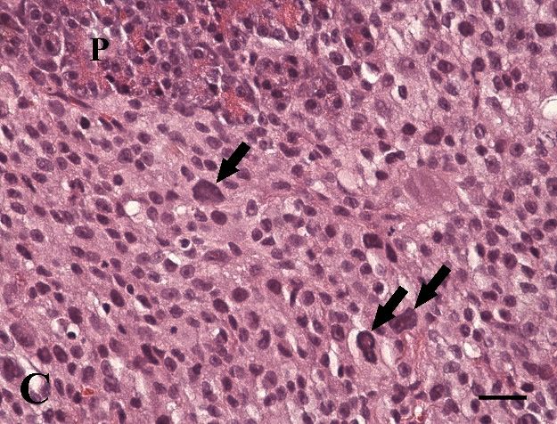
Vet. Sci. 2020, 7, 55                                                                                                                  3 of 13

µg/L, indicative for pancreatitis: >5.4 µg/L) and feline trypsin-like immunoreactivity (fTLI, reference
interval: 12–82 µg/L, indicative for pancreatitis >100 µg/L) values were determined by an ELISA assay
at LABOKLIN GmbH & Co. KG.

      Table 1. Primary antibodies and pretreatment used in immunohistochemistry of 17 feline non-epithelial
      pancreatic tumors.
           Marker               Type of Antibody                       Source             Dilution           Pretreatment
                                 Monoclonal mouse
                                                              Dako, Glostrup, Denmark,                   Peroxidase blocking;
           Vimentin                 anti-vimentin                                          1:1000
                                                                       M0725                          steam cook in EDTA buffer
                                      (Clone V9)
                                 Monoclonal mouse
        Smooth muscle                                         Dako, Glostrup, Denmark,                   Peroxidase blocking;
                                     anti-human                                            1:100
            actin                                                      M0851                         pressure cook in citrate buffer
                                     (Clone 1A4)
                                 Monoclonal mouse
                                                              Dako, Glostrup, Denmark,                   Peroxidase blocking;
           Desmin                    anti-human                                            1:100
                                                                       M0760                          steam cook in EDTA buffer
                                     (Clone D33)
        Von Willebrand            Polyclonal rabbit           Dako, Glostrup, Denmark,                   Peroxidase blocking;
                                                                                           1:2000
            factor                   anti-human                        A0082                         pressure cook in citrate buffer
                                 Monoclonal mouse
                                                              Dako, Glostrup, Denmark,                   Peroxidase blocking;
            CD31                     anti-human                                            1:100
                                                                       M0823                          steam cook in EDTA buffer
                                   (Clone JC70A)
                                 Monoclonal mouse
                                                              Dako, Glostrup, Denmark,                   Peroxidase blocking;
             CD3                     anti-human                                            1:100
                                                                       M7254                          steam cook in EDTA buffer
                                   (Clone F7.2.38)
                                 Monoclonal mouse             Bio-Rad Laboratories Inc,
                                                                                                         Peroxidase blocking;
            CD79a                    anti-human                  Munich, Germany,          1:3000
                                                                                                      steam cook in EDTA buffer
                                   (Clone HM57)                     MCA2538GA
                                  Polyclonal rabbit           Dako, Glostrup, Denmark,                   Peroxidase blocking;
         CD117/c-kit                                                                       1:150
                                     anti-human                        A4502                          steam cook in EDTA buffer
                                 Monoclonal mouse
       Feline Leukemia                                        Bio-Rad Laboratories Inc,                  Peroxidase blocking;
                         anti-feline leukemia virus (Clone                                 1:200
          Virus gp70                                         Munich, Germany, MCA1897                 steam cook in EDTA buffer
                                        C11D8)
                                 Monoclonal mouse
       Feline Leukemia                                        Bio-Rad Laboratories Inc,                  Peroxidase blocking;
                             anti-feline leukemia virus                                    1:100
           Virus p27                                         Munich, Germany, MCA2551                 steam cook in EDTA buffer
                                 (Clone PF12J-10A)

3. Results
     In pancreatic tissue from 17 cats, round (n = 13) and spindle (n = 4) cell tumors were diagnosed
histopathologically (Table 2). Round cell tumors included 12 lymphomas and one mast cell tumor.
Spindle cell tumors were classified as two hemangiosarcomas, one pleomorphic sarcoma, and one
fibrosarcoma. Clinical pathology was available for five cats with lymphoma and one cat with
pleomorphic sarcoma (Table 3).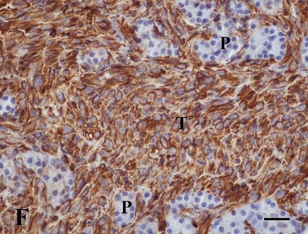
Vet. Sci. 2020, 7, 55 4 of 13
Table 2. Signalment, histopathological/immunohistochemical data and clinical outcome of cats with non-epithelial tumors (n = 17).
Staining, IHC
Age Mi/ Further Infiltrated Survival
Case No. Sex Breed Clinical Signs Pancreatic Sample Size (cm) Diagnosis Expression
(years) HPF Organs Time
+ −
1 1 MN DSH Inappetence, ascites TS 0.8 × 0.3 × 0.3 Large BCL 13 Kidney, liver, LN CD79a CD3, FeLV 2 months
2 12 F SB Inappetence CM 3.0 × 3.0 × 3.0 Large BCL 5 Om CD79a CD3, FeLV U
Vomiting, inappetence,
3* 10 MN BSH CM 4.0 × 4.0 × 4.0 Large BCL 9 Stomach, mAT CD79a CD3, FeLV Eutha
palpable abd. mass
CD79a,
4 13 MN Si Vomiting, diarrhea TS 1.0 × 0.4 × 0.3 Small TCL 0 Intestine CD3 7 months
FeLV
Lethargy, Intermediate
5* 15 MN DSH TS 1.5 × 1.5 × 1.5 0 Intestine, mLN CD3, FeLV CD79a Eutha
painful abd. TCL
Intermediate
6 11 F DSH Diarrhea TS 0.3 × 0.3 × 0.3 0 - CD3 CD79a Eutha
TCL
Intermediate CD79a,
7* 16 MN DSH Lethargy, ascites CP 8.5 × 7.0 × 2.6 3 Intestine, pAT, mLN CD3 Eutha
TCL FeLV
Intermediate
8 10 MN DSH U CP 9.0 × 6.0 × 3.0 4 Liver, intestine, mLN CD3 CD79a Eutha
TCL
CP 7.0 × 6.5 × 0.5 with two CD79a,
9* 15 MN DSH Lethargy, seizures Large TCL 1 Intestine CD3 Eutha
masses 0.5 × 0.5 × 0.5 FeLV
Two TS CD79a,
10 11 M Cart U Large TCL 3 Om, spleen CD3 Eutha
1.0 × 0.5 × 0.1 and 1.1 × 0.5 × 0.4 FeLV
Two TS
11 8 FS DSH U Large TCL 3 Intestine, mAT CD3, FeLV CD79a U
3.4 × 3.0 × 3.0 and 5.0 × 3.4 × 2.4
Vomiting, CD79a,
12 * 10 MN DSH TS 0.4 × 0.4 × 0.4 Large TCL 4 - CD3 U
painful abd. FeLV
CM 0.3 × 0.3 × 0.3 and 1.0 × 0.6
13 16 F BLH Palpable abd. mass Mast cell tumor 1 Spleen Giemsa c-kit, FeLV 2 months
× 0.5
Vim, vWF,
14 13 FS DSH Palpable abd. mass CM 2.0 × 1.3 × 1.0 Hemangio-sarcoma 3 - Eutha
CD31
Vim, CD31,
15 12 FS DSH U TS 5.5 × 2.0 × 0.4 Hemangio-sarcoma 1 Om, intestine, LN Eutha
vWF
CP 17.0 × 9.0 × 5.0 with PS with Om, adherent on
desmin,
16 * 17 F DSH Palpable abd. mass, lethargy multiple masses up to myofibro-blastic 9 capsule of spleen, Vim, actin Eutha
c-kit
2.0 × 2.0 × 2.0 parts intestine, liver
Two TS
17 13 MN DSH Inappetence, ascites Fibrosarcoma 2 Spleen Vim 2 weeks
1.2 × 0.8 × 0.6 and 3.0 × 3.0 × 2.0
− = no/negative, + = positive, * = clinical pathology is available, abd. = abdominal/abdomen, BCL = B-cell lymphoma, BLH = British Longhair, BSH = British Shorthair, Cart = Carthusian,
CM = complete mass, CP = complete pancreas, DSH = Domestic Short Hair, Eutha = euthanasia after surgery, F = female, FeLV = feline leukemia virus, F = female spayed,
HPF = high power field, IHC = immunohistochemistry, LN = lymph node, M = male, Mi = mitoses, mAT = mesenteric adipose tissue, mLN = mesenteric lymph node, MN = male
neutered, Om = omentum, pAT = peripancreatic adipose tissue, PS = pleomorphic sarcoma, SB = Sacred Birman, Si = Siamese, TCL = T-cell lymphoma, TS = tissue sample, U = unknown,
Vim = vimentin, vWF = von Willebrand factor.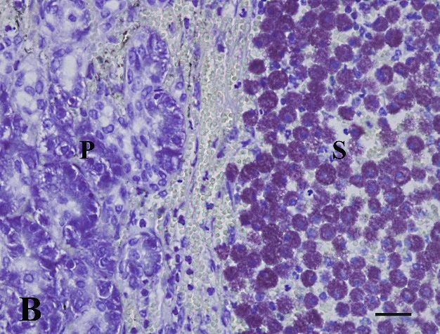
Vet. Sci. 2020, 7, 55                                                                                                       5 of 13

                 Table 3. Clinical pathology data of cats with non-epithelial pancreatic tumors (n = 6).

                                    fPLI      fTLI       DGGR          Alpha          SAA       Leuko       Neutro     Lymph
      Case
                    Diagnosis      (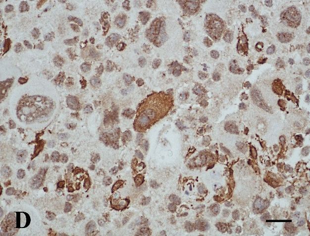
Vet. Sci. 2020, 7, x 6 of 13
Vet. Sci. 2020, 7, 55 6 of 13
Figure 1b) or B-cell (n = 3) lymphoma. In two cats with T-cell lymphoma (Nos. 5, 11), FeLV was
detected. The remaining ten cats were tested negative.
Seven
Seven cats
cats were
were euthanized
euthanized during
during surgery.
surgery. AA one-year-old
one-year-old Domestic
Domestic Shorthair
Shorthair (No. 1) had
(No. 1) had aa
survival time of two months and a 13-year-old Siamese (No. 4) survived seven months. Treatment
survival time of two months and a 13-year-old Siamese (No. 4) survived seven months. Treatment
data
data for
for these
these cases
cases were
were not
not available.
available.
Figure 1.
Figure 1. (a)
(A)T-cell
T-celllymphoma.
lymphoma.The
Thepancreas
pancreas(P)(P)
close to the
close small
to the intestine
small (I) is(I)enlarged,
intestine white
is enlarged, and
white
firm. (16-year-old, male neutered Domestic Shorthair, No. 7, formalin fixed tissue);
and firm. (16-year-old, male neutered Domestic Shorthair, No. 7, formalin fixed tissue); (B) T-cell (b) T-cell
lymphoma with
lymphoma withintense
intenseCD3-expression
CD3-expressionofofneoplastic
neoplastic lymphocytes
lymphocytes (L)(L) in the
in the pancreas
pancreas (P).(P). (15-year-
(15-year-old,
old, male neutered Domestic Shorthair, No. 9, IHC, CD3. Bar,
male neutered Domestic Shorthair, No. 9, IHC, CD3. Bar, 50 µm). 50 µm).
Tumor, nn =
3.1.2. Mast Cell Tumor, =1
A 16-year-old female British Longhair cat (No. 13) was presented to the clinician for routine
(No. 13)
vaccination, and during palpation
palpation a mass
mass inin the
the abdomen
abdomen waswas identified.
identified. Ultrasonography
Ultrasonography showed
marked splenomegaly. During During laparotomy,
laparotomy, a diffusely enlarged (22.0 (22.0 ×× 7.5 ×
× 4.0 cm) spleen was
removed. TwoTwo dark brown masses (0.3 × × 0.3 × × 0.3 and 1.0 × × 0.6 ×× 0.5
0.5 cm) were found within the
pancreas.
pancreas. Microscopically
Microscopicallytheytheywere
wereidentified
identifiedas accessory spleens.
as accessory Both, spleen
spleens. and accessory
Both, spleen spleens
and accessory
showed
spleens marked
showeddiffuse
marked infiltration by well-differentiated
diffuse infiltration large mast cells
by well-differentiated (Figure
large mast2A). Nuclear
cells (Figureatypia
2a).
and anisokaryosis
Nuclear atypia andwere mild. The mitotic
anisokaryosis countThe
were mild. wasmitotic
1/HPF. count
No vessel
was infiltration
1/HPF. Nocouldvesselbeinfiltration
observed,
but some
could mast cellsbut
be observed, infiltrated
some mast the cells
mildly hyperplastic
infiltrated pancreatic
the mildly tissue. pancreatic
hyperplastic Giemsa staining
tissue.showed
Giemsa
intense
stainingmetachromatic
showed intensegranularity
metachromaticof mast cells (Figure
granularity 2B). cells
of mast However,
(Figure there
2b). was no c-kit
However, expression
there was no
detectable. FeLV detectable.
c-kit expression testing wasFeLVnegative. Nowas
testing further treatment
negative. was performed.
No further treatmentThe wascat was euthanized
performed. The cat2
months after diagnosis
was euthanized 2 monthsdueafter
to progressive
diagnosis duerenal toand cardiac insufficiency.
progressive renal and cardiac insufficiency.
Vet. Sci. 2020, 7, x 7 of 13
Figure 2. (a) Mast cell tumor. The accessory spleen is prominent as a mass (S) within the pancreas (P).
Figure 2. (A) Mast cell tumor. The accessory spleen is prominent as a mass (S) within the pancreas (P).
(16-year-old, female spayed British Longhair, No. 13, HE. Bar, 1000 µm); (b) The accessory spleen (S)
(16-year-old, female spayed British Longhair, No. 13, HE. Bar, 1000 µm); (B) The accessory spleen (S)
within the pancreas (P) is diffusely infiltrated by lots of mast cells (the same cat as in Figure 2a, Giemsa
within the pancreas (P) is diffusely infiltrated by lots of mast cells (the same cat as in (A), Giemsa stain.
stain. Bar, 25 µm).
Bar, 25 µm).
3.2. Spindle Cell Tumors, n = 4
In two cats (Nos. 14, 15), pancreatic masses were hemangiosarcomas (Table 2). One cat was a 13-
year-old Domestic Shorthair (No. 14) with a palpable abdominal mass. Clinical findings in Cat No.
15 were not reported. Blood samples were not investigated in these cases. In both cats, histopathology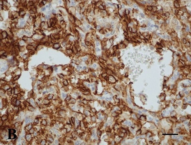
Vet. Sci. 2020, 7, 55 7 of 13
3.2. Spindle Cell Tumors, n = 4
In two cats (Nos. 14, 15), pancreatic masses were hemangiosarcomas (Table 2). One cat was
a 13-year-old Domestic Shorthair (No. 14) with a palpable abdominal mass. Clinical findings in
Cat No. 15 were not reported. Blood samples were not investigated in these cases. In both cats,
histopathology showed infiltrative vasoformative growth of neoplastic endothelial cells, marked
hemorrhage, thrombosis, mixed inflammation, and necrosis in the pancreas (Figure 3A). Mitotic count
was 1/HPF and 3/HPF, respectively. The neoplastic cells co-expressed vimentin, vWF and CD31
(Figure 3B). They were tested negative for FeLV. Both cats were euthanized during surgery.
Vet. Sci. 2020, 7, x 8 of 13
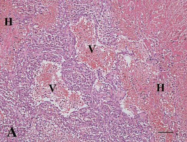
Figure 3. (a) Hemangiosarcoma. Tumor cells form vascular spaces (V) and there is marked
Figure 3. (A) Hemangiosarcoma. Tumor cells form vascular spaces (V) and there is marked inflammation
inflammation and hemorrhage (H) (12-year-old, female spayed Domestic Shorthair, No. 15. HE. Bar,
and hemorrhage (H) (12-year-old, female spayed Domestic Shorthair, No. 15. HE. Bar, 100 µm);
100 µm); (b) hemangiosarcoma. Intense CD31 expression in neoplastic cells. (same case as Figure 3a,
(B) hemangiosarcoma. Intense CD31 expression in neoplastic cells. (same case as (A), IHC, CD31. Bar,
IHC, CD31. Bar, 25 µm); (c) pleomorphic sarcoma. Pleomorphic, poorly differentiated spindle cells
25 µm);
with (C) giant
single pleomorphic sarcoma.
nuclei (arrow) Pleomorphic,
in the pancreas (P)poorly differentiated
(17-year-old, spindle cells
female Domestic with single
Shorthair, No. 16.giant
nuclei
HE. Bar, 25 µm); (d) pleomorphic sarcoma. Varying expression intensity of smooth muscle actin25
(arrow) in the pancreas (P) (17-year-old, female Domestic Shorthair, No. 16. HE. Bar, inµm);
(D) pleomorphic sarcoma. Varying expression intensity of smooth muscle actin in pleomorphic
pleomorphic tumor cells. (Same case as Figure 3c (IHC, actin. Bar, 25 µm); (e) fibrosarcoma. Mitotic tumor
figures (arrows) and intense anisokaryosis of neoplastic spindle cells between exocrine pancreatic
acini (P). (13-year-old, male neutered Domestic Shorthair, No. 17, HE. Bar, 25 µm); (f) fibrosarcoma.
Intense vimentin expression by the tumor cells (T) infiltrating the pancreas (P) without vimentin
expression (same case as Figure 3e, IHC, vimentin. Bar, 25 µm).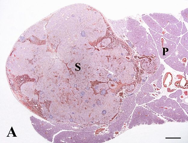
Vet. Sci. 2020, 7, 55                                                                                         8 of 13

      cells. (Same case as (C) (IHC, actin. Bar, 25 µm); (E) fibrosarcoma. Mitotic figures (arrows) and intense
      anisokaryosis of neoplastic spindle cells between exocrine pancreatic acini (P). (13-year-old, male
      neutered Domestic Shorthair, No. 17, HE. Bar, 25 µm); (F) fibrosarcoma. Intense vimentin expression
      by the tumor cells (T) infiltrating the pancreas (P) without vimentin expression (same case as (E), IHC,
      vimentin. Bar, 25 µm).

      One 17-year-old Domestic Shorthair (No. 16) was lethargic and had a palpable abdominal mass.
Pancreas-specific results were within reference intervals, but the SAA value was increased and CBC
revealed mild leukocytosis with neutrophilia and lymphopenia (Table 3). Multiple non-encapsulated
firm masses (0.1 × 0.1 × 0.1 to 2.0 × 2.0 × 2.0 cm) within the pancreas were noted. Further neoplastic
masses were not identified by clinical investigation of the skin or during laparotomy. Microscopically,
the multinodular pleomorphic sarcoma with myofibroblastic differentiation was characterized by
poorly differentiated spindle cells as well as pleomorphic and multinucleated giant cells within
the pancreatic interstitium (Figure 3C). Mitotic count was 9/HPF. Vascular infiltration was present.
There were multifocal mild mixed inflammation and small areas of necrosis. The pleomorphic sarcoma
showed a diffuse expression of vimentin and inhomogeneous co-expression of actin (Figure 3D).
Other sarcomas such as rhabdomyosarcoma, gastrointestinal stromal tumor, or mast cell tumor were
excluded by negative immunohistochemical reaction with anti-desmin and c-kit antibodies and Giemsa
stain negative. Furthermore, FeLV immunohistochemistry was negative. The cat was euthanized
during surgery.
      One 13-year-old Domestic Shorthair (No. 17) was presented with poor appetite and ascites.
No other intraabdominal masses or cutaneous sarcomas were noted by the clinician. Macroscopically,
the submitted tissue samples were brown and firm. Histopathologically, a fibrosarcoma with interstitial
infiltrative growth of moderately differentiated neoplastic spindle cells with heterogeneous morphology
(Figure 3E) was diagnosed. Mitotic count was 2 mitoses/HPF. Neoplastic cells exclusively expressed
vimentin (Figure 3F). FeLV was negative. The cat was euthanized two weeks after surgery.

4. Discussion
      Pancreatic neoplasms are rare in both humans [4] and domestic animals [3,28]. Epithelial solid
and cystic pancreatic neoplasms have been previously described in a larger series of 70 cats [29].
Non-epithelial neoplasms (e.g., lymphoma, sarcoma) are even rarer in all species [1,3,30].
      In general, lymphoma is the most common hematopoietic feline tumor [31]. A clinical diagnosis of
gastrointestinal lymphoma can be challenging because of its unspecific clinical presentation, especially
if neither masses nor lymphocytosis are present. During the clinical work up of such uncertain cases,
clinical pathology is commonly performed. In our study, cats with lymphoma for which blood values
were available (n = 5) showed high (n = 3) or questionable (n = 2) fPLI values, as well as increased
DGGR lipase (n = 3) and SAA values (n = 4). This may be explained by an accompanying purulent
pancreatitis in four cases. Further studies about pancreas-specific laboratory parameters in cats with
lymphomas are necessary to investigate the prevalence of pancreatic involvement. Furthermore,
inflammation was also reported in numerous feline epithelial pancreatic tumors [29] and as far as
serological data were available, fPLI value was increased in several cases [32]. One may conclude
that increased serological pancreatic enzymes can be indicative of an acute pancreatitis. However,
varying underlying pancreatic neoplasms should be taken into consideration especially if empiric
treatment is unsuccessful or if there is imaging evidence of neoplasia. Without histopathological
investigation, pancreatic neoplasms may be misdiagnosed as an unsuccessfully treated pancreatitis.
However, one cat with T-cell lymphoma (No. 7) showed increased fPLI and DGGR lipase values and
normal SAA value without histological signs of pancreatitis. This is consistent with the literature,
where false negative (sensitivity) and false positive (specificity) results have been described previously
for serological pancreatitis diagnostic in some cases [33].
      Morphological differentiation between inflammatory and neoplastic small lymphocytes in the
pancreas may require further investigation. Immunohistochemical assessment is an important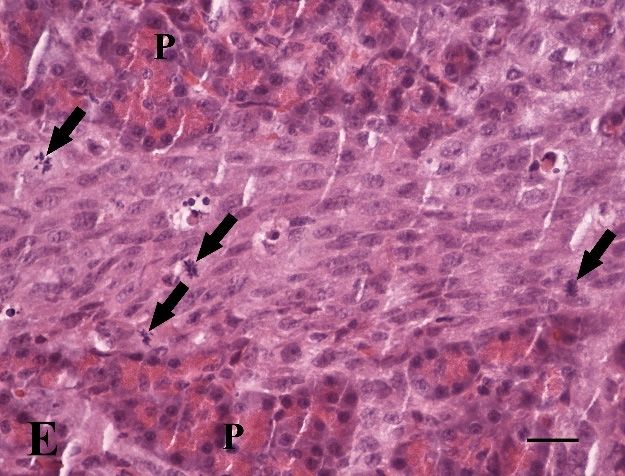
Vet. Sci. 2020, 7, 55                                                                               9 of 13

diagnostic tool [34]. In contrast, polymerase chain reaction for antigen receptor gene rearrangement
analysis (PARR) is well-known to have some limitations in cats [35]. In our study, T-cell lymphomas
were more frequent (75%) than B-cell lymphomas. This correlates with the adjoining anatomy of
small intestine and pancreas as interstitial T-cell lymphomas are more frequent than B-cell lymphomas
in cats [36]. Thus, the pancreatic sites of lymphomas were interpreted as part of a multicentric
systemic disease. Immunohistochemistry for FeLV was positive for two cats with T-cell lymphoma
(No. 5, 11). This is in accordance with the literature, where retroviral diseases (FeLV or feline
immunodeficiency virus) were listed as a risk factor in lymphomas [14]. In conclusion, without
cytological/histopathological examination and/or imaging techniques, feline pancreatic lymphoma
is easily missed if solitary pancreatitis is suspected because of clinical pathology and non-specific
clinical findings. This has a strong potential impact on therapy and prognosis, especially considering
the treatability of lymphomas by chemotherapy. Survival time of lymphoma subtypes may vary.
Sato et al. [37] showed a shorter survival time in alimentary (48 days) lymphomas than in cats with
nasal (135 days) or mediastinal (143 days) lymphomas. Wolfesberger et al. [38] reported the highest
survival time in intestinal T-cell lymphomas (1.7 years) compared to large B-cell lymphoma (4.5 months)
or peripheral T-cell lymphoma (6.1 months). In our study, seven out of 12 cats with lymphomas were
either euthanized or died during or shortly after surgery. Only two cats with lymphomas lived on for
2 months (No. 1, B-cell lymphoma) and 7 months (No. 4, T-cell lymphoma), respectively.
      Feline mast cell tumors are predominantly cutaneous neoplasms but can also appear in the
spleen [24]. In our study, a mast cell tumor in accessory splenic tissue was described for the first time.
Accessory splenic tissue is usually an incidental finding during laparotomy and appears as small dark
brown masses within the pancreas [39]. Ectopic splenic tissue was seen in 24 out of 540 cases (4%) of
routinely submitted feline pancreas samples in our laboratory (unpublished data). As in our case (cat
No. 13), a high percentage of mast cell tumors in cats are negative for different immunohistochemical
markers, including c-kit [40], which can impede a diagnosis in poorly differentiated cases. Prognostic
factors are not entirely clear in cats, but splenectomy has been recommended for feline splenic mast cell
tumors [41,42]. Careful exploration of the pancreas is recommended because of the risk of incomplete
resection of splenic tissue in the case of affected accessory spleens, as in our case.
      Sarcomas of the feline pancreas are very rare. Because of the retrospective nature of our study,
there were incomplete data on signalment, therapy, and clinical outcome. Previous clinical and
surgical reports were often unavailable. Therefore, a distinction between those sarcomas that were
primary pancreatic neoplasms and metastases from elsewhere cannot be made with certainty in all
cases. Nevertheless, because of the tumors being exclusively reported within the pancreas, both
macroscopically and histologically, there is a high likelihood of them being primaries.
      Feline visceral hemangiosarcomas were mostly seen in the liver, intestine, and lymph nodes but
rarely (2/26 cats) in the pancreas [20]. Both cats with hemangiosarcomas included in our study died
or were euthanized shortly after surgery. According to the literature, prognosis for feline visceral
hemangiosarcoma with a multifocal presentation is poor, with a median survival time of 77 days [20].
Correlation between the degree of differentiation, mitotic rate or size of feline hemangiosarcoma and
the clinical outcome could not be found [43].
      The present study included a fibrosarcoma and a pleomorphic sarcoma in the feline pancreas.
Nomenclature of pleomorphic sarcomas is still controversial. The terms “undifferentiated pleomorphic
sarcoma,” “anaplastic sarcoma with giant cells,” or “malignant fibrous histiocytoma” are used
synonymously in veterinary pathology [15]. In cat No. 16, immunohistochemistry revealed small areas
of myofibroblastic differentiation within the neoplasm. In literature, this combination of histological
type of sarcoma and immunohistochemical expression pattern has been described for feline cutaneous
post-vaccinal sarcomas [44,45]. Cutaneous sarcomas may undergo malignant transformation of
fibrocytes due to an inflammation [46]. Furthermore, there is one case report of a fibrosarcoma arising
at the site of a retained surgical sponge in a cat [47]. In human medicine, inflammatory pseudotumors
have been reported as a benign change in various organs, predominantly in the lungs [48]. The term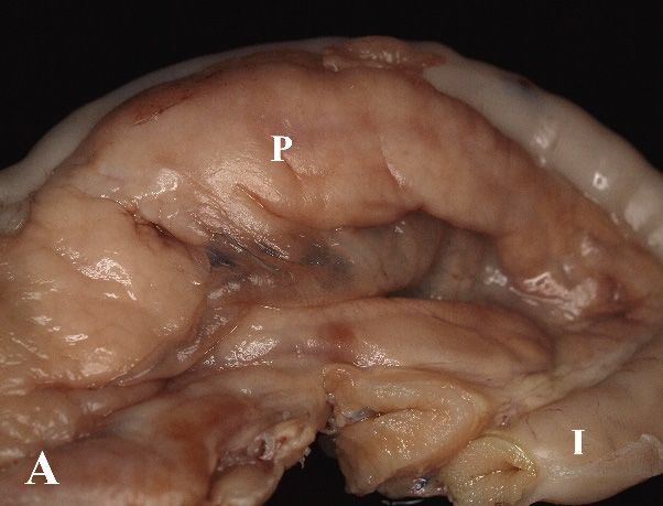
Vet. Sci. 2020, 7, 55                                                                                               10 of 13

“inflammatory fibrosarcoma” was suggested for some of these cases with metastases [49]. The cat
with the pleomorphic sarcoma in our study had increased SAA values of an unknown cause and the
pathogenesis of the tumor remained unclear. In human medicine, recurrence of metastatic sarcomas in
the pancreas is common and a median overall survival time of 21 months [17] as well as moderate
complication rates [18] have been described. The cats in our study were euthanized during surgery
because of the poor prognosis.

5. Conclusions
     In summary, our study described the characteristics of five different pancreatic tumor types of round
or spindle cell origin in 17 cats. It includes the first descriptions of a pancreatic pleomorphic sarcoma
and a mast cell tumor in accessory spleens within the pancreas. Epithelial pancreatic neoplasms
have been reported previously [29] and metastases of other carcinomas may affect the pancreas
also (unpublished data). In general, pancreatic neoplasms may be accompanied by inflammation,
and histopathological investigation is required to differentiate pancreatitis from an inflamed pancreatic
neoplasm. This may have a strong impact on therapy and prognosis.

Author Contributions: Conceptualization, H.A.-L.; data curation, M.S.; investigation, K.T.; methodology, K.T.;
resources, M.S.; supervision, K.S. and H.A.-L.; visualization, K.T.; writing—original draft, K.T.; writing—review
and editing, M.S., K.S., and H.A.-L. All authors have read and agreed to the published version of the manuscript.
Funding: This research received no external funding.
Acknowledgments: The authors thank Olga Seelbach for technical assistance and Anna-Sophia Müller for
correction as native speakers.
Conflicts of Interest: The authors declare no conflict of interest.

References
1.    Head, K.W.; Cullen, J.M.; Dubielzig, R.R.; Else, R.W.; Misdorp, W.; Patnaik, A.K.; Tateyama, S.; Van der Gaag, I.
      Histological classification of tumours of the pancreas of domestic animals. In World Health Organization
      International Histological Classification of Tumors of Domestic Animals, Histological Classification of Tumours of the
      Alimentary System of Domestic Animals; Head, K.W., Cullen, J.M., Dubielzig, R.R., Else, R.W., Misdorp, W.,
      Patnaik, A.K., Tateyama, S., Van der Gaag, I., Eds.; Armed Forces Institute of Pathology: Washington, DC,
      USA, 2003; pp. 111–118.
2.    Jubb, K.V.F.; Stent, A.W. Exocrine pancreas. In Pathology of Domestic Animals, 6th ed.; Maxie, M.G., Ed.;
      Elsevier: St. Louis, MO, USA, 2016; Volume 2, pp. 353–368.
3.    Munday, J.S.; Loehr, C.V.; Kiupel, M. Tumors of the alimentary tract. In Tumors in Domestic Animals, 5th ed.;
      Meuten, D.J., Ed.; Wiley Blackwell: Ames, IA, USA, 2017; pp. 499–601.
4.    Siegel, R.L.; Miller, K.D.; Jemal, A. Cancer statistics, 2020. CA Cancer J. Clin. 2020, 70, 7–30. [CrossRef]
      [PubMed]
5.    Kircher, C.H.; Nielsen, S.W. Tumours of the pancreas. Bull. World Health Organ. 1976, 53, 195–202. [PubMed]
6.    Bennett, P.F.; Hahn, K.A.; Toal, R.L.; Legendre, A.M. Ultrasonographic and cytopathological diagnosis of
      exocrine pancreatic carcinoma in the dog and cat. J. Am. Anim. Hosp. Assoc. 2001, 37, 466–473. [CrossRef]
      [PubMed]
7.    Seaman, R.L. Exocrine pancreatic neoplasia in the cat: A case series. J. Am. Anim. Hosp. Assoc. 2004, 40,
      238–245. [CrossRef]
8.    Linderman, M.J.; Brodsky, E.M.; De Lorimier, L.P.; Clifford, C.A.; Post, G.S. Feline exocrine pancreatic
      carcinoma: A retrospective study of 34 cases. Vet. Comp. Oncol. 2013, 11, 208–218. [CrossRef]
9.    Gieger, T. Alimentary lymphoma in cats and dogs. Vet. Clin. N. Am. Small Anim. Pract. 2011, 41, 419–432.
      [CrossRef]
10.   Hecht, S.; Penninck, D.G.; Keating, J.H. Imaging findings in pancreatic neoplasia and nodular hyperplasia in
      19 cats. Vet. Radiol. Ultrasound 2007, 48, 45–50. [CrossRef]
Vet. Sci. 2020, 7, 55 11 of 13
11. Lingard, A.E.; Briscoe, K.; Beatty, J.A.; Moore, A.S.; Crowley, A.M.; Krockenberger, M.; Churcher, R.K.;
Canfield, P.J.; Barrs, V.R. Low-grade alimentary lymphoma: Clinicopathological findings and response to
treatment in 17 cases. J. Feline Med. Surg. 2009, 11, 692–700. [CrossRef]
12. Takeuchi, Y.; Takahashi, M.; Tsuboi, M.; Fujino, Y.; Uchida, K.; Ohno, K.; Nakayama, H.; Tsujimoto, H.
Intestinal T-cell lymphoma with severe hypereosinophilic syndrome in a cat. J. Vet. Med. Sci. 2012, 74,
1057–1062. [CrossRef]
13. Kerns, A.T.; Brakel, K.A.; Premanandan, C.; Saffire, A.; Moore, S. Extranodal non-B, non-T-cell lymphoma
with bilateral tympanic bulla involvement in a cat. JFMS Open Rep. 2018, 4, 2055116918756724. [CrossRef]
14. Louwerens, M.; London, C.A.; Pedersen, N.C.; Lyons, L.A. Feline lymphoma in the post-feline leukemia
virus era. J. Vet. Intern. Med. 2005, 19, 329–335. [PubMed]
15. Hendrick, M.J. Mesenchymal tumors of the skin and soft tissues. In Tumors in Domestic Animals, 5th ed.;
Meuten, D.J., Ed.; Wiley Blackwell: Ames, IA, USA, 2017; pp. 142–175.
16. Yamamoto, R.; Suzuki, K.; Uchida, K.; Onda, N.; Shibutani, M.; Mitsumori, K. Pancreatic carcinosarcoma in a
cat. J. Comp. Pathol. 2012, 147, 223–226. [CrossRef] [PubMed]
17. Huddy, J.R.; Sodergren, M.H.; Deguara, J.; Thway, K.; Jones, R.L.; Mudan, S.S. Pancreaticoduodenectomy
for the management of pancreatic or duodenal metastases from primary sarcoma. Anticancer Res. 2018, 38,
4041–4046. [CrossRef] [PubMed]
18. Tseng, W.W.; Tsao-Wei, D.D.; Callegaro, D.; Grignani, G.; D’Ambrosio, L.; Bonvalot, S.; Ethin, C.G.;
Cardona, K.; Mullen, J.T.; Canter, R.J.; et al. A collaborative effort from the Trans-Atlantic Retroperitoneal
Sarcoma Working Group (TARPSWG). Pancreaticoduodenectomy in the surgical management of primary
retroperitoneal sarcoma. Eur. J. Surg. Oncol. 2018, 44, 810–815. [CrossRef] [PubMed]
19. Smith, A.N. Hemangiosarcoma in dogs and cats. Vet. Clin. N. Am. Small Anim. Pract. 2003, 33, 533–552.
[CrossRef]
20. Culp, W.T.N.; Drobatz, K.J.; Glassman, M.M.; Baez, J.L.; Aronson, L.R. Feline visceral hemangiosarcoma.
J. Vet. Intern. Med. 2008, 22, 148–152. [CrossRef]
21. Sharpe, A.; Cannon, M.J.; Lucke, V.M.; Day, M.J. Intestinal haemangiosarcoma in the cat: Clinical and
pathological features of four cases. J. Small Anim. Pract. 2000, 41, 411–415. [CrossRef]
22. Hendrick, M.J.; Mahaffey, E.A.; Moore, F.M.; Vos, J.H.; Walder, E.J. Tumors of fibrous tissue. In World Health
Organization International Histological Classification of Tumors of Domestic Animals, Histological Classification
of Mesenchymal Tumors of the Skin and Soft Tissues of Domestic Animals; Schulman, F.Y., Ed.; Armed Forces
Institute of Pathology: Washington, DC, USA, 1998A; pp. 15–18.
23. Hendrick, M.J.; Mahaffey, E.A.; Moore, F.M.; Vos, J.H.; Walder, E.J. Tumors of vascular tissue. In World Health
Organization International Histological Classification of Tumors of Domestic Animals, Histological Classification
of Mesenchymal Tumors of the Skin and Soft Tissues of Domestic Animals; Schulman, F.Y., Ed.; Armed Forces
Institute of Pathology: Washington, DC, USA, 1998B; pp. 22–25.
24. Hendrick, M.J.; Mahaffey, E.A.; Moore, F.M.; Vos, J.H.; Walder, E.J. Mast cell tumors. In World Health
Organization International Histological Classification of Tumors of Domestic Animals, Histological Classification of
mesenchymal tumors of the skin and soft tissues of Domestic Animals; Schulman, F.Y., Ed.; Armed Forces Institute
of Pathology: Washington, DC, USA, 1998C; pp. 28–29.
25. Valli, V.E.; Jacobs, R.M.; Parodi, A.L.; Vernau, W.; Moore, P.F. Tumors of lymphoid system. In World Health
Organization International Histological Classification of Tumors of Domestic Animals, Histological Classification of
Hematopoietic Tumors of Domestic Animals; Armed Forces Institute of Pathology: Washington, DC, USA, 2002;
pp. 25–47.
26. Gabor, L.J.; Canfield, P.J.; Malik, R. Immunophenotypic and histological characterization of 109 cases of
feline lymphosarcoma. Aust. Vet. J. 1999, 77, 436–441. [CrossRef]
27. De Cock, H.E.V.; Forman, M.A.; Farver, T.B.; Marks, S.L. Prevalence and histopathologic characteristics of
pancreatitis in cats. Vet. Pathol. 2007, 44, 39–49. [CrossRef]
28. Priester, W.A. Data from eleven United States and Canadian colleges of veterinary medicine on pancreatic
carcinoma in domestic animals. Cancer Res. 1974, 34, 1372–1375.
29. Törner, K.; Aupperle-Lellbach, H.; Staudacher, A.; Staudacher, M.; Steiger, K. Primary solid and cystic
tumours of the exocrine pancreas in cats. J. Comp. Pathol. 2019, 165, 5–19. [CrossRef] [PubMed]Vet. Sci. 2020, 7, 55 12 of 13
30. Bosmann, F.T.; Carneiro, F.; Hruban, R.H.; Theise, N.D. Tumours of the pancreas. In World Health Organization
Classification of Tumours of the Digestive System, 4th ed.; Bosmann, F.T., Carneiro, F., Hruban, R.H., Theise, N.D.,
Eds.; IARC Press: Lyon, France, 2010; pp. 279–337.
31. Vezzali, E.; Parodi, A.L.; Marcato, P.S.; Bettini, G. Histopathologic classification of 171 cases of canine and
feline non-Hodgkin lymphoma according to the WHO. Vet. Comp. Oncol. 2009, 8, 38–49. [CrossRef]
[PubMed]
32. Törner, K.; Staudacher, M.; Tress, U.; Weber, C.N.; Stadler, C.; Grassinger, J.M.; Müller, E.; Aupperle-Lellbach, H.
Histopathology and feline pancreatic lipase immunoreactivity in inflammatory, hyperplastic and neoplastic
pancreatic diseases in cats. J. Comp. Pathol. 2020, 174, 63–72. [CrossRef]
33. Lidbury, J.A.; Suchodolski, J.S. New advances in the diagnosis of canine and feline liver and pancreatic
disease. Vet. J. 2016, 215, 87–95. [CrossRef] [PubMed]
34. Waly, N.E.; Gruffydd-Jones, T.J.; Stokes, C.R.; Day, M.J. Immunohistochemical diagnosis of alimentary lymphomas
and severe intestinal inflammation in cats. J. Comp. Pathol. 2005, 133, 253–260. [CrossRef] [PubMed]
35. Burkhard, M.J.; Bienzle, D. Making sense of lymphoma diagnostics in small animal patients. Vet. Clin. N.
Am. Small Anim. Pract. 2013, 43, 1331–1347. [CrossRef] [PubMed]
36. Pohlman, L.M.; Higginbotham, M.L.; Welles, E.G.; Johnson, C.M. Immunophenotypic and histologic
classification of 50 cases of feline gastrointestinal lymphoma. Vet. Pathol. 2009, 46, 259–268. [CrossRef]
37. Sato, H.; Fujino, Y.; Chino, J.; Takahashi, M.; Fukushima, K.; Goto-Koshino, Y.; Uchida, K.; Ohno, K.;
Tsujimoto, H. Prognostic analyses on anatomical and morphological classification of feline lymphoma. J. Vet.
Med. Sci. 2014, 76, 807–811. [CrossRef]
38. Wolfesberger, B.; Skor, O.; Hammer, S.E.; Flickinger, I.; Kleiter, M.; Rütgen, B.C.; Schwendenwein, I.; Tichy, A.;
Hittmair, K.M.; Degasperi, B.; et al. Does categorisation of lymphoma subtypes according to the World
Health Organization classification predict clinical outcome in cats? J. Feline Med. Surg. 2017, 19, 897–906.
[CrossRef]
39. Ramírez, G.A.; Altimira, J.; García-González, B.; Vilafranca, M. Intrapancreatic ectopic splenic tissue in dogs
and cats. J. Comp. Pathol. 2013, 148, 361–364. [CrossRef]
40. Mallett, C.L.; Northrup, N.C.; Saba, C.F.; Rodriguez, C.O.; Rassnick, K.M.; Gieger, T.L.; Childress, M.O.;
Howerth, E.W. Immunohistochemical characterization of feline mast cell tumors. Vet. Pathol. 2012, 50,
106–109. [CrossRef] [PubMed]
41. Sabattini, S.; Bettini, G. Prognostic value of histologic and immunohistochemical features in feline cutaneous
mast cell tumors. Vet. Pathol. 2010, 47, 643–653. [CrossRef] [PubMed]
42. Evans, B.J.; O’Brien, D.; Allstadt, S.D.; Gregor, T.P.; Sorenmo, K.U. Treatment outcomes and prognostic factors
of feline splenic mast cell tumors: A multi-institutional retrospective study of 64 cases. Vet. Comp. Oncol.
2018, 16, 20–27. [CrossRef] [PubMed]
43. Schultheiss, P.C. A retrospective study of visceral and nonvisceral hemangiosarcoma and hemangiomas in
domestic animals. J. Vet. Diagn. Investig. 2004, 16, 522–526. [CrossRef] [PubMed]
44. Hendrick, M.J.; Brooks, J.J. Postvaccinal sarcomas in the cat: Histology and immunohistochemistry. Vet. Pathol.
1994, 31, 126–129. [CrossRef]
45. Aberdein, D.; Munday, J.S.; Dyer, C.B.; Knight, C.G.; French, A.F.; Gibson, I.R. Comparison of the histology
and immunohistochemistry of vaccination-site and non-vaccination-site sarcomas from cats in New Zealand.
N. Z. Vet. J. 2007, 55, 203–207. [CrossRef]
46. Jelinek, F. Postinflammatory sarcoma in cats. Exp. Toxicol. Pathol. 2003, 55, 167–172. [CrossRef]
47. Haddad, J.L.; Goldschmidt, M.H.; Patel, R.T. Fibrosarcoma arising at the site of a retained surgical sponge in
a cat. Vet. Clin. Pathol. 2010, 39, 241–246. [CrossRef]
48. Coffin, C.M.; Dehner, L.F.; Meis-Kindblom, J.M. Inflammatory myofibroblastic tumor, inflammatory
fibrosarcoma, and related lesions: An historical review with differential diagnostic considerations.
Semin. Diagn. Pathol. 1998, 15, 102–110.
49. Meis, J.M.; Enzinger, F.M. Inflammatory fibrosarcoma of the mesentery and retroperitoneum a tumor closely
simulating inflammatory pseudotumor. Am. J. Surg. Pathol. 1991, 15, 1146–1156. [CrossRef]
© 2020 by the authors. Licensee MDPI, Basel, Switzerland. This article is an open access
article distributed under the terms and conditions of the Creative Commons Attribution
(CC BY) license (http://creativecommons.org/licenses/by/4.0/).You can also read