Clinical and prognostic features of MMP-2 and VEGF in AEG patients
←
→
Page content transcription
If your browser does not render page correctly, please read the page content below
Open Medicine 2021; 16: 786–794
Research Article
Qing-Kang Zheng, Qing Yin, Nan Zhang, Zhi-Gang Sun*
Clinical and prognostic features of MMP-2 and
VEGF in AEG patients
https://doi.org/10.1515/med-2021-0252
received October 1, 2020; accepted February 24, 2021
1 Introduction
Abstract: Adenocarcinoma of the esophagogastric junc- Adenocarcinoma of the esophagogastric junction (AEG)
tion (AEG) has been increased in recent years and has incidents have increased in recent years and become a
become a worldwide problem that seriously affects worldwide problem that seriously affects human health
human health. The purpose of the study is to investigate [1,2]. AEG is defined as the malignant tumor whose center
the clinical and prognostic characteristics of the matrix is within 5 cm of the proximal and distal ends of the
metalloproteinase-2 (MMP-2) and vascular endothelial esophagogastric junction (EGJ). It includes proximal gas-
growth factor (VEGF) expression in AEG patients. A total tric cancer, cardiac cancer, and distal esophageal adeno-
of 69 patients were enrolled in this study. The result carcinoma [3]. Due to its particular anatomical location,
showed that the high expression of MMP-2 was signifi- the pathological type of AEG is adenocarcinoma, while
cantly associated with tumor differentiation (P < 0.05)
the epidemiological characteristics and clinical symp-
and depth of invasion (pT, P < 0.05). The high expression
toms of AEG are consistent with esophageal squamous
of VEGF was significantly associated with pT (P < 0.05)
cell carcinoma [4,5]. At present, more and more scholars
and lymph node metastasis (pN, P < 0.05). There was a
regard AEG as an independent malignant tumor that is
positive correlation between MMP-2 and VEGF expression
different from gastric cancer and esophageal adenocarci-
(P < 0.01). The 5-year survival rate for the 69 AEG patients
noma [6,7]. Because of the high recurrences rates, the
was 40.6% and it was significantly associated with tumor
prognosis of AEG patients remains poor even after the
differentiation (P < 0.05), pN (P < 0.01), pTNM stage (P <
curative surgery treatment [8,9]. The TNM staging system
0.01), MMP-2 expression (P < 0.05), and VEGF expression
lacks sufficient implied rate, because significantly dif-
(P < 0.05). Cox multivariate regression demonstrated
ferent survival rate is often observed in the same TNM
that tumor differentiation and pN were independent fac-
stage. Therefore, it is meaningful to combine some bio-
tors for the 5-year survival rate. Our study showed that
markers with TNM staging to distinguish AEG patients
MMP-2 and VEGF could work synergistically in AEG
development. with poor prognosis.
Matrix metalloproteinases (MMPs), a family of extra-
Keywords: adenocarcinoma of the esophagogastric junc- cellular zinc-dependent endoproteinases [10], play pivotal
tion, MMP-2, VEGF, immunohistochemistry roles in tumor infiltration, invasion, and angiogenesis
[11,12]. Among the MMPs, MMP-2 acts as a key enzyme
that could be related to tumor metastasis and physiologic
functions [13]. Vascular endothelial growth factor (VEGF)
is an angiogenetic factor produced by cancer cells and
* Corresponding author: Zhi-Gang Sun, Department of Thoracic could stimulate the growth of endothelial cells [12]. It
Surgery, Jinan Central Hospital, Cheeloo College of Medicine,
could promote endothelial cells proliferation and migra-
Shandong University, Jinan 250013, People’s Republic of China,
e-mail: sunszg@126.com, tel: +86-133-7058-2825 tion, enhance the permeability of blood vessels, promote
Qing-Kang Zheng: School of Clinical Medicine, Weifang Medical stromal proteolysis, and reduce endothelial cell apop-
University, Weifang 261053, People’s Republic of China tosis [13]. It has been reported that VEGF could induce
Qing Yin: Department of Medical Education, Jinan Central Hospital, multiple proteases expression including MMPs which
Cheeloo College of Medicine, Shandong University, Jinan 250013,
leads to extracellular matrixaround vessels [14]. The pur-
People’s Republic of China
Nan Zhang: Department of Oncology, Jinan Central Hospital,
pose of the study is to investigate the clinic and prog-
Cheeloo College of Medicine, Shandong University, Jinan 250013, nostic characteristics of MMP-2 and VEGF expression in
People’s Republic of China postoperative AEG patients using both univariate and
Open Access. © 2021 Qing-Kang Zheng et al., published by De Gruyter. This work is licensed under the Creative Commons Attribution 4.0
International License.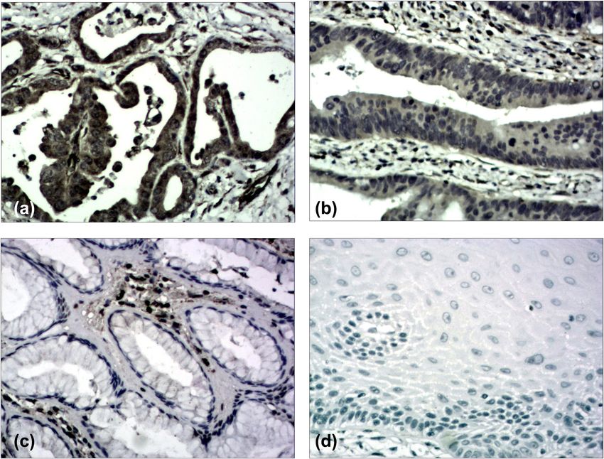
MMP-2, VEGF, and AEG 787
multivariate analysis, and the MMP-2 and VEGF expres- data of the AEG patients were shown in Table 1. This
sion was detected using immunohistochemistry (IHC) at research was approved by the Ethics Committee of Jinan
protein level. Central Hospital and was in accordance with the ethical
standards of the Helsinki Declaration of 1975, as revised
in 2000. And all the patients consented to the study.
2 Materials and methods
2.2 Immunohistochemistry
2.1 Patients
All the AEG specimens were obtained from the 69 cases.
Total of 69 AEG cases were enrolled into the study con- The tissue specimens were fixed in 10% neutral buffered
ducted at the Department of Thoracic Surgery and General formalin and processed routinely. MMP-2 and VEGF were
Surgery, Jinan Central Hospital between January 2010 detected by the streptavidin-peroxidase (SP) method
and May 2013. The inclusion criteria were as follows: using the same paraffin-embedded tissue samples, which
(1) patients underwent surgery and affirmed AEG by were cut into 4-mm-thick slices. The primary antibody
pathology; (2) the TNM staging system of AEG was based was applied using mouse antihuman monoclonal MMP-2
on the International Union Against Cancer (2009) guide- antibodies (1:150, Catalogue TA806846, Zhongshan Jinqiao
line; (3) the subjects had no preoperative chemotherapy Biotechnology, Beijing, P. R. China) or mouse antihuman
or radiotherapy treatment; (4) the cases had no seriously monoclonal VEGF antibodies (1:100, Catalogue ZM-0265,
surgical contraindications that could affect prognosis. (5) Zhongshan Jinqiao Biotechnology, Beijing, P. R. China).
The follow-up data of the cases were complete. The clinic The IHC protocols were described previously [15–17].
Table 1: Correlation between MMP-2 and VEGF expression and clinicopathological features of the patients with adenocarcinoma of the
esophagogastric junction
Clinical features Patients MMP-2 VEGF
Low High P Low High P
69 21 48 20 49
Gender *0.561 *0.554
Male 50 14 36 16 34
Female 19 7 12 4 15
Age, year *0.788 0.234788 Qing-Kang Zheng et al.
2.3 Immunohistochemical findings multiplying diffusion and intensity scores. Those with final
evaluation scores ≤4 were classified as low expression group and
those with ≥5 as high expression group (Figure 2) [17].
Cell counts were performed by counting 200 cells in each
area of at least 5 randomly selected areas with 400× mag-
nification using a light microscope. MMP-2 expression
was mainly located in the cytoplasm and plasma mem- 2.4 Statistical analysis
brane of the tumor cells and categorized under the fol-
lowing conditions: low expression, less than 50% of cells; Enumeration data were performed by χ2 test or Fisher’s
high expression, 50% or more of cells (Figure 1) [16]. exact probability test. The correlation between MMP-2
VEGF expression was mainly located in the cytoplasm and VEGF expression was analyzed using Spearman’s
of the tumor cells and was categorized as follows: staining rank correlation coefficient. Univariate analysis was per-
the rate of tumor cells was scored between 0 and 4: formed with Kaplan–Meier survival curves. Multivariate
0 (below 5%), 1 (6–25%), 2 (26–50%), 3 (51–75%), and 4 analysis was performed by the Cox proportional hazard
(above 75%); staining intensity was scored between 0 and model. All statistical data were analyzed using SPSS (IBM
3: 0 (negative), 1 (mildly positive), 2 (moderately positive), SPSS, Statistics 25, USA), and P < 0.05 indicated a statis-
and 3 (strongly positive). The final score was obtained by tically significant difference.
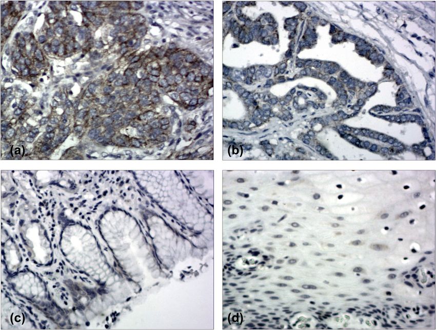
Figure 1: Immunohistochemical staining of adenocarcinoma of the esophagogastric junction (AEG) tissue sections demonstrating Matrix
metalloproteinase-2 (MMP-2) (Original magnification ×200). (a) AEG specimen with high expression of MMP-2. (b) AEG specimen with low
expression of MMP-2. (c) The corresponding normal gastric tissue specimen with no MMP-2 expression (contrast). (d) The corresponding
normal esophageal tissue specimen with no MMP-2 expression (contrast).MMP-2, VEGF, and AEG 789
Figure 2: Immunohistochemical staining of adenocarcinoma of the esophagogastric junction (AEG) tissue sections demonstrating vascular
endothelial growth factor (VEGF) (Original magnification ×200). (a) AEG specimen with high expression of VEGF. (b) AEG specimen with low
expression of VEGF. (c) The corresponding normal gastric tissue specimen with no VEGF expression (contrast). (d) The corresponding
normal esophageal tissue specimen with no VEGF expression (contrast).
2.5 Follow-up stage were demonstrated for MMP-2 (P > 0.05). The high
expression of VEGF was significantly associated with pT
Overall, 32 cases had postsurgical chemotherapy, 3 cases (pT1 + pT2 58.9% vs pT3 + pT4 82.9%; P < 0.05) and pN
had postsurgical radiotherapy, and 19 cases had postsur- (pN – 50.0% vs pN + 85.4%; P < 0.05). No statistically signifi-
gical radiotherapy combined with chemotherapy. Patients cant correlations with gender, age, tumor differentiation,
who died of tumor were included in the prognostic and pTNM stage were demonstrated for VEGF (P > 0.05).
analysis. There was a positive correlation between MMP-2 and VEGF
expression (P < 0.01) (Table 1).
The 5-year survival rate for all the 69 AEG patients
was 40.6%. A univariate analysis was conducted using
3 Results the log-rank test, and the 5-year survival rate was signifi-
cantly associated with differentiation (P < 0.05), pN (P <
The high expression of MMP-2 was significantly asso- 0.01), pTNM stage (P < 0.01), MMP-2 expression (P < 0.05),
ciated with tumor differentiation (Well + Moderately 56.3% and VEGF expression (P < 0.05) (Figure 3, Table 2). The
vs Poorly 81.1%; P < 0.05) and depth of invasion (pT; pT1 + 5-year survival rate of patients with low MMP-2 expres-
pT2 55.9% vs pT3 + pT4 82.9%; P < 0.05). No statistically sion in AEG tissues was significantly higher than those
significant correlations with gender, age, pN, and pTNM with high MMP-2 expression (61.9% vs 31.3%; P = 0.013).790 Qing-Kang Zheng et al. Figure 3: (a) The Kaplan-Meier survival curve of 69 cases of AEG patients. (b) Survival curves of AEG patients with tumor differentiation. (c) Survival curves of AEG patients with negative or positive pN. (d) Survival curves of AEG patients with different pTNM. (e) Survival curves of AEG patients with low or high expression of MMP-2 expression. (f) Survival curves of AEG patients with low or high expression of VEGF expression. Similarly, the 5-year survival rate of patients with low P = 0.048). The Cox multivariate regression result showed VEGF expression in AEG tissues was significantly higher that both tumor differentiation and pN were independent than those with high VEGF expression (60.0% vs 32.7%; factors for the 5-year survival rate (Table 3).
MMP-2, VEGF, and AEG 791
Table 2: Univariate analysis with respect to 5-year survival of the By contrast, Shen et al. [24] had the opposite conclusion.
patients with adenocarcinoma of the esophagogastric junction They found that MMP-2 overexpression was a predictive
factor for poor prognosis of gastric cancer. Qian et al. [25]
Clinical features Patients 5-year survival (%) and Liu et al. made the [26] same conclusions as Shen’s
Patients Rate (%) P value by studying non-small cell lung cancer (NSCLC) and
endometrial cancer, respectively. However, little research
69 28 40 6
has been done for the MMP-2 clinical features in AEG
Gender 0.617 patients. Lu et al. [27] detected MMP-2 expression using
Male 50 20 40.0
IHC in tumors specimens from 120 AEG patients. They
Female 19 8 42.1
Age, year 0.223
found that 51.7% of the cases had MMP-2 overexpression.792 Qing-Kang Zheng et al.
Table 3: Results of Cox regression multivariate 5-year survival analysis of the patients with adenocarcinoma of the esophagogastric
junction
B SE Wald P HR 95.0% CI for HR
Gender −0.554 0.384 2.083 0.149 0.575 0.271–1.219
Age 0.104 0.356 0.086 0.769 1.110 0.553–2.229
Differentiation 0.885 0.364 5.911 0.015 2.424 1.187–4.950
pT 0.803 0.437 3.370 0.066 2.232 0.947–5.259
pN 2.494 0.632 15.545 0.001 12.104 3.504–41.809
pTNM −0.671 0.487 1.899 0.168 0.511 0.1197–1.328
Chemotherapy −0.245 0.429 0.325 0.568 0.783 0.338–1.815
Radiotherapy 0.050 0.369 0.019 0.891 1.052 0.510–2.168
MMP-2 0.448 0.436 1.053 0.305 1.565 0.665–3.681
VEGF −0.441 0.471 0.876 0.349 0.644 0.256–1.619
B, regression coefficient; SE, standard error; Wald, Wald value; HR, hazard ratio; CI, confidence interval; pT, tumor invasion; pN, lymph
node metastasis, pTNM, tumor stage; MMP-2, matrix metalloproteinases 2; VEGF, vascular endothelial growth factor.
epithelial cells. However, it had no prognostic value for [36] confirmed that there was a significantly positive cor-
esophagogastric cancer patients. Park et al. [33] also relation between VEGF and MMP-2 in gastric cancer
detected VEGF expression in serum levels of ligands tissue of patients with metastases. So far, the correlation
from 147 patients who underwent potentially curative between VEGF and MMPs in AEG has not been reported in
resection for gastric and esophagogastric adenocarcinoma. PubMed. In our study, 69.6% AEG cases had high MMP-2
They found that VEGF levels were higher in patients with expressions, 71.0% AEG with high VEGF expression, and
R1 vs R0 resections. The increased VEGF levels were cor- 56.5% AEG cases had both high expressions of VEGF and
related with decreased overall survival rate. Moreover, MMP-2. Our study showed that MMP-2 expression was
the serum VEGF was found as a significantly independent positively related to VEGF expression in AEG tissues.
prognostic factor for overall survival. The controversy of We concluded that MMP-2 and VEGF could work syner-
the above findings could be possibly due to the different gistically in AEG development.
tissue specimens used and different stage or analytic This is the first report to study the relationship
method employed. Even using the same analytic method, between VEGF and MMP-2 in AEG at clinical level. In
the result may differ depending on the site selected for our study, all the patients successfully underwent radical
assessment. In our study, the VEGF protein expression in operation with regional lymph node dissection. The
all the patients was detected by IHC. Our data showed tumor did not invade other organs, and both edges of
that the high expression of VEGF was associated with resection were confirmed to be free of residual cancer
both tumor depth of invasion (pT) and lymph node cells by routine histological examination, to ensure
metastasis (pN). The 5-year survival rate of AEG patients complete resection. To eliminate the impact of mixed
was associated with VEGF expression by univariate factors correlated with prognosis on statistical analysis,
analysis. To eliminate the impact of mixed factors on the Cox regression multivariate analysis was performed
statistical analysis, we used multivariate analysis to to determine the independent prognostic factors. As a
determine prognostic factors, and our result showed result, the comparability was increased and statistical
that tumor differentiation and pN were relevant inde- bias was decreased, making the results of this study
pendent factors for a poor prognosis. more objective.
Recently, some reports showed there was a relation- However, the present study still has several limita-
ship between VEGF and MMPs in tumor progression. tions. First, in China the indications for treatment not
Zhang et al. [34]found that in vitro induction and activity only depend on doctors’ preferences, but also patients’
of MMP-2 stimulated by VEGF might be the main willingness and economic status. These factors may have
mechanism by which VEGF gave impetus to ovarian influenced the relatively poor survival result observed.
cancer cells invasion. Wang et al. [35] confirmed that In the study, 32 patients received postoperative chemo-
anti-basic fibroblast growth factor (anti-bFGF)-induced therapy, 3 patients received postoperative radiotherapy,
invasion of human lung cancer cells could be rescued and 19 patients received combined chemoradiotherapy.
by inhibiting the AKT/MMP-2/VEGF loop. Partyka et al. However, no statistically significant correlations withMMP-2, VEGF, and AEG 793
postoperative chemotherapy and radiotherapy were demon- during 1988-2012: a singleinstitution, high-volume experience
strated for the 5-year survival rate either in univariate or in China. Ann Surg. 2016;263:88–95. doi: 10.1097/
multivariate analysis. Second, this is a retrospective study SLA.0000000000001148.
[3] Siewert JR, Stein HJ. Classification of adenocarcinoma of the
with a small sample size, which could limit the value of
oesophagogastric junction. Br J Surg. 1998;85:1457–9.
the findings. A randomized-controlled prospective study doi: 10.1046/j.1365-2168.1998.00940.x.
with a larger sample size will be considered in further [4] Hosoda K, Yamashita K, Katada N, Watanabe M. Overview
investigations. of multimodal therapy for adenocarcinoma of the esophago-
In conclusion, high MMP-2 expression was signifi- gastric junction. Gen Thorac Cardiovasc Surg.
2015;63:549–56. doi: 10.1007/s11748-015-0575-2.
cantly associated with tumor differentiation and depth
[5] Huang Q. Carcinoma of the gastroesophageal junction in
of invasion in AEG patients. In addition, the high expres- Chinese patients. World J Gastroenterol. 2012;18:7134–40.
sion of VEGF was significantly associated with tumor doi: 10.3748/wjg.v18.i48.7134.
depth of invasion and lymph node metastasis. There was [6] Hasegawa S, Yoshikawa T. Adenocarcinoma of the esophago-
a positive correlation between MMP-2 and VEGF expres- gastric junction: incidence, characteristics, and treatment
strategies. Gastric Cancer. 2010;13:63–73. doi: 10.1007/
sions. Collectively, the results suggest that MMP-2 and
s10120-010-0555-2.
VEGF could work synergistically in AEG development.
[7] Zhang H, Wang W, Cheng Y, Song Y, Zhu K, Dang C.
Adenocarcinomas of the esophagogastric junction: experi-
Funding information: This study was supported by the ences at a single institution in China. World J Surg Oncol.
Shandong Provincial Natural Science Foundation (grant 2013;11:155. doi: 10.1186/1477-7819-11-155.
no. ZR2020MH204), the 19th batch of science and tech- [8] Roviello G, Petrioli R, Marano L, Polom K, Marrelli D, Perrella A,
et al. Angiogenesis inhibitors in gastric and gastroesophageal
nology innovation development plan of Jinan in 2020
junction cancer. Gastric Cancer. 2016;19:31–41. doi: 10.1007/
(Clinical medicine science and technology innovation s10120-015-0537-5.
plan, grant no. 202019032), and the second group of science [9] Fuchs CS, Niedzwiecki D, Mamon HJ, Tepper JE, Ye X,
and technology projects of Jinan Health Committee (grant Swanson RS, et al. Adjuvant chemoradiotherapy with epiru-
no. 2020-3-15). The funders had no role in study design, bicin, cisplatin, and fluorouracil compared with adjuvant
data collection and analysis, decision to publish, or pre- chemoradiotherapy with fluorouracil and leucovorin after
curative resection of gastric cancer: results from CALGB 80101
paration of the manuscript.
(Alliance). J Clinl Oncol. 2017;35:3671–7. doi: 10.1200/
JCO.2017.74.2130.
Author contributions: SZG conceived and supervised the [10] Egeblad M, Werb Z. New functions for the matrix metallo-
study; ZQK and ZN designed and performed experiments; proteinases in cancer progression. Nat Rev Cancer.
YQ analyzed data; ZQK and YQ wrote the manuscript; and 2002;2:161–74. doi: 10.1038/nrc745.
[11] Rak B, Mehlich D, Garbicz F, Domosud Z, Paskal W,
SZG and ZNrevised the manuscript. All the authors read
Marczewska JM, et al. Post-transcriptional regulation of
and approved the final version of the manuscript. MMP16 and TIMP2 expression via miR-382, miR-410 and miR-
200b in endometrial cancer. Cancer Genomics Proteomics.
Conflict of interest: The authors declare that they have no 2017;14:389–401. doi: 10.21873/cgp.20049.
conflicts of interest. [12] Peng J, Shao N, Peng H, Chen LQ. Prognostic significance of
vascular endothelial growth factor expression in esophageal
carcinoma: a meta-analysis. J BUON. 2013;18:398–406.
Data availability statement: The datasets generated during
doi: 10.1016/j.prp.2006.12.002.
and/or analyzed during the current study are available [13] Honkavuori-Toivola M, Santala M, Soini Y, Turpeenniemi-
from the corresponding author on reasonable request. Hujanen T, Talvensaari-Mattila A. Combination of strong MMP-
2 and weak TIMP-2 immunostainings is a significant prog-
nostic factor in endometrial carcinoma. Dis Markers.
2013;35:261–6. doi: 10.1155/2013/416870.
[14] Abdel-Rahman O. Targeting vascular endothelial growth factor
(VEGF) pathway in gastric cancer: preclinical and clinical
References aspects. Crit Rev Oncol Hematol. 2015;93:18–27. doi: 10.1016/
j.critrevonc.2014.05.012.
[1] Hatta W, Tong D, Lee YY, Ichihara S, Uedo N, Gotoda T. [15] Shao JB, Li Z, Zhang N, Yang F, Gao W, Sun ZG. Hypoxia-
Different time trend and management of esophagogastric inducible factor 1α in combination with vascular endothelial
junction adenocarcinoma in three Asian countries. Dig Endosc. growth factor could predict the prognosis of postoperative
2017;29(Suppl 2):18–25. doi: 10.1111/den.12808. patients with oesophageal squamous cell cancer. Pol J Pathol.
[2] Liu K, Yang K, Zhang W, Chen X, Chen X, Zhang B, et al. 2019;70:84–90. doi: 10.5114/pjp.2019.87100.
Changes of esophagogastric junctional adenocarcinoma and [16] Xiang ZL, Zeng ZC, Fan J, Tang ZY, Zeng HY, Gao DM. Gene
gastroesophageal reflux disease among surgical patients expression profiling of fixed tissues identified hypoxia-794 Qing-Kang Zheng et al.
inducible factor-1α, VEGF, and matrix metalloproteinase-2 as [26] Liu C, Li Y, Hu S, Chen Y, Gao L, Liu D, et al. Clinical significance
biomarkers of lymph node metastasis in hepatocellular carci- of matrix metalloproteinase-2 in endometrial cancer: a sys-
noma. Clin Cancer Res. 2011;17:5463–72. doi: 10.1158/ tematic review and meta-analysis. Medicine (Baltimore).
1078-0432. 2018;97:e10994. doi: 10.1097/MD.0000000000010994.
[17] Berk V, Deniz K, Bozkurt O, Ozaslan E, Karaca H, Inanc M, et al. [27] Lu X, Duan L, Xie H, Lu X, Lu D, Lu D, et al. Evaluation of MMP-9
Predictive significance of VEGF and HIF-1α expression in patients and MMP-2 and their suppressor TIMP-1 and TIMP-2 in ade-
with metastatic colorectal cancer receiving chemotherapy nocarcinoma of esophagogastric junction. Onco Targets Ther.
combinations with bevacizumab. Asian Pac J Cancer Prev. 2016;9:4343–9. doi: 10.2147/OTT.S99580.
2015;16:6149–54. doi: 10.7314/apjcp.2015.16.14.6149. [28] Abdel-Rahman O. Targeting vascular endothelial growth factor
[18] Siqueira AS, Carvalho MR, Monteiro AC, Freitas VM, Jaeger RG, (VEGF) pathway in gastriccancer: preclinical and clinical
Pinheiro JJ. Matrix metalloproteinases, TIMPs and growth fac- aspects. Crit Rev Oncol Hematol. 2015;93:18–27. doi: 10.1016/
tors regulating ameloblastoma behaviour. Histopathology. j.critrevonc.2014.05.012.
2010;57:128–37. doi: 10.1111/j.1365-2559.2010.03596.x. [29] Kleespies A, Guba M, Jauch KW, Bruns CJ. Vascular endothelial
[19] Tester AM, Waltham M, Oh SJ, Bae SN, Bills MM, Walker EC, growth factor in esophageal cancer. J Surg Oncol.
et al. Pro-matrix metalloproteinase-2 transfection increases 2004;87:95–104. doi: 10.1002/jso.20070.
orthotopic primary growth and experimental metastasis of [30] Peng J, Shao N, Peng H, Chen LQ. Prognostic significance of
MDA-MB-231 human breast cancer cells in nude mice. Cancer vascular endothelial growth factor expression in esophageal
Res. 2004;64:652–8. doi: 10.1158/0008-5472.can-0384-2. carcinoma: a meta-analysis. J BUON. 2013;18:398–406.
[20] Kamat AA, Fletcher M, Gruman LM, Mueller P, Lopez A, doi: 10.4161/onco.23973.
Landen CN, et al. The clinical relevance of stromal [31] Liu L, Ma XL, Xiao ZL, Li M, Cheng SH, Wei YQ. Prognostic value
matrix metalloproteinase expression in ovarian cancer. of vascular endothelial growth factor expression in resected
Clin Cancer Res. 2006;12:1707–14. doi: 10.1158/1078- gastric cancer. Asian Pac J Cancer Prev. 2012;13:3089–97.
0432.CCR-05-2338. doi: 10.7314/apjcp.2012.13.7.3089.
[21] Kallakury BV, Karikehalli S, Haholu A, Mueller P, Lopez A, [32] Gray RT, O’Donnell ME, Maxwell P, McGuigan JA, Spence GM.
Landen CN, et al. Increased expression of matrix metallopro- Long-term follow-up of immunocytochemical analysis of vas-
teinases 2 and 9 and tissue inhibitors of metalloproteinases 1 cular endothelial growth factor (VEGF), and its two receptors,
and 2 correlate with poor prognostic variables in renal cell VEGF-R1 (Flt-1) and VEGF-R2 (Flk-1/KDR), in oesophagogastric
carcinoma. Clin Cancer Res. 2001;7:3113–9. doi: 10.1093/ cancer. Int J Biol Markers. 2013;28:63–70. doi: 10.5301/
carcin/22.10.1727. JBM.2012.10433.
[22] Trudel D, Fradet Y, Meyer F, Harel F, Têtu B. Significance of [33] Park DJ, Yoon C, Thomas N, Ku GY, Janjigian YY, Kelsen DP,
MMP-2 expression in prostate cancer: an immunohisto- et al. Prognostic significance of targetable angiogenic and
chemical study. Cancer Res. 2003;63:8511–5. growth factors in patients undergoing resection for gastric and
[23] Pellikainen JM, Ropponen KM, Kataja VV, Kellokoski JK, gastroesophageal junction cancers. Ann Surg Oncol.
Eskelinen MJ, Kosma VM. Expression of matrix metalloproteinase 2014;21:1130–7. doi: 10.1245/s10434-013-3429-0.
(MMP)-2 and MMP-9 in breast cancer with a special reference to [34] Zhang A, Meng L, Wang Q, Xi L, Chen G, Wang S, et al.
activator protein-2, HER2, and prognosis. Clin Cancer Res. Enhanced in vitro invasiveness of ovarian cancer cells through
2004;10:7621–8. doi: 10.1158/1078-0432.CCR-04-1061. up-regulation of VEGF and induction of MMP-2. Oncol Rep.
[24] Shen W, Xi H, Wei B, Chen B, Lin C. The prognostic role of 2006;15:831–6. doi: 10.3892/or.15.4.831.
matrix metalloproteinase 2 in gastric cancer: a systematic [35] Wang Z, Xu H, Zhang J, Jin H, Wei P. Basic fibroblast growth
review with meta-analysis. J Cancer Res Clin Oncol. factor blockade enhances lung cancer cell invasion by acti-
2014;140:1003–9. doi: 10.1007/s00432-014-1630-6. vating the AKT/MMP-2/VEGF pathway. Basic Clin Pharmacol
[25] Qian Q, Wang Q, Zhan P, Peng L, Wei SZ, Shi Y, et al. The role of Toxicol. 2019;17:43–50. doi: 10.1111/bcpt.13290.
matrix metalloproteinase 2 on the survival of patients with [36] Partyka R, Gonciarz M, Jałowiecki P, Förster S, Hark K, Green JE,
non-small cell lung cancer: a systematic review with meta- et al. VEGF and meta metalloproteinase 2 (MMP 2) expression
analysis. Cancer Invest. 2010;28:661–9. doi: 10.3109/ in gastric cancer tissue. Med Sci Monit. 2012;18:BR130–4.
07357901003735634. doi: 10.12659/MSM.882614.You can also read


