Clinical features and outcomes of Chinese patients with anti γ aminobutyric acid B receptor encephalitis
←
→
Page content transcription
If your browser does not render page correctly, please read the page content below
EXPERIMENTAL AND THERAPEUTIC MEDICINE 20: 617-622, 2020

            Clinical features and outcomes of Chinese patients with
               anti‑γ‑aminobutyric acid B receptor encephalitis
                            XIU‑HE ZHAO, XUE YANG, XUE‑WU LIU and SHENG‑JUN WANG

              Department of Neurology, Qilu Hospital, Shandong University, Jinan, Shandong 250012, P.R. China

                                         Received July 18, 2019; Accepted March 10, 2020

                                                     DOI: 10.3892/etm.2020.8684

Abstract. Antibodies against γ‑aminobutyric acid B (GABAB)            patients with LE present with mesial temporal lobe epilepsy,
receptor are associated with limbic encephalitis (LE). It is esti-    memory disturbance and neuropsychiatric symptoms (1‑3).
mated that ~1/2 of patients with LE have small‑cell lung cancer.      LE occurs in paraneoplastic and non‑paraneoplastic
The present study analyzed the specific GABAB receptor                settings (2). Furthermore, LE with autoantibodies against
antibodies in serum and cerebrospinal fluid (CSF) samples of          synaptic antigens includes leucine‑rich glioma inactivated
12 patients. The clinical manifestations, therapy and outcome         protein 1 (LGI1), α‑amino‑3‑hydroxy‑5‑methyl‑4‑isoxazol
were retrospectively compared. The median onset age was               epropionic acid (AMPA) receptor, metabotropic glutamate
65.1 years and all patients presented with new‑onset seizures.        receptor 5 and γ ‑aminobutyric acid B (GABAB) recep-
In total, 11 (91.6%) patients had memory deficits, 7 (58.3%)          tors (1‑3). Among these autoantibodies associated with LE,
patients had psychiatric problems and 4 (33.3%) patients had          the autoantibody to GABAB receptor was first described by
a disturbance of consciousness. Furthermore, lung cancer was          Lancaster et al (4).
detected in 7 patients (58.3%) by CT scan. Lymphocytic pleocy-            Anti‑GABAB receptor encephalitis is a relatively rare
tosis and protein concentration elevation in CSF were detected        disease: Only 100 cases have been reported in the literature
in 3 (25%) and 4 (33.3%) patients, respectively. Furthermore,         since 2010 (4‑9). In addition, ~1/2 of patients with anti-
MRI scan results identified 4 (33.3%) patients with abnormali-        bodies against GABAB receptor encephalitis have small
ties in the mesial temporal region. The lung cancer tissues of        cell lung cancer (SCLC). The majority of patients exhibit
3 patients were positively stained for anti‑GABAB receptor on         neurological improvement after immunotherapy and tumor
immunohistochemistry. All patients received antiepileptic drugs       therapy (4‑9).
and immunotherapy. In total, 3 patients with lung cancer were             To date, only a small number of cases of positivity for
subjected to tumor resection. Those patients without cancer           antibodies against GABAB have been reported in the Asian
exhibited neurological improvement at the follow‑up. The              population (7‑9). Therefore, the present study investigated
present results suggested that seizures and memory deficits were      the clinical manifestations, therapy and outcomes in Chinese
the major manifestations in Chinese patients with anti‑GABAB          patients with GABAB receptor antibodies.
receptor antibodies who were responsive to immunotherapy.
The lung cancer tissues from patients with anti‑GABAB                 Materials and methods
receptor antibodies were positively stained for anti‑GABAB
receptor. Collectively, the present results suggested that patients   Patients and methods. In total, 12 patients diagnosed with
with underlying lung cancer have a relatively poor prognosis.         anti‑GABAB receptor encephalitis at Qilu Hospital of
                                                                      Shandong University (Jinan, China) between August 2015
Introduction                                                          and December 2018 were included in the study. This study
                                                                      was approved by the Ethics Committee of Qilu Hospital of
Limbic encephalitis (LE) is characterized by autoimmune               Shandong University (Jinan, China; no. KYLL‑2017‑550).
inflammation of structures of the limbic system. In the clinic,       Written informed consent was obtained from each patient
                                                                      or a relative serving as a legal representative. The diagnostic
                                                                      criteria were based on characteristic neurological syndromes
                                                                      suspected to be autoimmune‑associated and the detec-
                                                                      tion of specific GABAB receptor antibodies, as previously
Correspondence to: Dr Sheng‑Jun Wang, Department of
Neurology, Qilu Hospital, Shandong University, 107 Wen Hua Xi         reported (10,11). All neurological syndromes, including LE
Road, Jinan, Shandong 250012, P.R. China                              and other neuropsychiatric manifestations, including ataxia,
E‑mail: junwang9999@sina.com                                          opsoclonus‑myoclonus syndrome and brainstem encephalitis,
                                                                      were considered during patient selection. Detailed informa-
Key words: γ‑aminobutyric acid, encephalitis, Chinese, seizure,       tion, including clinical symptoms and results of laboratory
treatment                                                             examinations, cerebrospinal fluid (CSF) assay, electroen-
                                                                      cephalogram (EEG), radiologic examination (CT and MRI),
                                                                      as well as therapies and outcome information, were obtained.
618 ZHAO et al: FEATURES OF CHINESE ANTI-GABABR ENCEPHALITIS
Detection of autoimmune antibodies. Cell‑based indirect of ataxia, opsoclonus‑myoclonus syndrome and brainstem
immunofluorescence tests were used to detect the following encephalitis.
autoantibodies: Anti‑N‑methyl‑D‑aspartate receptor,
anti‑GABAB receptor, anti‑AMPA receptor, anti‑LGI1 and Diagnostic examinations. No changes in the routine hema-
anti‑contactin‑associated protein‑like 2, and paraneoplastic tological and biochemical examinations among patients
antibodies anti‑Yo (anti‑Purkinje cell antibody), anti‑Hu were identified. Only two patients (16.7%) presented with
(anti‑neuronal nuclear antibody 1), anti‑Ri (anti‑neuronal nuclear mild hyponatremia (6 cells/mm3) in three cases
compared with positive and negative controls under a fluores- (25%; range, 1‑130/mm3). Furthermore, the protein concentra-
cence microscope(Olympus IX‑70; Olympus Corporation). tion of CSF was elevated in four patients (33.3%; >0.45 g/l;
range, 0.24‑0.84 g/l). In total, three out of the 10 patients tested
Immunohistochemical staining. Anti‑GABAB receptor in the for CSF oligoclonal bands had positive results (30%; Table I).
tumor tissues were evaluated by immunohistochemical staining MRI scans demonstrated abnormalities in mesial temporal
with specific antibodies. After deparaffinization in xylene and regions on T2‑weighted and fluid‑attenuated inversion
graded alcohol concentrations, endogenous peroxidase was recovery MRI sequences in four patients (33.3%) (Fig. 1).
blocked in 0.3% hydrogen peroxide. Non‑specific binding was Furthermore, three patients exhibited bilateral abnormalities
blocked by incubation in 10% bovine serum albumin (Sigma and one patient had unilateral abnormalities. It was identified
Aldrich; Merck KGaA). Sections were incubated with primary that two patients (16.7%) had diffused cortical atrophy. In addi-
polyclonal antibody against human GABAB receptor (cat. tion, EEG examination results were available for 11 patients.
no. sc‑393270; 1:200 dilution; Santa Cruz Biotechnology, Inc.). It was demonstrated that there were temporal lobe epileptic
A horseradish peroxidase‑labeled secondary antibody (cat. activities in six patients (6/11; 54.5%) and general slow waves
no. sc‑2005; 1:500 dilution; Santa Cruz Biotechnology, Inc.) in 10 patients (10/11; 90.9%; Table I).
was then added. The slides were stained with diaminobenzi- All patients received tumor screening by CT scans.
dine and then counterstained with hematoxylin. The stained Lung cancer was detected in seven patients (58.3%) (Fig. 1).
slides were dehydrated and observed under a microscope. In Furthermore, tissue pathology exams indicated that two
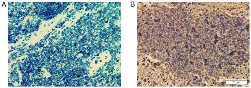
total, lung cancer tissues from three patients were stained. patients had SCLC and one patient had neuroendocrine adeno-
The lung cancer tissues from one other patient who had SCLC carcinoma (Table I). It was revealed that the lung cancer tissues
without the manifestations of anti‑GABAB receptor encepha- of these three patients were positively stained for anti‑GABAB
litis was also stained and used as a control. receptors by immunohistochemistry (Fig. 2). However, the
other four patients with tumors refused further pathological
Treatment and follow‑up. Patients received antiepileptic drug examinations and surgical treatments.
therapy, immunotherapy and tumor therapy when required.
The therapeutic effects were assessed using the modified Treatment and outcome assessment. All patients received anti-
Rankin Scale (mRS) (12). epileptic drug treatments, including oxcarbazepine, sodium
valproate, topiramate and levetiracetam. In addition, all
Results patients received immunotherapy, which included intravenous
immunoglobulin and/or the steroid hormones methylpred-
Clinical manifestations. In total, nine patients were male nisolone or dexamethasone. In total, three patients with lung
(75%) and three were female (25%). The age of symptom onset cancer received tumor resection and chemotherapy. The
ranged from 54 to 74 years (median, 65.1 years). The time of neurological function scores evaluated by mRS and the scores
symptom onset to diagnosis was from 1 to 36 weeks (median, of the patients were 3.37±0.52 (range, 3‑4) prior to therapy and
6.9 weeks). Seizures occurred in all 12 patients and nine (75%) 2.38±0.92 (range, 1‑4) after therapy. The mean follow‑up dura-
patients presented with seizures as the initial symptoms. tion was 11.3 months (range, 3‑30 months). Mortality occurred
Furthermore, three patients developed status epilepticus prior in seven patients at follow‑up. Furthermore, after therapy,
to the treatments. No seizure was recorded during EEG exams. patients without tumors exhibited neurological improvement,
The seizure frequency prior to treatment ranged from 2 to including seizure control, and had no relapse at follow‑up
15 times per week (median, 4.2 times per week). Furthermore, (range, 3‑24 months; Table I).
memory deficits and psychiatric symptoms (including behav-
ioral, mood and personality changes) were documented in 11 Discussion
(91.6%) and seven (58.3%) patients, respectively. Awareness
impairment was observed in four patients (33.3%) (Table I). The present study assessed a number of Chinese patients with
However, none of the patients had the clinical manifestations anti‑GABAB receptor encephalitis. This rare disease primarilyTable I. Clinical manifestations of encephalitis patients with anti‑GABABR antibody.
Case1 Case2 Case3 Case4 Case5 Case6 Case7 Case8 Case9 Case10 Case11 Case12
Sex M M M M M F F M M M M F
Age (years) 62 70 64 54 66 62 69 74 67 65 69 60
TOSD (weeks) 8 3 2 2 7 2 8 9 2 3 1 36
Psychiatric symptoms ‑ + + + + ‑ + + ‑ ‑ + ‑
Memory deficits + + + + + + ‑ + + + + +
Awareness impairment + + + ‑ ‑ ‑ ‑ ‑ ‑ ‑ ‑ +
Seizures + + + + + + + + + + + +
Status epilepticus + ‑ ‑ + ‑ ‑ ‑ ‑ ‑ ‑ + ‑
Seizure frequency prior 3 5 4 15 3 3 2 3 4 2 3 3
to therapy (times/week)
Anti‑GABABR antibody +/+++ ++/+++ +++/+++ ++/++ ++/++ ++/+ +++/++ ++/+ ++/++ ++/+ ++/+ +++/++
(grading), serum/CSF
Anti‑GABABR antibody + ND ND ND ND + ND ND ND ND ND ND
(grading), lung cancer tissue
Other positive autoimmune TG;TPO ‑ ‑ ‑ ‑ ‑ TG;TPO ‑ ‑ ‑ Hu Hu; TG;
antibodies TPO
Serum sodium (mmol/l) 144 138 123 140 146 142 141 142 147 139 135 140
CSF WC (/mm3; RR:0‑6) 2 130 4 62 2 6 4 1 34 1 6 4
CSF protein (g/l; RR:0‑0.45) 0.74 0.84 0.26 0.24 0.33 0.35 0.70 0.45 0.42 0.44 0.62 0.42
CSF OB + ‑ ‑ ‑ ND ‑ ‑ ND ‑ + ‑ +
Lung tumor (CT scan) + + ‑ + + + ‑ ‑ ‑ + ‑ +
Tumor tissue pathology Aden ND ‑ ND ND SCLC ‑ ND ND ‑ ND SCLC
Brain MRI limbic lobes + ‑ + ‑ + + ‑ ‑ ‑ ‑ ‑ ‑
abnormality
Cortical atrophy + ‑ ‑ ‑ ‑ + ‑ ‑ ‑ ‑ ‑ ‑
EEG generalized slow waves + + + ND + ‑ + + + + + +
Epileptic waves ‑ ‑ + ND + ‑ ‑ + + ‑ + +
EXPERIMENTAL AND THERAPEUTIC MEDICINE 20: 617-622, 2020
Immunotherapy drugs IVIg+Dex Mpd IVIg+Dex IVIg+Dex Mpd IVIg IVIg+Dex IVIg+Mpd IVIg+Mpd IVIg IVIg+Dex IVIg
Anti‑epileptic drugs LEV LEV LEV+VPA LEV OXA+LEV LEV LEV LEV+VPA LEV+VPA VPA LEV+VAP LEV+TOP
Tumor treatment mRS Yes No ND No No Yes ND ND ND No No No
(before/after treatments) 4/2 4/4 4/2 3/3 3/3 3/2 3/1 3/2 3/1 3/3 3/2 4/4
Follow‑up duration (Months) 30 6 18 4 5 8 18 6 20 10 8 3
Follow‑up results No relapse Died No relapse Died Died Died No relapse No relapse No relapse Died Died Died
Grading: (+), 1:1‑1:10; (++), 1:10‑1:100; (+++), >1:100. GABABR, γ‑aminobutyric acid B receptor; M, male; F, female; TOSD, time of onset of symptoms to diagnosis; CSF, cerebrospinal fluid; ND, not done
or no date available; TG, anti‑thyroglobulin antibody; TPO, anti‑thyroid peroxidase antibody; WC, white cell; RR, reference range; OB, oligoclonal bands; SCLC, small cell lung cancer; Aden, adenocarcinoma;
CT, computer tomography; MRI, magnetic resonance imaging; EEG, electroencephalogram; Mpd, methylprednisolone; IVIg, intravenous immunoglobulin; Dex, dexamethasone; LEV, levetiracetam; VPA, sodium
valproate; OXA, oxcarbazepine; TOP, topiramate; mRS, modified Rankin scale.
619620 ZHAO et al: FEATURES OF CHINESE ANTI-GABABR ENCEPHALITIS Figure 1. Images of one limbic encephalitis patient with anti‑γ‑aminobutyric acid B receptor antibody. (A) Transverse and (B) coronal sections of MRI fluid‑attenuated inversion recovery sequence indicate increased signals in the bilateral mesial temporal lobes (white asterisks). (C and D) Thorax CT revealing one mass in the right lung hilus on (C) pulmonary and (D) mediastinal window image (black asterisks). Figure 2. GABAB receptors are expressed in the tumor tissue of a patient with small cell lung cancer and anti‑GABAB receptor encephalitis. (A) No nuclei with brown staining are detected in the lung tumor tissues from one patient with small cell lung cancer without the manifestations of anti‑GABAB receptor encephalitis on immunohistochemistry stain. (B) Numerous cells with brown colored nuclei were observed in lung tumor tissues of one patient (Case 6) with small cell lung cancer and anti‑GABAB receptor encephalitis on immunohistochemistry stain. GABAB, γ‑aminobutyric acid B. affects middle‑aged and aged males who have a high risk of to immunotherapy (4‑9,13). In the present study, all patients receptor encephalitis, usually manifesting as LE, and has had seizures as the major symptom. Consistent with previous symptoms including seizures, memory deficits, psychosis studies, manifestations including memory deficits, psychiatric and altered consciousness (4‑9). Furthermore, seizures are changes and confusion were observed in the present study (4‑9). frequently the initial and most prominent symptom, which are The GABAB receptor is a G protein‑coupled receptor for the usually refractory to anti‑epileptic drugs but exhibit a response inhibitory neurotransmitter GABA. The GABAB receptor is
EXPERIMENTAL AND THERAPEUTIC MEDICINE 20: 617-622, 2020                                       621

able to mediate pre‑synaptic and post‑synaptic GABAergic inhi-       of patients with GABAB receptor encephalitis (1). These
bition and suppress high activity states. Autoantibodies binding     patients may also have other autoantibodies, including
to GABAB receptor may promote synaptic activity states with          anti‑Hu, anti‑voltage‑gated calcium channel and anti‑thyroid
excessive synchronization in neuronal networks, which leads to       antibodies (4‑9). Furthermore, co‑existence of anti‑GABAB
epileptic seizures (1‑3,14). It has been previously demonstrated     receptor antibodies and onconeuronal antibodies in patients
that mice with GABAB receptor dysfunction developed seizures         with SCLC are frequently associated with poor prognosis (4‑9).
and learning difficulties (15).                                      In the present study, two patients were also determined to have
    Hyponatremia was detected in two patients. One patient           anti‑Hu antibodies. As the diagnosis of tumors is established
(Case 3) with obvious hyponatremia (123 mmol/l) had a                after the diagnosis of anti‑GABAB receptor encephalitis,
symptom of vomiting. No malignant tumor was detected in              screening for cancer is important once the clinical diag-
this patient during the follow‑up. CSF cytology of patients          nosis is confirmed. Furthermore, it has been suggested that
with anti‑GABAB receptor encephalitis has no specific                tumor screening should be performed after the encephalitis
features compared with that of other types of autoimmune or          diagnosis (20).
viral encephalitis. Consistent with previous studies, certain            For the treatment of GABAB receptor encephalitis
patients in the present study had lymphocytic pleocytosis and        with malignancy, immunotherapy and tumor treatment are
a mildly elevated protein concentration (4‑9). Furthermore,          necessary (4‑9). The first line of immunotherapy includes
in the majority of patients, EEG exam results indicated slow         corticosteroids, Igs and plasmapheresis, either alone or in
or epileptic activity in the temporal lobes. In addition, MRI        combination (11). Furthermore, it is strongly recommended that
scans identified that 1/3 of patients had hyperintense signals       the therapy should be started once the anti‑GABAB receptor
in the mesial temporal lobes, which was consistent with the          encephalitis is diagnosed. Seizures caused by anti‑GABAB
results of previous studies (4‑9). It has been reported that brain   receptor encephalitis are frequently refractory to any antiepi-
MRI scans may exhibit dynamic changes in volume and signal           leptic drugs, but respond well to immunotherapy (21). In line
intensity in the amygdala and hippocampus, which indicates           with this, the present results suggested that patients without
considerable inflammation and subsequent degeneration (4‑9).         cancer also responded well to immunotherapy.
Furthermore, brain 18‑fluoro‑deoxyglucose positron emission              In conclusion, it was indicated that seizures and memory
tomography hypermetabolism has been identified in certain            deficits are the major manifestations of anti‑GABAB
patients (7). In addition, MRI changes in patients cannot            receptor encephalitis in Chinese patients. Therefore, testing
provide specific information for the diagnosis of anti‑GABAB         for anti‑GABAB receptor antibodies may be used for elderly
receptor encephalitis. Therefore, negative brain MRI scan            patients with LE or new‑onset refractory seizures. Most
results may not exclude the diagnosis of this disease.               patients with anti‑GABAB receptor encephalitis without
    Antibodies against GABAB receptors are mainly from the           cancer responded well to immunotherapy. However, patients
IgG1 subclass, which may induce neuronal damage directly via         with underlying lung cancer had a relatively poor prognosis.
complement activation and antibody‑dependent cell‑mediated
cytotoxicity (2). In the central nervous system, the GABAB           Acknowledgements
receptor is primarily expressed in the hippocampus, amygdala,
thalamus and cerebellum (1‑3). Furthermore, ~1/2 of patients         Not applicable.
with anti‑GABAB receptor encephalitis have a paraneoplastic
etiology, which is usually SCLC and is frequently identified         Funding
after the development of neurologic symptoms (4‑9). Thymus
carcinoid, melanoma and gastric adenocarcinoma are also              This work was supported by grants from the Natural Science
reported in patients with anti‑GABAB receptor encephalitis           Foundation of China (grant no. 81873786), the Natural Science
and patients with SCLC usually have a poor prognosis after           Foundation of Shandong Province (grant no. ZR2017MH082),
immunotherapy (4‑9,16,17). Similarly, at the follow‑up for the       Innovative Research Project of Resident Standardization
present study, high mortality was reported in patients with          Training of Qilu Hospital, Shandong University (grant
lung cancer. In addition, the present immunohistochemistry           no. ZPZX2019A04) and Undergraduate Teaching Reform and
results indicated that GABAB receptor was expressed in lung          Research Project of Cheeloo College of Medicine, Shandong
cancer tissues; to the best of our knowledge, this has not been      University (grant no. qlyxjy‑201917).
previously reported. Pulmonary neuroendocrine cells may
produce GABA and GABAB receptors are expressed in airway             Availability of data and materials
epithelium (18). Therefore, the present results supported the
hypothesis that the ectopic expression of neuronal proteins          The datasets used and/or analyzed during the current study are
by the tumor reduces immune tolerance for these proteins,            available from the corresponding author on reasonable request.
which then contributes to the development of the autoimmune
encephalitis (19). The GABAB receptors become autoimmune             Authors' contributions
antigens, which leads to extensive infiltration of cytotoxic
T cells and neuronal degeneration. This effect also triggers         XZ was responsible for the analysis of the data and the drafting
B‑cell immune responses, thus leading to the production of           of the manuscript. XY was responsible for the autoimmune
autoantibodies with neuronal functional alterations (1‑3).           antibody detection experiments and immunohistochemistry
    Autoantibodies recognizing the extracellular domain of           staining. XL was responsible for the analysis of the radiology
the GABAB receptor may be detected in serum and CSF                  data and the revision of the manuscript. SW was responsible for
622 ZHAO et al: FEATURES OF CHINESE ANTI-GABABR ENCEPHALITIS
the design, data analysis, critical revision and final approval of 7. Kim TJ, Lee ST, Shin JW, Moon J, Lim JA, Byun JI, Shin YW,
Lee KJ, Jung KH, Kim YS, et al: Clinical manifestations and
the manuscript. All authors read and approved the final version outcomes of the treatment of patients with GABAB encephalitis.
of the manuscript. J Neuroimmunol 270: 45‑50, 2014.
8. Guan HZ, Ren HT, Yang XZ, Lu Q, Peng B, Zhu YC, Shao XQ,
Hu YQ, Zhou D and Cui LY: Limbic encephalitis associated with
Ethics approval and consent to participate Anti‑γ‑aminobutyric Acid B receptor antibodies: A case series
from china. Chin Med J (Engl) 128: 3023‑3028, 2015.
This study was approved by the Ethics Committee of 9. Qiao S, Zhang YX, Zhang BJ, Lu RY, Lai QL, Chen LH and
Wu J: Clinical, imaging, and follow‑up observations of patients
Qilu Hospital of Shandong University (Jinan, China; with anti‑GABA B receptor encephalitis. Int J Neurosci 127:
no. KYLL‑2017‑550). Written informed consent was 379‑385, 2017.
10. Zuliani L, Graus F, Giometto B, Bien C and Vincent A: Central
obtained from each patient or a relative serving as a legal nervous system neuronal surface antibody associated syndromes:
representative. Review and guidelines for recognition. J Neurol Neurosurg
Psychiatry 83: 638‑645, 2012.
11. Graus F, Titulaer MJ, Balu R, Benseler S, Bien CG, Cellucci T,
Patient consent for publication Cortese I, Dale RC, Gelfand JM, Geschwind M, et al: A clinical
approach to diagnosis of autoimmune encephalitis. Lancet
Not applicable. Neurol 15: 391‑404, 2016.
12. van Swieten JC, Koudstaal PJ, Visser MC, Schouten HJ and van
Gijn J: Interobserver agreement for the assessment of handicap in
Competing interests stroke patients. Stroke 19: 604‑607, 1988.
13. Hainsworth JB, Shishido A, Theeler BJ, Carroll CG and
Fasano RE: Treatment responsive GABA(B)‑receptor limbic
The authors declare that they have no competing interests. encephalitis presenting as new‑onset super refractory status
epilepticus (NORSE) in a deployed U.S. soldier. Epileptic
Disord 16: 486‑493, 2014.
References 14. Emson PC: GABA(B) receptors: Structure and function. Prog
Brain Res 160: 43‑57, 2007.
1. Lancaster E and Dalmau J: Neuronal autoantigens‑‑pathogenesis, 15. Prosser HM, Gill CH, Hirst WD, Grau E, Robbins M, Calver A,
associated disorders and antibody testing. Nat Rev Neurol 8: Soffin EM, Farmer CE, Lanneau C, Gray J, et al: Epileptogenesis
380‑390, 2012. and enhanced prepulse inhibition in GABA(B1)‑deficient mice.
2. Melzer N, Meuth SG and Wiendl H: Paraneoplastic and Mol Cell Neurosci 17: 1059‑1070, 2001.
nonparaneoplastic autoimmunity to neurons in the central 16. Boronat A, Sabater L, Saiz A, Dalmau J and Graus F: GABA(B)
nervous system. J Neurol 260: 1215‑1233, 2013. receptor antibodies in limbic encephalitis and anti‑GAD
3. Dalmau J, Geis C and Graus F: Autoantibodies to synaptic associated neurologic disorders. Neurology 76: 795‑800, 2011.
receptors and neuronal cell surface proteins in autoimmune 17. Jia XT, Pan Y, Di Z, Gu N, Liu Z and Kang YM: Anti‑GABAB
diseases of the central nervous system. Physiol Rev 97: 839‑887, receptor encephalitis in a patient with gastric adenocarcinoma.
2017. Neurol Sci 39: 1981‑1984, 2018.
4. Lancaster E, Lai M, Peng X, Hughes E, Constantinescu R, 18. Mizuta K, Osawa Y, Mizuta F, Xu D and Emala CW: Functional
Raizer J, Friedman D, Skeen MB, Grisold W, Kimura A, et al: expression of GABAB receptors in airway epithelium. Am J
Antibodies to the GABA(B) receptor in limbic encephalitis with Respir Cell Mol Biol 39: 296‑304, 2008.
19. DeLuca I, Blachère NE, Santomasso B and Darnell RB: Tolerance
seizures: Case series and characterisation of the antigen. Lancet to the neuron‑specific paraneoplastic HuD antigen. PLoS One 4:
Neurol 9: 67‑76, 2010. e5739, 2009.
5. Höftberger R, Titulaer MJ, Sabater L, Dome B, Rózsás A, 20. Titulaer MJ, Soffietti R, Dalmau J, Gilhus NE, Giometto B, Graus F,
Hegedus B, Hoda MA, Laszlo V, Ankersmit HJ, Harms L, et al: Grisold W, Honnorat J, Sillevis Smitt PA, Tanasescu R, et al:
Encephalitis and GABAB receptor antibodies: Novel findings Screening for tumours in paraneoplastic syndromes: Report of
in a new case series of 20 patients. Neurology 81: 1500‑1506, an EFNS task force. Eur J Neurol 18: e19‑e3, 2011.
2013. 21. Dubey D, Samudra N, Gupta P, Agostini M, Ding K, Van
6. Dogan Onugoren M, Deuretzbacher D, Haensch CA, Ness PC, Vernino S and Hays R: Retrospective case series of
Hagedorn HJ, Halve S, Isenmann S, Kramme C, Lohner H, the clinical features, management and outcomes of patients with
Melzer N, Monotti R, et al: Limbic encephalitis due to GABAB autoimmune epilepsy. Seizure 29: 143‑147, 2015.
and AMPA receptor antibodies: A case series. J Neurol Neurosurg
Psychiatry 86: 965‑972, 2015.You can also read