CLINICAL GUIDELINES FOR - BREAST CANCER CONTROL AND MANAGEMENT - Knowledge Hub
←
→
Page content transcription
If your browser does not render page correctly, please read the page content below

CLINICAL GUIDELINES FOR BREAST CANCER CONTROL AND MANAGEMENT

CLINICAL GUIDELINES FOR
BREAST CANCER CONTROL AND
MANAGEMENT
CLINICAL GUIDELINES FOR BREAST CANCER CONTROL AND MANAGEMENT
CLINICAL GUIDELINES FOR BREAST CANCER CONTROL AND MANAGEMENT

          NATIONAL DEPARTMENT OF HEALTH
CLINICAL GUIDELINES FOR BREAST CANCER CONTROL AND MANAGEMENT

                                   i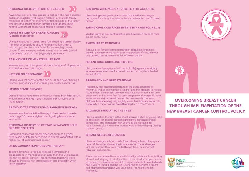
CLINICAL GUIDELINES FOR BREAST CANCER CONTROL AND MANAGEMENT

National Department of Health

Switchboard: 		       012 395 8000

Physical Address: Civitas Building
			               Corner of Thabo Sehume and Struben Streets
			Pretoria

Postal address: 		 Private Bag X828
			Pretoria 0001

              Published by the Department of Health, Private Bag X828, Pretoria, 0001, South Africa

                           Copyright: © Department of Health, Republic of South Africa
                                                  April 2018

                                                www.doh.gov.za

                                                          1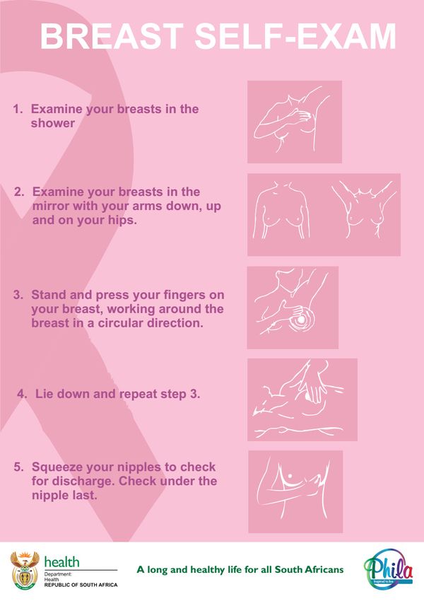
CLINICAL GUIDELINES FOR BREAST CANCER CONTROL AND MANAGEMENT

Table of Contents
Foreword...........................................................................................................................................................................3
Acknowledgements...........................................................................................................................................................4
Abbreviations and Acronyms.............................................................................................................................................5
Definition of terms.............................................................................................................................................................7
List of tables....................................................................................................................................................................10
List of figures...................................................................................................................................................................10
1.     Introduction..............................................................................................................................................................12
2.     Key area 1: Prevention and early detection, screening and genetic assessment...................................................14
3.     Key area 2: Timely access to care...........................................................................................................................24
4.     Key area 3: Assessment, diagnosis and staging.....................................................................................................39
5.     Key Area 4: Treatment of breast cancer..................................................................................................................49
6.     Key area 5: Palliative care in breast cancer............................................................................................................60
7.     Key area 6: Follow-up and surveillance in breast cancer........................................................................................66
8.     Key Area 7: Data, monitoring and research.............................................................................................................73
9.     Key area 8: Community outreach and engagement................................................................................................75
Annexures.......................................................................................................................................................................77

                                                                                       2
CLINICAL GUIDELINES FOR BREAST CANCER CONTROL AND MANAGEMENT

Foreword

                                   Ms Precious Matsoso
                                   Director General

Breast cancer, along with cervical cancer, has been identified as a national priority in South Africa. Breast cancer is the
most prevalent cancer and a leading cause of death among South African women. The increasing incidence of breast
cancer is a major health concern. 19.4 million women aged 15 years old and older live at-risk of contracting the disease.
Per the National Cancer Registry in 2012, 8 203 new cases of breast were observed. Given the recent advances
in medicine and technology, however, we have a tremendous opportunity to attack breast cancer energetically and
effectively with a revised national programme.

The Breast Cancer Clinical Guidelines is an important document aimed at providing detailed information regarding the
standards laid out in the Breast Cancer Prevention and Control Policy – its companion document. These standards
include awareness, prevention, and treatment and care in the South African context. It provides clinicians with the step-
by-step guidance from the initial contact with the patient to the discharge back into the community. The implementation
of these clinical guidelines will reduce the breast cancer related mortalities.

The clinical guidelines will also provide an opportunity to respond to the health system priorities related to cancers.
It provides guidelines on the required minimum standard to establish Regional Breast Units (RBUs), including list of
essential equipment, essential medicines, and personnel required. It further provides guidance on collaboration with
civil society and private partners in fighting the battle against cancer.

Ms P Matsoso
Director General
29/10/ 2019

                                                               3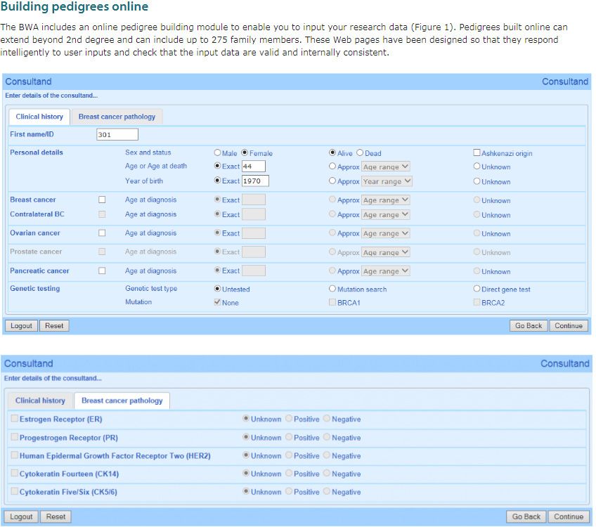
CLINICAL GUIDELINES FOR BREAST CANCER CONTROL AND MANAGEMENT

Acknowledgements
The National Department of Health would like to acknowledge the irreplaceable contribution of the writing group of breast
cancer clinicians responsible for the clinical guidelines. The authors of this document would like to make clear however,
that any conclusions in this document and standards derived from this evidence are not necessarily the opinion of this
writing group and should not be taken as such.

The contributors consisted of breast surgeons, plastic and reconstructive surgeons, oncologists, radiologists, nuclear
physicians, pathologists, and geneticists. In addition, experts in the various fields, including civil society organisations
and other interest groups, were also contacted when necessary for further input. The clinical guidelines provide additional
context based on information that may not be available in the Breast Cancer Prevention and Control Policy. It is aligned
to the health care situation in South Africa and the needs envisaged by the people of this country.

NDoH leads: Dr M Makua and Dr Y Pillay

Project Manager and Editorial lead: Mr J Tillus and Clinton Health Access Initiative

Clinical experts: Dr A Sherriff, Dr A Hoosen, Dr D Shamley, Dr T Thebe, Dr E Panieri, Dr G Demetriou, Dr J Smilg, Dr J
Edge, Dr L Cairncross, Dr P Ruff, Dr S Nietz, Dr S Rayne, Dr S Cacala, Dr L Stopforth, Ms T Wainstein, Dr Z Mohamed,
Dr Z Jafta, Dr R Krause, Dr I Buccimazza and Dr H Cubasch

National Department of Health contributors: Mr G Steel, Ms J Riddin, Ms D Chweneyagae and Ms S Singh

Partners and CSOs: Ms S Meyer (Cancer Alliance) and Ms L Turner (Breast Health Foundation)

Other contributors: Ministerial Advisory Committee for Cancers (MACC) representatives, Affordable Medicines
representatives, Provincial Heads of Breast units, Medical Oncology units and Radiation Oncology units, Breastcare,
Bettercare Books, BIGOSA and The Breast Course for Nurses

                                                            4
CLINICAL GUIDELINES FOR BREAST CANCER CONTROL AND MANAGEMENT

Abbreviations and Acronyms

ADH          Atypical ductal hyperplasia
ALH          Atypical lobular hyperplasia
ALND         Axillary lymph node dissection
ASR          Age standardised rate
AUS          Axillary ultrasonography
BCCCP        Breast Cancer Comprehensive Control Policy
BCN          Breast care nurse
BCS          Breast conserving surgery
BI-RADS      Breast imaging-reporting and data system
BMI          Body mass index
BPM          Bilateral prophylactic mastectomy
BPSO         Bilateral prophylactic salpingo-oophorectomy
BRCA         Breast cancer gene mutation
BRCA1        Breast cancer gene 1
BRCA2        Breast cancer gene 2
BCRL         Breast cancer related lymphoedema
BSE          Breast self-examination
CBC          Contralateral breast cancer
CBE          Clinical breast examination
CI           Confidence interval
CISH         Chromogenic in-situ hybridization
CMF          Cyclophoshamide, methrotrexate and fluorouracil
CNB          Core needle biopsy
CPM          Contralateral prophylactic mastectomy
CT           Computerized tomography
DCIS         Ductal carcinoma in situ
DFS          Disease free survival
DH           District Hospital
DOH          Department of Health
EDA          Emotional distress assessment
ER/PR        Estrogen-receptor/progesterone receptor
ESMO         European Society for Medical Oncology
FDG-PET      Fluorodeoxyglucose-positron emission tomography
FEC          5-fluorouracil, Epirubicin, and cyclophosphamide
FISH         Fluorescent in-situ hybridization
FNAC         Fine needle aspiration cytology
H&E          Haematoxylin and eosin
HER-2        Human epidermal growth factor receptor 2
HERA         Herceptin Adjuvant
HR           Hazard ratio
IDC          Invasive ductal carcinoma
IHC          Immunohistochemistry
ILC          Invasive lobular carcinoma
LABC         Locally advanced breast cancer
LCIS         Lobular carcinoma in situ
LHRH         Luteinizing-hormone-releasing hormone
LN           Lobular neoplasia
LTR          Lifetime risk
MDT          Multi-disciplinary team

                                                        5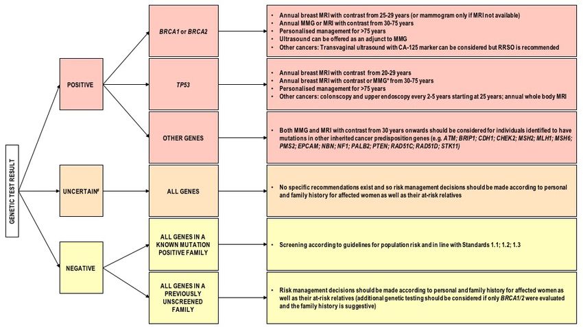
CLINICAL GUIDELINES FOR BREAST CANCER CONTROL AND MANAGEMENT

MMG        Mammogram
MRI        Magnetic resonance imaging
NCCN       National Comprehensive Cancer Network
NDoH       National Department of Health
NICE       National Institute for Health and Care Excellence
OS         Overall survival
PCHCT      Palliative and hospice care team
Pcr        Pathological complete response
PCR        Polymerase Chain reaction
PET/CT     Positron emission tomography/computerized tomography
PHC        Primary Health Care
PISCBE     Provider Initiated Screening Clinical Breast Examination
PPV        Positive predictive value
QoL        Quality of life
RBu        Regional breast unit
RCT        Randomized control trial
RR         Relative Risk
RRSO       Risk reducing salpingo-oophorectomy
SBA        Specialist breast assessment
SBU        Specialized breast unit
SISH       Silver-enhanced in-situ hybridization
SLNB       Sentinel lymph node biopsy
SR         Systematic review
TRAM       Transverse rectus abdominismyocutaneous
WHO/IARC   World Health Organization/International Agency for Research on Cancer

                                                    6
CLINICAL GUIDELINES FOR BREAST CANCER CONTROL AND MANAGEMENT

Definition of terms

               Term                                                     Definition
Adjuvant systemic therapy           Chemotherapy, monoclonal antibodies, radiotherapy and hormonal blockade
                                    given after surgery to help decrease the risk of the cancer recurring.
Benign disease                      Condition, tumour, or growth that is not cancerous. This means that it does not
                                    spread to other parts of the body. It does not invade nearby tissue
Breast care nurse                   Nurse who specialises in breast care. Breast care nurses improve the continuity
                                    of care for women, and provide important information, support and referral for a
                                    wide range of needs experienced by women
Breast conserving surgery           An operation to remove the cancer and some normal tissue around it, but not
                                    the breast itself. It is also called breast-sparing surgery, lumpectomy, partial
                                    mastectomy, quadrantectomy, and segmental mastectomy
Early and locally advanced breast   Early breast cancer is defined as tumours of not more than 5 cm diameter,
cancer                              with either impalpable or palpable but not fixed lymph nodes and no evidence
                                    of distant metastases. Locally advanced breast cancer is defined as invasive
                                    breast cancer that has one or more of the following features: may be large
                                    (typically bigger than 5 cm) may have spread to several lymph nodes in the
                                    armpit (axilla) or other areas near the breast
Emotional distress                  A negative emotional reaction—which may include fear, anger, anxiety, and
                                    suffering. Breast cancer treatment can be both physically and emotionally
                                    exhausting. There are many changes taking place that may be difficult to cope
                                    with. “Chemobrain” is a term coined to describe the mental changes caused by
                                    chemotherapy treatment. Patients have experienced memory deficits and the
                                    inability to focus. Breast cancer treatments can also leave patients fatigued,
                                    which is normal.
High burden disease                 Impact of a health problem as measured by financial cost, mortality, morbidity,
                                    or other indicators
Image guided core needle biopsy     Diagnostic procedure in which a core needle biopsy can be performed using
                                    ultrasonic or stereotactic guidance to confirm cancer in the breast
Lymph node surgery                  Procedure whereby a surgeon operates to remove a primary cancer, one or
                                    more of the nearby (regional) lymph nodes may be removed as well
Lymphedema                          Condition of localized fluid retention and tissue swelling caused by a
                                    compromised lymphatic system, which normally returns interstitial fluid to the
                                    bloodstream
Mammogram/mammography               An X-ray of the breast that is taken with a device that compresses and flattens
                                    the breast
Mastectomy                          Surgical removal of one or both breasts, partially or completely
MDT                                 An integrated team approach to health care in which medical and allied health
                                    care professionals consider all relevant treatment options and develop an
                                    individual treatment plan for each patient collaboratively
Metastatic disease                  Condition where cancer cells break away from where they first formed (primary
                                    cancer), travel through the blood or lymph system, and form new tumours
                                    (metastatic tumours) in other parts of the body
Micro-metastatic disease            Small collection of cancer cells that has been shed from the original tumour and
                                    spread to another part of the body through the lymphovascular system
Movement related pain               Pain that occurs with movement
Navigator                           An individual who guides patients with a suspicious finding (e.g., test shows
                                    they may have cancer) through and around barriers in the complex cancer care
                                    system to help ensure timely diagnosis and treatment.
Neo-adjuvant therapy                Chemotherapy given prior to definitive surgery in an attempt to reduce cancer
                                    size prior to operation
Palliative care services            Approaches that improve the quality of life of patients and their families facing
                                    the problem associated with life-threatening illness, through the prevention and
                                    relief of suffering by means of early identification and impeccable assessment
                                    and treatment of pain and other problems, physical, psychosocial and spiritual

                                                           7
CLINICAL GUIDELINES FOR BREAST CANCER CONTROL AND MANAGEMENT
Post mastectomy radiation Radiation therapy seeks to eradicate occult disease that remains in
postmastectomy chest wall or regional nodal basins, including the
supraclavicular, axillary, and internal mammary regions to reduce the risk of
postmastectomy locoregional recurrence and to improve overall survival
Provincial oncology unit Oncology unit located at a provincial-level facility
Radiation therapy Type of cancer treatment that uses beams of intense energy to kill cancer cells.
Radiation therapy most often uses X-rays.
Reconstructive surgery options Surgical procedure that restores shape to your breast after a mastectomy
Regional breast unit An RBU is a facility (primary or secondary) that has the adequate staffing and
equipment to render the essential packages of services for prevention and
early diagnosis
Sentinel lymph nodes biopsy Procedure in which the sentinel lymph node is identified, removed, and
examined to determine whether cancer cells are present. A negative SLNB
result suggests that cancer has not developed the ability to spread to nearby
lymph nodes or other organs
Specialised breast unit Tertiary or quaternary with MDT capabilities
Stage 1 disease A cancer is relatively small and contained within the organ it started in
Stage 2 disease Tumour is larger than in stage 1, but the cancer has not started to spread into
the surrounding tissues. Sometimes stage 2 means that cancer cells have
spread into lymph nodes close to the tumour. This depends on the particular
type of cancer
Stage 3 disease Cancer is larger. It may have started to spread into surrounding tissues and
there are cancer cells in the lymph nodes in the area
Stage 4 disease Cancer has spread from where it started to another body organ. This is also
called secondary or metastatic cancer
Synoptic histological assessment clinical documentation method that uses structured checklists to help clinicians
produce more complete, consistent and valuable medical reports
Triple assessment Combination of three tests, i.e. clinical examination, radiological imaging
(mammography, ultrasonography) and pathology used to accurately diagnose
all palpable breast lumps
8CLINICAL GUIDELINES FOR BREAST CANCER CONTROL AND MANAGEMENT
List of tables
Table 1: Possible outcomes of genetic testing results and implications for the individual and family.............................21
Table 2: Risk categorisation for screening.......................................................................................................................23
Table 3: Minimum requirement for an RBU and SBU......................................................................................................24
Table 4: List of Regional Breast Units by province..........................................................................................................26
Table 5: List of Specialised Breast Units (SBU) by province...........................................................................................27
The signs and symptoms as outlined in Table 6 may vary from subliminal signs to limb swelling and
limited limb functionality...................................................................................................................................................67
Table 7: Stages, Signs and Symptoms of BCRL.............................................................................................................67
Table 8: Lymphedema Risk Checklist to be followed by lymphedema (LE) therapist.....................................................68
Table 9: Referral pathways for lymphedema management at all levels of care..............................................................69
List of figures
Figure 1: The High-5 Method for patient risk assessment. Source: National Department of Health South Africa...........15
Figure 2: Symptomatic patient care algorithm.................................................................................................................18
Figure 3: Algorithm for image-based screening for women who have undergone genetic testing..................................22
Figure 4: Spoke-Hub model demonstrating a centralised model of care for breast cancer. Until SBUs (with specialised
and multi-disciplinary capabilities) can be established across the country, RBUs with linkages and coordinated
relationships with SBUs must be established. Linkages between SBUs within a province and between provinces may
also exist as needed........................................................................................................................................................28
Figure 5: Service requirements for a Specialist Breast Unit (SBU).................................................................................31
Figure 6 Service requirements for a Regional Breast Unit (RBU)...................................................................................33
Standard 2.11, Standard 2.12, and Standard 2.13 speak to the required timeliness and quality of patient care as
affirmed by medical literature and the experience of specialists in South Africa. These standards will also form
the basis for evaluation of service standards during the peer review visits. The timelines of these standards are
summarised in Figure 7 below.........................................................................................................................................37
Figure 8: Timeline of patient care upon presentation of breast symptoms......................................................................38
Figure 9: Breast cancer staging......................................................................................................................................44
Figure 10: Standard Operating Procedure: Integrating Nurse & Patient Navigators into breast cancer care in the Public
Health Sector of South Africa..........................................................................................................................................46
Figure 11: Protocol for surgery........................................................................................................................................49
Figure 12: Protocol for SLNB and ALND.........................................................................................................................50
Figure 13: SPADI Pain Domain.......................................................................................................................................70
Figure 14: Quick access Early Warning System to detect upper limb deterioration: Only Pain items on SPADI............71
Figure 15: Examples of incorrect elevation of the scapula at rest and during arm abduction.........................................71
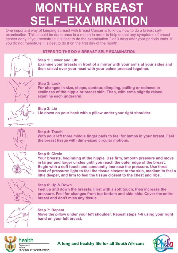
Figure 16: Examples of an appropriate self-breast examination. Source: Groote Schuur Hospital................................82
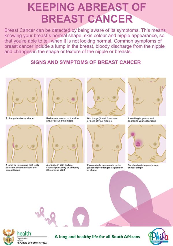
Figure 17: Example of awareness message on signs and symptoms of breast cancer. Source: National Department of
Health South Africa..........................................................................................................................................................84
Figure 18: Example of awareness message for monthly self-examinations. Source: National Department of Health
South Africa.....................................................................................................................................................................85
9CLINICAL GUIDELINES FOR BREAST CANCER CONTROL AND MANAGEMENT
Figure 19: Example of awareness message for self-breast examination. Source: National Department of Health South
Africa...............................................................................................................................................................................86
Figure 20: Example of awareness message for monthly self-breast examination. Source: National Department of
Health South Africa..........................................................................................................................................................87
Figure 21: Example of a Z-fold information pamphlet created for the Breast Cancer Prevention and Control Policy
(2017). Front....................................................................................................................................................................88
Figure 22: Example of a Z-fold information pamphlet created for the Breast Cancer Prevention and Control Policy
(2017). Back....................................................................................................................................................................89
Figure 23: Printable copy of the High-5 method. Source: National Department of Health South Africa..........................90
Figure 24: Example of risk breast cancer calculator. Source: IBIS Breast Cancer Risk Evaluation Tool........................92
10CLINICAL GUIDELINES FOR BREAST CANCER CONTROL AND MANAGEMENT
1. Introduction
1.1. Background and context
• These guidelines provide additional concept of regional breast units, which was not included in the policy
guidelines. This provides an opportunity for the equitable access to breast care and notes that not all breast
conditions will result in breast cancer.
• The guidelines also expand on the concept of breast care nurse. This is not formally adopted by the regulatory
authority (South African Nursing Council), but may refer to any professional nurse who completed the course
on breast care.
• There is an accredited breast care course which is available online with intermittent face-to-face session.
• The department is working in collaboration with NGOs to develop the Breast Care course, which will be available
for nurses working in public health facilities.
• This guideline also provides the concept of patient navigator, which is not a formal qualification or post but a
delegation of the nurse/counsellor who will take responsibility of being a contact between the patient and the
health care system.
• The guideline further introduces the provider-initiated clinical breast examination. It will require a development
of on-site skills demonstration to health care workers. The National Department of Health will provide the
training package and program for health care workers in PHC and district hospitals.
• The implementation of these guidelines is essential as they will form the basis of the implementation of the
National Cancer Campaign in the next 3 years.
1.2. Goals and objectives
GOALS:
• Improve survival
• Decrease time to presentation, diagnosis and treatment
• Decrease stage at point of treatment
• Improve quality of life in survivorship and palliation
• Effectively monitor and evaluate program implementation and the impact of breast cancer interventions
STRATEGIC OBJECTIVES:
• To improve early detection rates by promoting community awareness, and educating communities and health
care workers on breast healthcare and breast cancer management.
• To facilitate referral pathways for patients with breast healthcare concerns.
• To provide guidelines for establishing appropriate facilities for the management and care of breast conditions
• To set standards for optimal care and management of breast conditions
• To provide a framework for auditing standards and outcomes
1.3. Guiding principles
GLOBAL FRAMEWORK
• South Africa recognises the United Nations’ Resolution adopted by the General Assembly on September 25,
2015: Transforming our world: the 2030 agenda for sustainable development. The development of the policy
is thus guided by sustainable development goal (SDG) 3: “Ensure healthy lives and promote well-being for
all at all ages”. One critical SDG target states that governments must ensure universal access to sexual and
reproductive health-care services, including family planning, information and education, and the integration of
reproductive health into national strategies and programmes by 2030.
NATIONAL FRAMEWORKS
• In recognizing that health and development of the country are integrally linked, health reform in South Africa is
firmly embedded in the country’s National Development Plan 2030 Our Future – make it work. The NDP aims for
an inter-connectedness with the World Health Commission on the Social Determinants of Health which are con-
sidered key to any equitable health service delivery platform and includes the need to: improve the conditions
of daily life, tackle inequitable distribution of power, money and resources and measure the problem, evaluate
actions and expand the knowledge base (NCD DOC).
• South Africa is in the process of introducing the National Health Insurance (NHI), in line with the National Devel-
opment Plan. The NHI is a health financing system whose aim is to ensure that all South Africans have access
to affordable, quality health services, based on health needs, rather than socioeconomic status. NHI importantly
recognises that there is a need for massive reorganisation of the health care system to create a new platform
for service provision which will also forms the basis for this policy development.
11CLINICAL GUIDELINES FOR BREAST CANCER CONTROL AND MANAGEMENT
POLICIES, STRATEGIC PLANS AND PROGRAMMES
• Strategic plans for maternal, new-born, child and women’s health and nutrition (MNCWH&N) in South Africa
(2012-2016), and the National Contraception and Fertility planning policy and service delivery guidelines (2012)
cover other SRH priorities, and provide platforms for the implementation of the policy. All the above guidelines
allow the full integration of this policy with other existing policies in the department to comprehensively address
the non-communicable diseases. Integration: The policy provides synergy with other existing policy guidelines
that aim to ensure universal access to sexual and reproductive health services.
Outcome focus:
• The main focus is on promoting early detection and treatment. This policy includes prevention, screening,
diagnosis, treatment, care, and palliative care services. It includes the service delivery package in the community,
PHC, district, regional and tertiary hospitals and private institutions.
Community engagement and involvement:
Included in the policy is the role of civil society organisations and the various ways of raising community the awareness
around breast cancer.
1. Key area 1: Prevention and early detection, screening and genetic assessment
Standard 1.1: Women over 40 years attending a Primary Health Clinic will have a clinical breast examination (Provider
Initiated Screening Clinical Breast Exams or “PISCBE”) biannually.
All women irrespective of the reason for the visit to the facility should receive provider initiated screening clinical
breast examination (PISCBE). The examination should be done systematically, followed by the recording of the
results. If any abnormality is detected irrespective of the severity, that woman should immediately be given a referral
letter detailing the findings to the regional breast unit. Refer to Annexures for examples printed material with step-by-
step instruction.
Standard 1.2: Breast care education in all primary healthcare (PHC) facilities
The opportunistic breast education will include, but not limited to, the following:
A. WHAT IS BREAST CANCER?
Breast cancer is caused by abnormal cells in the body that grow and increase in number without stopping causing a
mass. In breast cancer these cells start in the breast and can spread to other parts of the body.
B. RISK FACTORS FOR BREAST CANCER:
• Breast cancer is very common in women and most women have no specific cause or reason for developing
breast cancer
• Although women over 50 are more likely to get breast cancer, cancer can also develop in women in their 30’s
and 40’s. Very rarely, men can develop breast cancer too
• Risk of breast cancer is higher if you have a close family member with breast cancer such as your mother, sister,
daughter and all individuals mentioned in standard 1.5 of this document.
• Having a distant relative with breast cancer (grandmother, cousin, aunt, niece) will only slightly increase your
risk
• Breast cancer risk is higher for women who have increased exposure to female hormones (oestrogen). In-
creased exposure to oestrogen occurs in women who have no children or children when they are older or wom-
en who use oestrogen containing oral contraceptives or hormone replacement therapy
• Being overweight increases your risk of breast cancer
• Lack of physical exercise
• High saturated fats in diet
• High amount of sugar in diet
• Smoking and drinking may increase your breast cancer risk
C. SIGNS OF BREAST CANCER:
• Breast cancer usually presents as a painless lump in the breast
• Other signs of breast cancer are:
o A bloody or clear nipple discharge
o Nipple retraction (pulled in)
o Skin changes such as dimpling or swelling “orange peel”
o Lumps under the arm (lymph nodes)
o Scaly rash on the nipple
12CLINICAL GUIDELINES FOR BREAST CANCER CONTROL AND MANAGEMENT
D. BREAST CANCER CAN BE CURED IF DETECTED EARLY:
• It is important to know what is normal for your breasts. You may notice this when in the bath or shower or
when looking in the mirror
• If you notice a change in your breast, consult your local clinic or doctor immediately to have this investigated
• Breast self-examination may help you be aware of any early changes in your breasts
See Annexure Standard 1.2 for examples of breast cancer and breast self-examination pamphlets for women.
Standard 1.3: Awareness messages should be disseminated for communities and health care workers that
any woman who notices a change in breast should report to the facility.
Breast cancer is still associated with myths and stigma; and it is therefore essential that factual messages are
communicated to raise awareness amongst the general public, including families and community leaders.
See Annexure Standard 1.3 examples of awareness messages.
Standard 1.4: All eligible women should have their risk of breast cancer determined and be managed according
to local protocol.
During a routine 6-monthly check-up, the patient must be assessed for risk of breast cancer. The figure below
demonstrates “The High-5” Method, which is an easy-to-remember set of questions that must be asked of the patient.
Depending on the response to question 1, a breast exam will be conducted. As the breast exam is being done, the
clinical personnel must ask the remaining four questions. The breast risk assessment must be routinely done every 6
months (Hi-5) Questions:
Figure 1: The High-5 Method for patient risk assessment. Source: National Department of Health South Africa
If YES is answered to any of the above questions, then follow-up with the following:
- When was your last breast exam? Pay particular attention to patients whose last exam was conducted beyond
6 months. If so, conduct clinical breast exam.
- Changes to your breast, such as a lump in breast or armpit? Use this as an educational opportunity. Look for
the following items (check box):
yy Lump
yy Nipple discharge
yy Changes in colour
yy Skin changes
yy Any change in the size of the breast and/or swelling
- Do you experience any abnormal vaginal bleeding? Ask for bleeding after (check box):
yy After sexual intercourse
yy Post-menopause
yy In-between menstrual cycle
13CLINICAL GUIDELINES FOR BREAST CANCER CONTROL AND MANAGEMENT
- Have you ever had a pap smear? When was your last pap smear? Were there any abnormalities communi-
cated to you?
- Do you have a family history of cancer? Use this as an opportunity to explore further.
See Annexure 1.4 for a printable copy of the High-Five Method risk assessment form.
In case of high risk patients, clinicians can use another risk calculator index available. Annexure Standard 1.7
provides examples of risk assessment calculators.
• Women with breast symptoms presenting to any health facility (first point of contact) should have a history and
breast examination performed and then should be referred DIRECTLY to either a RBU or SBU depending on
accessibility
• Women at symptomatic breast clinics will present with symptoms within four identifying symptom clusters:
Mastalgia, Breast Lump, Infection, Nipple Discharge and a proportion of women will be asymptomatic but
concerned about their personal risk of breast cancer
• Referrals to a RBU/SBU should fall into the following referral categories:
o Immediate Referral – to be seen at the next clinic SYMPTOMS/SIGNS suggestive of breast cancer
o Early referral – to be seen within 21 days: SYMPTOMS/SIGNS indeterminate but may be breast cancer
o Routine referral – to be seen within 60 days: NORMAL/ BENIGN pathology
• Determining low and intermediate and high risk clinical findings at primary level of care requires appropriate
training of primary care clinicians in risk factors for breast cancer, signs and symptoms of breast cancer, the
spectrum of benign breast disease and adequate clinical breast examination
URGENT REFERRALS:
Women with the following findings on clinical examination should be referred to the Breast Clinic (preferably at an
SBU) immediately and be seen at the next weekly clinic:
• Clinically suspicious breast lump (fixed, hard, irregular)
• Axillary or supraclavicular lymphadenopathy
• Skin ulceration or nodules
• Skin tethering
• Nipple retraction
• Eczematous nipple changes
• Patients with a pathological nipple discharge (spontaneous, unilateral, single duct, bloody/serous)
The following patients warrant referral to a SBU/RBU within 21 days:
• Patients >25 with a palpable breast lump
• Patients a non lactational breast infection or abscess
The following patients should be referred to an SBU/RBU within 60 days:
• Patients with mastalgia and a normal clinical examination
• Patients presenting with a concern about family history with a normal clinical examination
• Patients with lactation associated infection with non-resolution or recurrent symptoms
14CLINICAL GUIDELINES FOR BREAST CANCER CONTROL AND MANAGEMENT
Figure 2: Symptomatic patient care algorithm
Patient with Breast Symptom
First point of
Contact(PHC/DH)
Clinical Breast Examination
DIRECT REFERRAL to
BREAST CLINIC at
SBU/RBU
High Suspicion Intermediate Suspicion ca Low suspicion ca/
Malignancy Indeterminate examination Normal examination
Urgent Referrals Early Referrals Routine Referrals
Seen Next Clinic Within 21 Days Within 60 Days
• Clinically suspicious breast • Palpable breast lump in women • Palpable breast lump in
lump (fixed, hard, irregular) >25 years womenCLINICAL GUIDELINES FOR BREAST CANCER CONTROL AND MANAGEMENT
Standard 1.5: Referral to genetic services is offered to women whose family history meets the criteria for
referral.
ELIGIBILITY CRITERIA FOR REFERRAL TO GENETIC SERVICES:
Individuals who fulfil these criteria must be referred to Genetic Services for assessment and management:
yy A person with breast/ovarian cancer who has any of the following:
o Known mutation in a cancer predisposition gene (e.g. BRCA1/2, p53) in the family
o Cancer diagnosedCLINICAL GUIDELINES FOR BREAST CANCER CONTROL AND MANAGEMENT
Table 1: Possible outcomes of genetic testing results and implications for the individual and family
Standard 1.6: Women who are known to carry a gene mutation should have annual image-based screening with MRI.
Figure 3: Algorithm for image-based screening for women who have undergone genetic testing
*In general, the use of MMG is discouraged in individuals who carry a pathogenic TP53 mutation. However, if MRI is not
available the risk of a cancer diagnosis outweighs the risk of radiation exposure from MMG.
#A variant of uncertain significance is defined as a change in the DNA sequence of a gene which is poorly understood
with respect to its contribution to disease causation. Additional research is required to classify the sequence change is
either benign or pathogenic.
Other important considerations:
• This algorithm must be considered in conjunction with other risk management strategies for inherited forms of breast
cancer (i.e. SBE; CBE; chemoprevention; prophylactic mastectomy; prophylactic oophorectomy; transvaginal
ultrasound and CA-125 markers). The personal and family history of the at-risk individual, in addition to their
personal desires, psychosocial wellbeing, and support systems should also be evaluated and considered in the
decision-making process and tailoring of the risk management programme.
17CLINICAL GUIDELINES FOR BREAST CANCER CONTROL AND MANAGEMENT
• This algorithm can be applied to both individuals who have not been diagnosed with cancer and those who have
been diagnosed with cancer and have undergone a mastectomy as part of their treatment.
• Such considerations should be made by an at-risk individual in conjunction with a multi-disciplinary team
consisting of a genetics professional (genetic counsellor; medical geneticist), radiologist, surgeon, oncologist,
gynaecologist (preferably with an interest in oncology), and possibly a psychologist.
• Wherever possible, members of the MDT should make the at-risk individuals aware of the risks posed to their
unaffected relatives and encourage increased awareness, referral to genetic services and increased screening
availability.
Standard 1.7: Women at high risk of developing breast cancer should be considered for annual breast MRI in
addition to mammography and CBE
While a variety of imaging modalities have been developed for breast cancer screening, mammography is both the
best studied and the only imaging technique that has shown a mortality benefit. Ultrasonography is commonly used for
diagnostic follow-up of an abnormality seen on screening mammography. It clarifies features of a potential lesion, and
it may be an adjunct to mammography in women with increased breast density. Magnetic resonance imaging (MRI),
performed in combination with mammography, is primarily targeted to screening in high-risk patients and not general
population-based screening.
Table 2: Risk categorisation for screening
Risk Category Lifetime Risk (%) Screening Age to start Age to stop Frequency
High Risk as > 30%+ Mammography 40 70 Annual
determined by (MRI for patients under (or 5 years before
a risk calculator. 30 years of age can be the age at which
See Annexures for considered) the relative was
examples of risk diagnosed with
calculators. Women with a BRACA2 breast cancer if
mutation this calculated age
is earlier than 40
years)
Risk-factors to pay attention to in the high-risk group include:
• Known BRCA mutation carriers
• First-degree relatives of known BRCA mutation carriers
• Radiation to the chest between age 10 and 30
• Genetic mutation in genes causing the Li-Fraumeni, Cowden and Bannayan-Riley syndromes
18CLINICAL GUIDELINES FOR BREAST CANCER CONTROL AND MANAGEMENT
2. Key area 2: Timely access to care
Standard 2.1: RBUs should meet the minimum standards to provide accurate diagnosis of benign and malignant dis-
ease. SBUs should have minimum staffing and equipment to accurate treatment of benign and malignant disease.
Table 3: Minimum requirement for an RBU and SBU
Facility Minimum equipment and service
Minimum staffing requirements Minimum equipment requirements
Type delivery package
• Surgeon in breast surgery (Clinician with • Ultrasound (spec) yy Navigation
appropriate qualifications or exposure/ • Biopsy guns and needle yy Counselling
experience) • Theatre and equipment yy Guided biopsy
Regional Breast Unit
• Radiologist / Sonographer trained in breast • Teleconferencing facility yy Breast surgery (excluding com-
biopsy procedure • Database plex Oncoplastic and Sentinel
• Anaesthetist (Qualified professional with • Mammogram nodes surgery)
(RBU)
diploma in anaesthesia)
• Pathologist
• Mammographer (2X per machine)
• Registered nurses
• Administration
• Patient advocacy representation (under
nurse supervision)
• 2X Specialist surgeon with exposure (ex- yy Gamma probe yy Access to oncologists (Medical
tensive – 5yrs/ 50 /year) yy Radiology Department with MRI, and Radiation)
• Plastic Surgeon (Qualification or Exposure Mammogram, Ultrasound, CT yy Access to nuclear medicine
– experience) in Breast surgery Scan, Bone Scan (Bone scanner, Gamma Cam-
• Radiologist yy Specimen mammography era, etc.)
Specialised Breast Unit
(SBU): Tertiary hospital
• Mammographer
• Breast Specialist Nurse1 once established/
Registered oncology trained nurses
• Administrator
• Patient advocacy representation (under the
supervision of nurse)
• Anaesthetist (anaesthetics diploma) –
• Pathologists
• Lymphedema specialist
• Physiotherapist
• Access to Oncologists (Medical and Radi-
ation)
19CLINICAL GUIDELINES FOR BREAST CANCER CONTROL AND MANAGEMENT
Standard 2.2: SBUs should allow rapid referral and access for Specialist Breast Assessment (SBA) within 1 –
62 days according to referral triage.
Open access is required at certain institutions; however as regional structures improve, appointments via referral
pathways will be preferable. Nevertheless, patients should not wait longer than 62 days.
HUB AND SPOKE SYSTEM:
Timely transition from screening and early detection to treatment (radiotherapy, chemotherapy, surgery, or palliative)
is imperative for the survivorship of a patient suspected of breast cancer. And in instances of advanced disease
presentation, direct referral to a SBU after passing through a RBU must be expedient and uninterrupted. Patient care
is jeopardized by the lengthy pathways for continuum of care. Hindrances to timely treatment are primarily due to, but
not limited, the following:
yy Shortage of qualified human resource (both specialized and support) for clinical assessment, medical imaging
and nuclear medicine, surgery, clinical laboratory and pathology, radiotherapy, systemic therapy and palliative
and end of life care at all levels
yy Availability and maintenance of medical devices and equipment
yy Poor infrastructure
yy Patient transport and accommodation
yy Burdensome costs to the patient (and his/her loved ones)
yy Disjointed referral requirements (repeated referrals for unnecessary tests, referrals to another facility or the
same facility that in actuality serve as an impasse, etc.)
Given the aforementioned limitations in the continuum of care, linkages must be established in the interim between
RBUs and SBUs.
An RBU is a facility (primary or secondary) that has the adequate staffing and equipment to render the essential
packages of services1 for secondary prevention and early diagnosis. Until South Africa can establish SBUs2 (at the
minimum) in each province, each RBU must coordinate with an SBU (tertiary or quaternary with MDT capabilities) for
direct referrals. It is important to note that an RBU-SBU relationship is a contextual decision dependent on the services
and capabilities available. The prerequisites for a direct referral must be agreed between the RBU and SBU. The
aim is that patients who have been properly worked up and diagnosed are not overburdened by the disjointed referral
pathways (for any of the aforementioned limitations) while the disease progresses. And human resource capabilities
ought to be maximized at all levels.
Table 4: List of Proposed Regional Breast Units by province
Province Proposed Regional Breast Units pending accreditation
Eastern Cape Nelson Mandela Academic Hospital
Cecilia Makiwane Hospital
Free State Boitumelo Regional Hospital
Dihlabeng Provincial Hospital
Bongani Regional Hospital
Botshabelo District Hospital
Mofumahadi Manapo Mopeli Regional Hospital
Gauteng Pholosong Hospital
Tambo Memorial Hospital
Sebokeng Hospital
Kalafong Hospital
KwaZulu-Natal Addington Hospital
Port Shepstone Regional Hospital
R.K.Khan Hospital
Ngwelezana Hospital
Limpopo Mankweng Hospital
Mpumalanga Emalahleni Hospital
Northern Cape Kimberley Hospital
Upington Hospital
North West Klerksdorp Tshepong Complex
Potchefstroom Hospital
Western Cape Karl Bremer Hospital
Khayelitsha Hospital
Mitchell’s Plain District Hospital
Paarl Provincial Hospital
Somerset Hospital
Victoria Hospital
Worcester Hospital
Package of services for screening and early diagnosis include, but are not limited to, screening, imaging, and clinical assessment
There are currently only nine SBUs in South Africa
20CLINICAL GUIDELINES FOR BREAST CANCER CONTROL AND MANAGEMENT
Table 5: List of Proposed Specialised Breast Units (SBU) by province
Province Proposed Specialised Breast Units pending accreditation
Eastern Cape Frere Hospital
Livingstone Hospital
Free State Universitas Annex
Gauteng Steve Biko Academic Hospital
Charlotte Maxeke Johannesburg Academic Hospital
Chris Hani Baragwanath Hospital
Helen Joseph Hospital
Dr George Mukhari Academic Hospital
KwaZulu-Natal Grey’s Hospital
Inkosi Albert Luthuli Central Hospital
Limpopo Polokwane Hospital
Western Cape George Hospital
Groote Schuur Hospital
Tygerberg Hospital
It is envisaged that all these centres will require an accreditation process in order to ensure that they meet the
minimum standards as set in this guideline (or identify the level of support required to meet the standard.) Figure 4:
Spoke-Hub model demonstrating a centralised model of care for breast cancer. Until SBUs (with specialised and multi-
disciplinary capabilities) can be established across the country, RBUs with linkages and coordinated relationships
with SBUs must be established. Linkages between SBUs within a province and between provinces may also exist as
needed.
21CLINICAL GUIDELINES FOR BREAST CANCER CONTROL AND MANAGEMENT
Standard 2.3: SBUs have multidisciplinary capability for diagnosis and appropriate management of benign
breast disease.
Specialist Breast Units (SBUs) must have multidisciplinary capacity for diagnosis and appropriate management of benign
breast disease and malignant breast disease. Some institutions will offer both the RBU and SBU service packages. In
such instance, the diagnostic procedure will be done at the RBU level, which may be called a diagnostic clinic.
BREAST CLINICS/DIAGNOSTIC CLINICS (RBU SERVICE PACKAGE)
Diagnostic clinic
• Specialist Breast Units should run diagnostic Breast Clinics for women who are referred from primary care fa-
cilities or health practitioners with breast complaints
• These diagnostic breast clinics should over time develop into one stop diagnostic units where patients are
able to access same day clinical evaluation, radiological assessment and tissue diagnosis as appropriate to
each clinical scenario.
• A one-stop diagnostic model implies that after a single visit a patient may be allocated into the relevant referral
pathway for malignant disease, benign disease or discharge.
• The advantages of one stop diagnostic clinics:
o Immediate referral to oncology for malignant disease
o Decrease in multiple clinic visits for investigation and results
o Decrease in transport costs and loss of income
o Increased efficiency of evaluation and discharge where appropriate
o Alleviation of anxiety by eliminating waiting time for a diagnosis
o Reducing loss to follow up, particularly for patients with malignant disease
yy On Site Resources required for a one stop diagnostic clinic for symptomatic women:
o Breast surgeon or Breast Clinician trained in the diagnosis of breast conditions supported by a
medical and nursing team appropriate to the volume of patients seen
o Capacity to perform FNAB and core needle breast biopsies
o On site cytology and cytotechnologist for preliminary cytological analysis where available
o A Breast Specialist Nurse3
o Breast ultrasound and a clinician trained in ultrasound and ultrasound guided biopsies
o Mammography
Radiology services
• SBU must have at least one radiologist trained in breast imaging and image guided biopsy supported by an
appropriate team of radiographers and administrative staff
• Mammography and ultrasound services should be resourced appropriately, proportional to the volume of be-
nign and malignant disease seen in the SBU annually
• Mammography and ultrasound for women with signs suspicious for breast cancer should ideally be at the date
of the initial visit but (in case of delay at initial visit) should not exceed 14 days thereafter
• Mammography and ultrasound for patients with clinically benign disease should be at the date of the initial vis-
it but (in case of delay at initial visit) should not exceed 62 days thereafter
• Patients who require breast imaging as part of surveillance for gene mutations or strong family history should
ideally have this done at booked intervals or within 60 days of initial visit in the context of a normal clinical ex-
amination
MANAGEMENT OF BENIGN AND MALIGNANT BREAST DISEASE AT THE SBU:
yy After initial evaluation at the RBU (on-site/off-site), a woman with benign disease will either be discharged, un-
dergo appropriate surgery or have a follow up appointment
yy Women with malignant disease will be referred for evaluation at the MDT (see Standard 2.5)
yy The MDT will make the decision regarding initial management of malignant disease (surgery, chemotherapy,
hormonal treatment, etc.)
Surgical services
yy SBU should have at least two specialist surgeons supported by an appropriate team of doctors, nurses and
administrators to perform necessary surgery for women with benign and malignant breast disease
yy Adequate theatre time must be allocated proportional to the number of breast cancer cases operated on at
each SBU (100 patients per year = one full day list per week)
yy This theatre time and staffing allocation must be calculated to ensure that where surgery is the first treatment
modality for breast cancer, treatment takes place within 62 days of first point of contact with the health system
PHC/DH or within 31 days of the decision to treat
3 See Annexure Standard 2.3 for description of the qualifications for a breast nurse
22CLINICAL GUIDELINES FOR BREAST CANCER CONTROL AND MANAGEMENT
Oncology Services
yy Oncology services should be staffed with at least one oncologist supported by a team of medical and nursing
practitioners
yy Appropriate resources should be allocated proportional to number of breast cancer patient seen per year
yy The allocation of oncology resources should be calculated to ensure that where chemotherapy is the first
treatment modality, treatment takes place within 62 days of first point of contact with the health system at
PHC/DH or within 31 days of decision to treat.
yy Radiotherapy resources should be allocated to ensure that adjuvant treatment occurs within 60 days of
surgery and no more than 90 days after surgery.
Pathology Services
yy Each SBU should have at least one pathologist supported by an appropriate team and be able to fully interpret
breast biopsies
yy Pathology services should be resourced proportional to the number of breast cancer patients seen annually to
ensure that breast biopsy results are available within ten days of biopsy
yy Where available, cytotechnologists should be allocated to Breast Clinics within the SBU for immediate
provisional interpretation of cytology slides facilitating early triage into benign or malignant pathways
Figure 5: Service requirements for a Specialist Breast Unit (SBU)
Radiology Pathology
Surgery and Plastic
Surgery
Breast Specialist Anaesthetics
Nurse SPECIALIST
BREAST
UNIT Comprehensive Cancer
Breast Clinic (RBU) Care
First Consultation
Clinical Examination; Cytology/histology;
Imaging
MDT Palliative Care
Benign Indeterminate Malignant
Further
investigation
Discharge
Follow-up
Surgery
23CLINICAL GUIDELINES FOR BREAST CANCER CONTROL AND MANAGEMENT
Standard 2.4: Regional Breast Units have direct link to MDT.
Regional Breast Units (RBU) have multidisciplinary capacity for the diagnosis of benign and malignant disease and
capacity for surgical management of benign disease and basic surgical management of malignant disease once
referred back from MDT evaluation at the SBU.
Regional Breast Units (RBU) have multidisciplinary capacity for the diagnosis of benign and malignant disease and
capacity for surgical management of benign disease and basic surgical management of malignant disease once
referred back from MDT evaluation at the SBU.
Diagnosis
yy In areas where SBU’s have not been established or are geographically too far to access, Regional Breast
Units should be capacitated to undertake timely and accurate diagnosis of patients with breast symptoms
yy Appropriate district and/or regional hospitals should be identified for the establishment of RBU which should
be capacitated to run diagnostic Breast Clinics
yy These RBU based Breast Clinics should also over time develop into one stop diagnostic clinics
yy On Site Resources required for a one stop diagnostic Breast Clinic for symptomatic women:
o Dedicated allocation of an outpatient facility and regular weekly time slot in the designated regional
facility (usually secondary hospital)
o Breast surgeon or Breast Clinician trained in the diagnosis of breast conditions supported by a medi-
cal and nursing team appropriate to the volume of patients seen
o Capacity to perform FNAB and core breast biopsies
o On site cytology and cytotechnologist for preliminary cytological analysis where available
o A Breast Specialist Nurse (* see appendix for description)
o Breast ultrasound and a clinician trained in breast ultrasound and ultrasound guided biopsies
o Mammography
• Patients who have been diagnosed with benign disease may be managed at the RBU
• Once evaluated, the following patients should be referred to the SBU:
o Patients with confirmed malignancy
o Patients with a clinical, radiological or pathological suspicion of malignant
o Patients with indeterminate or uncertain results
o High risk patients who require genetic testing or ongoing surveillance
o Women over 55 with a breast lump
• After assessment at the SBU including surgical, oncological and MDT review, a patient may be referred back
to the RBU with a recommendation for the appropriate surgery if this is within the capabilities of the RBU sur-
gical team
• Where SBU’s and RBU’s are remote, it is possible for these MDT reviews to be done via telemedicine or an
alternative distance communication platform
24You can also read