Clinical management of suspected or confirmed Covid-19 disease - Version 5 (24th August 2020) - NICD
←
→
Page content transcription
If your browser does not render page correctly, please read the page content below
Clinical management of suspected or
     confirmed Covid-19 disease
       Version 5 (24th August 2020)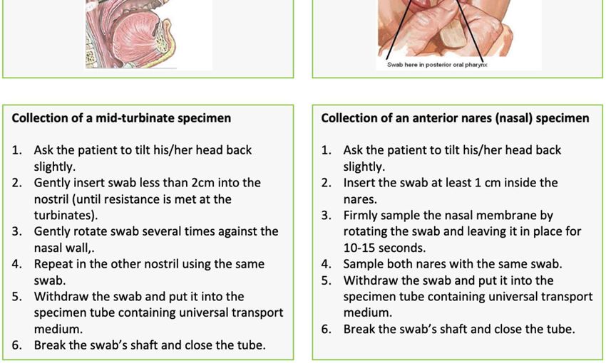
2
Table of Contents

Epidemiology and clinical characteristics ............................................................................. 4
Testing .................................................................................................................................. 7
Management of the patient with asymptomatic or mild disease ...................................... 12
Respiratory support for hospitalised Covid-19 patients ..................................................... 15
Drug therapy ...................................................................................................................... 25
Palliative Care of patients with Covid-19............................................................................ 29
Special populations: children, newborns, pregnant and breastfeeding women, and people
living with HIV .................................................................................................................... 32
De-isolation and return to work ......................................................................................... 37
Infection prevention and control (IPC) ............................................................................... 40
Recording and reporting ..................................................................................................... 41

Guidelines committee (in alphabetical order): Lesley Bamford, Tom Boyles, Lucille Blumberg, Greg
Calligaro, Angelique Coetzee, Cheryl Cohen, Andrew Gray, Dean Gopalan, Ivan Joubert, Tamara Kredo,
Ahmad Haeri Mazanderani, Tendesayi Kufa-Chakeza, Halima Dawood, Nelesh Govender, Lance
Lasersohn, Shelley-Ann McGee, Manala Makua, Shaheen Mehtar, Kerrigan McCarthy, Marc
Mendelson, Jeremy Nel, Wolfgang Preiser, Jantjie Taljaard, Francois Venter.

NEMLC Covid-19 subcommittee (in alphabetical order): Marc Blockman, Karen Cohen, Andrew Gray,
Tamara Kredo, Jeremy Nel, Gary Maartens, Andrew Parish (chairperson), Helen Rees, Gary Reubenson
(vice chairperson), Renee de Waal.

Paediatrics subcommittee (in alphabetical order): Lesley Bamford, Adrie Bekker, Heloise Buys, Ute
Feucht, Marian Jacobs, Prakash Jeena, Fikile Mabena, Nomalinda Makubalo, Carol Marshall, Ntombi
Mazibuko, Neil McKerrow, Jame Nuttall, Shakti Pillay, Robert Pattinson, Gary Reubenson, Natasha
Rhoda

                                                                     3
Epidemiology and clinical characteristics

      The mean incubation period for Covid-19 is 4-5 days. Patients may be infectious for 2-3 days
      prior to the onset of symptoms however.

      The strongest risk factor for severe disease is advanced age. Other risk factors include
      cardiopulmonary comorbidities, obesity, HIV, and diabetes mellitus.

      The spectrum of Covid-19 clinical presentations include asymptomatic infection, a respiratory
      tract infection that may range from mild to severe, and atypical manifestations such as
      diarrhoea, skin manifestations, hyperglycaemic syndromes and large vessel strokes.

SARS-CoV-2 is a betacoronavirus closely related to SARS-CoV and MERS-CoV. It is an enveloped, non-
segmented, positive sense RNA virus. It is thought to have originated in bats but the animal
responsible for transmission to humans remains unknown.

Epidemiology
The median incubation period for Covid-19 is estimated to be 4-5 days, with an interquartile range of
2-7 days. Based on patients’ viral shedding patterns and on epidemiological modelling, patients
appear to be infectious for 2-3 days prior to the onset of symptoms, and the contribution of pre-
symptomatic infections to the overall pandemic may be substantial. 1-7 The basic reproductive number
for the virus is approximately 2.2 (meaning that on average each person spread the infection to two
others).8 A male preponderance of cases has been noted globally both in terms of absolute case
numbers, and in severe disease.9-11 Risk factors for severe disease include older age, cardiopulmonary
comorbidities, obesity, HIV, and diabetes mellitus. Very few cases which required hospitalisation have
been reported among children under the age of 15 years (~1%).

Clinical characteristics – what to look for
Truly asymptomatic Covid-19 patients (as distinguished from pre-symptomatic patients) have been
described, but their proportion is not well characterised yet.6, 12 Among symptomatic patients in China,
81% developed mild disease, an estimated 14% developed severe disease (with hypoxaemia, marked
tachypnoea and extensive lung infiltrates), while 5% became critically ill (with respiratory failure,
septic shock and/or multiorgan dysfunction).13 Because of the strong effect of age on disease severity,
the proportions of mild, severe, and critical cases seen in a country will partially depend on that
country’s population age structure however.

The most common presenting symptom has been fever in approximately 90%, but importantly this
may only be present in a minority of patients on admission.11, 14 A cough is present in two-thirds of
patients, but sputum production is only reported by one third of patients, as is dyspnoea. Myalgia, a
sore throat, nausea, vomiting, and diarrhoea are all present in less than one fifth of cases.11, 14, 15
Anosmia (loss of sense of smell) and dysgeusia (alteration of the sense of taste) have also emerged as
relatively common, early, and moderately specific symptoms.16, 17 Atypical manifestations are
increasingly being recognised, including large vessel strokes in young patients, diabetic
ketoacidosis/hyperglycaemic hyperosmolar syndrome, unexplained abdominal pain, various
dermatological manifestations, and a multisystem inflammatory syndrome in children. 18-20

Abnormalities are visible on chest X-ray in at least 60% of hospitalised Covid-19 patients, with chest
CT scans being more sensitive.11, 14, 21 These are typically bilateral patchy ground glass opacities, though
other patterns have been described.11, 22 However, a normal chest X-ray or chest CT scan does not rule

                                                     4
out Covid-19. This is especially true of patients with mild disease, in whom a majority of chest X-rays
may be normal.23

Outcomes and prognosis
The vast majority of cases will make a full recovery although this may take several weeks, particularly
in severe cases. In a minority of cases, Covid-19 has been associated with rapid progression to acute
respiratory distress syndrome (ARDS), multiple organ failure and sometimes death. Internationally,
the case fatality ratio has ranged between 0.7-7%, and is partially determined by the particular
population’s age distribution, the pandemic’s burden on the healthcare system at the time, and the
extent to which mild or asymptomatic cases are diagnosed.9, 24 Long-term sequelae, if any, are
currently unknown.

References
1.     Du Z, Xu X, Wu Y, Wang L, Cowling BJ, Meyers LA. Serial Interval of COVID-19 among Publicly
       Reported Confirmed Cases. Emerg Infect Dis. 2020;26(6).
2.     Yu P, Zhu J, Zhang Z, Han Y, Huang L. A familial cluster of infection associated with the 2019
       novel coronavirus indicating potential person-to-person transmission during the incubation
       period. J Infect Dis. 2020.
3.     Tindale L, Coombe M, Stockdale JE, Garlock E, Lau WYV, Saraswat M, et al. Transmission
       interval estimates suggest pre-symptomatic spread of COVID-19. medRxiv.
       2020:2020.03.03.20029983.
4.     Nishiura H, Linton NM, Akhmetzhanov AR. Serial interval of novel coronavirus (COVID-19)
       infections. Int J Infect Dis. 2020;93:284-6.
5.     Nishiura H, Kobayashi T, Suzuki A, Jung SM, Hayashi K, Kinoshita R, et al. Estimation of the
       asymptomatic ratio of novel coronavirus infections (COVID-19). Int J Infect Dis. 2020.
6.     Arons MM, Hatfield KM, Reddy SC, Kimball A, James A, Jacobs JR, et al. Presymptomatic SARS-
       CoV-2 Infections and Transmission in a Skilled Nursing Facility. New England Journal of
       Medicine. 2020.
7.     Gandhi M, Yokoe DS, Havlir DV. Asymptomatic Transmission, the Achilles’ Heel of Current
       Strategies to Control Covid-19. New England Journal of Medicine. 2020.
8.     Li Q, Guan X, Wu P, Wang X, Zhou L, Tong Y, et al. Early Transmission Dynamics in Wuhan,
       China, of Novel Coronavirus-Infected Pneumonia. N Engl J Med. 2020.
9.     Onder G, Rezza G, Brusaferro S. Case-Fatality Rate and Characteristics of Patients Dying in
       Relation to COVID-19 in Italy. JAMA. 2020.
10.    Chen T, Wu D, Chen H, Yan W, Yang D, Chen G, et al. Clinical characteristics of 113 deceased
       patients with coronavirus disease 2019: retrospective study. BMJ. 2020;368:m1091.
11.    Guan WJ, Ni ZY, Hu Y, Liang WH, Ou CQ, He JX, et al. Clinical Characteristics of Coronavirus
       Disease 2019 in China. N Engl J Med. 2020.
12.    Mizumoto K, Kagaya K, Zarebski A, Chowell G. Estimating the asymptomatic proportion of
       coronavirus disease 2019 (COVID-19) cases on board the Diamond Princess cruise ship,
       Yokohama, Japan, 2020. Euro Surveill. 2020;25(10).
13.    Wu Z, McGoogan JM. Characteristics of and Important Lessons From the Coronavirus Disease
       2019 (COVID-19) Outbreak in China: Summary of a Report of 72314 Cases From the Chinese
       Center for Disease Control and Prevention. JAMA. 2020.
14.    Goyal P, Choi JJ, Pinheiro LC, Schenck EJ, Chen R, Jabri A, et al. Clinical Characteristics of Covid-
       19 in New York City. N Engl J Med. 2020.
15.    Wang D, Hu B, Hu C, Zhu F, Liu X, Zhang J, et al. Clinical Characteristics of 138 Hospitalized
       Patients With 2019 Novel Coronavirus-Infected Pneumonia in Wuhan, China. JAMA. 2020.
16.    Giacomelli A, Pezzati L, Conti F, Bernacchia D, Siano M, Oreni L, et al. Self-reported olfactory
       and taste disorders in SARS-CoV-2 patients: a cross-sectional study. Clin Infect Dis. 2020.

                                                     5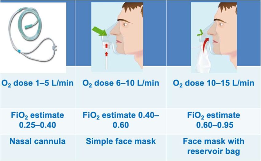
17.   Yan CH, Faraji F, Prajapati DP, Boone CE, DeConde AS. Association of chemosensory
      dysfunction and Covid-19 in patients presenting with influenza-like symptoms. Int Forum
      Allergy Rhinol. 2020.
18.   Oxley TJ, Mocco J, Majidi S, Kellner CP, Shoirah H, Singh IP, et al. Large-Vessel Stroke as a
      Presenting Feature of Covid-19 in the Young. N Engl J Med. 2020.
19.   Galvan Casas C, Catala A, Carretero Hernandez G, Rodriguez-Jimenez P, Fernandez Nieto D,
      Rodriguez-Villa Lario A, et al. Classification of the cutaneous manifestations of COVID-19: a
      rapid prospective nationwide consensus study in Spain with 375 cases. Br J Dermatol. 2020.
20.   Riphagen S, Gomez X, Gonzalez-Martinez C, Wilkinson N, Theocharis P. Hyperinflammatory
      shock in children during COVID-19 pandemic. Lancet. 2020.
21.   Wong HYF, Lam HYS, Fong AH-T, Leung ST, Chin TW-Y, Lo CSY, et al. Frequency and Distribution
      of Chest Radiographic Findings in COVID-19 Positive Patients. Radiology.0(0):201160.
22.   Salehi S, Abedi A, Balakrishnan S, Gholamrezanezhad A. Coronavirus Disease 2019 (COVID-19):
      A Systematic Review of Imaging Findings in 919 Patients. AJR Am J Roentgenol. 2020:1-7.
23.   Weinstock MB EA, Russell JW, et al. Chest x-ray findings in 636 ambulatory patients with
      COVID-19 presenting to an urgent care center: a normal chest x-ray is no guarantee. J Urgent
      Care Med. 2020;14(7):13-8.
24.   World Health Organization. Report of the WHO-China Joint Mission on Coronavirus Disease
      2019       (COVID-19)2020.        Available       from:    https://www.who.int/docs/default-
      source/coronaviruse/who-china-joint-mission-on-covid-19-final-report.pdf.

                                               6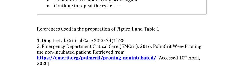
Testing
PCR-based tests are recommended for the diagnosis of acute Covid-19 infection. Upper
respiratory tract samples should be sent on all patients. Sputum or (if the patient is intubated)
bronchoalveolar lavage samples should be sent when available.
Due to very poor sensitivity within the first 1-2 after symptom onset, serology is not
recommended for the diagnosis of acute Covid-19 infection.
Patients seeking healthcare services for potential Covid-19 should preferably phone ahead of time to
their doctor, clinic, emergency room, or closest testing centre, so that adequate precautions can be
taken. Patients should wear masks while in transit to the hospital (cloth masks can suffice until they
are given a surgical mask on arrival). Patients who do not self-identify as potentially having Covid-19
should be screened and identified as soon as possible upon arriving at a health facility, to avoid
prolonged contact with other patients and healthcare workers.
A suspected Covid-19 case includes any person presenting with an acute (≤14 days)
respiratory tract infection or other clinical illness compatible with Covid-19, or an asymptomatic
person who is a close contact to a confirmed case.
In the context of Covid-19, the key respiratory syndrome consists of ANY of:
Cough
Sore throat
Shortness of breath
Anosmia or dysgeusia
… with or without other symptoms (which may include fever, weakness, myalgia, or
diarrhoea).
An acute exacerbation of a chronic pulmonary condition (e.g. COPD, asthma) should also be regarded
as potentially being due to Covid-19.
Atypical manifestations are increasingly being recognised, including large vessel strokes in young
patients, diabetic ketoacidosis/hyperglycaemic hyperosmolar syndrome, unexplained abdominal
pain, various dermatological manifestations, and a multisystem inflammatory syndrome in children.1-
3
A close contact is defined as a person having had face-to-face contact (≤1 metre) or having been in a
closed space with a confirmed Covid-19 case for at least 15 minutes. This includes, amongst others:
All persons living in the same household as a Covid-19 case, and people working closely in the
same environment as a case.
Healthcare workers or other people providing direct care for a Covid-19 case while not
wearing recommended personal protective equipment or PPE (e.g., gowns, gloves, N95
respirator, eye protection).
A contact in an aircraft sitting within two seats (in any direction) of the case, travel
companions or persons providing care, and crew members serving in the section of the aircraft
where the case was seated
7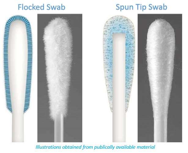
Testing
Testing for acute Covid-19 infection should be by means of polymerase chain reaction (PCR) assays.
Samples to be sent are:
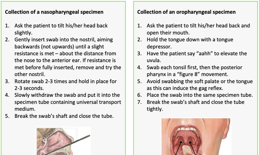
Upper respiratory tract samples – A sample from the upper respiratory tract should be sent
from all patients. A single site is sufficient. Currently, a nasopharyngeal swab is the preferred
specimen, but in patients where this is not possible (e.g. recent nasal surgery, or severe
coagulopathy), an oropharyngeal, nasal mid-turbinate, or anterior nares swab can be
collected instead.4, 5
Lower respiratory tract samples – send when available. Lower respiratory tract samples may
have a higher sensitivity than upper respiratory tract samples.4, 6 Sputum, tracheal aspirates,
or bronchoalveolar lavage fluid are all acceptable samples to send. Sputum induction should
not be performed however.
Where both upper and lower respiratory tract samples are available, both should be sent.
Appropriate personal protective equipment (PPE) should be worn by all healthcare workers when
obtaining specimens (see IPC section).
Obtaining samples for SARS-CoV-2 testing
Healthcare workers obtaining respiratory samples require appropriate personal protective
equipment, including eye protection (goggles or visor), gloves, an apron or gown, and an N95
respirator (or equivalent, e.g. FFP2 mask). Meticulous hand hygiene is also essential. See
section 6 for further details.
Collecting a good quality specimen is vital – see box below.
Appropriate swabs are flocked or spun, and consist of polyester, nylon or rayon material with
a plastic or aluminium shaft. Cotton swabs, calcium alginate swabs, and swabs with a wooden
shaft are not recommended, as they may contain substances that inactivate SARS-CoV-2 and
inhibit PCR testing.
Transport of specimens
Nasopharyngeal, mid-turbinate and anterior nares samples should ideally be placed in
viral/universal transport medium (UTM) and kept between 2-8°C until they are processed at
the laboratory. Due to constraints in the supply of viral/universal transport medium, dry
swabs can be sent provided that the sample will reach the laboratory within 2 days. Dry swabs
can be sent at ambient temperature.
8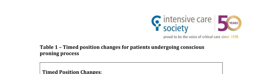
Lower respiratory tract samples can be sent in standard specimen containers and do not
require viral/universal transport medium.
Transport time to testing laboratory
2 days: transport in UTM, preferably at 2-8°C.
medium needed) and can be transported at If UTM is not available, can use normal saline
ambient temperature as an alternative.
9
Repeat testing
PCR tests may produce false negative results due to factors such as poor sampling technique,
suboptimal specimen storage (e.g. unavailability of viral/universal transport medium, or specimen not
stored at cold temperatures), the site the sample is obtained from, and the time point at which the
swab is taken (viral loads are usually highest early on in the disease course). If a high clinical suspicion
for Covid-19 persists despite an initial negative test, repeat testing should be considered in
consultation with an infectious diseases expert, particularly in hospitalised patients for whom
management might be significantly altered. However, it is equally important to maintain a broad
differential diagnosis and to always consider alternative diagnoses (see box below).

A single positive PCR test is sufficient proof of Covid-19 infection. There is no role for repeat
“confirmatory” PCR testing on patients who test positive despite the absence of symptoms, as PCR-
based tests have excellent specificity, and asymptomatic and presymptomatic Covid-19 patients are
now well described.

       The differential diagnosis of suspected cases includes influenza (remembering the seasonality),
       both conventional and atypical bacterial pneumonias, and in patients with HIV and a CD4 count
Point of care antigen tests
We do not currently recommend point of care antigen-based tests, due to concerns about poor
sensitivity and specificity.9
References
1. Oxley TJ, Mocco J, Majidi S, Kellner CP, Shoirah H, Singh IP, et al. Large-Vessel Stroke as a
Presenting Feature of Covid-19 in the Young. N Engl J Med. 2020.
2. Galvan Casas C, Catala A, Carretero Hernandez G, Rodriguez-Jimenez P, Fernandez Nieto D,
Rodriguez-Villa Lario A, et al. Classification of the cutaneous manifestations of COVID-19: a
rapid prospective nationwide consensus study in Spain with 375 cases. Br J Dermatol. 2020.
3. Riphagen S, Gomez X, Gonzalez-Martinez C, Wilkinson N, Theocharis P. Hyperinflammatory
shock in children during COVID-19 pandemic. Lancet. 2020.
4. Wang W, Xu Y, Gao R, Lu R, Han K, Wu G, et al. Detection of SARS-CoV-2 in Different Types of
Clinical Specimens. JAMA. 2020.
5. Centre for Evidence-Based Medicine. Comparative accuracy of oropharyngeal and
nasopharyngeal swabs for diagnosis of COVID-19. 2020 Accessed: 19 April 2020. Available
from: https://www.cebm.net/covid-19/comparative-accuracy-of-oropharyngeal-and-
nasopharyngeal-swabs-for-diagnosis-of-covid-19/.
6. Yu F, Yan L, Wang N, Yang S, Wang L, Tang Y, et al. Quantitative Detection and Viral Load
Analysis of SARS-CoV-2 in Infected Patients. Clin Infect Dis. 2020.
7. Zhao J, Yuan Q, Wang H, Liu W, Liao X, Su Y, et al. Antibody responses to SARS-CoV-2 in
patients of novel coronavirus disease 2019. Clin Infect Dis. 2020.
8. Guo L, Ren L, Yang S, Xiao M, Chang, Yang F, et al. Profiling Early Humoral Response to
Diagnose Novel Coronavirus Disease (COVID-19). Clin Infect Dis. 2020.
9. World Health Organization. Advice on the use of point-of-care immunodiagnostic tests for
COVID-19. 2020 [20th April 2020]. Available from: https://www.who.int/news-
room/commentaries/detail/advice-on-the-use-of-point-of-care-immunodiagnostic-tests-for-
covid-19.
11Management of the patient with asymptomatic or mild disease
Patients who are asymptomatic or who meet criteria for mild disease can be managed at home
provide they can safely self-isolate.
Patients who self-isolate at home should be given strict advice on how to reduce possible
transmission to others.
Paracetamol is recommended for symptomatic treatment of patients with fever or pain in
preference to nonsteroidal anti-inflammatory drugs (NSAIDs).
Patients with Covid-19 who are medically well, or who are assessed as having only mild disease, may
be managed at home, provided they can safely do so.
Criteria for management at home (for age >12 years1):
Mild disease1
•SpO2 ≥95%
•Respiratory rateand living area, they should stay in their room with the door closed, only coming out when
necessary, wearing a surgical mask if they do so.
Where contact is unavoidable, the patient should wear a surgical mask, and maintain a distance
of at least 1 metre (preferably 2 metres) from other people.
Patients should clean their hands with soap and water frequently. Alcohol-based sanitizers may
also be used, provided they contain at least 70% alcohol.
Patients should practice good cough and sneeze hygiene, by using a tissue, and then immediately
discarding the tissue in a lined trash can, followed by washing hands immediately.
Patients should not have visitors in their home. Only those who usually live in their home should
be allowed to stay.
Patients should avoid sharing household items like dishes, cups, eating utensils and towels. After
using any of these, the items should be thoroughly washed with soap and hot water.
All high-touch surfaces like table tops, counters, toilets, phones, computers, etc. should be
appropriately and frequently cleaned.
If patients need to wash laundry at home before the PCR results are available, then they should
wash all laundry at the highest temperature compatible with the fabric using laundry detergent.
This should be above 60°C. If possible, they should tumble dry and iron using the highest setting
compatible with the fabric. Disposable gloves and a plastic apron should be used when handling
soiled materials if possible and all surfaces and the area around the washing machine should be
cleaned. Laundry should not be taken to a launderette. The patient should wash his/her hands
thoroughly with soap and water after handling dirty laundry (remove gloves first if used).
Patients should know who to call and/or where to go if they develop any worsening symptoms,
so that they can be safely reassessed.
In addition to this advice, a patient information sheet should be provided (see Appendix 1 for an
example).
Symptomatic treatment for Covid-19 patients managed at home
For patients requiring symptomatic relief of fever or pain, we suggest using paracetamol as a first-
choice agent rather than a nonsteroidal anti-inflammatory drug (NSAID).
o There is no good evidence that NSAIDs worsen Covid-19 infection, so patients
currently requiring NSAIDs for other indications should not discontinue NSAIDs for
Covid-related reasons.2
Whether nebulisers increase the risk of transmission of SARS-CoV2 is currently unknown. Evidence
reviews conducted prior to the Covid-19 outbreak have not found clear evidence of increased
transmission of respiratory viruses.3, 4 Furthermore, the aerosol generated by nebulisers is derived
from the nebulising chamber rather than the patient.5 Nonetheless for patients with asthma or
chronic pulmonary obstructive pulmonary disease (COPD) who may experience an acute
exacerbation of their illness due to Covid-19, the use of metered dose inhalers, with or without a
spacer, is preferred to the use of a nebuliser.
o Patients who do require a nebuliser should use it in a room that is isolated from other
household members and/or other patients. Good ventilation for this area is
recommended; this may be facilitated by opening the windows in the room.
o Spacers need to be disinfected between patients with either soap and water followed
by a wipe down with 70% alcohol, or by using a chlorine-based disinfectant (soak for
30 mins then rinse well with water to avoid chlorine being absorbed into the spacer).
Cough suppressants, such as codeine-containing cough mixtures, are not indicated, and are not
available in public sector health facilities. Opioids, such as morphine, should not be used for this
reason alone, and where they are indicated they should only be used with due caution and careful
monitoring.
Recent work suggested that angiotensin converting enzyme inhibitors (ACEi) or angiotensin
receptor blockers (ARBs) might upregulate ACE2 receptors, the binding site for SARS-CoV-2, within
13tissues including the lung and heart, prompting theoretical concerns that this might place patients
at risk of worse outcomes with Covid-19.6 To date, this remains purely theoretical, with no
evidence of worse clinical outcomes.7 Furthermore, discontinuing or switching ACEi or ARBs to
alternative agents may be deleterious to patient care. Pending further evidence, we therefore do
not recommend discontinuing ACEi or ARBs unless there are other medical reasons to do so.
References
1. Wang D, Hu B, Hu C, Zhu F, Liu X, Zhang J, et al. Clinical Characteristics of 138 Hospitalized
Patients With 2019 Novel Coronavirus-Infected Pneumonia in Wuhan, China. JAMA. 2020.
2. SAHPRA. The Use of Non-Steroidal Anti-Inflammatory Drugs in patients with Covid-19. Media
release. 2020.
3. Tran K, Cimon K, Severn M, Pessoa-Silva CL, Conly J. Aerosol generating procedures and risk
of transmission of acute respiratory infections to healthcare workers: a systematic review.
PLoS One. 2012;7(4):e35797.
4. Wan GH, Tsai YH, Wu YK, Tsao KC. A large-volume nebulizer would not be an infectious source
for severe acute respiratory syndrome. Infect Control Hosp Epidemiol. 2004;25(12):1113-5.
5. Public Health England. COVID-19. Guidance for infection prevention and control in healthcare
settings. 2020.
6. Fang L, Karakiulakis G, Roth M. Are patients with hypertension and diabetes mellitus at
increased risk for COVID-19 infection? Lancet Respir Med. 2020.
7. Zhang P, Zhu L, Cai J, Lei F, Qin JJ, Xie J, et al. Association of Inpatient Use of Angiotensin
Converting Enzyme Inhibitors and Angiotensin II Receptor Blockers with Mortality Among
Patients With Hypertension Hospitalized With COVID-19. Circ Res. 2020.
14Respiratory support for hospitalised Covid-19 patients
Version 5 – what’s new?
Expanded and enhanced sections on high flow nasal cannula oxygen (HFNO) and self-
proning.
Supplemental oxygen remains the mainstay of therapy for most hospitalised patients. Target
SpO2 ≥90% in non-pregnant adults, titrating to reach targets by means of a nasal cannula,
simple face mask or face mask with reservoir bag.
The use of the prone position in non-intubated, conscious patients who are hypoxaemic may be
beneficial.
Patients who have respiratory failure despite maximal facemask oxygen should be promptly
identified and evaluated for possible escalation of respiratory support. Possible modalities
include high flow nasal cannula oxygen, continuous positive airway pressure, or intubation and
mechanical ventilation.
General principles
Give supplemental oxygen therapy immediately to patients with low oxygen saturation.1
Oxygen therapy is likely to be the single most effective supportive measure in Covid-19 patients.
Target SpO2 ≥90% in non-pregnant adults and SpO2 ≥92% in pregnant patients.1 Children with
emergency signs (obstructed or absent breathing, severe respiratory distress, central cyanosis,
shock, coma or convulsions) should receive oxygen therapy during resuscitation to target SpO2
≥94%; otherwise, the target SpO2 is ≥92%.
Titrate oxygen therapy up and down to reach targets by means of a nasal cannula, simple face
mask or face mask with reservoir bag, as appropriate. Nasal cannulae should not be reused. Face
masks and reservoir bags must be heat disinfected between each patient use if they are used for
more than one patient.
For paediatric oxygen recommendations, see section 5.1
Judicious fluid management in patients with Covid-19 is needed.
Patients who are relatively hypovolaemic (e.g. due to prolonged high fever), will need appropriate
fluid replacement. However, overly aggressive fluid resuscitation may worsen oxygenation. This may
15especially problematic in settings where there is limited availability of mechanical ventilation, and in patients with established ARDS.2, 3 Recognize severe hypoxemic respiratory failure when a patient with respiratory distress is failing standard oxygen therapy. Patients may continue to have increased work of breathing or hypoxemia (SpO2
From: Nishimura M. et al.4
Advantages
HFNC is considered to have a number of physiological effects including:
low levels of positive end-expiratory pressure (PEEP), at best up to 10 cm H20, that may assist
in increasing lung volume and recruitment of alveoli9, 10
reduction of anatomical dead space as the high flow washes out CO211
maintenance of a constant FiO2 as the difference between inspiratory flow and delivered flow
is small12
humidification contributing to good muco-ciliary function and patient comfort;13
decreased work of breathing14
Other general advantages in Covid-19 patients include that it:
may be implemented and managed by non-ICU specialists outside ICU
does not require invasive monitoring
does not need as intensive nursing care as for invasive ventilation
can be combined with awake self-proning
may be a lower-resource alternative to mechanical ventilation in some patient
Is relatively well tolerated and not too cumbersome allowing patient self-care or assisted care
while applying the therapy, including daily functions such as eating.
Concerns
Is there an increased risk of transmission?
The two main concerns relate to the risk of aerosolization and the adequacy of oxygen supplies. All
respiratory therapy has the potential to create aerosols. A caution with HFNC initially arose because
of a concern for possible generation of droplets and aerosols created or propelled by oxygen therapy
via this delivery system with a consequent increased risk of disease transmission. Dispersion studies
have shown that, compared to oxygen therapy with a mask or standard nasal cannulae at 5 L/min, the
utilization of HFNC is no riskier with respect to either dispersion or microbiological contamination into
17the environment.15, 16 The risk may be further mitigated by the additional application of, for example, a surgical mask to the patient. How do we ensure adequate oxygen supplies? As high flows (up to 60L/min) are used with HFNC systems, a concern rose on the adequacy of hospital oxygen supplies if the therapy was applied to a large number of patients within the same facility. The high flow of oxygen exceeds the requirements for routine general ward beds (4-15L/min), and for ICU or ventilated patients (30L/min).17 This concern relates to storage and delivery of oxygen and questions the ability of banks or storage tanks for liquid oxygen to cope with demand in maintaining a constant flow and pressure to reticulation and supply points. Medical engineering consultation is required about oxygen supply at individual hospitals including number of HFNC units that can be supported. Indications For patients who are deteriorating or not improving on conventional oxygen therapy and supportive care, but who do not appear to be in imminent danger of collapse, HFNC oxygen therapy likely offers benefit. Consider HFNC in awake, co-operative patient if SpO2
Patients should be monitored with continuous pulse oximetry to enable monitoring of
response and for early identification of rapid deterioration.
Initial settings: Flow 50-60L/min and FiO2 0.8-1.0, titrated to aim initially for SpO2 >90% are
recommended.
Particularly where hospital supply is constrained, be vigilant about using the minimum O2 flow
necessary to maintain SpO2. Titrate FiO2 to 1.0 prior to increasing flow greater than 35L/min.
Patients receiving a trial of HFNC should be in a monitored setting and cared for by personnel
experienced with HFNC and capable of performing endotracheal intubation in case the patient
acutely deteriorates or does not improve after a short trial (about 1 hour).
Once HFNC has been initiated, need to assess the patient regularly and as clinically indicated
to determine if the patient needs to be intubated.
There should be a low threshold for intubation where there is clinical decline (which may
include a rising O2 requirement, consistently or rapid increase in respiratory rate, consistently
or rapidly declining SpO2, increased work of breathing/exhaustion, and altered mental state).
Intubation should not be delayed if the patient acutely deteriorates or does not improve after
a short trial (1 hour).
Initiation of HFNC does not by default imply that a patient’s care will be escalated to invasive
ventilation. Although some patients may proceed to invasive ventilation, many will not with
These decisions should be made in accordance with local facility triage team protocols as well
as national guidelines. If resources and staffing allow, HFNC may still be a reasonable
intervention in patients who do not meet local facility triage team protocols as well as national
guidelines criteria for ICU admission. In this situation, it may represent the limit of care.
If the target SpO2 is achieved and the patient is clinically improving (decrease in respiratory
rate and respiratory distress), weaning should be commenced. Flow may be gradually reduced
by 5-10 L/min and FiO2 by 0.05-0.1 every 2-4 hours. Switching to conventional O2 therapy
should be considered when FiO2 < 0.4 and flow < 20 L/min.
Mechanical ventilation
Patients with hypoxaemic respiratory failure may require intubation and mechanical ventilatory
support. Detailed recommendations on ventilation strategies are beyond the scope of this guideline.
Always consult an intensivist if possible, or alternatively a practitioner experienced with mechanical
ventilation. Nonetheless, the general principles to consider include:
Individualise ventilatory strategies based on respiratory mechanics and disease progression.
Use lung-protective ventilation strategies for patients with established ARDS who have low
lung compliance.
Aim for an initial tidal volume of 4-6ml/kg.19 Higher tidal volume up to 8 ml/kg predicted body
weight may be needed if minute ventilation requirements are not met in a patient with good
lung compliance.
Strive to achieve the lowest plateau pressure possible. Plateau pressures above 30 cmH20 are
associated with an increased risk of pulmonary injury.19
Hypercapnia is permitted if meeting the pH goal of >7.15-7.20.
Application of prone ventilation 12-16 hours a day is strongly recommended for patients with
severe ARDS.19
In patients with moderate or severe ARDS, identifying optimal PEEP levels will require titration
of PEEP.19
The use of deep sedation may be required to control respiratory drive, achieve tidal volume
targets, and assist with patient-ventilator dyssynchrony.
In patients with moderate-severe ARDS (PaO2/FiO2sedation, such that tidal volume limitation cannot be reliably achieved; or refractory
hypoxemia.
Avoid disconnecting the patient from the ventilator, which results in loss of PEEP and
atelectasis. Use closed system catheters for airway suctioning and clamp endotracheal tube
when disconnection is required (for example, transfer to a transport ventilator). A high
efficiency particulate filter on the expiratory limb of the ventilator circuit should be used.
Self-proning
Prone positioning has been shown to improve oxygenation in spontaneously breathing, non-intubated
non-Covid-19 patients with hypoxemic acute respiratory failure.21 Consequently, its potential value in
the management of patients with Covid-19 pneumonia has been explored.22, 23 A management
strategy involving early intervention and awake proning with high-flow nasal cannula or non-invasive
mechanical ventilation to prevent alveolar collapse resulted in lower intubation and mortality rates
than observed in other locations.24 Other studies have demonstrated that application of self-proning
with HFNC may help avoid intubation.25, 26
Physiological effects of proning
The physiological benefits of prone positioning that should apply to all patients regardless of whether
they are intubated or not, include:
Improved Ventilation/Perfusion (VQ) matching and reduced hypoxaemia (secondary to more
homogeneous aeration of lung and ameliorating the ventral-dorsal trans-pulmonary pressure
gradient – more uniform lung ventilation, better distribution of air flow and better matching
of areas that receive oxygen and appropriate blood flow.)
Reduced shunt (perfusion pattern remaining relatively constant while lung aeration becomes
more homogenous – better matching of areas that have blood flow to receiving oxygen)
Recruitment of the posterior lung segments due to reversal of atelectasis;
Improved secretion clearance.
Different approaches to positional adjustment in Covid-19
Various approaches have been attempted.
Complete pronation (with the patient lying on their abdomen, ideally for ~16-18 hours per
day) as in proned intubated patients would be optimal. However, this can be difficult in many
patients e.g. with obesity.
Another approach is to rotate positions, including lying on either side and sitting bolt upright
which may be easier for many patients to tolerate.
Some centres encourage mobilization via walking of selected Covid-19 patients.
Proning for a few hours with a return to supine position may lead only to transient
improvements in oxygenation. Longer-lasting benefit might result from longer periods of
pronation, or strategies involving ongoing rotation between several different positions. The
key principle is to avoid spending much time in a flat, supine position.
As one suggested approach, we suggest following the UK Intensive Care Society’s proning
recommendations as outlined below.27
Awake pronation appears to be a safe, inexpensive, and versatile strategy which can be used at all
levels across a variety of different healthcare settings.
20Figure 1 – Flow diagram decision tool
for Conscious Proning process
FiO2 28% or requiring basic respiratory support to achieve SaO2 92 – 96% (88-92% if risk
of hypercapnic respiratory failure) AND suspected/confirmed COVID-19.
YES
NO
Consider prone position if ability to; NO
Continue - Communicate and co-operate with procedure. Continue
supine - Rotate to front and adjust position independently supine
- No anticipated airway issues
YES
Absolute contraindications
- Respiratory distress (RR 35, PaCO2 6.5, accessory muscle use)
- Immediate need for intubation
- Haemodynamic instability (SBP < 90mmHg) or arrhythmia
- Agitation or altered mental status YES Continue
- Unstable spine/thoracic injury/recent abdominal surgery Supine or
Relative Contraindications: consider
- Facial injury escalation
- Neurological issues (e.g. frequent seizures) to medical
- Morbid obesity team
- Pregnancy (2/3rd trimesters)
- Pressure sores / ulcers
Assist patient to prone position (See Table 1)
- Explain procedure/benefit
- Ensure oxygen therapy and basic respiratory support
secure with adequate length on the tubing
- Pillows may be required to support the chest
- Reverse trendelenberg position may aid comfort
- Monitor oxygen saturations If drop then ensure O2
connected and working
- Sedation must not be administered to facilitate proning
Monitor Oxygen Saturations for 15 minutes:
SaO2 92-96% (88-92% if risk of hypercapnic respiratory failure) and nil obvious distress
YES NO
Continue proning process (See Table 1): If deteriorating oxygen saturations:
- Change position every 1-2 hrs aiming to - Ensure oxygen is connected to patient
achieve a prone time as long as possible - Increase inspired oxygen
- When not prone aim to be sat at between - Change patients position
30-60 degrees upright - Consider return to supine position
- Monitor oxygen saturations after every - Escalate to critical care if appropriate
position change Discontinue if:
- Titrate down oxygen requirements as able - No improvement with change of position
- Patient unable to tolerate position
- RR 3 , looks tired, using accessory
muscles
2122
References
1.     World Health Organization. Clinical management of severe acute respiratory infection when
       novel coronavirus (nCoV) infection is suspected 2020 [Available from:
       https://www.who.int/publications-detail/clinical-management-of-severe-acute-respiratory-
       infection-when-novel-coronavirus-(ncov)-infection-is-suspected.
2.     Schultz MJ, Dunser MW, Dondorp AM, Adhikari NK, Iyer S, Kwizera A, et al. Current
       challenges in the management of sepsis in ICUs in resource-poor settings and suggestions
       for the future. Intensive Care Med. 2017;43(5):612-24.
3.     Rhodes A, Evans LE, Alhazzani W, Levy MM, Antonelli M, Ferrer R, et al. Surviving Sepsis
       Campaign: International Guidelines for Management of Sepsis and Septic Shock: 2016.
       Intensive Care Med. 2017;43(3):304-77.
4.     Nishimura M. High-flow nasal cannula oxygen therapy in adults. J Intensive Care.
       2015;3(1):15.
5.     Rochwerg B, Granton D, Wang DX, Helviz Y, Einav S, Frat JP, et al. High flow nasal cannula
       compared with conventional oxygen therapy for acute hypoxemic respiratory failure: a
       systematic review and meta-analysis. Intensive Care Med. 2019;45(5):563-72.
6.     Wang K, Zhao W, Li J, Shu W, Duan J. The experience of high-flow nasal cannula in
       hospitalized patients with 2019 novel coronavirus-infected pneumonia in two hospitals of
       Chongqing, China. Ann Intensive Care. 2020;10(1):37.
7.     Lalla U, Allwood BW, Louw EH, Nortje A, Parker A, Taljaard JJ, et al. The utility of high-flow
       nasal cannula oxygen therapy in the management of respiratory failure secondary to COVID-
       19 pneumonia2020.
8.     Zucman N, Mullaert J, Roux D, Roca O, Ricard JD, Contributors. Prediction of outcome of
       nasal high flow use during COVID-19-related acute hypoxemic respiratory failure. Intensive
       Care Med. 2020.
9.     Parke R, McGuinness S, Eccleston M. Nasal high-flow therapy delivers low level positive
       airway pressure. Br J Anaesth. 2009;103(6):886-90.
10.    Corley A, Caruana LR, Barnett AG, Tronstad O, Fraser JF. Oxygen delivery through high-flow
       nasal cannulae increase end-expiratory lung volume and reduce respiratory rate in post-
       cardiac surgical patients. Br J Anaesth. 2011;107(6):998-1004.
11.    Frizzola M, Miller TL, Rodriguez ME, Zhu Y, Rojas J, Hesek A, et al. High-flow nasal cannula:
       impact on oxygenation and ventilation in an acute lung injury model. Pediatr Pulmonol.
       2011;46(1):67-74.
12.    Ritchie JE, Williams AB, Gerard C, Hockey H. Evaluation of a humidified nasal high-flow
       oxygen system, using oxygraphy, capnography and measurement of upper airway pressures.
       Anaesth Intensive Care. 2011;39(6):1103-10.
13.    Oto J, Nakataki E, Okuda N, Onodera M, Imanaka H, Nishimura M. Hygrometric properties of
       inspired gas and oral dryness in patients with acute respiratory failure during noninvasive
       ventilation. Respir Care. 2014;59(1):39-45.
14.    Pham TM, O'Malley L, Mayfield S, Martin S, Schibler A. The effect of high flow nasal cannula
       therapy on the work of breathing in infants with bronchiolitis. Pediatr Pulmonol.
       2015;50(7):713-20.
15.    Hui DS, Chow BK, Lo T, Tsang OTY, Ko FW, Ng SS, et al. Exhaled air dispersion during high-
       flow nasal cannula therapy versus CPAP via different masks. Eur Respir J. 2019;53(4).
16.    Li J, Fink JB, Ehrmann S. High-flow nasal cannula for COVID-19 patients: low risk of bio-
       aerosol dispersion. Eur Respir J. 2020;55(5).
17.    World Health Organization. Oxygen sources and distribution for COVID-19 treatment
       centres. 2020. Available from: https://www.who.int/publications/i/item/oxygen-sources-
       and-distribution-for-covid-19-treatment-centres.

                                                 23
18. World Health Organization. WHO-ICRC Basic Emergency Care: approach to the acutely ill and
injured. 2020. Available from: https://www.who.int/publications/i/item/basic-emergency-
care-approach-to-the-acutely-ill-and-injured.
19. Fan E, Del Sorbo L, Goligher EC, Hodgson CL, Munshi L, Walkey AJ, et al. An Official American
Thoracic Society/European Society of Intensive Care Medicine/Society of Critical Care
Medicine Clinical Practice Guideline: Mechanical Ventilation in Adult Patients with Acute
Respiratory Distress Syndrome. Am J Respir Crit Care Med. 2017;195(9):1253-63.
20. National Heart L, Blood Institute PCTN, Moss M, Huang DT, Brower RG, Ferguson ND, et al.
Early Neuromuscular Blockade in the Acute Respiratory Distress Syndrome. N Engl J Med.
2019;380(21):1997-2008.
21. Scaravilli V, Grasselli G, Castagna L, Zanella A, Isgro S, Lucchini A, et al. Prone positioning
improves oxygenation in spontaneously breathing nonintubated patients with hypoxemic
acute respiratory failure: A retrospective study. J Crit Care. 2015;30(6):1390-4.
22. Elharrar X, Trigui Y, Dols AM, Touchon F, Martinez S, Prud'homme E, et al. Use of Prone
Positioning in Nonintubated Patients With COVID-19 and Hypoxemic Acute Respiratory
Failure. JAMA. 2020.
23. Farkas J. PulmCrit Wee- Proning the non-intubated patient. 2020 [Available from:
https://emcrit.org/pulmcrit/proning-nonintubated/.
24. Sun Q, Qiu H, Huang M, Yang Y. Lower mortality of COVID-19 by early recognition and
intervention: experience from Jiangsu Province. Ann Intensive Care. 2020;10(1):33.
25. Caputo ND, Strayer RJ, Levitan R. Early Self-Proning in Awake, Non-intubated Patients in the
Emergency Department: A Single ED's Experience During the COVID-19 Pandemic. Acad
Emerg Med. 2020;27(5):375-8.
26. Ding L, Wang L, Ma W, He H. Efficacy and safety of early prone positioning combined with
HFNC or NIV in moderate to severe ARDS: a multi-center prospective cohort study. Crit Care.
2020;24(1):28.
27. Intensive Care Society. ICS Guidance for Prone Positioning of the Conscious COVID Patient
2020. [26/04/2020]. Available from: https://emcrit.org/wp-content/uploads/2020/04/2020-
04-12-Guidance-for-conscious-proning.pdf.
24Drug therapy
Version 5 – what’s new?
Recommendations relating to the use of dexamethasone, heparin and remdesivir.
Recommendation against the use of chloroquine, hydroxychloroquine, or
lopinavir/ritonavir outside of a clinical trial.
Dexamethasone is recommended for patients requiring supplemental oxygen or mechanical
ventilation.
Heparin venous thromboembolism prophylaxis is recommended for all hospitalised patients.
Therapeutic dosing is suggested for patients requiring ≥60% supplemental oxygen, or those
with a D-dimer >6 times the upper limit of normal.
Due to remdesivir’s high cost and marginal benefit, routine use of the drug in hospitalised
patients with Covid-19 is not recommended in the public sector outside of clinical trials.
Corticosteroids
We recommend dexamethasone (6mg per day for 10 days) for the following indications:
Patients with Covid-19 who are mechanically ventilated.
Patients with Covid-19 who require supplemental oxygen but who are not mechanically
ventilated.
If dexamethasone is not available, an alternative corticosteroid may be used, such as:
Betamethasone 6mg daily p.o. or intravenous, for 10 days
Prednisone 40 mg daily p.o. for 10 days
For patients able to tolerate drugs them, oral corticosteroid formulations may reduce the need for
intravenous access. Dexamethasone tablets are available via the section 21 application process.
Rationale: The Recovery trial, a large-scale randomised controlled open label multi-center adaptive
trial recently reported preliminary results for its dexamethasone arm.1 Patients on invasive ventilation
had an absolute reduction in mortality of 12% (95% CI 5.5-17.9%), with 9 ventilated patients needing
to be treated to avert 1 death. There was a smaller benefit seen in those patients requiring
supplemental oxygen, with a 3% reduction in mortality (95% CI 0.89-5.25%). 33 such patients would
need to be treated to prevent 1 death.
Note: it is unclear whether these benefits can be extrapolated to the HIV population. In HIV-
positive patients, especial care should be taken to exclude tuberculosis and Pneumocystis jirovecii
pneumonia coinfection.
We recommend against using dexamethasone for the treatment of Covid-19 in patients who do not
require supplemental oxygen or mechanical ventilation.
Note: systemic corticosteroids should not be withheld from patients who require them for
another reason such as an acute exacerbation of asthma or chronic obstructive pulmonary
disease.
25Rationale: In the Recovery trial, there was evidence of potential harm when dexamethasone was given
to patients not requiring supplemental oxygen. Those receiving dexamethasone had a 4% absolute
increase in mortality (relative risk 1.22, 95% CI 0.93-1.61), though the result did not reach statistical
significance.1 In addition, glucocorticoids given to patients with other viral pneumonias such as SARS,
MERS and influenza glucocorticoids, showed delayed viral clearance, no survival benefit and possible
harms, such as hyperglycaemia and an increased risk of secondary nosocomial infections. 2-4
Venous thromboembolism prophylaxis and pre-emptive therapy
We recommend that all hospitalised patients with Covid-19 receive prophylaxis against venous
thromboembolic disease (VTE), in the absence of any contraindications.
We suggest that patients hospitalised with Covid-19 be considered for unfractionated or low
molecular weight heparin at therapeutic doses (e.g. enoxaparin 1mg/kg 12-hourly based on actual
weight) in the following scenarios:
The patient requires supplemental oxygen at ≥60% oxygen concentration, or requires
mechanical ventilation.
The patient’s serum D-dimers are greater than 6-times the upper limit of normal (i.e. above
1.5 mg/L).
Rationale: A recent evidence review concluded that there was insufficient evidence for therapeutic-
intensity doses of either unfractionated or low molecular weight heparin in patients with Covid-19 in
the absence of proven VTE disease. However, the high incidence of VTE disease seen in several cohorts
of patients with severe Covid-19 was noted.5, 6 In one cohort, therapeutic-intensity anticoagulation
amongst Covid-19 patients in ICU was associated with a lower incidence of VTE.6 Local experience has
also seen a number of concerning cases of severe venous and arterial thromboembolic disease. Based
on expert opinion, the panel therefore suggested that a severely ill subset of hospitalised Covid-19
patients be given therapeutic intensity heparin. The weakness of the evidence for this practice was
acknowledged however, and guidance will be updated once further evidence is available.
Note:
1. The risks of therapeutic anticoagulation need to be considered on a case-by-case basis for each
patient.
2. Where available, factor Xa monitoring may be beneficial, since heparin resistance has been
described in severely-ill Covid-19 patients.7
3. We do not recommend continuing anticoagulation therapy after discharge, as the risks of a major
bleed outside a hospital may outweigh any potential benefits.
4. In patients on chronic antiplatelet therapy who are given therapeutic-intensity anticoagulation,
consider temporarily withholding the antiplatelet therapy unless there is a compelling indication for
it to be continued.
5. The possibility of pulmonary embolism, stroke, or myocardial infarction should be considered in
any hospitalised patient with Covid-19 whose condition rapidly deteriorates.
Remdesivir
Owing to remdesivir’s anticipated high cost and marginal benefit, we suggest that remdesivir not be
used in patients with Covid-19 within the public sector, outside of a clinical trial.
Remdesivir may be accessed via the section 21 application process for patients in the private sector,
or for individual patients within the state sector, in accordance with the MEURI framework.
Rationale: One randomised control trial (RCT) showed that remdesivir shortened median time to
recovery from 15 to 11 days, while an earlier RCT (which was underpowered as it could not complete
recruitment) demonstrated no statistically significant benefits in any outcomes. 8, 9 A meta-analysis of
26the two RCTs showed that remdesivir decreased the risk of disease progression to requiring
ventilation.10 However, there were no statistically significant differences in mortality.
Other drugs
The National Essential Medicines List (NEMLC) Covid-19 subcommittee has produced rapid evidence
reviews of a large number of potential therapeutic and prophylactic agents. These are updated
regularly, and are available at: http://www.health.gov.za/index.php/national-essential-medicine-list-
committee-nemlc/category/633-covid-19-rapid-reviews
There is insufficient evidence to recommend any of the following drugs for the treatment of Covid-
19 outside of a clinical trial:
Interferon beta
Intravenous immunoglobulin
Tocilizumab
Azithromycin
Convalescent plasma
Favipiravir
Due to evidence of futility from large-scale randomised control trials, we recommend against using
the following drugs to treat patients with Covid-19 outside of a clinical trial:
Chloroquine or hydroxychloroquine
Lopinavir/ritonavir
The guideline group are aware that many medicines are being used based on in vitro and observational
data, such as vitamin D, vitamin C, beta-2-agonists and statins. None of these are currently
recommended for the prevention or treatment of Covid-19, and some may do more harm than good.
In addition, the evidence for the use of colchicine is currently undergoing review, and caution is
advised until this review is complete. The evidence for all potential pharmacological interventions is
constantly being monitored and the guidelines will be updated accordingly.
Where investigational therapeutics are given outside of a clinical trial, this should be done under the
Monitored Emergency Use of Unregistered Interventions (MEURI) framework, which provide an
appropriate structure to offer individuals investigational interventions on an emergency basis in the
context of an outbreak with a high mortality.11 The principles of this include:
Data providing preliminary support for the intervention’s efficacy and safety are available, at
least from laboratory or animal studies.
The relevant human research ethics committee has approved the therapeutics’ use.
The patient’s informed consent is obtained.
Adequate resources are devoted to minimizing the risk of administering the therapeutic
agent.
The results of the intervention are documented and shared with the wider medical and
scientific community.
Prophylaxis
There is currently insufficient evidence to recommend any drug as prophylaxis for Covid-19 other
than in a clinical trial. The evidence for chloroquine or favipiravir as prophylaxis has specifically been
reviewed by the NEMLC Covid-19 subcommittee and found to be insufficient to warrant a
recommendation for their use.
27References
1. Horby P, Lim WS, Emberson J, Mafham M, Bell J, Linsell L, et al. Effect of Dexamethasone in
Hospitalized Patients with COVID-19: Preliminary Report. medRxiv.
2020:2020.06.22.20137273.
2. Ni YN, Chen G, Sun J, Liang BM, Liang ZA. The effect of corticosteroids on mortality of patients
with influenza pneumonia: a systematic review and meta-analysis. Crit Care. 2019;23(1):99.
3. Stockman LJ, Bellamy R, Garner P. SARS: systematic review of treatment effects. PLoS Med.
2006;3(9):e343.
4. Arabi YM, Mandourah Y, Al-Hameed F, Sindi AA, Almekhlafi GA, Hussein MA, et al.
Corticosteroid Therapy for Critically Ill Patients with Middle East Respiratory Syndrome. Am J
Respir Crit Care Med. 2018;197(6):757-67.
5. Helms J, Tacquard C, Severac F, Leonard-Lorant I, Ohana M, Delabranche X, et al. High risk of
thrombosis in patients with severe SARS-CoV-2 infection: a multicenter prospective cohort
study. Intensive Care Medicine. 2020;46(6):1089-98.
6. Klok FA, Kruip M, van der Meer NJM, Arbous MS, Gommers D, Kant KM, et al. Incidence of
thrombotic complications in critically ill ICU patients with COVID-19. Thromb Res.
2020;191:145-7.
7. White D, MacDonald S, Bull T, Hayman M, de Monteverde-Robb R, Sapsford D, et al. Heparin
resistance in COVID-19 patients in the intensive care unit. J Thromb Thrombolysis. 2020.
8. Beigel JH, Tomashek KM, Dodd LE, Mehta AK, Zingman BS, Kalil AC, et al. Remdesivir for the
Treatment of Covid-19 — Preliminary Report. New England Journal of Medicine. 2020.
9. Wang Y, Zhang D, Du G, Du R, Zhao J, Jin Y, et al. Remdesivir in adults with severe COVID-19:
a randomised, double-blind, placebo-controlled, multicentre trial. Lancet.
2020;395(10236):1569-78.
10. Living mapping and living systematic review of Covid-19 studies 2020 [Available from:
https://covid-nma.com/living_data/index.php.
11. World Health Organization. Guidance For Managing Ethical Issues In Infectious Disease
Outbreaks 2016. Available from:
https://apps.who.int/iris/bitstream/handle/10665/250580/9789241549837-
eng.pdf;jsessionid=2C3A0BBB41D97192E283FF36FF1D7644?sequence=1.
28Palliative Care of patients with Covid-19
Palliative care of Covid-19 patients includes the alleviation of symptoms that are causing
distress, and the promotion of a dignified death.
The most common physical symptoms requiring palliation include breathlessness, anxiety,
increased secretions, cough and fever. These can be at least partly alleviated via the judicious
use of symptomatic treatment.
In terms of the National Policy Framework and Strategy on Palliative Care, palliative care is
the holistic multi-disciplinary care of a patient and family affected by a life-limiting or life-
threatening illness. It is applicable from the time of diagnosis onwards for all adults and
children across their lifespan, and includes bereavement care for the family. 1 It is an approach
which aims to improve the quality of life of patients, caregivers, and families by preventing
and alleviating suffering through early identification, assessment and management of pain
and other physical, spiritual or psychosocial conditions. 2
While the palliative care principles of symptom control, and psychosocial and spiritual support are
relevant from the time of diagnosis, this particular section is relevant to the care of severely ill patients
who are not candidates for more intensive management and are deteriorating despite best supportive
care. The goal in these patients is to alleviate the symptoms that cause distress and to promote a
dignified death.
Spiritual and psychological well-being are paramount, since many of these patients may die alone
in a room without their loved ones present. To this end:
o Explain the prognosis to the patient and their family.
o Encourage them to talk to their family/friends on their phones if possible.
o Guide families on how to communicate with the patient, if this is needed.
o Refer patients and families to the palliative care team (if available) for further counselling
as required. If a palliative team is not available, seek resources such as NGOs, hospices or
faith-based organisations which are willing to assist with counselling services.
o Ensure the necessary privacy for both patients and their family.
The most common physical symptoms requiring palliation include breathlessness, anxiety,
increased secretions, cough and fever, as well as constipation from opioid use.
o Interventions include non-pharmacological and pharmacological strategies.
o Do not withhold medications for fear of respiratory depression.
o Doses in this guideline are a starting point and can be increased as necessary.
o The side effects of the medicines should be explained to the patient. Some may cause
increased confusion and should be explained to the family (telephonically).
Stop vital signs monitoring and routine blood tests during this phase of illness as this is
uncomfortable for the patient and causes unnecessary contact.
If the patient is unable to eat, do not use artificial (NGT/PEG etc) nutrition. Offer oral fluids as
tolerated.
Unnecessary medications for control of chronic illnesses may be stopped, unless essential for
acute symptom management.
Regular mouth care (cleaning and keeping moist) and skin care (regular turning) is essential.
29You can also read