Clinico-Epidemiological Profile and Therapeutic Response of Alopecia Areata in A Tertiary Care Teaching Hospital
←
→
Page content transcription
If your browser does not render page correctly, please read the page content below
Maitreyee Panda et al /J. Pharm. Sci. & Res. Vol. 6(4), 2014, 169-174
Clinico-Epidemiological Profile and Therapeutic
Response of Alopecia Areata in A Tertiary Care
Teaching Hospital
Dr. Maitreyee Panda1, dr. Monalisa Jena2*, Dr. Nibedita Patro1, Dr. Mrutunjay Dash3, Dr. Ajaya Kumar Jena1, Dr. Swati Mishra2
Department of Skin & VD1, Department of Pharmacology2, Department of Paediatrics3,
IMS & SUM Hospital, S’O’A University,
K-8, Kalinga nagar, Bhubaneswar-751003,
Odisha.
Abstract
Background: Alopecia areata is a common disorder of hair occurring on any hair bearing site of the body and having a great
psychosocial impact.
Aims: To assess the demographic pattern, clinical aspects, associations and therapeutic response of AA in a tertiary care teaching
hospital.
Materials & methods: All patients diagnosed with alopecia areata were included in the study. With due consent, they were subjected to
detailed history taking, physical examination, routine blood and radiological investigations and histopathological examination in
doubtful cases. After confirmation of the diagnosis, they were subjected to two modes of therapy, oral mini pulse (OMP) steroid therapy
in patients having diffuse involvement and intralesional steroid therapy in patients having localized involvement.Results: Age of onset
was < 40 years in 97% of patients with a male predominance. Alopecia areata was the commonest (88%) variant noted, while alopecia
totalis (1%), alopecia universalis (6%) and ophiasis pattern (5%) were also identified. Scalp (58.3%) was the commonest site involved
followed by face (24%) and multiple sites (16%). Other associated autoimmune diseases were atopy (10%), lichen planus (3%) and
vitiligo (2%). 72.2% of patients treated with intralesional steroid and 60% of patients treated with oral pulse steroid showed marked
improvement.
Conclusion: Alopecia areata is a disease of the young. Poor prognostic factors are extensive involvement, early age of onset, and
positive family history in first degree relatives. Mild localized disease can be managed with intralesional steroid alone. Pulsed oral
steroid, is a well tolerated mode of therapy in extensive disease.
Key words: Alopecia areata, Alopecia totalis, Alopecia universalis, Nail affection, Ophiasis.
INTRODUCTION: sulfasalazine and biological. Phototherapy has been tried in
Alopecia areata (AA) is a common, unpredictable, non the form of psoralen plus UVA (PUVA) and narrow band
scarring form of hair loss without any visible signs of UVB. Topical modalities include, topical immunotherapy
inflammation or skin symptoms. It is recurrent type of hair (1-chloro, 2,4-dinitrobenzene [DNCB], Diphencyprone
loss that can affect any hair bearing area. Limited scalp [DPCP], squaric acid dibutylester [SABDE]), topical
involvement is most common presentation, but more severe steroids, calcineurin inhibitors, capsaicin, mesotherapy,
forms of the disorder, involving the entire scalp or body topical irritant (anthralin) and vasodilator minoxidil.
also exist. Onset may be at any age & sex.(1) Highest Treatment goal is hair re-growth that is cosmetically
prevalence is found between 30-59 years of age.(2) At any acceptable to the patient, but hair loss is not prevented by
given time, approximately 0.2% of population has alopecia these treatment modalities.(3,8) The unpredictable severity
areata & approximately 1.7% of the population will and course of disorder exerts high emotional disturbances
experience episodes of alopecia areata during their life and may result in psychiatric co morbidity.(9)
time.(3,4) It is a T cell mediated autoimmune disorder, most Our study was a clinic-epidemiological study of alopecia
likely to occur in genetically predisposed individuals. areata in a tertiary care teaching hospital to note the varying
Course of the disease is unpredictable with spontaneous patterns, demographic profile, associated diseases and
remission and relapses. 34-50% of patients will recover therapeutic response to oral mini pulse (OMP) steroid
within one year although almost all will experience more therapy and intralesional steroids.
than one episode of the disease.(3) Clinically it may present
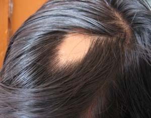
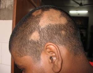
with many patterns such as AA monolocularis (Fig. 1a), MATERIALS AND METHODS:
AA multilocularis (Fig. 1b), AA universalis, alopecia This study was undertaken in the department of
totalis, ophiasis, reticularis, sisaipho etc. A new variant dermatology, IMS & SUM Hospital, Bhubaneswar in
recently described is ‘Acute diffuse and total alopecia’.(5) It collaboration with the department of Pharmacology, during
is characterized by a generalized thinning, rapid a period of two years from November 2011 to October
progression, tissue eosinophilia, brief clinical course and 2013.
favorable prognosis.(6) Scarring is characteristically Selection criteria:
absent.(7) There are lack of adequate efficacy of medications During this period those patients attending the dermatology
available for the treatment of alopecia areata. The various OPD having hair loss either patchy or diffuse with smooth
modalities of treatment are immunosuppressants such as bald surface and having no features of scarring, scaling or
steroids, cyclosporine A, methotrexate, azathioprine, inflammation on the bald area were included in the study.
169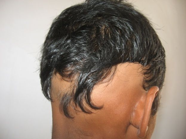
Maitreyee Panda et al /J. Pharm. Sci. & Res. Vol. 6(4), 2014, 169-174

All age groups & both sexes of patients who had not                 100% growth, good with 51- 75% and unsatisfactory with
received any treatment for last 3 months were included.             15 yrs = 4 tab (4mg)                               decades.
It was given on two consecutive days of a week usually
     Saturday and Sunday after breakfast.                           Table 3: Table showing sex wise distribution of cases
2. Those patients having one or two patches of small size                                Total no of     No of males      No of females
                                                                     Age groups
     < 4cm each were given intralesional triamcinolone                                    patients         with %            with %
     injection.                                                           0-10                8           4(50%)             4(50%)
Triamcinolone acetonide was injected into the deep dermis                 11-20               9           5(55%)            4(45%)
using a 0.5 inch long 30 gauze needle at multiple sites, 1cm              21-30              32           24(75%)           8(25%)
apart and 0.1 ml into each site. This was repeated every 4-6              31-40              21           17(80%)           4(20%)
weeks. The triamcinolone concentration used was 10mg/ml                   41-50               2           2(100%)             0(0)
for scalp and 2.5mg/ml for the face.                                      Total              72           52(72%)           20(28%)
With both the above treatments the patients were followed
up at an interval of 1 month for a period of 6months.               In our study (Table 4) majority of patients had lesion on the
During the follow up, the percentage of improvement in the          scalp (58.3%). Out of these, 16 had single lesion and 26
form of re-growth of hair was recorded through serial               had multiple lesions. Face was involved in 17 patients, 15
clinical photographs. Any side effect of the treatment was          having single lesion & 2 having multiple lesion. Both scalp
recorded. Hair growth was classified as excellent with 76-          and body was involved in 11 and only body in 2 patients.

                                                                                                                              170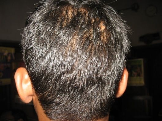
Maitreyee Panda et al /J. Pharm. Sci. & Res. Vol. 6(4), 2014, 169-174

Table4: Table Showing Location (site) wise Distribution              Table 9: Associated disease:
of Cases                                                             Associated disease        No of patients       Percentage (%)
                No of patients     No of patients                    Atopy                           7                    10
   Location       with single       with multiple        Total       Lichen Planus                   2                     3
                lesion with %      lesions with %                    Vitiligo                        1                    1.5
Scalp           16(38%)            26(62%)          42(58.3%)
Face            15(88.2%)          2(11.8%)         17(24%)          Out of 42 patients tried with intralesional triamcinolone
Scalp + Body    0(0%)              11(100%)         11(16%)          acetonide (Table 10), 6 patients didn’t report for follow up.
Body            0(0%)              2(100%)          2(3%)            Thus out of 36 patients, 26 (72.2%) responded to our trial.
                                                    Total = 72       Maximum number of patients (20) had excellent result by
                                                                     the end of 6 month. As regards to side effects, only 3
Out of 62 patients (Table 5), most (34) of the patients had          patients had folliculitis at the site of administration. One
oval lesion (54%). Fourteen (23%) had circular and 14                patient had local irritation which subsided with regular
(23%) had reticular lesions.                                         follow up. One patient had transient atrophy at the site of
                                                                     application.
Table 5: Shape of lesion (excluding AU,AT and
Ophiasis)                                                            Table 10: Response to intralesional steroid at 6 months
      Shape              No of patients        Percentage (%)        (n=36)
Oval                          34                     54                         Response                        No. of patients
Circular                      14                     23              Excellent                                        20
Reticular                     14                     23              Good                                              6
Total                         62                    100              Unsatisfactory                                    2
                                                                     Non responsive                                    8
Out of 72 patients 7 had nail involvement (Table 6). Most
common nail involvement was pitting (57%) followed by                Out of 30 patients with OMP therapy (Table 11), 5 patients
longitudinal ridging (29%) and dystrophy (14%).                      didn’t report for follow up. Thus out of 25 patients, 15
                                                                     (60%) showed good response. Side effects noted were
Table 6: Nail involvement                                            acneiform eruptions, gastric upset and cushingoid facies.
         Nail change                  No of patients with %
Pitting                                       4(57)                  Table no 11: Response to OMP steroid at 6 months
Longitudinal ridging                          2(29)                  (n=25)
Dystrophy                                     1(13)                             Response                        No of patients
total                                           7                    Excellent                                        9
                                                                     Good                                             6
Majority (Table 7) of patients (72%) presented within 6              Unsatisfactory                                   4
months of disease onset.                                             Non responsive                                   6

Table 7: Duration of disease:
     Duration            No of patients       Percentage (%)
0-6months                     52                    72
6 m- 1 yr                      7                    10
>1yr                          13                    18

Out of 72 patients (Table 8), 7 (10%) had family history of
AA. It was further noticed that those patients having
positive family history had extensive disease. Three
patients (43%) had extensive AA, 3 (43%) had AU and 1                                                              1a
(14%) had ophiasis pattern of involvement.

Table 8: Associated Family history of AA according to
clinical variants:
                       No of patients with
     Variants                                 Percentage (%)
                          positive F/H
Extensive AA                    3                   43
AU                              3                   43
Ophiasis                        1                   14
Total                           7
                                                                                                                  1b
Out of 72 patients (Table 9), 10 had associated diseases
such as atopy in 7, followed by LP in 2 and vitiligo in 1
patient.                                                                       Figure 1a : AA MONOLOCULARIS and 1b : AA
                                                                                             MULTILOCULARIS

                                                                                                                             171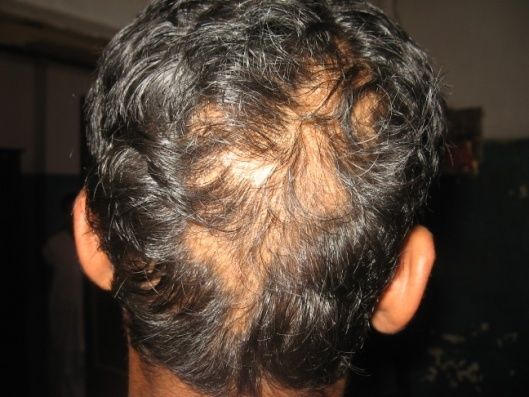
Maitreyee Panda et al /J. Pharm. Sci. & Res. Vol. 6(4), 2014, 169-174

                                                                                            6a
       Figure 2: ALOPECIA UNIVERSALIS

                                                                                   6b
                                                             Figure 6a : PRETREATMENT PHOTO OF ALOPECIA
                                                              AREATA, 6b : HAIR GROWTH POST 8 WEEKS OF
                                                                              OMP THERAPY
              Figure 3: OPHIASIS
                                                                                     DISCUSSION:
                                                            The etiology of AA is still an enigma. The most accepted
                                                            hypothesis is a hair follicle specific T-cell mediated
                                                            autoimmune disorder occurring in genetically predisposed
                                                            individuals with a suggested polygenic inheritance.(5) It is
                                                            linked with certain HLA class II alleles. The HLA-
                                                            DQB1*03 allele may be an important marker for
                                                            susceptibility to the disease.(10,11)
                                                            Diagnosis is mostly clinical. A skin biopsy specimen is
                                                            confirmatory for AA. In acute cases peribulbar and
                                                            intrabulbar lymphocytic inflammatory infiltrate around
                                                            anagen follicles, resembling ‘swarm of bees’ is
                                                            characteristic. Dermoscopic findings include yellow dots,
                                                            black dots, broken hairs, tapering hair (exclamation mark)
         Figure 4: ALOPECIA TOTALIS                         and short vellus hairs.
                                                            Extrafollicular involvement comprises of ungual and ocular
                                                            alterations. Nail changes are seen in 29% of adults and 50%
                                                            of children with AA. Nail changes (Fig. 5) mainly include
                                                            superficial geometric pitting, punctuate leukonychia and
                                                            trachyonychia.(12) Punctate lens opacities, early cataracts
                                                            and fundal abnormalities may occur in 40% to 50% of
                                                            patients with AA.(13)
                                                            The presence of exclamatory mark hairs at periphery,
                                                            positive hair pull test (>6 hairs), daily hair count (>100
                                                            hairs), hair pluck test (more telogen hairs) and positive
                                                            dermoscopic findings suggest active disease.(14) Predictors
                                                            of poor prognosis include, younger age of onset, family
                                                            history of AA, atopy, severe disease like alopecia totalis
                                                            and alopecia universalis, ophiasis, duration more than 1
Figure 5: PITTING AND LONGITUDINAL RIDGING IN
                                                            year & associated nail and autoimmune disease.(15,16)
             SEVERE ALOPECIA AREATA
                                                            In our study out of 72 patients, AA classic variant having
                                                            patchy lesions constituted the maximum percentage (88%)

                                                                                                               172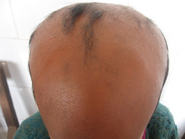
Maitreyee Panda et al /J. Pharm. Sci. & Res. Vol. 6(4), 2014, 169-174

of all cases followed by 7% AU, 5% Ophiasis and 3% AT.                                     CONCLUSION:
In various studies conducted in the past it was seen that AA        Alopecia areata is a relatively benign non scarring form of
classic variant constituted the major proportion of cases in        hair follicle specific autoimmune disease, triggered by
comparison to other variants.(17,18) Age range of the patients      environmental factor in genetically susceptible individuals.
of AA in our study was 1 and ½ years to 48 years among              Treatment is still an enigma and large number of treatment
which maximum cases were seen in 3rd decade of life                 modalities speaks of their lack of adequate efficacy.
(44%) followed by 4th decade (29%). The peak incidence of           We conclude that both intralesional steroid and OMP
manifestation in all clinical variants of AA if grouped             steroid are effective modalities in the treatment of alopecia
together was in between the age of 20 and 50 years and              areata with limited side effects. But as our study was done
onset to occur is at any age. S Manzoor et. al.(19) reported        in a limited number of cases and was an uncontrolled one
that maximum number of patients were in the age group of            we recommend a large scale controlled study which is
third decade and 4th decade, which was consistent with our          needed to be done to conclude that both are really effective
findings. Out of 72 patients 72% (52 patients were male             modality of treatment in AA. This condition has a definite
and 28% (20 patients) were female. Thus the sex ratio in            psychological impact and affects the quality of life of many
our study is 2.6:1. The reported sex incidence has varied           patients. Proper counseling and appropriate treatment helps
widely from males out numbering females by 3:1(20),                 to attain cosmetically acceptable hair regrowth and
through equality(21), to twice as common in females.(22)            improves quality of life.
Manzoor S et al.(19), in an Indian study had found the male
to female ratio to be 2.63:1. Male preponderance could be                                        REFFERENCES:
due to more number of male patients attending the hospital.         1.    Mac Donald Hull SP,Wood M L, Hutchinson P E, Saidden M,
                                                                          Messenger A G. Guidelines for management of AA. British Journal
Majority of patients had lesion on the scalp (58.3%)                      of Dermatology 2003; 149: 692–699.
followed by face i,e beard, moustache and eyebrows (24%),           2.    McMichael AJ, Pearce DJ, Wasserman D, Camacho FT, Fleischer
scalp and body (16%) and body alone (3%). In an Indian                    AB Jr, Feldman SR, Balkrishnan R. Alopecia in the United States:
study by Jain S et. al. 2003(18), maximum number of cases                 outpatient utilization and common prescribing patterns. J Am Acad
                                                                          Dermatol. 2007 Aug;57(2 Suppl):S49-51.
were seen in scalp. Lesion of AA can have various shapes.           3.    Price V H: Alopecia Areata: Clinical aspect. J. Invest Dermatol
In our study maximum number of patients excluding AU,                     1991; 96:685.
AT and Ophiasis had oval and circular lesions (77%). Only           4.    Satavi K: Prevalence of AA in the first national health & nutrition
14 patients had reticular lesion. This is consistent with the             examination survey. Arch Dermatol 1992; 128(5): 702.
                                                                    5.    Martinez-Mir A, Zlotogorski A, Gordon D, Petukhova L, Mo J,
finding of Sadollah S et.al.(23) who in their study of 956                Gilliam TC, et.al. Genomewide Scan for Linkage Reveals Evidence
patients of AA reported that their commonest clinical                     of Several Susceptibility Loci for Alopecia Areata. Am J Hum
presentations were round and oval configuration. In our                   Genet. Feb 2007; 80(2) :316-28.
study, out of 72 patients 10% (7 patients) had nail                 6.    Lew BL, Shin MK, Sim WY. Acute diffuse and total alopecia: A
                                                                          new subtype of alopecia areata with a favourable prognosis. J Am
involvement among which the commonest presentation was                    Acad Dermatol 2009; 60: 85-93.
pitting (57%) followed by longitudinal ridging (29%) and            7.    Sharpio J, Madani S. Alopecia Areata: Diagnosis & management. Int
dystrophy (14%). S. Jain et al 2003 (18) in their study of 150            J Dermatol 1999; 38(1): 19-24.
patients reported nail changes in 13% of patients, the              8.    Fiedler VC. Alopecia Areata: A review of therapy, efficacy, safety &
                                                                          mechanism. Arch Dermatol. 1992; 128: 1519-29.
commonest being pitting.                                            9.    Gupta MA, Gupta AK Depression and suicidal ideation in
In our study the minimum duration of the disease was 15                   dermatology patients with acne, alopecia areata, atopic dermatitis
days and maximum was 6 years. However, majority of                        and psoriasis. Br J Dermatol. 1998 Nov; 139(5):846-50.
patients presented within 6 months of onset (72%). This is          10.   Petukhova L, Duvic M, Hordinsky M, Norris D, Price V, Shimomura
                                                                          Y, et. al. Genome wide association study in alopecia areata
consistent with the findings of Jain S etal 2003 (18) who                 implicates both innate and adaptive immunity. Nature 2010; 466:
reported that 78% of patients presented within 6 months of                113-17.
onset. Familial incidence of AA varies from 10 to 20% in            11.   Barahmani N, Andrade M, Slusser JP, Wei Q, Hordinsky M, Price
different studies.(21,24) In our study 10% of patients were               VH, et. al. Human leukocyte antigen class II alleles are associated
                                                                          with risk of alopecia areata. J Invest Dermatol 2008; 128: 240-43.
having positive family history. This is to be more frequent         12.   Dotz WI, Lieber CD, Vogt PJ. Leukonychia punctata and pitted nails
with severe forms of AA. Some cases of AA are associated                  in alopecia areata. Arch Dermatol. 1985 Nov;121(11):1452-4.
with autoimmune diseases. In our study 10 patients had              13.   Pandhi D, Singal A, Gupta R, Das G. Ocular alterations in patients
associated diseases among which 10% had atopy, 3% with                    of alopecia areata. J Dermatol 2009; 36: 262-8.
                                                                    14.   Seetharam KA. Alopecia areata: An update. Indian J Dermatol
Lichen planus and 1.5% vitiligo. Jain S et.al.(18) had                    Venereol Leprol. 2013; 79: 563-75.
observed atopic manifestation in 11.37% of their patients.          15.   Madani S, Shapiro J. Alopecia areata update. J Am Acad Dermatol
Out of 30 patients tried with OMP, 5 patients didn’t turn up              2000; 42: 549-66.
for follow up. Of the remaining 25 patients, 60% (15                16.   Alkhalifah A, Alsantali A, Wang E, Mc Elwee KJ, Shapiro J.
                                                                          Alopecia areata update: Part I. Clinical picture, histopathology and
patients) responded to our trial. Ahu Birol et. al.(25) had               pathogenesis. J Am Acad Dermatol 2010; 62: 177-88.
reported response rate of 80% by giving pulse steroid for a         17.   Sharma VK, Dawn G, Kumar B. Profile of AA in northern India.int J
period of 6months. Patients had acneiform eruptions,                      Dermatol 1996 Jan; 35(1): 22-7.
gastric upsets & cushingoid facies at the end of 6 months.          18.   Jain S, Marfatia YS. AA: Pattern in industrial city of Baroda. Indian
                                                                          Journal of Dermatology, Venereology and Leprology 2003; 69(2):
Binod K Khaitan et. al.(26) have reported these side effects              81-2.
in few patients. Treatment with intralesional steroid showed        19.   Manzoor S, Masood G. AA in Kashmir: A study of 200 patients. Int
good result in 72.2% of patients in our study which was                   J Trichology 2001; 67(6): 324-5.
consistent with results (65-70%) found in other studies.(27)

                                                                                                                                    173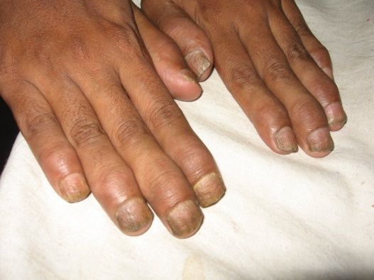
Maitreyee Panda et al /J. Pharm. Sci. & Res. Vol. 6(4), 2014, 169-174

20. Bastos AA, Poiares Baptista A. Algunas considerations sobre 300       25. Ahu Birol, Emel Erkek etal. The efficacy of intermittent low dose
    casos de pelada. Trals Soc Portuges Dermatol Vehereol. 1967; 15:          systemic corticosteroid in the treatment of AA. Turkey J Med Sci.
    135-9.                                                                    2004; 34: 55-8.
21. Muller, H.A. and Winkelmann, R. K. (1963). Alopecia areata. New       26. Khaitan BK, Mittal R, Verma KK. Extensive Alopecia areata treated
    Clinical Applications Volume 9, 1988, pp 1-27                             with betamethasone oral mini pulse therapy: An open uncontrolled
22. Friedmann PS. AA and autoimmunity. Br J Dermatol. 1981; 105:              study. Indian J Dermatol Venerol Leprol. 2004; 70(6): 350-3.
    153-7.                                                                27. Alkhalifah A, Alsantali A, Wang E, Mc Elwee KJ, Shapiro J.
23. Sadollah S. Determination of clinical patients of AA in relation to       Alopecia areata update: Part II. Treatment. J Am Acad Dermatol
    some varieties in Kerman, Iran. Int J of Dermatol. 2006; 3(2).            2010; 62: 191-202.
24. Gip L, Lodin A, Molin L. Alopecia areata – A follow up
    investigation of out patient material. Act Derm Venereol. 1969; 49:
    180-8.

                                                                                                                                    174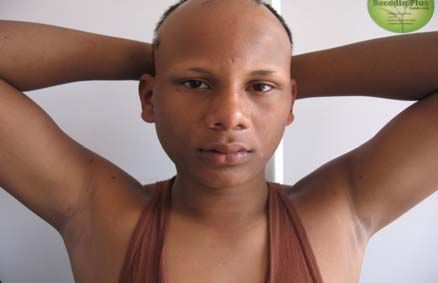
You can also read