Comparison of the oncogenomic landscape of canine and feline hemangiosarcoma shows novel parallels with human angiosarcoma - Journals Gateway | ...
←
→
Page content transcription
If your browser does not render page correctly, please read the page content below
© 2021. Published by The Company of Biologists Ltd | Disease Models & Mechanisms (2021) 14, dmm049044. doi:10.1242/dmm.049044

RESEARCH ARTICLE

Comparison of the oncogenomic landscape of canine
and feline hemangiosarcoma shows novel parallels
with human angiosarcoma
Kim Wong1, *, Latasha Ludwig2,*, Oscar Krijgsman3, David J. Adams1, Geoffrey A. Wood2 and
Louise van der Weyden1,‡

ABSTRACT                                                                                              and thorium dioxide), chronic lymphedema and various familial
Angiosarcoma (AS) is a highly aggressive tumor of blood and                                           syndromes (Young et al., 2010). The multifocal and aggressive
lymphatic vessels in humans that shares many similarities with                                        nature of this tumor results in many patients showing advanced
spontaneously occurring hemangiosarcoma (HSA) in dogs and cats.                                       disease at presentation and having a high risk of local recurrence
To investigate the genetic suitability of HSA as a model for AS, we                                   and metastasis, thus resulting in a poor prognosis (30-40%
sequenced ∼1000 cancer genes in 41 cases of HSA and matched                                           disease-specific survival) (Mark et al., 1996; Fayette et al., 2007;
germline tissue: 15 canine visceral HSAs, 13 canine skin HSAs and                                     Buehler et al., 2014). The rarity of AS, together with the fact that
13 feline skin HSAs. Analysis of visceral HSAs from dogs presenting                                   this tumor type encompasses a heterogeneous group of sarcomas
with concurrent splenic and cardiac neoplasms showed that the                                         with specific behaviors depending on the primary site (Fayette
tumors were not independent primaries, consistent with the highly                                     et al., 2007), has made identification of the genetics underlying
metastatic nature of HSA. Comparison of HSA to AS revealed that                                       the pathogenesis of AS somewhat challenging. The driver genes
several driver genes were recurrently mutated in both species, such                                   of AS are beginning to emerge from whole-genome, whole-exome
as TP53, PIK3CA, ATRX, GRIN2A and LRP1B. Similar to AS, a UV                                          and targeted sequencing studies (Behjati et al., 2014; Murali et al.,
mutational signature was found in a subset of canine cutaneous                                        2015; Zehir et al., 2017; Painter et al., 2020), yet this is still a disease
HSAs and both species show differing mutational profiles between                                      with significant unmet clinical needs, as prospective studies
tissue sites. Our characterization of canine and feline HSA                                           addressing the various treatment options are limited and the
demonstrates many important parallels to AS and provides hope                                         development of new therapies is hampered by a lack of good
that future studies on these cancers will benefit of all three species.                               preclinical models.
                                                                                                         Although genetically engineered mouse models have proven
KEY WORDS: Dog, Cat, Visceral, Skin, Hemangiosarcoma,                                                 useful in understanding the biology and signaling pathways of AS in
Comparative genomics                                                                                  humans (Dill et al., 2012; Chadwick et al., 2018; Salter et al., 2019),
                                                                                                      spontaneous hemangiosarcoma (HSA) in companion ( pet) animals
INTRODUCTION                                                                                          may represent more relevant clinical models. Dogs, in particular, are
Angiosarcoma (AS) is a rare tumor that accounts for 1-2% of soft-                                     relevant models, as they spontaneously develop these tumors, have a
tissue sarcomas in humans, which in themselves comprise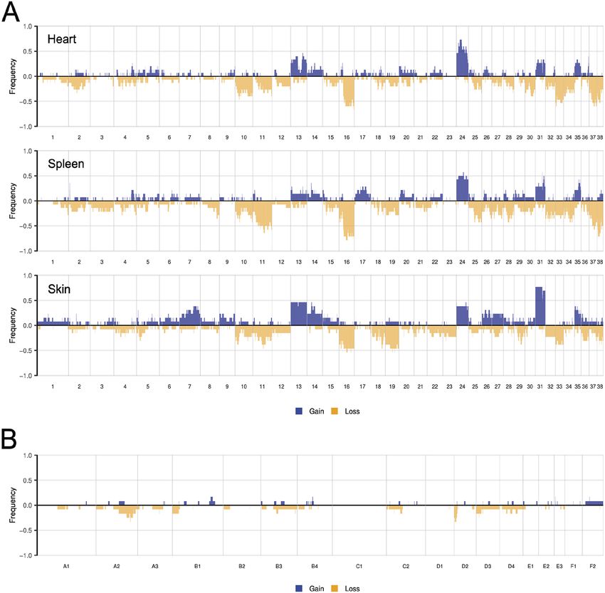
RESEARCH ARTICLE                                                            Disease Models & Mechanisms (2021) 14, dmm049044. doi:10.1242/dmm.049044

tumor development, particularly in the lungs, liver, mesentery and          and splenic HSA lesions (where both lesions were sequenced) and
omentum. It is not uncommon for dogs to present with concurrent             one was a cardiac lesion only. The canine and feline cutaneous cases
visceral HSA tumors of the spleen and heart (Waters et al., 1988;           ranged from dermal to subcutaneous regions of the skin, from
Boston et al., 2011; Yamamoto et al., 2013), and it is not known            different body sites (including the ventral abdomen, limbs and
whether these represent independent primary tumors or metastatic            back). A summary of all cases and their signalment data is provided
disease. Dogs can also develop cutaneous HSA, particularly in non-          in Table S1. The tissue was sampled by taking cores from formalin-
pigmented or light-haired skin (often on the ventral abdomen), with         fixed paraffin-embedded (FFPE) blocks. All cores were taken in
UV radiation proposed to play a causative role (Hargis et al., 1992).       duplicate, and the DNA was extracted from each core using one of
Visceral HSA is associated with a significantly worse prognosis             two different methods (detailed in the Materials and Methods) to
than cutaneous HSA (Schultheiss, 2004), owing to local infiltration,        provide replicates to aid in variant calling. The methods showed a
rupture of the primary tumor and/or metastases (Brown et al., 1985).        high degree of concordance in variant calling (Fig. S1). Using a list
In cats, HSA is overall less common, with an incidence of 0.5-2% of         of 1039 human cancer-associated genes in the OncoKB database
all tumors (Smith, 2003), and more commonly found in the skin               (https://www.oncokb.org/cancerGenes), we identified the canine
than the visceral organs, with the latter being relatively rare             and feline orthologs of these genes, where possible, and were able to
(Johannes et al., 2007). In both dogs and cats, HSAs involving the          include a total of 962 and 986 genes for the canine and feline bait
subcutis (cutaneous extending into subcutis or subcutaneous form)           libraries, respectively (Table S2). To explore the cancer gene
are more biologically aggressive than the cutaneous form, and more          mutational landscape of these tumors, we generated profiles of
likely to locally recur (Johannes et al., 2007), whereas cats that had      somatic point mutations, multi-nucleotide variants, indels and
aggressive surgical excision of their cutaneous tumors showed good          somatic copy number alterations (SCNAs) for these genes.
long-term prognosis (McAbee et al., 2005).                                  Complete lists of the somatic mutations identified in each sample
   Whole-exome sequencing of canine HSAs has previously been                are provided in Table S3.
performed in two studies, including splenic tumors from a variety of
breeds (n=20) (Wang et al., 2017) and visceral tumors (spleen, heart        Concurrent canine splenic and cardiac HSA lesions are not
and liver) from Golden retrievers (n=47) (Megquier et al., 2019). A         independent tumors
more recent study performed amplicon-based targeted resequencing            Post-mortem studies on dogs with clinical signs of cardiac (right
of splenic tumors (n=50) using a 30-gene ‘HSA panel’ (Wang et al.,          atrium) HSA have commonly reported the presence of concurrent
2020). However, there were differences in the candidate driver genes        splenic HSA [17/51 (33%) cases (Yamamoto et al., 2013) and 9/31
identified in each of these studies, suggesting that HSA is a               (29%) cases (Boston et al., 2011)]. Conversely, studies on dogs with
heterogeneous tumor type, which is in agreement with studies of             clinical signs of splenic HSA have reported the presence of
AS (Boichard et al., 2020). Furthermore, there have been no detailed        concurrent cardiac HSA [6/25 cases (24%) (Waters et al., 1988) and
genetic analyses of canine skin HSA [only studies looking at                2/23 (8.7%) cases (Boston et al., 2011)]. However, it is not known
mutations in PDGFRA/B (Abou Asa et al., 2015) and c-KIT (Chen               whether these represent independent/synchronous primary tumors
et al., 2016), specifically], and no genetic analyses of feline skin HSA.   or metastatic disease. To answer this question, we obtained tumor
Thus, we asked whether deep sequencing of a targeted gene panel             samples from 14 dogs with concurrent splenic and cardiac HSA
would provide novel insights into the genetics of canine and feline         lesions. The paired samples from each dog had shared mutations
HSA, and whether they are relevant models of AS in humans. This             (defined as mutations in the same genomic position with the same
has the potential to open up avenues for the application of human           nucleotide change; Fig. 1), and almost all of these mutations were
therapies to these animals, as a number of drugs utilized in veterinary     not seen in tumors from other dogs. For example, although almost
medicine were originally developed for human use (reviewed in               all tumors from different dogs had different TP53 mutations,
Paoloni and Khanna, 2008). Likewise, cancer clinical trials in              concurrent cardiac and splenic tumors from each dog shared the
companion animals have potential applications to human patients,            same TP53 mutation (Fig. S2); four of the concurrent HSAs had two
especially when the cancer type is more common in animals. This line        identical TP53 mutations. It is highly unlikely that this pattern of
of thinking is concordant with the ‘One Medicine’ concept that              TP53 mutations occurred by chance in independent tumors, in
promotes the view that human and veterinary medicine share                  agreement with the overall overlap of mutations shown in Fig. 1.
many commonalities and both can contribute to the advancement               Thus, we can conclude that the concurrent lesions do not represent
of the other (Schwabe, 1964). This has evolved into the ‘One                two independent tumors, and it is more likely that one is a metastasis
Health Initiative’ that promotes interdisciplinary collaboration            of the other.
in aspects of health for humans, animals and the environment                                                                                           Disease Models & Mechanisms
(https://onehealthinitiative.com/). To be a relevant model of AS in         Cancer gene mutation landscape of canine visceral HSA
clinical trials, alterations in cancer-associated driver genes are of       tumors
most interest. Thus, we performed targeted sequencing of ∼1000              For clarity, and as we have shown that the cardiac and splenic
cancer-associated genes in paired splenic and cardiac HSA tumors            tumors are not independent tumors, they will be discussed together
from the same dogs, as well as cutaneous HSA tumors from both               and referred to as visceral HSA. Our analysis of canine visceral
dogs and cats.                                                              HSAs found that the tumor suppressor TP53 was the most
                                                                            frequently mutated gene (14/15 cases, 93%; Fig. 2A); all but one
RESULTS                                                                     of the mutations occurred in the DNA-binding domain, which is the
Sequencing of canine and feline HSA                                         most frequently mutated domain in TP53 in human cancers
We performed targeted panel sequencing of cancer-associated                 (Laptenko and Prives, 2006). Interestingly, six cases had multiple
genes in tumor-germline (matched normal tissue) pairs of 15                 mutations in the gene (Fig. S2). The oncogene PIK3CA was the
primary canine visceral HSA cases, 13 primary canine cutaneous              second most frequently mutated gene in canine visceral HSA (9/15
HSA cases and 13 primary feline cutaneous HSA cases. Fourteen of            cases, 60%; Fig. 2A), with the majority of mutations (6/9) identified
the canine visceral cases were from dogs with concurrent cardiac            as H1047R in the kinase domain (Table S3A and Fig. S2). The

                                                                                                                                                  2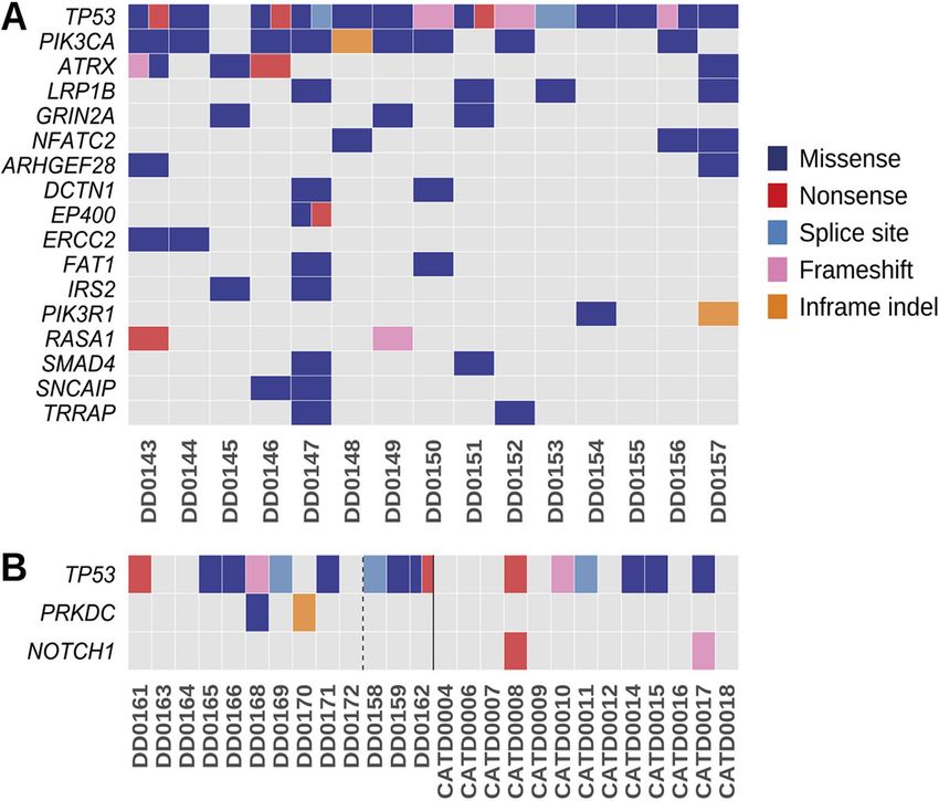
RESEARCH ARTICLE                                                        Disease Models & Mechanisms (2021) 14, dmm049044. doi:10.1242/dmm.049044

                                                                                                           Fig. 1. Shared and unique somatic
                                                                                                           mutations in canine cardiac and
                                                                                                           splenic hemangiosarcoma (HSA)
                                                                                                           from the same patient. Shown here are
                                                                                                           the number of unique and shared
                                                                                                           mutations [defined as mutations in the
                                                                                                           same genomic position with the same
                                                                                                           nucleotide change(s)] in tumors from
                                                                                                           dogs that presented with concurrent
                                                                                                           cardiac and splenic HSA.

PIK3CA protein is highly conserved between human and dog                FAT1, IRS2, PIK3R1, RASA1, SMAD4, SNCAIP and TRRAP
(99.8% identity), including amino acid H1047. PIK3CA H1047R is          (Fig. 2A).
a hotspot missense mutation in many human cancers that increases
the catalytic activity of PI3K (Samuels et al., 2004). Interestingly,   Cancer gene mutation landscape of canine cutaneous HSA
one canine HSA (DD0144) with a PIK3CA mutation also carried             tumors
a mutation in another class I PI3K gene, PIK3C2G, and two               Similar to visceral HSAs, the most frequently mutated gene in the
cases (DD0154, DD0157) without mutations in PIK3CA had                  canine skin HSA cohort was TP53 (9/13 cases, 69%; Fig. 2B).
mutations in its regulatory subunit, PIK3R1 (Table S3A). The next       However, in contrast to visceral HSAs, the only other recurrently
most frequently mutated genes in the canine visceral HSAs were the      mutated gene, when excluding samples with a UV signature, was
tumor suppressor genes ATRX (with DD0143 carrying one                   the protein kinase PRKDC (2/13 cases, 15%). Interestingly, in three
independent and two linked mutations in the gene) and LRP1B             of the canine skin HSA samples (DD0158, DD0159, DD0162), we
(4/15 cases, 26%), followed by GRIN2A and NFATC2 (3/15 cases,           identified an elevated mutation rate and the COSMIC mutational
20%; Fig. 2A). Finally, there were 11 genes mutated in 2/15             signatures SBS7a and SBS7b (Fig. S3), which have been found
(13%) of cases, specifically, ARHGEF28, DCTN1, EP400, ERCC2,            predominantly in skin cancers and have a proposed etiology of UV

                                                                                  Fig. 2. Cancer gene landscape of canine visceral, canine
                                                                                  cutaneous and feline cutaneous HSA tumors. (A,B) Mutations in
                                                                                  canine visceral HSA (A) and canine and feline cutaneous HSA (B).
                                                                                  Canine samples are left of the solid line; feline, right. Canine
                                                                                  cutaneous HSA samples with a UV mutational signature are right of
                                                                                  the dashed line. Mutations in visceral HSA tumors were present in
                                                                                  either the heart sample, spleen sample or both. Shown are genes
                                                                                  mutated in two or more samples, excluding those with a UV

                                                                                                                                                       Disease Models & Mechanisms
                                                                                  mutational signature. A full summary of the cases analyzed in this
                                                                                  study is provided in Table S1.

                                                                                                                                                  3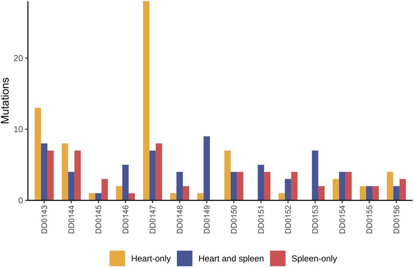
RESEARCH ARTICLE                                                          Disease Models & Mechanisms (2021) 14, dmm049044. doi:10.1242/dmm.049044

light exposure (Alexandrov et al., 2020). These tumors were located       few SCNAs compared to the canine skin HSAs, and the most
on the hindlimb and ventral abdomen of these dogs, which are sun-         penetrant SCNAs were smaller regions of chromosomes A2 and D2
exposed sites (if the dog ‘sun bathes’ belly up), thus suggesting a       (Fig. 3B).
potential role for UV light exposure in the development of some
cutaneous canine HSAs. This is consistent with that reported for          Analysis of germline sequence data for pathogenic
face and scalp AS in humans (Painter et al., 2020; Boichard et al.,       mutations
2020). As a result of exposure to UV light, these three canine cases      In addition to our analysis of somatic mutations, we looked for
have a significantly increased number of somatic mutations relative       putative pathogenic germline variants in the canine and feline
to the other cases, which confounds driver gene identification, as the    orthologs of established human AS susceptibility genes (Cao et al.,
majority of the recurrent mutations are likely to be passenger            2019), specifically, ERCC2, IDH1, IDH2, PIK3CA, POT1, PTEN,
mutations. However, if these tumors are taken into account, then          RB1, TP53 and XPC. The AS susceptibility genes POLH and
other recurrently mutated genes in the skin HSAs include RELN and         PTHR1 were not included in OncoKB and thus were not part of our
the tumor suppressor gene, PTPRD (4/13 cases, 30%; 3/4 cases had          targeted gene panel (see Materials and Methods). We identified
a UV mutational signature; Table S3A). Other recurrently mutated          missense variants in ERCC2 (six dogs), IDH1 (three dogs) and
genes included PIK3CA, LRPB1, PLCG2, KMT2D and ERBB4                      IDH2 (two dogs; Table S4A). We also identified a nonsense
(3/13 cases, 23%; 2/3 cases had a UV mutational signature;                mutation in TP53 in two dogs. However, the location of this variant
Table S3A). Finally, SMARCA1 and the tumor suppressor gene                (C>T, chromosome 5 at position 32,565,554) falls within only one
PTEN were also recurrently mutated (2/13 cases, 15%; 1/2 cases had        of the three canine TP53 transcripts annotated in Ensembl (v98,
a UV mutational signature; Table S3A).                                    TP53-202, exon 1), and it is only represented at very low levels in
                                                                          RNA-sequencing (RNA-seq) data from canine visceral HSA
Cancer gene mutation landscape of feline cutaneous                        samples (Megquier et al., 2019). In addition, the gene model may
HSA tumors                                                                be erroneous, as there is evidence of at least one additional intron/
Similar to canine skin HSA tumors, the most recurrently mutated           exon upstream of exon 1 in TP53-202 (Fig. S5). Finally, we
gene in the feline skin HSA cohort was TP53 (6/13 cases, 46%;             identified missense variants (at five different locations) in XPC
Fig. 2B; Table S3B). However, the only other recurrently mutated          in 15 dogs; however, as we found that a number of other sites in
gene was the tumor suppressor gene NOTCH1 (2/13 cases, 15%),              XPC were not true variants but instead reference genome
with tumors harboring nonsense and frameshift mutations.                  CanFam3.1 errors that have been corrected in CanFam4, it is
NOTCH1 was not mutated in any canine skin HSA cases,                      likely that the majority of these are also due to reference genome
although one canine visceral HSA case harbored a frameshift               errors. In the cat germlines, we identified missense mutations
mutation in NOTCH1. There were additional similarities to canine          in ERCC2, RB1, IDH1, IDH2, POT1, TP53 and XPC (Table S4B).
skin HSA, as RELN, PIK3CA and KLHL6 were mutated in both                  In three cases, RB1 had an additional in-frame deletion. It would
species (in one case each, if excluding the canine cases with a UV        be of interest to further investigate the variants in these orthologs
mutational signature; Table S3).                                          of established human AS predisposition genes to determine
                                                                          whether they play a role in germline predisposition to HSA in
Analysis of SCNAs                                                         dogs and cats.
We next analyzed the copy number profiles of canine visceral,
canine cutaneous and feline cutaneous HSA. The frequencies of             Comparative analysis with AS in humans
SCNAs in each cohort are shown in Fig. 3, and representative copy         AS is a heterogeneous tumor type, with differences in the relative
number profiles of individual samples are shown in Fig. S4. Canine        frequencies of the mutated genes reported between studies (Behjati
visceral HSAs had substantial chromosomal gains and losses, the           et al., 2014; Murali et al., 2015; Zehir et al., 2017; Painter et al.,
most frequent of which were gains along chromosomes 13, 14, 24,           2020). For the comparison of canine and feline HSA to human AS,
31 and 35, and losses along chromosomes 10, 11, 16, 33, 37 and 38         we used the data from the Angiosarcoma Project (July 2020; 62
(Fig. 3A). This pattern is similar to that identified in primary canine   patients) (Painter et al., 2020). As some patients had samples from
visceral HSA in a study using oligonucleotide array comparative           multiple time points, we selected one sample per patient for the
genomic hybridization (CGH) (Thomas et al., 2014), in which               comparison (Materials and Methods; Table S5). In all three species,
copy number gain of chromosomes 13, 24 and 31 and loss of                 the tumor suppressor gene TP53 is the most recurrently mutated
chromosome 16 were most prevalent. We also noted differences in           gene (Fig. 4), although the frequency is higher in dogs (69-93%)
the penetrance of recurrent copy number gains and losses, which           and cats (46%) than in humans (21%). Interestingly, 6/28 (21%)             Disease Models & Mechanisms
may be attributed to the differences in the sensitivities of the          canine HSAs had two TP53 mutations compared to 3/62 (4.8%)
technologies used and/or differences in SCNA penetrance amongst           human HSAs. The oncogene PIK3CA is recurrently mutated in
breeds (Thomas et al., 2014). The cardiac and splenic tumors              canine HSA [both in our study and others (Megquier et al., 2019)]
had very similar copy number profiles, lending additional support         and in AS patients (9/62 patients, 10.8%; Fig. 4), although it is
to our assertion that concurrent cardiac and splenic tumors are           exclusively mutated in AS from the breast (9/9 patients) (Painter
not independent tumors. Although genome-wide copy number                  et al., 2020). In our cohort, we found that more visceral HSA cases
profiling has previously focused on visceral HSA, we were able to         had PIK3CA mutations (9/15, 60%) than skin HSA samples (3/13,
compare visceral and cutaneous HSA and observe differences in the         23%); Fig. S2), which may also explain why PIK3CA was only
penetrance of copy number gains of chromosome 13, 14, 24 and 31           mutated in a single feline skin sample (1/13, 7.6%). The putative
(Fig. 3). However, as the visceral and cutaneous HSA samples              tumor suppressor gene, LRP1B (Fig. 4), is also mutated in both
were obtained from a variety of breeds, we are unable to attribute        human AS and canine HSA.
these differences to tissue site or breed differences. It would be           The transcriptional regulator ATRX is recurrently mutated in
of interest, in future studies, to use larger cohorts of cutaneous        human AS (5/62 cases, 6%; Fig. 4) (Painter et al., 2020) and
HSA to study these differences. The feline skin HSAs had relatively       immunohistochemical staining revealed that a subset of patient

                                                                                                                                                4
RESEARCH ARTICLE Disease Models & Mechanisms (2021) 14, dmm049044. doi:10.1242/dmm.049044
Fig. 3. Somatic copy number
alterations (SCNAs) of canine
visceral, canine cutaneous and
feline cutaneous HSA tumors.
(A,B) Penetrance plots showing the
frequency of SCNAs in canine
cardiac (top; n=15), splenic (middle;
n=14) and skin HSAs (bottom; n=13)
(A), and feline skin HSAs (n=13) (B).
samples have loss of ATRX expression [7/118 cases, 6% (Panse MTOR, PCLO, PGR, PIK3CA, RELN, SETD2 and TERT; Fig. 4) or
et al., 2018)]. Loss of ATRX expression was more frequently mutated in one AS sample (BIRC3, CALR, LATS2, PAX3 and TEK)
observed in tumors located in deep soft tissues than in other body (Painter et al., 2020). Thus, 16/21 (76%) of the genes with protein-
sites, and associated with a significantly worse event-free survival altering mutations in feline skin HSA are also mutated in AS.
(Panse et al., 2018). We report, for the first time, that ATRX is also When considering only AS of the head/face/neck/scalp (HFNS)
recurrently mutated in canine HSA (4/15 visceral samples and 1/13 in the Painter et al. (2020) cohort, the most frequently mutated gene
skin samples) (Fig. 4). Previously, mutation of ATRX had only been is TP53 (9/19 patients, 47%), as was the case in the canine and feline
reported in 1/47 canine visceral HSA samples (Megquier et al., skin HSA samples (Fig. 4). Importantly, NOTCH1 is frequently
2019). Interestingly, loss of atrx was found to cooperate with p53 mutated in AS of the HFNS (5/19 samples, 26%) and was also
deficiency in promoting the development of sarcomas in zebrafish recurrently mutated in feline skin HSA (one frameshift mutation and
(Oppel et al., 2019), and in our cohort, 3/4 (75%) canine HSAs with one nonsense mutation) (Fig. 4). Critically, decreased NOTCH1
ATRX mutations also had TP53 mutations (Fig. 4; Fig. S2). We also expression by immunohistochemistry has been reported in AS (29/ Disease Models & Mechanisms
provide the first reports of recurrent mutation of FAT1, GRIN2A, 123 cases, 24%) and is statistically associated with a cutaneous site
PLCO and RELN in canine HSA (with the latter two genes also of origin (Panse et al., 2018). Additionally, inhibition of NOTCH
mutated in feline HSA), which are also recurrently mutated in AS signaling induces the development of malignant vascular tumors in
(Fig. 4). Several receptor-type protein tyrosine phosphatases mice (Liu et al., 2011; Dill et al., 2012). Interestingly, however,
(PTPRs) are recurrently mutated in AS; we identified mutations in NOTCH1 mutations were not seen in any of the canine skin HSA
PTPRB, PTPRD, PTPRO, PTPRS and PTPRT (Fig. 4) in canine samples. Yet, it is worth mentioning that decreased NOTCH2
HSA, while previously, only mutations in PTPRD have been expression has been reported in AS (16/103 cases, 16%) (Panse
reported (Megquier et al., 2019). et al., 2018), NOTCH2 mutations have been reported in skin and
The genetic landscape of feline HSA has not been reported HFNS AS (Painter et al., 2020), and NOTCH2 mutations were seen
previously. Of the genes in our feline gene panel, we found protein- in 2/13 of the canine skin HSA samples (albeit that both cases had a
altering mutations in 21 genes (Table S3B). Both TP53 and UV mutational signature and elevated mutation rate). Taken
NOTCH1 were recurrently mutated in feline HSA and in human AS together, this would suggest a tumor-suppressive role for the
(Fig. 4). In addition, we found mutations in other genes that were NOTCH signaling pathway in the pathogenesis of AS. There are
mutated in feline HSA and recurrently mutated in AS (ATRX, GLI1, other parallels between canine skin HSA and human skin AS. For
5RESEARCH ARTICLE Disease Models & Mechanisms (2021) 14, dmm049044. doi:10.1242/dmm.049044
Fig. 4. Comparative mutational landscape of human angiosarcoma (AS) and canine and feline HSA tumors. Shown are genes mutated in at least two of the
three species. The human AS data from 62 samples were obtained from the Angiosarcoma Project (Painter et al., 2020). The genes mutated in human AS are
represented as a percentage of total cases.
example, a UV mutational signature was found in 3/13 canine skin trials of the PI3K inhibitor, ZSTK474, showed prolonged stable
samples (Fig. 1; Fig. S3), and AS of the HNFS is also associated disease for sarcoma patients (Lockhart et al., 2013), and several
with a strong UV mutational signature (Painter et al., 2020), groups have reported on the potential efficacy of PI3K inhibitors in Disease Models & Mechanisms
suggesting that, for AS of HNFS and canine cutaneous HSA, UV- sarcoma mouse and zebrafish models, such as BKM120
light-induced DNA damage may be a causative factor. (Buparlisib) in undifferentiated pleomorphic sarcoma (Kim et al.,
2012), and BEZ235 in well-differentiated liposarcoma (Gutierrez
Identification of actionable mutations et al., 2011) and chondrosarcoma (Zhang et al., 2013). Furthermore,
By specifically sequencing canine and feline orthologs of human a novel dual PI3K/mTOR inhibitor, VDC-597, showed dose-
cancer genes and comparing altered genes with AS, we were able to dependent inhibition of both Akt1 and 4eBP1, reduction of
identify several potential actionable mutations. In all three species, proliferation, migration and VEGF production, and promotion of
we observed mutations within the PI3K/AKT/mTOR pathway, tumor cell apoptosis in three canine HSA cell lines (Pyuen et al.,
including PIK3CA, PIK3C2B, PIK3C2G, PIK3R1, MTOR and 2018). NOTCH1 is recurrently mutated in human AS and feline
PTEN. The PI3K/AKT/mTOR pathway is an important intracellular HSA, and mutated in one canine visceral HSA sample, and there
signaling pathway responsible for regulation of the cell cycle, and are many clinical trials using different types of drugs targeting
there are a range of PIK3 inhibitors in clinical trials, either dual NOTCH, including receptor/ligand antibodies, gamma secretase
PI3K/mTOR inhibitors, pan-PI3K inhibitors or isoform-specific inhibitors (GSIs) and NOTCH transcription complex inhibitors,
inhibitors (reviewed in Yang et al., 2019). For example, clinical with a Phase III trial of the GSI Nirogacestat currently underway
6RESEARCH ARTICLE Disease Models & Mechanisms (2021) 14, dmm049044. doi:10.1242/dmm.049044
(reviewed in Moore et al., 2020). In addition, as a subset of HNFS 24 are seen in a higher proportion of visceral cases relative to skin
AS (Painter et al., 2020) and cutaneous HSA tumors harbor a cases, yet a higher proportion of gains across chromosomes 13, 14
high mutation rate and a strong UV mutational signature, immune and 31 are seen in the skin cases relative to the visceral cases.
checkpoint inhibition may be beneficial in these patients. Indeed, Further studies are required to confirm whether these differences are
pilot clinical studies assessing the effectiveness of therapeutic inherent to the tumor tissue site or due, in part, to differences in
antibodies against canine PD-L1 and PD-1 for advanced breeds.
spontaneous cancers in dogs have shown some success in canine In addition to tissue-specific differences in the mutational
oral melanoma and undifferentiated sarcoma patients (Maekawa profiles, it is important to note that there are also species-specific
et al., 2017; Igase et al., 2020). Finally, PIK3CA has been shown to differences. For example, the tumor suppressor gene POT1 is
be associated with sensitivity to ionizing radiation when mutated mutated in 11/62 (17.7%) of AS cases (seven from HFNS and four
(Yard et al., 2016); thus, given the frequency of PIK3CA mutations from breast) (Painter et al., 2020); however, we, and others
in breast AS (Painter et al., 2020) and canine visceral HSA, (Megquier et al., 2019), have not observed any somatic mutations
radiotherapy may also be beneficial in these patients. of POT1 in canine HSA. Similarly, the tumor suppressor gene
ROBO1 is mutated in 8/62 AS cases (six from HFNS and two from
DISCUSSION breast) (Painter et al., 2020), but not in this or previous HSA studies
In our cohort, recurrently mutated genes found in canine HSA (Megquier et al., 2019). The significance of this is uncertain at this
included TP53, PIK3CA and LRP1B, which is consistent with stage, as mutations in these genes may be found in larger cohorts, if
previous reports in canine HSA (Megquier et al., 2019; Wang et al., they are low penetrance driver genes in HSA. Alternatively, given
2020) and in AS. Critically, despite the small cohort size, we have the tissue-specific preferences of some AS driver genes, sequencing
also identified cancer-associated genes that are recurrently mutated of canine/feline HSAs from other tissue sites may reveal these as
in AS, but not previously reported as such in canine HSA, including driver genes in HSA.
ATRX, FAT1, GRIN2A and RELN, further strengthening the Retrospective post-mortem studies on dogs that presented with
support for canine HSA as a relevant model for AS in humans. In clinical signs of cardiac HSA have reported the presence of
addition, targeted sequencing of canine cutaneous HSA revealed concurrent splenic HSA and vice versa (Waters et al., 1988; Boston
strong parallels with the genetics of AS of HFNS, including et al., 2011; Yamamoto et al., 2013). However, some dogs that
the identification of a UV mutational signature. Sequencing of present with multiple splenic HSAs have no involvement of the
feline cutaneous HSA for the first time also revealed parallels to heart or any other primary site, and some dogs that present with
both canine cutaneous HSA and human AS. Genetic parallels cardiac HSAs have no gross metastases in the spleen or other
between canine/feline HSA and AS, in addition to the clinical and primary sites (Yamamoto et al., 2013). Thus, there is strong
histopathological parallels, makes dogs and cats attractive as evidence that primary HSA can arise in either the heart or spleen;
relevant models for AS in humans. however, it is unclear whether the common occurrence of
The tissue site in which the AS arises plays a strong role in the concurrent splenic and cardiac HSA represents two independent
biology of the disease and thus the prognosis of the patient (Fayette primary tumors or one metastasis originating from the other. We
et al., 2007), and the same is true for canine and feline HSA. showed that paired (concurrent) splenic and cardiac HSAs from
Visceral HSA is associated with a poorer prognosis than cutaneous each dog shared mutations and had similar SCNAs, which indicates
HSA [with respect to cutaneous HSA lesions that can be completely they are not independent primaries and thus favors the likelihood
excised with clear margins, which have a good to fair prognosis that one represents metastasis from the other site. This is consistent
(Schultheiss, 2004)]. This may be partly attributed to the relative with AS, in which a significant number of patients have metastases
accessibility of these tissues for diagnosis and treatment (such as at the time of presentation, with one study reporting metastases at
skin versus visceral). However, it could also be attributed to the presentation in 26/81 (32%) AS patients (Buehler et al., 2014). More
differences in the mutational profile of the tumors at each site. For extensive sequencing across the exome or genome is required to
example, PIK3CA and KDR mutations are predominantly found in decipher the evolutionary histories of the canine splenic or cardiac
AS tumors of the breast (7/7 and 9/11 samples with mutations in HSA and determine which are the metastases. However, these
these genes, respectively, were from breast tissue) (Painter et al., findings have significant clinical implications, as early detection of
2020). Having sequenced orthologs of human cancer genes in HSA by screening blood samples (as is in development at the
canine HSAs from multiple tissue locations, specifically visceral University of Minnesota; ‘Shine On’ Project, https://vetmed.umn.
and skin, for the first time, we are able to see that, similar to AS, edu/centers-programs/clinical-investigation-center/current-clinical-
there are differences in the mutational landscape in canine HSA trials/early-detection-target-hemangiosarcoma-cells-dogs-shine- Disease Models & Mechanisms
from different tissue sites. First, despite similar cohort sizes, the project), followed by removal of the spleen or right atrium to prevent
visceral HSA cohort has more genes affected by protein-altering an aggressive HSA from growing and metastasizing, is much more
mutations than the cutaneous cohorts; 21 and 27 genes in the feline likely to be successful (curative) if there is not a high risk of dual
HSA and canine HSA cohorts, respectively (excluding samples primaries forming.
with a UV mutational signature) relative to 113 genes in the canine A limitation of our study is the small cohort size. It would have
visceral cohort. Second, some of the canine cutaneous samples had been interesting to determine whether some of the genetic alterations
a UV mutational signature, which was not seen in the canine visceral we observed are associated with parameters such as treatment
HSA samples. Third, although both the visceral and cutaneous response and patient survival; however, because the genetic
samples have some mutated genes in common, such as TP53 and alterations identified are very heterogeneous, such analysis would
PIK3CA, there are some tissue-specific differences. For example, require a significantly larger cohort of HSA from each tissue site. In
PIK3CA was mutated in 9/15 visceral HSA samples (with 6/9 at addition, given that we did not find any protein-altering single-
amino acid hotspot position 1047) but only 3/13 skin samples (with nucleotide variants (SNVs) or indels in our gene panel in 3/13 feline
1/3 at position 1047). Finally, there are also differences between cases (CATD0004, CATD0006, CATD0018), or a UV mutational
SCNAs in different tissues. For example, gains across chromosome signature in any feline case, it would suggest that future studies may
7RESEARCH ARTICLE Disease Models & Mechanisms (2021) 14, dmm049044. doi:10.1242/dmm.049044
have to use substantially larger cohorts in order to be able to identify transcription start codon were designed against the canine reference genome
rare driver genes and definitively determine whether a UV mutational CanFam3.1 (Hoeppner et al., 2014) (ELID: S3250944) and the feline
signature exists in a proportion of feline cutaneous HSA tumors. reference genome FelCat9.0 (Buckley et al., 2020) (ELID: S3250994). For
However, it should be noted that very few synonymous mutations genes with a ‘one-to-many’ orthologous relationship, all orthologs were
included in the bait design. For human cancer genes without a canine or
(A oxidation artefacts by comparing the mutation spectra
from our samples to COSMIC mutational signatures (v3, SBS45 and
Bait design SBS52) (Alexandrov et al., 2020) and applied a variant allele frequency
For gene panel design, we started with the 1039 human cancer genes in the (VAF) and sequence-context-specific filter. A C>A variant was classified as
OncoKB database (7 May 2019 update) and the TERT promoter region. A a false positive if it had a VAF≤0.1 and occurred in the sequence context
list of canine and feline orthologs of these genes was obtained from Ensembl CCN or TCN (or sequence context NGG or NGA for G>T variants). As
(v98). Agilent SureSelect bait libraries for exon target capture of these genes DNA sequence data from at least two cores were available for most samples,
(and up to 125 bp flanking sequence) and 500 bp upstream of the TERT we were able to leverage this when filtering the somatic variant calls further.
8RESEARCH ARTICLE Disease Models & Mechanisms (2021) 14, dmm049044. doi:10.1242/dmm.049044 Knowing that DNA from FFPE tissue contains artefactual C>T variants et al., 2013). The STAR genome index files were created using gene models from cytosine deamination, we defined a high-confidence C>T variant as from Ensembl v98. Alignments to TP53 were visualized using the one with 10 or more supporting reads and a VAF>0.05. Other SNV types Integrative Genome Viewer (Robinson et al., 2011). were classified as high-confidence calls if there were eight or more supporting reads and a VAF≥0.04. Any variant that did not meet one of Acknowledgements these criteria was discarded, unless the same variant was called in a replicate The authors would like to thank all the pets and their owners who participated in this sample with high-confidence. Indels with a VAF
RESEARCH ARTICLE                                                                        Disease Models & Mechanisms (2021) 14, dmm049044. doi:10.1242/dmm.049044

Chadwick, M. L., Lane, A., Thomas, D., Smith, A. R., White, A. R., Davidson, D.,        Li, H. (2013). Aligning sequence reads, clone sequences and assembly contigs with
  Feng, Y., Boscolo, E., Zheng, Y., Adams, D. M. et al. (2018). Combined mTOR             BWA-MEM. arXiv 1303.3997 [q-bio.GN] .
  and MEK inhibition is an effective therapy in a novel mouse model for                 Liu, Z., Turkoz, A., Jackson, E. N., Corbo, J. C., Engelbach, J. A., Garbow, J. R.,
  angiosarcoma. Oncotarget 9, 24750-24765. doi:10.18632/oncotarget.25345                  Piwnica-Worms, D. R. and Kopan, R. (2011). Notch1 loss of heterozygosity
Chen, Y.-C., Liao, J.-W., Hsu, W.-L. and Chang, S.-C. (2016). Identification of the       causes vascular tumors and lethal hemorrhage in mice. J. Clin. Invest. 121,
  two KIT isoforms and their expression status in canine hemangiosarcomas. BMC            800-808. doi:10.1172/JCI43114
  Vet. Res. 12, 142. doi:10.1186/s12917-016-0772-y                                      Lockhart, A. C., Olszanski, A. J., Allgren, R. L., Yaguchi, S., Cohen, S. J., Hilton,
Cibulskis, K., Lawrence, M. S., Carter, S. L., Sivachenko, A., Jaffe, D., Sougnez,        J. F., Wang-Gillam, A. and Shapiro, G. I. (2013). Abstract B271: A first-in-human
  C., Gabriel, S., Meyerson, M., Lander, E. S. and Getz, G. (2013). Sensitive             Phase I study of ZSTK474, an oral pan-PI3K inhibitor, in patients with advanced
  detection of somatic point mutations in impure and heterogeneous cancer                 solid malignancies. Mol. Can. Ther. 12, B271-B.
  samples. Nat. Biotechnol. 31, 213-219. doi:10.1038/nbt.2514                           Lydiatt, W. M., Shaha, A. R. and Shah, J. P. (1994). Angiosarcoma of the head and
Dill, M. T., Rothweiler, S., Djonov, V., Hlushchuk, R., Tornillo, L., Terracciano,        neck. Am. J. Surg. 168, 451-454. doi:10.1016/S0002-9610(05)80097-2
  L., Meili-Butz, S., Radtke, F., Heim, M. H. and Semela, D. (2012). Disruption of      Maekawa, N., Konnai, S., Takagi, S., Kagawa, Y., Okagawa, T., Nishimori, A.,
  Notch1 induces vascular remodeling, intussusceptive angiogenesis, and                   Ikebuchi, R., Izumi, Y., Deguchi, T., Nakajima, C. et al. (2017). A canine
  angiosarcomas in livers of mice. Gastroenterology 142, 967-977.e2. doi:10.              chimeric monoclonal antibody targeting PD-L1 and its clinical efficacy in canine
  1053/j.gastro.2011.12.052                                                               oral malignant melanoma or undifferentiated sarcoma. Sci. Rep. 7, 8951. doi:10.
Dobin, A., Davis, C. A., Schlesinger, F., Drenkow, J., Zaleski, C., Jha, S., Batut,       1038/s41598-017-09444-2
  P., Chaisson, M. and Gingeras, T. R. (2013). STAR: ultrafast universal RNA-seq        Mark, R. J., Poen, J. C., Tran, L. M., Fu, Y. S. and Juillard, G. F. (1996).
  aligner. Bioinformatics 29, 15-21. doi:10.1093/bioinformatics/bts635                    Angiosarcoma. A report of 67 patients and a review of the literature. Cancer
Dunn, T., Berry, G., Emig-Agius, D., Jiang, Y., Lei, S., Iyer, A., Udar, N., Chuang,      77, 2400-2406. doi:10.1002/(SICI)1097-0142(19960601)77:113.0.CO;2-Z
  variant caller for somatic and germline next-generation sequencing data.              McAbee, K. P., Ludwig, L. L., Bergman, P. J. and Newman, S. J. (2005). Feline
  Bioinformatics 35, 1579-1581. doi:10.1093/bioinformatics/bty849                         cutaneous hemangiosarcoma: a retrospective study of 18 cases (1998-2003).
Fayette, J., Martin, E., Piperno-Neumann, S., Le Cesne, A., Robert, C.,                   J. Am. Anim. Hosp. Assoc. 41, 110-116. doi:10.5326/0410110
  Bonvalot, S., Ranchère, D., Pouillart, P., Coindre, J. M. and Blay, J. Y.            McLaren, W., Gil, L., Hunt, S. E., Riat, H. S., Ritchie, G. R., Thormann, A.,
  (2007). Angiosarcomas, a heterogeneous group of sarcomas with specific                  Flicek, P. and Cunningham, F. (2016). The ensembl variant effect predictor.
  behavior depending on primary site: a retrospective study of 161 cases. Ann             Genome Biol. 17, 122. doi:10.1186/s13059-016-0974-4
  Oncol. 18, 2030-2036. doi:10.1093/annonc/mdm381                                       Megquier, K., Turner-Maier, J., Swofford, R., Kim, J. H., Sarver, A. L., Wang, C.,
Fosmire, S. P., Dickerson, E. B., Scott, A. M., Bianco, S. R., Pettengill, M. J.,         Sakthikumar, S., Johnson, J., Koltookian, M., Lewellen, M. et al. (2019).
  Meylemans, H., Padilla, M., Frazer-Abel, A. A., Akhtar, N., Getzy, D. M. et al.         Comparative genomics reveals shared mutational landscape in canine
  (2004). Canine malignant hemangiosarcoma as a model of primitive angiogenic             hemangiosarcoma and human angiosarcoma. Mol. Cancer Res. 17,
  endothelium. Lab Invest. 84, 562-572. doi:10.1038/labinvest.3700080                     2410-2421. doi:10.1158/1541-7786.MCR-19-0221
Gutierrez, A., Snyder, E. L., Marino-Enriquez, A., Zhang, Y. X., Sioletic, S.,          Moore, G., Annett, S., McClements, L. and Robson, T. (2020). Top notch targeting
  Kozakewich, E., Grebliunaite, R., Ou, W. B., Sicinska, E., Raut, C. P. et al.           strategies in cancer: a detailed overview of recent insights and current
  (2011). Aberrant AKT activation drives well-differentiated liposarcoma. Proc. Natl.
                                                                                          perspectives. Cells 9, 1503. doi:10.3390/cells9061503
  Acad. Sci. USA 108, 16386-16391. doi:10.1073/pnas.1106127108                          Murali, R., Chabdramohan, R., Moller, I., Scholz, S. L., Berger, M.,
Hargis, A. M., Ihrke, P. J. and Spangler, I. W. (1992). A retrospective
                                                                                          Huberman, K., Viale, A., Pirun, M., Socci, N. D., Bouvier, N. et al. (2015).
  clinicopathologic study of 212 dogs with cutaneous hemangiomas and
                                                                                          Targeted massively parallel sequencing of angiosarcomas reveals frequent
  hemangiosarcomas.          Vet.    Pathol.      29,     316-328.       doi:10.1177/
                                                                                          activation of the mitogen activated protein kinase pathway. Oncotarget 6,
  030098589202900406
                                                                                          36041-36052. doi:10.18632/oncotarget.5936
Hoeppner, M. P., Lundquist, A., Pirun, M., Meadows, J. R., Zamani, N., Johnson,
                                                                                        Oppel, F., Tao, T., Shi, H., Ross, K. N., Zimmerman, M. W., He, S., Tong, G.,
  J., Sundströ m, G., Cook, A., FitzGerald, M. G., Swofford, R. et al. (2014). An
                                                                                          Aster, J. C. and Look, A. T. (2019). Loss of atrx cooperates with p53-deficiency to
  improved canine genome and a comprehensive catalogue of coding genes and
                                                                                          promote the development of sarcomas and other malignancies. PLoS Genet. 15,
  non-coding transcripts. PLoS One 9, e91172. doi:10.1371/journal.pone.0091172
                                                                                          e1008039. doi:10.1371/journal.pgen.1008039
Igase, M., Nemoto, Y., Itamoto, K., Tani, K., Nakaichi, M., Sakurai, M., Sakai, Y.,
                                                                                        Painter, C. A., Jain, E., Tomson, B. N., Dunphy, M., Stoddard, R. E., Thomas,
  Noguchi, S., Kato, M., Tsukui, T. et al. (2020). A pilot clinical study of the
                                                                                          B. S., Damon, A. L., Shah, S., Kim, D., Gó mez Tejeda Zañudo, J. et al. (2020).
  therapeutic antibody against canine PD-1 for advanced spontaneous cancers in
                                                                                          The Angiosarcoma Project: enabling genomic and clinical discoveries in a rare
  dogs. Sci Rep. 10, 18311. doi:10.1038/s41598-020-75533-4
Johannes, C. M., Henry, C. J., Turnquist, S. E., Hamilton, T. A., Smith, A. N.,           cancer through patient-partnered research. Nat. Med. 26, 181-187. doi:10.1038/
  Chun, R. and Tyler, J. W. (2007). Hemangiosarcoma in cats: 53 cases (1992-              s41591-019-0749-z
  2002). J. Am. Vet. Med. Assoc. 231, 1851-1856. doi:10.2460/javma.231.12.1851          Panse, G., Sa Chrisinger, J., Leung, C. H., Ingram, D. R., Khan, S., Wani, K., Lin,
Kim, S., Dodd, R. D., Mito, J. K., Ma, Y., Kim, Y., Riedel, R. F. and Kirsch, D. G.       H., Lazar, A. J. and Wang, W.-L. (2018). Clinicopathological analysis of ATRX,
  (2012). Efficacy of phosphatidylinositol-3 kinase inhibitors in a primary mouse         DAXX and NOTCH receptor expression in angiosarcomas. Histopathology 72,
  model of undifferentiated pleomorphic sarcoma. Sarcoma 2012, 680708.                    239-247. doi:10.1111/his.13337
Kim, J.-H., Graef, A. J., Dickerson, E. B. and Modiano, J. F. (2015). Pathobiology      Paoloni, M. and Khanna, C. (2008). Translation of new cancer treatments from pet
  of Hemangiosarcoma in dogs: research advances and future perspectives. Vet              dogs to humans. Nat. Rev. Cancer 8, 147-156. doi:10.1038/nrc2273
  Sci. 2, 388-405. doi:10.3390/vetsci2040388                                            Plassais, J., Kim, J., Davis, B. W., Karyadi, D. M., Hogan, A. N., Harris, A. C.,
Kim, S., Scheffler, K., Halpern, A. L., Bekritsky, M. A., Noh, E., Kä llberg, M.,        Decker, B., Parker, H. G. and Ostrander, E. A. (2019). Whole genome
  Chen, X., Kim, Y., Beyter, D., Krusche, P. et al. (2018). Strelka2: fast and            sequencing of canids reveals genomic regions under selection and variants
                                                                                          influencing morphology. Nat. Commun. 10, 1489. doi:10.1038/s41467-019-
                                                                                                                                                                                Disease Models & Mechanisms
  accurate calling of germline and somatic variants. Nat. Methods 15, 591-594.
  doi:10.1038/s41592-018-0051-x                                                           09373-w
Kuhn, R. M., Haussler, D. and Kent, W. J. (2013). The UCSC genome browser and           Prymak, C., McKee, L. J., Goldschmidt, M. H. and Glickman, L. T. (1988).
  associated tools. Brief Bioinform. 14, 144-161. doi:10.1093/bib/bbs038                  Epidemiologic, clinical, pathologic, and prognostic characteristics of splenic
Kuilman, T., Velds, A., Kemper, K., Ranzani, M., Bombardelli, L., Hoogstraat,             hemangiosarcoma and splenic hematoma in dogs: 217 cases (1985). J. Am. Vet.
  M., Nevedomskaya, E., Xu, G., de Ruiter, J., Lolkema, M. P. et al. (2015).              Med. Assoc. 193, 706-712.
  CopywriteR: DNA copy number detection from off-target sequence data. Genome           Pyuen, A. A., Meuten, T., Rose, B. J. and Thamm, D. H. (2018). In vitro effects of
  Biol. 16, 49. doi:10.1186/s13059-015-0617-1                                             PI3K/mTOR inhibition in canine hemangiosarcoma. PLoS One 13, e0200634.
Lahat, G., Dhuka, A. R., Hallevi, H., Xiao, L., Zou, C., Smith, K. D., Phung, T. L.,      doi:10.1371/journal.pone.0200634
  Pollock, R. E., Benjamin, R., Hunt, K. K. et al. (2010). Angiosarcoma: clinical       Robinson, J. T., Thorvaldsdó ttir, H., Winckler, W., Guttman, M., Lander, E. S.,
  and molecular insights. Ann. Surg. 251, 1098-1106. doi:10.1097/SLA.                     Getz, G. and Mesirov, J. P. (2011). Integrative genomics viewer. Nat. Biotechnol.
  0b013e3181dbb75a                                                                        29, 24-26. doi:10.1038/nbt.1754
Laptenko, O. and Prives, C. (2006). Transcriptional regulation by p53: one protein,     Salter, D. M., Griffin, M., Muir, M., Teo, K., Culley, J., Smith, J. R., Gomez-
  many possibilities. Cell Death Differ. 13, 951-961. doi:10.1038/sj.cdd.4401916          Cuadrado, L., Matchett, K., Sims, A. H., Hayward, L. et al. (2019). Development
Larson, N. B. and Fridley, B. L. (2013). PurBayes: estimating tumor cellularity and       of mouse models of angiosarcoma driven by p53. Dis Model Mech. 12,
  subclonality in next-generation sequencing data. Bioinformatics 29, 1888-1889.          dmm038612. doi:10.1242/dmm.038612
  doi:10.1093/bioinformatics/btt293                                                     Samuels, Y., Wang, Z., Bardelli, A., Silliman, N., Ptak, J., Szabo, S., Yan, H.,
LeBlanc, A. K. and Mazcko, C. N. (2020). Improving human cancer therapy through           Gazdar, A., Powell, S. M. and Riggins, G. J. (2004). High frequency of mutations
  the evaluation of pet dogs. Nat Rev Cancer 20, 727-742. doi:10.1038/s41568-             of the PIK3CA gene in human cancers. Science 304, 554. doi:10.1126/science.
  020-0297-3                                                                              1096502

                                                                                                                                                                         10
RESEARCH ARTICLE Disease Models & Mechanisms (2021) 14, dmm049044. doi:10.1242/dmm.049044
Schultheiss, P. C. (2004). A retrospective study of visceral and non-visceral similarities with human angiosarcoma. PLoS One 15, e0229728. doi:10.1371/
hemangiosarcoma and hemangiomas in domestic animals. J. Vet. Diagn. Invest. journal.pone.0229728
16, 522-526. doi:10.1177/104063870401600606 Waters, D. J., Caywood, D. D., Hayden, D. W. and Klausner, J. S. (1988).
Schwabe, C. W. (1964). Veterinary Medicine and Human Health. Baltimore: Metastatic pattern in dogs with splenic haemangiosarcoma: clinical implications.
Williams & Wilkins. J. Small Anim. Pract. 29, 805-814. doi:10.1111/j.1748-5827.1988.tb01907.x
Smith, A. N. (2003). Hemangiosarcoma in dogs and cats. Vet. Clin. North Am. Small Wei, L., Liu, L. T., Conroy, J. R., Hu, Q., Conroy, J. M., Morrison, C. D.,
Anim. Pract. 33, 533-552. doi:10.1016/S0195-5616(03)00002-0 Johnson, C. S., Wang, J. and Liu, S. (2015). MAC: identifying and correcting
Spangler, W. L. and Culbertson, M. R. (1992). Prevalence, type, and importance of
annotation for multi-nucleotide variations. BMC Genom. 16, 569. doi:10.1186/
splenic diseases in dogs: 1480 cases (1985–1989). J. Am. Vet. Med. Assoc. 200,
s12864-015-1779-7
829-834.
Yamamoto, S., Hoshi, K., Hirakawa, A., Chimura, S., Kobayashi, M. and
Tamburini, B. A., Trapp, S., Phang, T. L., Schappa, J. T., Hunter, L. E. and
Modiano, J. F. (2009). Gene expression profiles of sporadic canine Machida, N. (2013). Epidemiological, clinical and pathological features of primary
hemangiosarcoma are uniquely associated with breed. PLoS One 4, e5549. cardiac hemangiosarcoma in dogs: a review of 51 cases. J. Vet. Med. Sci. 75,
doi:10.1371/journal.pone.0005549 1433-1441. doi:10.1292/jvms.13-0064
Thomas, R., Borst, L., Rotroff, D., Motsinger-Reif, A., Lindblad-Toh, K., Yang, J., Nie, J., Ma, X., Wei, Y., Peng, Y. and Wei, X. (2019). Targeting PI3K in
Modiano, J. F. and Breen, M. (2014). Genomic profiling reveals extensive cancer: mechanisms and advances in clinical trials. Mol Cancer 18, 26. doi:10.
heterogeneity in somatic DNA copy number aberrations of canine 1186/s12943-019-0954-x
hemangiosarcoma. Chromosome Res. 22, 305-319. doi:10.1007/s10577-014- Yard, B. D., Adams, D. J., Chie, E. K., Tamayo, P., Battaglia, J. S., Gopal, P.,
9406-z Rogacki, K., Pearson, B. E., Phillips, J., Raymond, D. P. et al. (2016). A genetic
Tischler, G. and Leonard, S. (2014). biobambam: tools for read pair collation based basis for the variation in the vulnerability of cancer to DNA damage. Nat. Commun.
algorithms on BAM files. Source Code Biol. Med. 9, 13. doi:10.1186/1751-0473-9- 7, 11428. doi:10.1038/ncomms11428
13 Young, R. J., Brown, N. J., Reed, M. W., Hughes, D. and Woll, P. J. (2010).
Vail, D. M. and MacEwen, E. G. (2000). Spontaneously occurring tumors of Angiosarcoma. Lancet Oncol. 11, 983-991. doi:10.1016/S1470-2045(10)70023-1
companion animals as models for human cancer. Cancer Invest. 18, 781-792. Zehir, A., Benayed, R., Shah, R. K., Syed, A., Middha, S., Kim, H. R., Srinivasan,
doi:10.3109/07357900009012210
P., Gao, J., Chakavarty, D., Devlin, S. M. et al. (2017). Mutational landscape of
Venkatraman, E. S. and Olshen, A. B. (2007). A faster circular binary segmentation
metastatic cancer revealed from prospective clinical sequencing of 10,000
algorithm for the analysis of array CGH data. Bioinformatics 23, 657-663. doi:10.
1093/bioinformatics/btl646 patients. Nat. Med. 23, 703-713. doi:10.1038/nm.4333
Wang, G., Wu, M., Maloneyhuss, M. A., Wojcik, J., Durham, A. C., Mason, N. J. Zhang, Y.-X., van Oosterwijk, J. G., Sicinska, E., Moss, S., Remillard, S. P., van
and Roth, D. B. (2017). Actionable mutations in canine hemangiosarcoma. PLoS Wezel, T., Bü hnemann, C., Hassan, A. B., Demetri, G. D., Bové e, J. V. et al.
One 12, e0188667. doi:10.1371/journal.pone.0188667 (2013). Functional profiling of receptor tyrosine kinases and downstream signaling
Wang, G., Wu, M., Durham, A. C., Radaelli, E., Mason, N. J., Xu, X. and in human chondrosarcomas identifies pathways for rational targeted therapy. Clin.
Roth, D. B. (2020). Molecular subtypes in canine hemangiosarcoma reveal Cancer Res. 19, 3796-3807. doi:10.1158/1078-0432.CCR-12-3647
Disease Models & Mechanisms
11You can also read