Computer Vision for Medical Infant Motion Analysis: State of the Art and RGB-D Data Set
←
→
Page content transcription
If your browser does not render page correctly, please read the page content below
Computer Vision for Medical Infant Motion
Analysis: State of the Art and RGB-D Data Set

   Nikolas Hesse1∗[0000−0003−1141−0614] , Christoph Bodensteiner1 , Michael
Arens1[0000−0002−7857−0332] , Ulrich G. Hofmann2[0000−0002−6264−3701] , Raphael
                 Weinberger3 , and A. Sebastian Schroeder3
    1
     Fraunhofer Institute of Optronics, System Technologies and Image Exploitation
                              IOSB, Ettlingen, Germany
 2
     University Medical Center Freiburg, Faculty of Medicine, University of Freiburg,
                                       Germany
   3
      Ludwig Maximilian University, Hauner Children’s Hospital, Munich, Germany

              Abstract. Assessment of spontaneous movements of infants lets trained
              experts predict neurodevelopmental disorders like cerebral palsy at a very
              young age, allowing early intervention for affected infants. An automated
              motion analysis system requires to accurately capture body movements,
              ideally without markers or attached sensors to not affect the movements
              of infants. A vast majority of recent approaches for human pose estima-
              tion focuses on adults, leading to a degradation of accuracy if applied
              to infants. Hence, multiple systems for infant pose estimation have been
              developed. Due to the lack of publicly available benchmark data sets, a
              standardized evaluation, let alone a comparison of different approaches
              is impossible. We fill this gap by releasing the Moving INfants In RGB-D
              (MINI-RGBD)† data set, created using the recently introduced Skinned
              Multi-Infant Linear body model (SMIL). We map real infant movements
              to the SMIL model with realistic shapes and textures, and generate RGB
              and depth images with precise ground truth 2D and 3D joint positions.
              We evaluate our data set with state-of-the-art methods for 2D pose es-
              timation in RGB images and for 3D pose estimation in depth images.
              Evaluation of 2D pose estimation results in a PCKh rate of 88.1% and
              94.5% (depending on correctness threshold), and PCKh rates of 64.2%,
              respectively 90.4% for 3D pose estimation. We hope to foster research in
              medical infant motion analysis to get closer to an automated system for
              early detection of neurodevelopmental disorders.

              Keywords: Motion analysis, infants, pose estimation, RGB-D, data set,
              cerebral palsy

1           Introduction
Advances in computer vision and the widespread availability of low-cost RGB-D
sensors have paved the way for novel applications in medicine [28], e.g. Alzheimer’s
        ∗
            nikolas.hesse@iosb.fraunhofer.de
        †
            Data set available for research purposes at http://s.fhg.de/mini-rgbd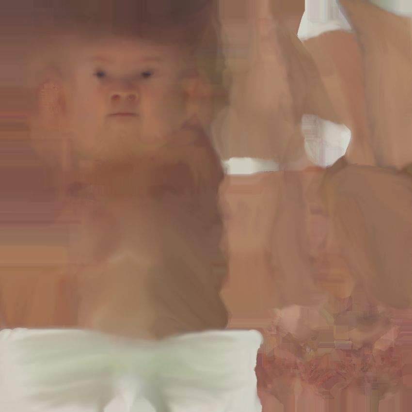
2      N. Hesse et al.

disease assessment [17], quantification of multiple sclerosis progression [22], as
well as gait [43] or motion analysis [8]. For the latter, accurately capturing hu-
man movements is the fundamental step. Human pose estimation from RGB or
depth images, especially using convolutional neural networks (CNNs), currently
receives a lot of attention from the research community [5,44,12,45]. However,
research is largely focused on adults. The ability to accurately capture infant
movements is fundamental for an automated assessment of motor development.
     In his pioneering work, Prechtl found that the quality of spontaneous move-
ments of infants is a good marker for detecting impairments of the young nervous
system [33]. This discovery led to the development of the General Movements
Assessment (GMA) [33,32,11] which allows detection of neurodevelopmental dis-
orders at a very young age. An automated system, relying on data captured by
cheap RGB or RGB-D cameras could enable the widespread screening of all in-
fants at risk of motion disorders. This would allow early intervention for affected
children, which is assumed to improve outcome [41].
     The application of state-of-the-art adult pose estimation systems to children
was recently studied [36]. Authors found that children are underrepresented in
most widely used benchmark data sets. Their analysis revealed that pose esti-
mation accuracy decreases when approaches that were trained on adult data are
applied to children. To mitigate this problem, they create a data set that was
collected from internet videos, comprising 1176 images of 104 different children
in unconstrained settings. For the evaluation of pose estimation approaches, they
manually annotate 2D joint positions of 22 body keypoints.
     Why does no infant data set exist? As mentioned above, computer vision re-
search is primarily focused on adults. Reasons might include the higher number
of potential applications for adults, with infant motion analysis being more of a
niche application. Furthermore, it is not easy to generate reliable ground truth
for infants. Manual annotation, especially in 3D, is error prone and cumbersome.
Capturing ground truth with standard motion capture systems using markers
may affect the infants’ behavior, while suffering from problems with occlusions
[27]. Researchers have used robotics to generate ground truth data [37,25], but
reproducing the complexity of real infant movements is not possible with jus-
tifiable efforts. Other than that, laws are more strict concerning the privacy of
children, since infants can not decide whether or not they want their image to
be published. This makes the creation of a data set containing real infant images
more challenging.
     We fill this gap by creating an RGB-D data set for the evaluation of motion
capture systems for infant motion analysis using the recently introduced Skinned
Multi-Infant Linear model (SMIL) [14]. Our Moving INfants In RGB-D (MINI-
RGBD) data set contains realistic motion, realistic appearance, realistic shapes,
and precise 2D and 3D ground truth, and covers a wide range of challenging
motions. To preserve the privacy of infants, we generate new textures and shapes
by averaging multiple textures and shapes of real infants. These are still highly
realistic, yet do not show any existing infant. We map real infant movements
to these new “synthetic infants” and render RGB and depth images to simulate
Computer Vision for Medical Infant Motion Analysis            3

                                                 Motion feature     Classification /
    Data acquisition       Motion capture
                                                  extraction          Prediction

                                                         Motion analysis

Fig. 1. Standard motion analysis pipeline. After data is acquired, motions are captured.
Motion features are extracted and used to classify or predict the medical outcome. In
this work, we focus on motion capture from RGB or depth images.

standard commodity RGB-D sensors. Our data set (described in Sec. 3), differs
from the data set of [36] in multiple ways: (i) it contains infants up to the age
of 7 months, (ii) we consider constrained settings for medical motion analysis,
i.e. infants lie in supine position, facing the camera, (iii) we provide sequences of
continuous motions instead of single frames, (v) we render data from a realistic
3D infant body model instead of annotating real images, (iv) we generate RGB
and depth images, and (vi) we provide accurate 2D and 3D ground truth joint
positions that are directly regressed from the model.
     In the following, we review the state of the art in infant motion analysis and
analyze evaluation procedures (Sec. 2). We describe the creation of our data set
in Sec. 3, and present pose estimation baseline evaluations for RGB and RGB-D
data in Sec. 4.

2    Medical Infant Motion Analysis - State of the Art
We review systems aiming at the automated prediction of cerebral palsy (CP)
based on the assessment of motions. Although this problem is approached in
different ways, the pipeline is similar for most systems, and can be divided into
motion capture and motion analysis (Fig. 1). Motion features are extracted from
captured movements, and used for training a classifier to predict the outcome.
The reviewed systems report high sensitivity and specificity for CP prediction,
mostly on study populations containing a small number of infants with confirmed
CP diagnosis. Yet, the majority of approaches shows a considerable lack of eval-
uation of motion capture methods. The majority of approaches only scarcely
evaluates the accuracy of motion capture methods. We believe that the sec-
ond step should not be taken before the first one, i.e. each system should first
demonstrate that it is capable of accurately capturing movements before predict-
ing outcome based on these captured movements. Of course, the non-existence
of a public benchmark data set makes it hard to conduct an extensive evaluation
of motion capture accuracy.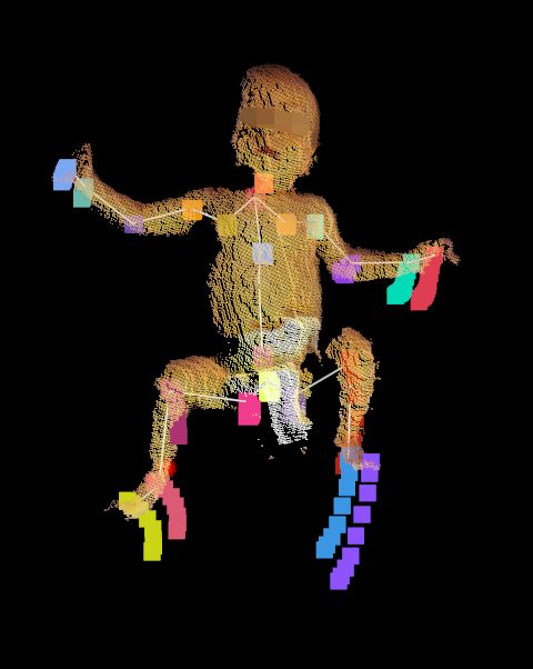
4      N. Hesse et al.

   In this section, we present an overview of methods used for infant motion cap-
ture and how these are evaluated. The reader is referred to [26] for an extensive
overview of the motion analysis stage of different approaches.

2.1   Wearable Motion Sensors
Although a recent study shows that wearable sensors do not seem to affect the
leg movement frequency [18], they supposedly have a negative influence on the
infant’s content. Karch et. al report that recordings for two thirds of partici-
pating infants had to be stopped after re-positioning the attached sensors due
to crying (and technical difficulties) [20]. Furthermore, approaches relying on
attached sensors generally suffer from practical limitations like time consum-
ing human intervention for setup and calibration, and add the risk of affecting
the movements of infants. Proposed systems using wearable sensors use wired
[13] and wireless accelerometers [40,10,9], electromagnetical sensors [21,20], or a
pressure mattress in combination with Inertial Measurement Units (IMU) [35].
In the following, we focus on vision-based approaches, and refer the reader to [7]
for an overview of monitoring infant movements using wearable sensors.

2.2   Computer Vision for Capturing Movements
Cameras, opposed to motion sensors, are cheap, easy to use, require no setup
or calibration, and can be easily integrated into standard examinations while
not influencing infants’ movements. This makes them more suitable for use in
clinical environments, doctor’s offices or even at home. Other than sensor-based
approaches, vision-based approaches do not measure motions directly. More or
less sophisticated methods are needed to extract motion information, e.g. by
estimating the pose in every image of a video. We describe the methods used
in the current state-of-the-art in infant motion analysis, as well as the evalua-
tion protocols for these methods. Our findings further support the need for a
standardized, realistic, and challenging benchmark data set.
    Video-based approaches. We review approaches that process RGB (or
infrared) images for the capture of infant motion. We include methods relying
on attached markers, despite posing some of the same challenges as wearable
sensors. They require human intervention for marker attachment, calibration
procedures and most of all possibly affect the infants’ behavior or content. Still,
they use computer vision for tracking the pose of the infants.
    One of the first approaches towards automated CP prediction was introduced
in 2006 by Meinecke et al. [27]. A commercial Vicon system uses 7 infrared cam-
eras to track 20 markers, distributed across the infant’s body. After an initial
calibration procedure, the known marker positions on a biomechanical model
are used to calculate the rotation of head and trunk, as well as the 3D po-
sitions of upper arms, forearms, thighs, lower legs, and feet from the tracked
markers on the infant. The system is highly accurate, authors report errors of
2 mm for a measurement volume of 2 m3 . However, the system suffers from
certain limitations. Due to the unconstrained movements of the infants close to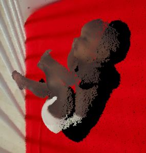
Computer Vision for Medical Infant Motion Analysis        5

the underground, especially the markers of the upper extremities were occluded
and therefore invisible to the cameras half of the measurement time. Attaching
additional markers exceeded the system’s capabilities and therefore, authors re-
frained from using motion features of upper extremities for CP prediction. The
high cost of the system, the complex setup and calibration, and the occlusion
problems stand against the highly accurate tracking of joints in 3D.
    Kanemaru et al. use a commercial marker tracking system (Frame-DIAS) to
record 2D positions of markers on arms, shoulders, hips and legs at 30 Hz using
a single camera [19]. They normalize the marker displacement data using the
body size of the infant and smooth the resulting 2D position data. The accuracy
of the capture system is not reported.
    Machireddy et al. [25] present a hybrid system that combines color-based
marker tracking in video with IMU measurements. The different sensor types
are intended to compensate for each others limitations. The IMU sensors are
attached to the infant’s hands, legs, and chest, together with colored patches.
The 2D positions of patches are tracked based on color thresholds. From the
known patch size and the camera calibration, an estimate for the 3D position of
each patch is calculated. The IMUs are synchronized with the camera, and the
output of all sensors is fused using an extended Kalman filter. Ground truth for
evaluation is generated by rotating a plywood model of a human arm using a
drill, equipped with one marker and one IMU. Authors present plots of ground
truth positions and estimated positions for a circular and a spiral motion. Exact
numbers on accuracy are not presented.
    Adde et al. take a more holistic approach [1]. Instead of tracking individual
limbs, they calculate the difference image between two consecutive frames to
generate what they call a motion image. They calculate the centroid of motion,
which is the center of the pixel positions forming the motion regions in the
motion image. Furthermore, they construct a motiongram by compressing all
motion images of a sequence either horizontally or vertically by summing over
columns, respectively rows, and stacking them to give a compact impression on
how much an infant moved, and where the movements happened. The accuracy
of the system is not evaluated.
    Stahl et al. use a motion tracking method based on optical flow between con-
secutive RGB frames [42]. They initialize points on a regular grid, distributed
across the image, and track them over time. They evaluate the approach by man-
ually selecting five points to be tracked from the grid as head, hands, and feet,
and manually correct tracking errors. They display the result of their evaluation
in one plot over 160 frames. Numbers on average accuracy are not given.
    Rahmati et al. present an approach for motion segmentation using weak su-
pervision [34]. Initialized by manual labeling, they track the body segmentation
trajectories using optical flow fields. In case a trajectory ends due to fast motion
or occlusion, they apply a particle matching algorithm for connecting a newly
started trajectory for the same body segment. They evaluate the accuracy on 20
manually annotated frames from 10 infant sequences, reporting an F-measure of
96% by calculating the overlap between ground truth and estimated segmenta-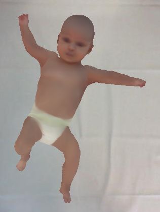
6      N. Hesse et al.

Table 1. Summary of motion capture methods and corresponding evaluation of depth-
based approaches for medical infant motion analysis. SD denotes standard deviation.

First     author, Method // tracked limbs Ground truth (GT) generation // Re-
year, reference                           ported avg. accuracy
Olsen 2014 [30] Geodesic distances // 11 Manual annotation (number of frames not
                 3D joint positions          specified) // 9 cm (extracted from plot)
Olsen 2014 [29] Model-based tracking // Manual annotation (number of frames not
                 11 3D joint positions       specified) // 5 cm (SD: 3 cm) (extracted
                                             from plot)
Hesse 2015 [16] Random ferns body part Manual annotation of 1082 frames // 4.1
                 classifier // 21 3D joint cm
                 positions
Hesse 2017 [15] Random ferns (extension 3D model fitting (+ visual verification),
                 of [16]) // 21 3D joint po- 5500 frames (3 seq.) // 1.2 cm (SD: 0.9
                 sitions                     cm)
Hesse 2018 [14] Model-based tracking // No GT, evaluation on 37 seq. (200K
                 full body pose and shape frames, ∼2 hours), pose errors determined
                                             from visual examination // average scan
                                             to model distance 2.51 mm, 34 pose errors
                                             lasting 90 s (≈ 1.2%)
Serrano     2016 Model based tracking // Robot leg kicking, angle comparison for
[37]             angles of hip, knee, and knee and ankle, 250 frames // 2 - 2.5 de-
                 ankle                       gree error
Cenci 2017 [6]   Movement blobs // arms No evaluation
                 and legs
Shivakumar       Optical flow + color- Manual annotation of 60 frames // 8.21
2017 [38]        based segmentation // cm, SD: 8.75 cm
                 3D positions of head,
                 torso, hands, feet

tion. They compare their tracking method to different state-of-the-art trackers
on the same data set, with their tracker showing superior results. Furthermore,
they evaluate their segmentation method on the Freiburg-Berkeley data set con-
taining moving objects (e.g. cats and ducks) and compare results to an optical
flow method. Their method achieves best results, at an F-measure of 77%.
    Depth-based approaches With the introduction of low-cost RGB-D sen-
sors, motion analysis approaches started taking advantage of depth information.
The most well-known RGB-D camera is probably the Microsoft Kinect, which
was introduced as a gesture control device for the gaming console XBox, but
soon became widely used in research due to its affordable price. The motion
tracking provided by the Kinect SDK has been used for motion analysis pur-
poses, but does not work for infants as it was purposed for gaming scenarios of
standing humans taller than one meter. We review approaches that aim at esti-
mating infants’ poses from RGB-D data and turn our attention to the respective
evaluation procedures. An overview of examined approaches is given in Tab. 1.
Computer Vision for Medical Infant Motion Analysis        7

    Olsen et al. transfer an existing pose estimation approach to infants [30]. The
underlying assumption is that extremities have maximum geodesic distance to
the body center. The body center is localized by filtering the infant’s clothing
color, based on a fixed threshold. They locate five anatomical extremities by
finding points on the body farthest from the body center. Assuming a known
body orientation, each of these points is assigned to one of the classes head, left
/ right hand, left / right foot, based on the spatial location and the orientation
of the path to body center. Intermediate body parts like elbows, knees and chest
are calculated based on fractional distances on the shortest path from body
center to extremities, resulting in 3D positions of eleven joints. For evaluation,
they annotate 3D joint positions on an unspecified number of frames. Annotated
joints lie in the interior of the body, while the estimated joints lie on the body
surface. Results are presented in a plot, numbers given here are read off this
plot. The average joint position error is roughly 9 cm. Highest errors occur for
hands and elbows (15 cm), lowest for body center, chest, and head (3 cm).
    In subsequent work, the same authors use a model-based approach for track-
ing eleven 3D joint positions [29]. They construct a human body model from
simplistic shapes (cylinders, sphere, ellipsoid). After determining size parame-
ters of the body parts, their previous method [30] is used for finding an initial
pose. They fit the body model to the segmented infant point clouds that are
computed from depth images. They optimize an objective function, defined by
the difference of closest points from point cloud and model, with respect to the
model pose parameters using the Levenberg-Marquardt algorithm. As in previ-
ous work, they evaluate the accuracy of their system on manually annotated 3D
joint positions of an unspecified number of frames. The results are compared
to their previous approach. Numbers are extracted from presented plots. The
model-based system achieves an average joint position error of 5 cm (standard
deviation (SD) 3 cm). Largest errors occur for right hand (7 cm) and stomach
(6 cm).
    Inspired by the approach used in the Kinect SDK, Hesse et al. propose a
system for the estimation of 21 joint positions using a random ferns body part
classifier [16]. A synthetic infant body model is used for rendering a large num-
ber of labeled depth images, from which a pixel-wise body part classifier based
on binary depth comparison features is trained. 3D joint positions are calculated
as the mean of all pixels belonging to each estimated body part. The system is
trained on synthetic adult data and evaluated on the PDT benchmark data set
containing adults. An average joint positions error of 13 cm is reported, com-
pared to 9.6 cm for the Kinect SDK. The authors manually annotated 3D joint
positions of an infant sequence consisting of 1082 frames. They report an average
joint position error of 4.1 cm, with left hand (14.9 cm) and left shoulder (7.3
cm) showing the largest errors. They explain the errors with wrongly classified
body parts for poses that were not included in the training set.
    The approach is extended in [15] by including a feature selection step, gener-
ating more infant-like poses for training data, integrating kinematic chain con-
straints, and by applying PCA on torso pixels to correct for body rotations.
8 N. Hesse et al.
Ground truth joint positions are generated for 5500 frames of 3 sequences by
fitting a body model and visually verifying the accuracy of the results. The best
average error of the proposed methods is reported as 1.2 cm (SD 0.9 cm), com-
pared to 1.8 cm (SD 3.1 cm) of the initial approach [16]. Additionally, a more
strict evaluation metric, the worst-case accuracy, is applied. It denotes the per-
centage of frames for which all joint errors are smaller than a given threshold.
For a threshold of 5 cm, 90% of frames are correct for [15], and 55% for [16], a
threshold of 3cm decreases the accuracy to 50%, and less than 30%, respectively.
In recent work, Hesse et al. propose a model-based approach for estimating
pose and shape of infants [14]. They learn an infant body model from RGB-D
recordings and present an algorithm for fitting the model to the data. They
optimize an objective function, consisting of scan to model distance, similar to
[29], but add more terms, e.g. integrating prior probabilities of plausible shapes
and poses. The average distance of scan points to model surface for 200K frames
of 37 infant sequences (roughly 2 hours) is 2.51 mm (SD 0.21 mm). From manual
inspection of all sequences, they report 18 failure cases in 7 sequences and 16
unnatural foot rotations lasting altogether 90 seconds, which corresponds to
1.2% of overall duration.
Serrano et al. track lower extremities using a leg model [37]. The approach
is semi-automatic and requires some manual intervention. The infant’s belly is
manually located from the point cloud and the tracker’s view is restricted to one
leg. After the length and width of each segment of the leg model are defined,
the model parameters (angles) are optimized using robust point set registration.
They generate ground truth for 250 frames using a robotic leg that simulates
kicking movements of infants. The average angle error of the proposed method
is reported with 2.5 degrees for the knee and 2 degrees for the ankle.
In [6], Cenci et al. use the difference image between two frames with a defined
delay in between. After noise filtering, the difference image is segmented into
motion blobs using a threshold. K-means clustering assigns each of the movement
blobs to one of four different body parts (arms and legs). A state vector is
generated for each frame, which contains information on which limb moves /
does not move in this frame. There is no evaluation of the correctness of assigning
blobs to limb classes.
Opposed to previous approaches, which rely on readily available depth sen-
sors, Shivakumar et al. introduce a stereo camera system, providing higher depth
resolution than existing sensors [38]. After initially locating the body center
based on a color threshold, an ellipse is fitted to the colored region and tracked.
In addition to the torso center, hands, legs and head regions are selected by
the user, which are then tracked based on their color. The positions of limbs
are defined as the pixel in the corresponding limb region that is farthest from
the body center. In case of overlap of multiple limb regions, a recovery step
distinguishes them. An optical flow method is used for estimating the motion
of the limb positions in the successive frame. An evaluation is presented on 60
manually annotated frames from three sequences, showing an average error of
8.21 cm (SD: 8.75 cm) over all limbs.Computer Vision for Medical Infant Motion Analysis 9
SMIL Generated RGB images
Shape shapes
RGB-D Generated Depth images
Alignment Texture textures
data
2D joint positions
Pose Renderer
Synthetic
infant 3D joint positions
Registration Rendering Data set
Fig. 2. Overview of data set creation pipeline. SMIL body model [14] is aligned to real
infant RGB-D data. Subsets of shapes and textures are used for generating realistic,
privacy preserving infant bodies. We animate the new “synthetic infants” with real
movements (poses) from the registration stage. We render RGB and depth images, and
create ground truth 2D and 3D joint positions to complete our new data set.
To summarize the evaluation protocols of reviewed approaches, comparison
to previous work was limited to works of the same authors. Ground truth was
mostly, if at all, generated by manual annotation of a small number of frames
or by relying on robotics. This emphasizes the need for an infant benchmark
RGB-D data set.
3 Moving INfants In RGB-D Data Set (MINI-RGBD)
An RGB-D data set for the evaluation of infant pose estimation approaches needs
to fulfill several requirements. It has to cover (i) realistic infant movements, (ii)
realistic texture, (iii) realistic shapes, and (iv) precise ground truth, while (v)
not violating privacy. Our presented data set fulfills all of these requirements.
The data set creation procedure can be divided into two stages, registration
and rendering (see Fig. 2). Two samples of rendered images and joint positions
are displayed in Fig. 3.
3.1 Registration
The capturing of shape and pose is achieved by registering SMIL to 12 RGB-D
sequences of moving infants that were recorded in a children’s hospital. Written
informed consent was given by parents and ethics approval was obtained from
Ludwig Maximilian University Munich. SMIL is based on SMPL [23], and shares
the same properties. The model can be regarded as a parametric mapping, with
pose and shape parameters serving as input, and output being a triangulated
mesh of the shaped and posed infant, consisting of 6890 vertices. The model
contains 23 body joints, each of which has three degrees of freedom (DOF).
Together with 3 DOF for the global orientation this gives 72 pose parameters.
We follow the protocol of [14] to register the SMIL model to point clouds
created from RGB-D sequences (which we will also call “scans”). We briefly
recap the method and refer the reader to [14] for additional details.10 N. Hesse et al.
(a) (b) (c) (d) (e) (f)
Fig. 3. Two samples from MINI-RGBD data set. (a) and (d): RGB image. (b) and (e):
point cloud created from depth image. (c) and (f): ground truth skeleton. Viewpoint
for (b), (c), (e), and (f) is slightly rotated to side. Best viewed in color.
To register the model to a scan, an objective function is optimized w.r.t. pose
and shape parameters of SMIL. The function consists of the following terms: (i)
distance between scan points and model mesh surface, (ii) a landmark error
terms that penalizes distances between model landmarks projected to 2D and
2D body, face, and hand landmark estimates from RGB images using OpenPose
library [5,39,44,31], (iii) a temporal pose smoothness term, (iv) a penalty for self
intersections, (v) a term for keeping the back-facing model vertices close to, but
not inside the background table, and (vi) prior probabilities on plausible shapes
and poses. This results in a posed and shaped model mesh that describes the
input point cloud data. The initialization frame is automatically selected based
on 2D pose estimates. For the rest of the sequence, the resulting parameters of
the last processed frame are used as initialization for the subsequent frame.
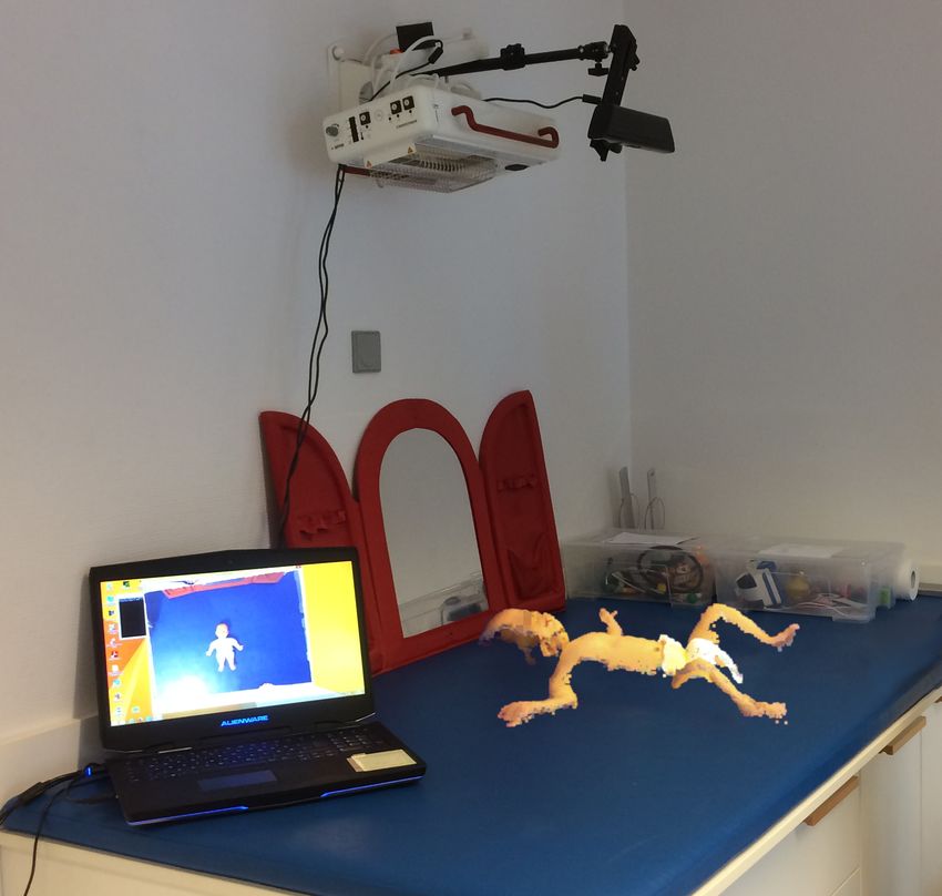
Going beyond the methods of [14], we generate one texture for each sequence,
similar to [3,4]. We create a texture map by finding closest points from textured
point cloud and registered model mesh, as well as a corresponding normal map for
each frame. We merge 300 randomly selected texture maps from each sequence
by averaging texture maps that are weighted according to their normal maps,
with higher weights for points with normals directed towards the camera. Infants
tend to lie on their backs without turning, which is why the merged texture
maps have blank areas depending on the amount of movement in the sequence.
We fill the missing areas by expanding the borders of existing body areas. To
preserve the privacy of the infants we do not use textures from single sequences,
but generate average textures from subsets of all textures. The resulting texture
maps (sample displayed in Fig. 4 (a)) are manually post-processed by smoothing
borders and visually enhancing areas of the texture for which the filling did not
create satisfying results. We create a variety of realistic body shapes by averaging
different subsets of shapes from the registration stage (Fig. 4 (b)).
3.2 Rendering
For each of the 12 sequences, we randomly select one of the average shapes
and one of the average textures. We map the pose parameters of the sequence,
obtained in the registration stage, to the new shape, and animate textured 3D
meshes of realistic, yet artificial infants. Based on plane parameters extracted
from the background table of the real sequences, we add a plane to simulate theComputer Vision for Medical Infant Motion Analysis 11
(a) (b) (c) (d)
Fig. 4. (a) Sample of generated texture. (b) Generated shapes in T-pose. (c) Plotted
joint positions from an “easy” sequence. Hand positions shown in light and dark green.
Foot positions in red and blue. (d) Hand and foot positions for a “difficult” sequence.
Color coding as in (c). Best viewed in color.
background. We texture the plane with one of various background images (e.g.
examination table, crib, changing table) to account for background variation.
We use OpenDR [24] to render RGB and depth images from the meshes and
backgrounds. We select 1000 consecutive frames from each sequence where the
infant is the most active. The rendered depth image is overly smooth, which is
why we add random noise of up to ±0.5 cm to simulate noise levels of depth
sensors. We use camera parameters similar to Microsoft Kinect V1, which is the
most frequently used sensor in approaches in Sec. 2, at a resolution of 640 * 480,
at 30 frames per second. The distance of the table to the camera is roughly 90 cm
for all sequences. The 3D joint positions are directly regressed from the model
vertices (see Fig. 3 (c) and (f)). To provide 2D ground truth, we project the
3D joints to 2D using the camera calibration. For completeness, we add depth
values for each joint. To simplify data set usage, we provide a segmentation mask
discriminating between foreground and background.
3.3 MINI-RGBD Data Set Summary
We generate 12 sequences, each with different shape, texture, movements, and
background, and provide 2D and 3D ground truth joint positions, as well as
foreground segmentation masks. Movements are chosen to be representative of
infants in the first half year of life, and we divide the sequences into different
levels of difficulty (see Fig. 4 (c) and (d) for examples): (i) easy: lying on back,
moving arms and legs, mostly besides body, without crossing (sequences 1-4), (ii)
medium: slight turning, limbs interact and are moved in front of the body, legs
cross (sequences 5-9), and (iii) difficult: turning to sides, grabbing legs, touching
face, directing all limbs towards camera simultaneously (sequences 10-12).
Different approaches utilize different skeletons. To properly compare these
approaches, we add one frame in T-pose (extended arms and legs, cf. Fig. 4
(b)) for each sequence to calculate initial offsets between estimation and ground
truth that can be used to correct for skeleton offsets.
The limitations of the underlying SMIL model include finger motions, facial
expressions and hair. These are not represented by the model, which is why the
hand is fixed as a fist, and the face has a neutral expression.12 N. Hesse et al.
PCKh 1.0 PCKh 2.0 PCKh 1.0 PCKh 2.0
100% 100%
80% 80%
60% 60%
40% 40%
20% 20%
0% 0%
1 2 3 4 5 6 7 8 9 10 11 12
Sequence number
(a) (b)
Fig. 5. RGB evaluation. Results for 2D pose estimation from OpenPose library. (a)
Percentage of correct keypoints in relation to head segment length (PCKh) per joint.
PCKh 1.0 denotes a correctness threshold of one time head segment length, PCKh 2.0
of twice the head segment length. (b) PCKh per sequence.
4 Evaluation
We provide baseline evaluations using state-of-the-art approaches for the RGB,
as well as the RGB-D part of our MINI-RGBD data set.
4.1 2D Pose Estimation in RGB Images
We use a state-of-the-art adult RGB pose estimation system from OpenPose
library [5,31] as baseline for evaluation of the RGB part of the data set. To
account for differences in skeletons between OpenPose and SMIL, we calculate
joint offsets for neck, shoulders, hips, and knees from the T-pose frame (Sec. 3.3),
and add these offsets to the estimated joint positions in every frame.
Error metrics. We apply the PCKh error metric from [2], which is com-
monly used for the evaluation of pose estimation approaches [5,44,12,36]. It
denotes the percentage of correct keypoints with the threshold for correctness
being defined as 50% of the head segment length. The SMIL model has a very
short head segment (head joint to neck joint, cf. Fig. 3, (c) and (f)), which is
why we present results using the full head segment length (PCKh 1.0), as well as
two times the head segment length (PCKh 2.0) as thresholds. The head segment
length for each sequence is calculated from the ground truth joint positions in
the T-pose frame. Average 2D head segment length over all sequences is 11.6 pix-
els. We calculate the PCKh values for each joint for each sequence, and average
numbers over all sequences, respectively over all joints.
OpenPose estimates 15 joint positions (nose, neck, shoulders, elbow, hands,
hips, knees, feet) that we map to corresponding SMIL joints. Unlike SMIL, Open-
Pose estimates the nose position instead of head position. We add the model
vertex of the tip of the nose as additional joint instead of using SMIL head joint.
Results. We display average PCKh per joint in Fig. 5 (a), and average PCKh
per sequence in Fig. 5 (b). The mean average precision, i.e. the average PCKh
over all joints and sequences, for PCKh 1.0 is 88.1% and 94.5% for PCKh 2.0.
PCKh rates are very consistent over most body parts, with a slight decrease of
PCKh 1.0 for lower limb joints, especially knees. Results for some body jointsComputer Vision for Medical Infant Motion Analysis 13
(e.g. nose, neck, shoulders, Fig. 5 (a)) as well as for some sequences (1, 2, 3, 6,
7, 11, Fig. 5 (b)) are close to perfect (according to the error metric). We observe
largest errors when the limbs are directed towards the camera.
OpenPose has reportedly shown impressive results on unconstrained scenes
containing adults [5], and confirms these on our synthetic infant data set. Being
trained on real images of unconstrained scenes, the results further validate the
high level of realism of our data set, but also show how challenging the data is,
and that there is still room for improvement (e.g. sequences 9, 10, 12).
4.2 3D Pose Estimation in Depth Images
We evaluate the system with the lowest reported average joint position error for
RGB-D data from our overview (Tab. 1), the extension of the pixelwise body part
classifier based on random ferns [16,15]. For each pixel of an input depth image,
the label is predicted as one of 21 body parts (spine, chest, neck, head, shoulders,
elbows, hands, fingers, hips, knees, feet, and toes). The 3D joint positions are
then calculated as the mean of each body part region. RGB is not used in this
approach. Similar to Sec. 4.1, we calculate joint offsets in the T-pose frame and
add them to the estimated joint positions throughout each sequence.
Error metrics. We use the PCKh error metric as described above, here in
3D. Average 3D head segment length over all sequences is 2.64 cm. Additionally,
we evaluate the average joint position error (AJPE), which denotes the euclidean
distance from estimated joint position to corresponding ground truth.
Results. We present results in Fig. 6. Mean average precision, i.e. average
PCKh over all joints and sequences, for PCKh 1.0 is 64.2%, and 90.4% for PCKh
2.0. Compared to the RGB evaluation, we experience a bigger difference between
PCKh 1.0 and PCKh 2.0. Very high PCKh 2.0 rates are achieved for torso and
head body parts, while lowest rates are obtained for joints related to extremities
(Fig. 6 (a)). PCKh 1.0 rates differ a lot from PCKh 2.0 for elbows, hands, and
feet. We observe that the estimated hand and foot regions are too large, leading
to the hand joints lying more in the direction of the elbow, respectively the
foot joints in direction of the knees. With an expansion of the threshold for
correctness (PCKh 2.0) these displacements are accepted as correct, leading to
large jumps from around 30% (PCKh 1.0) to 70 - 80% (PCKh 2.0).
The average joint position error (AJPE) over all sequences and joints is 2.86
cm. Joint position errors are largest for the extremities, at an average distance
to ground truth of up to 5 cm (Fig. 6 (c)). If the estimate for a joint was missing
in a frame, we ignored this joint for the calculation of AJPE, i.e. we only divided
the sum of joint errors by the number of actually estimated joints. The number
of frames with missing estimates, denoted by joint (in 12K frames, average for
left and right sides): neck 37, elbows 12, hands 62, fingers 156, feet 79, toes 607,
all others 0. For the calculation of PCKh metric, missing joints were considered
as lying outside the correctness threshold.
The evaluated approach shows high accuracy when arms and legs are moving
beside the body, but the accuracy decreases, especially for hands and feet, when
limbs move close to or in front of the body. This becomes extremely visible in14 N. Hesse et al.
PCKh 1.0 PCKh 2.0 PCKh 1.0 PCKh 2.0
100% 100%
80% 80%
60% 60%
40% 40%
20% 20%
0% 0%
spine
shoulder.R
chest
neck
head
elbow.R
hand.R
fingers.R
shoulder.L
elbow.L
hand.L
fingers.L
hips
hip.R
knee.R
foot.R
toe.R
hip.L
knee.L
foot.L
toe.L
1 2 3 4 5 6 7 8 9 10 11 12
Sequence number
(a) (b)
5 5
4 4
AJPE in cm
AJPE in cm
3 3
2 2
1 1
0 0
spine
chest
neck
head
shoulder.R
elbow.R
hand.R
fingers.R
shoulder.L
elbow.L
hand.L
fingers.L
hips
hip.R
knee.R
foot.R
toe.R
hip.L
knee.L
foot.L
toe.L
1 2 3 4 5 6 7 8 9 10 11 12
Sequence number
(c) (d)
Fig. 6. RGB-D evaluation. Results for 3D pose estimation based on random ferns
[16,15]. (a) Percentage of correct keypoints in relation to head segment length (PCKh)
per joint. PCKh 1.0 denotes a correctness threshold of one time head segment length,
PCKh 2.0 of twice the head segment length. (b) PCKh per sequence. (c) Average joint
position error (AJPE) per joint. (d) AJPE per sequence.
sequence 9, where the infant moves the left arm to the right side of the body
multiple times, leading to the highest overall AJPE of 4.7 cm (Fig. 6 (d)). Best
AJPE results are achieved for sequence 2, at 1.46 cm, which is close to results
reported in [15]. The varying accuracy for different sequences shows the levels
of difficulty and the variance of motion patterns included in the data set.
5 Conclusions
We presented an overview of the state-of-the-art in medical infant motion anal-
ysis, with a focus on vision-based approaches and their evaluation. We observed
non-standardized evaluation procedures, which we trace back to the lack of pub-
licly available infant data sets. The recently introduced SMIL model allows us to
generate realistic RGB and depth images with accurate ground truth 2D and 3D
joint positions. We create the Moving INfants In RGB-D (MINI-RGBD) data set,
containing 12 sequences of real infant movements with varying realistic textures,
shapes and backgrounds. The privacy of recorded infants is preserved by not
using real shape and texture, but instead generating new textures and shapes by
averaging data from multiple infants. We provide baseline evaluations for RGB
and RGB-D data. By releasing the data set, we hope to stimulate research in
medical infant motion analysis.
Future work includes the creation of a larger data set, suitable for training
CNNs for estimating 3D infant pose from RGB-D data.Computer Vision for Medical Infant Motion Analysis 15
References
1. Adde, L., Helbostad, J.L., Jensenius, A.R., Taraldsen, G., Støen, R.: Using
computer-based video analysis in the study of fidgety movements. Early human
development 85(9), 541–547 (2009)
2. Andriluka, M., Pishchulin, L., Gehler, P., Schiele, B.: 2D human pose estimation:
New benchmark and state of the art analysis. In: IEEE Conference on Computer
Vision and Pattern Recognition (CVPR). pp. 3686–3693 (2014)
3. Bogo, F., Black, M.J., Loper, M., Romero, J.: Detailed full-body reconstructions
of moving people from monocular RGB-D sequences. In: 2015 IEEE International
Conference on Computer Vision (ICCV) (2015)
4. Bogo, F., Romero, J., Loper, M., Black, M.J.: FAUST: Dataset and evaluation for
3D mesh registration. In: 2014 IEEE Conference on Computer Vision and Pattern
Recognition (CVPR) (2014)
5. Cao, Z., Simon, T., Wei, S.E., Sheikh, Y.: Realtime multi-person 2D pose estima-
tion using part affinity fields. In: 2017 IEEE Conference on Computer Vision and
Pattern Recognition (CVPR). pp. 1302–1310 (2017)
6. Cenci, A., Liciotti, D., Frontoni, E., Zingaretti, P., Carinelli, V.P.: Movements
analysis of preterm infants by using depth sensor. International Conference on
Internet of Things and Machine Learning (IML 2017) (2017)
7. Chen, H., Xue, M., Mei, Z., Bambang Oetomo, S., Chen, W.: A review of wearable
sensor systems for monitoring body movements of neonates. Sensors 16(12), 2134
(2016)
8. Chen, L., Wei, H., Ferryman, J.: A survey of human motion analysis using depth
imagery. Pattern Recognition Letters 34(15), 1995–2006 (2013)
9. Fan, M., Gravem, D., Cooper, D.M., Patterson, D.J.: Augmenting gesture recogni-
tion with erlang-cox models to identify neurological disorders in premature babies.
In: Proceedings of the 2012 ACM Conference on Ubiquitous Computing. pp. 411–
420. ACM (2012)
10. Gravem, D., Singh, M., Chen, C., Rich, J., Vaughan, J., Goldberg, K., Waffarn, F.,
Chou, P., Cooper, D., Reinkensmeyer, D., et al.: Assessment of infant movement
with a compact wireless accelerometer system. Journal of Medical Devices 6(2),
021013 (2012)
11. Hadders-Algra, M., Nieuwendijk, A.W., Maitijn, A., Eykern, L.A.: Assessment of
general movements: towards a better understanding of a sensitive method to eval-
uate brain function in young infants. Developmental Medicine & Child Neurology
39(2), 88–98 (1997)
12. Haque, A., Peng, B., Luo, Z., Alahi, A., Yeung, S., Fei-Fei, L.: Towards view-
point invariant 3D human pose estimation. In: European Conference on Computer
Vision. pp. 160–177. Springer (2016)
13. Heinze, F., Hesels, K., Breitbach-Faller, N., Schmitz-Rode, T., Disselhorst-Klug,
C.: Movement analysis by accelerometry of newborns and infants for the early de-
tection of movement disorders due to infantile cerebral palsy. Medical & biological
engineering & computing 48(8), 765–772 (2010)
14. Hesse, N., Pujades, S., Romero, J., Black, M.J., Bodensteiner, C., Arens, M., Hof-
mann, U.G., Tacke, U., Hadders-Algra, M., Weinberger, R., Müller-Felber, W.,
Schroeder, A.S.: Learning an infant body model from rgb-d data for accurate full
body motion analysis. In: International Conference on Medical Image Computing
and Computer-Assisted Intervention. Springer (2018)16 N. Hesse et al.
15. Hesse, N., Schröder, A.S., Müller-Felber, W., Bodensteiner, C., Arens, M., Hof-
mann, U.G.: Body pose estimation in depth images for infant motion analysis. In:
IEEE 39th Annual International Conference of the Engineering in Medicine and
Biology Society (EMBC) (2017)
16. Hesse, N., Stachowiak, G., Breuer, T., Arens, M.: Estimating body pose of infants
in depth images using random ferns. In: 2015 IEEE International Conference on
Computer Vision Workshop (ICCVW) (2015)
17. Iarlori, S., Ferracuti, F., Giantomassi, A., Longhi, S.: RGBD camera monitoring
system for alzheimer’s disease assessment using recurrent neural networks with
parametric bias action recognition. IFAC Proceedings Volumes 47(3), 3863–3868
(2014)
18. Jiang, C., Lane, C.J., Perkins, E., Schiesel, D., Smith, B.A.: Determining if wear-
able sensors affect infant leg movement frequency. Developmental neurorehabilita-
tion pp. 1–4 (2017)
19. Kanemaru, N., Watanabe, H., Kihara, H., Nakano, H., Takaya, R., Nakamura, T.,
Nakano, J., Taga, G., Konishi, Y.: Specific characteristics of spontaneous move-
ments in preterm infants at term age are associated with developmental delays at
age 3 years. Developmental Medicine & Child Neurology 55(8), 713–721 (2013)
20. Karch, D., Kang, K.S., Wochner, K., Philippi, H., Hadders-Algra, M., Pietz, J.,
Dickhaus, H.: Kinematic assessment of stereotypy in spontaneous movements in
infants. Gait & posture 36(2), 307–311 (2012)
21. Karch, D., Kim, K.S., Wochner, K., Pietz, J., Dickhaus, H., Philippi, H.: Quantifi-
cation of the segmental kinematics of spontaneous infant movements. Journal of
biomechanics 41(13), 2860–2867 (2008)
22. Kontschieder, P., Dorn, J.F., Morrison, C., Corish, R., Zikic, D., Sellen, A.,
D’Souza, M., Kamm, C.P., Burggraaff, J., Tewarie, P., et al.: Quantifying pro-
gression of multiple sclerosis via classification of depth videos. In: International
Conference on Medical Image Computing and Computer-Assisted Intervention.
pp. 429–437. Springer (2014)
23. Loper, M., Mahmood, N., Romero, J., Pons-Moll, G., Black, M.J.: SMPL: A
skinned multi-person linear model. ACM Trans. Graph. 34(6), 248:1–248:16 (2015)
24. Loper, M.M., Black, M.J.: OpenDR: An approximate differentiable renderer. In:
European Conference on Computer Vision (ECCV). pp. 154–169. Springer (2014)
25. Machireddy, A., van Santen, J., Wilson, J.L., Myers, J., Hadders-Algra, M., Song,
X.: A video/IMU hybrid system for movement estimation in infants. In: 39th An-
nual International Conference of the IEEE Engineering in Medicine and Biology
Society (EMBC). pp. 730–733. IEEE (2017)
26. Marcroft, C., Khan, A., Embleton, N.D., Trenell, M., Plötz, T.: Movement recog-
nition technology as a method of assessing spontaneous general movements in high
risk infants. Frontiers in neurology 5 (2014)
27. Meinecke, L., Breitbach-Faller, N., Bartz, C., Damen, R., Rau, G., Disselhorst-
Klug, C.: Movement analysis in the early detection of newborns at risk for devel-
oping spasticity due to infantile cerebral palsy. Human movement science 25(2),
125–144 (2006)
28. Morrison, C., Culmer, P., Mentis, H., Pincus, T.: Vision-based body tracking: turn-
ing Kinect into a clinical tool. Disability and Rehabilitation: Assistive Technology
11(6), 516–520 (2016)
29. Olsen, M.D., Herskind, A., Nielsen, J.B., Paulsen, R.R.: Model-based motion track-
ing of infants. In: Computer Vision-ECCV 2014 Workshops. pp. 673–685. Springer
(2014)Computer Vision for Medical Infant Motion Analysis 17
30. Olsen, M.D., Herskindt, A., Nielsen, J.B., Paulsen, R.R.: Body part tracking of
infants. In: 22nd International Conference on Pattern Recognition (ICPR). pp.
2167–2172. IEEE (2014)
31. OpenPose library: https://github.com/CMU-Perceptual-Computing-Lab/
openpose, accessed June 2018
32. Prechtl, H.F., Einspieler, C., Cioni, G., Bos, A.F., Ferrari, F., Sontheimer, D.:
An early marker for neurological deficits after perinatal brain lesions. The Lancet
349(9062), 1361–1363 (1997)
33. Prechtl, H.: Qualitative changes of spontaneous movements in fetus and preterm
infant are a marker of neurological dysfunction. Early human development 23(3),
151–158 (1990)
34. Rahmati, H., Dragon, R., Aamo, O.M., Adde, L., Stavdahl, Ø., Van Gool, L.:
Weakly supervised motion segmentation with particle matching. Computer Vision
and Image Understanding 140, 30–42 (2015)
35. Rihar, A., Mihelj, M., Pašič, J., Kolar, J., Munih, M.: Infant trunk posture and arm
movement assessment using pressure mattress, inertial and magnetic measurement
units (imus). Journal of neuroengineering and rehabilitation 11(1), 133 (2014)
36. Sciortino, G., Farinella, G.M., Battiato, S., Leo, M., Distante, C.: On the estimation
of children’s poses. In: International Conference on Image Analysis and Processing.
pp. 410–421. Springer (2017)
37. Serrano, M.M., Chen, Y.P., Howard, A., Vela, P.A.: Lower limb pose estimation
for monitoring the kicking patterns of infants. In: 38th Annual International Con-
ference of the IEEE Engineering in Medicine and Biology Society (EMBC). pp.
2157–2160. IEEE (2016)
38. Shivakumar, S.S., Loeb, H., Bogen, D.K., Shofer, F., Bryant, P., Prosser, L., John-
son, M.J.: Stereo 3D tracking of infants in natural play conditions. In: International
Conference on Rehabilitation Robotics (ICORR). pp. 841–846. IEEE (2017)
39. Simon, T., Joo, H., Matthews, I., Sheikh, Y.: Hand keypoint detection in single
images using multiview bootstrapping. In: 2017 IEEE Conference on Computer
Vision and Pattern Recognition (CVPR). pp. 4645–4653 (2017)
40. Singh, M., Patterson, D.J.: Involuntary gesture recognition for predicting cerebral
palsy in high-risk infants. In: International Symposium on Wearable Computers
(ISWC). pp. 1–8. IEEE (2010)
41. Spittle, A., Orton, J., Anderson, P.J., Boyd, R., Doyle, L.W.: Early developmental
intervention programmes provided post hospital discharge to prevent motor and
cognitive impairment in preterm infants. The Cochrane Library (2015)
42. Stahl, A., Schellewald, C., Stavdahl, Ø., Aamo, O.M., Adde, L., Kirkerød, H.: An
optical flow-based method to predict infantile cerebral palsy. IEEE Transactions
on Neural Systems and Rehabilitation Engineering 20(4), 605–614 (2012)
43. Sun, B., Liu, X., Wu, X., Wang, H.: Human gait modeling and gait analysis
based on Kinect. In: IEEE International Conference on Robotics and Automation
(ICRA). pp. 3173–3178. IEEE (2014)
44. Wei, S.E., Ramakrishna, V., Kanade, T., Sheikh, Y.: Convolutional pose machines.
In: 2016 IEEE Conference on Computer Vision and Pattern Recognition (CVPR).
pp. 4724–4732 (2016)
45. Zimmermann, C., Welschehold, T., Dornhege, C., Burgard, W., Brox, T.: 3D hu-
man pose estimation in RGBD images for robotic task learning. In: IEEE Interna-
tional Conference on Robotics and Automation (ICRA) (2018)You can also read