COVID-19 AND SARS-COV-2: EVERYTHING WE KNOW SO FAR - A COMPREHENSIVE REVIEW
←
→
Page content transcription
If your browser does not render page correctly, please read the page content below
Open Chemistry 2021; 19: 548–575
Review Article
Sumaira Naz, Muhammad Zahoor*, Muhammad Umar Khayam Sahibzada, Riaz Ullah*,
Ali S. Alqahtani
COVID-19 and SARS-CoV-2: Everything we know
so far – A comprehensive review
https://doi.org/10.1515/chem-2021-0049 vaccine against it. A number of vaccines are available
received August 23, 2020; accepted October 28, 2020 even in markets in different countries. More and more
Abstract: Coronavirus disease-2019 (COVID-19) emerged ways of personal protection, prevention, and mitigation
as a unique type of pneumonia outbreak in the Wuhan of the disease are being explored and shared. While the
city of China in 2019 and spread to all its provinces in a outbreak has been declared as pandemic, the response of
matter of days and then to every continent of the world scientists was timely and enormous; thousands of pub-
except Antarctica within 3–4 month. This paper aims to lications about various aspects and impact of the diseases
comprehensively consolidate the available information and its causative virus are there on the World Health
about COVID-19 and present all the possible information Organization database and many more studies are underway.
about this disease in form of a single paper to readers. The purpose of writing this review article is to provide a
Unparalleled research and exhaustive studies of every- comprehensive summary of the major aspects and important
thing about the disease and its causative virus, i.e., scientific findings so far, about COVID-19 and SARS-CoV-2, in
severe acute respiratory syndrome coronavirus 2 (SARS- a single article for ready reference.
CoV-2), are underway since its emergence. The genome Keywords: COVID-19, SARS-CoV-2 genome, epidemiology,
sequence of the virus was made available within a record risk factors and transmission of COVID-19, pathogenesis
short time by China, making possible immediate study of and clinical features of COVID-19, diagnosis, treatment,
its structure and characteristics. The routes of transmis- and vaccine of COVID-19, prevention and mitigation of
sion of the disease, signs and symptoms, incubation COVID-19, reinfection with COVID-19
period, pathogenesis, and pathophysiology have been
extensively studied and presented in an organized way
in this review paper. The number of confirmed cases and
case fatality and mortality rates are updated regularly. 1 Introduction
The different diagnostic mechanisms have been charac-
terized. Testing and management criteria and protocols Plagues and epidemics have been one of the greatest
have been adopted. Extensive efforts are underway for challenges to the survival of the human race through
finding a treatment of the disease and developing a the course of history. Due to the factors like rapid growth
in population and emerging of advanced means of com-
munication and transportation across the globe, the
humankind has become more susceptible to epidemic
* Corresponding author: Muhammad Zahoor, Department of diseases. That’s why the epidemics which took months
Biochemistry, University of Malakand, Chakdara, Dir Lower, and years to transfer from the infected area to other parts
18800, KPK, Pakistan, e-mail: mohammadzahoorus@yahoo.com of the world now reach in every nook and corner of the
* Corresponding author: Riaz Ullah, Department of Pharmacognosy
globe in a matter of days or weeks. In recent history, some
(MAPPRC), College of Pharmacy, King Saud University, PO Box 2457,
Riyadh 11451, Saudi Arabia, e-mail: rullah@ksu.edu.sa new epidemics appeared and took many precious lives
Sumaira Naz: Department of Biochemistry, University of Malakand, like the Spanish Flu in 1918, Severe Acute Respiratory
Chakdara, Dir Lower, 18800, KPK, Pakistan Syndrome (SARS) in 2002, Ebola in 2014, and Middle
Muhammad Umar Khayam Sahibzada: Department of Pharmacy, East Respiratory Syndrome (MERS) in 2015 [1]. The dawn
Sarhad University of Science and Information Technology,
of 2020 saw an outbreak of a new super spreading disease
Peshawar, 25000, KPK, Pakistan
Ali S. Alqahtani: Department of Pharmacognosy (MAPPRC), College
named COVID-19 (Coronavirus disease 2019) and the cau-
of Pharmacy, King Saud University, PO Box 2457, Riyadh 11451, sative agent was identified as a novel coronavirus named
Saudi Arabia initially as 2019 novel coronavirus (2019-nCoV) and later
Open Access. © 2021 Sumaira Naz et al., published by De Gruyter. This work is licensed under the Creative Commons Attribution 4.0
International License.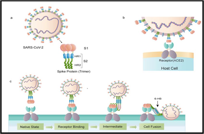
COVID-19 and SARS-CoV-2: A comprehensive review 549
on as severe acute respiratory syndrome coronavirus 2 alpha and beta CoV is known. SARS-CoV-2 is the seventh
(SARS-CoV-2). The disease emerged as a novel pneu- known pathogenic CoV, out of which three have caused severe
monia surfaced in Wuhan city of Hubei province, China, epidemics, i.e., SARS, MERS, and now COVID-19 [8].
in December 2019 and spread not only to all the provinces
of China, but to all continents of the world except Ant-
arctica in only 3–4 months since its first reported case.
The WHO declared the disease as a pandemic (a global 2.1 Genome and the virion
epidemic) on March 11, 2020 [2–5]. As the new virus
strengthens its hold over the globe with every emerging Figure 1 shows the structure of SARS-CoV-2. It is an
confirmed case of the disease, extensive research was enveloped, positive-sense RNA virus with a quite large
started worldwide by the scientists to study every aspect RNA genome-a characteristic common to all corona viruses.
of the virus and the disease to help design diagnostic The genome is arranged in such a manner that the replicase
tools, evolve strategies to combat or at least contain the locus is encoded within the 5′ end (which encodes replicase
disease, and finally come up with a treatment. The infor- gene), whereas structural proteins are encoded at the 3′ end.
mation about COVID-19 and the causative virus are avail- The structural proteins are present in the following order:
able everywhere on the internet in form of the research (i) spike (S), (ii) a small membrane (E), (iii) membrane (M),
articles, multiple websites, news, media, etc. Several and (iv) nucleocapsid and internal protein (I). The I is
reviews have been published, each addressing a parti- responsible for complexing with the genome RNA, making
cular aspect of COVID-19. A need was strongly felt for a helical capsid that is present in the viral envelope. Trimeric
such an article that encompasses all the necessary ele- transmembrane peplomers (the spike) protruding from the
ments on the topic, properly citing the scientific findings envelope give crown-like shape to the virion and hence the
and studies that are so far carried out in each area name “coronaviruses” to SARS-CoV-2 and other viruses of
regarding the recent pandemic. In our review article, we the same family have been adapted internationally [9].
have tried our best to present an overview of the published
data about almost all the main features of COVID-19. We
have added new insights and recommendations suggested
by the researchers and have tried to point out what are the 2.2 The spike ‘S’-tool for the entry of the
next challenges to be faced by scientific community. Apart virus into the host cell
from it, there is a ready reference material for the
researchers where they would find answer to most of their The spike is mainly responsible for the attachment of the
queries in one place. The general readers will also benefit virus with its receptor that has been reported to be angio-
from having a compendium of useful and authentic infor- tensin-converting enzyme 2 (ACE2) – the same receptor
mation presented in the simplest possible manner. that is already been reported for SARS-CoV [10]. The
S protein (sized 180–200 kDa) has three important com-
ponents: N-terminus, a transmembrane domain, and a
C-terminal segment [11]. Upon interaction of the virus
2 The virus-SARS-CoV-2
The causative virus of COVID-19, i.e., SARS-CoV-2, was
identified as a novel member of a previously well-known
subfamily of viruses coronavirinae of the family Coronaviruses
(CoVs; corona – viridae). The family, in turn, belongs to the
super family – Nidovirus (Nidovirales). The coronavirinae sub-
family is further divided into three genera, viz., alpha, beta,
and gamma [6]. The causative virus of COVID-19 and that of
SARS both belong to beta coronaviruses, the causative virus of
the latter was named as SARS-CoV and because of the striking
similarity (at least 86%) between the genomes of both, routes
of transmission, and symptoms of the disease between the
two, the former was named as SARS-CoV-2 [7]. So far, out of
all the three genera of the CoVs, the pathogenesis of only the Figure 1: Structure of SARS-CoV-2 [9].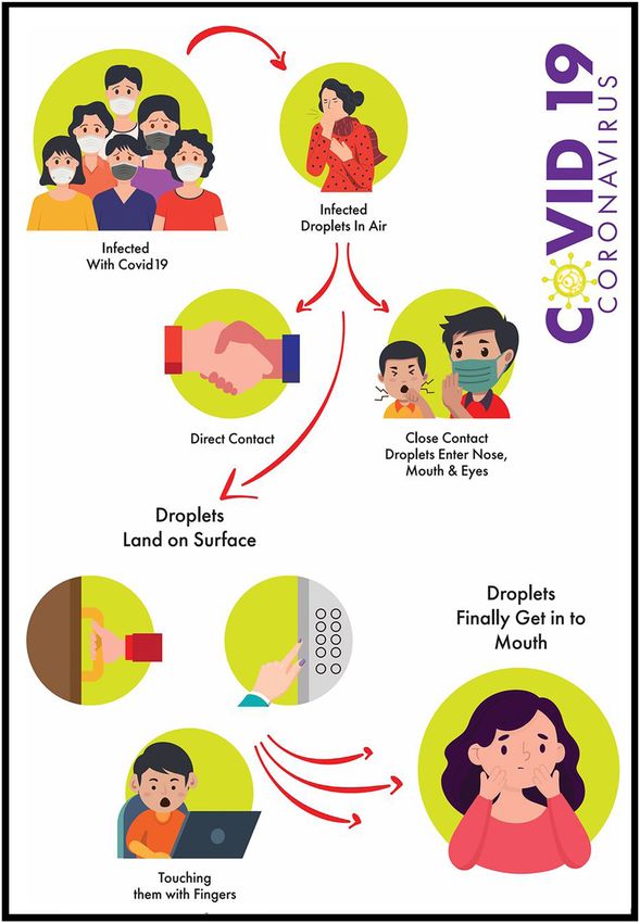
550 Sumaira Naz et al.
with the host cell, there occur changes in the S protein the virus and may contribute to its pathogenicity and trans-
which help in fusion of the virus with the host cell mem- missibility. Another important feature of the S is that it is
brane. Figure 2 represents structure of S and its binding lined with N-glycans that are required to neutralize anti-
to the receptor [12]. bodies (Abs) and interact with the host proteases. The cryo-
The S has 2 subunits, S1 and S2; the latter is cleaved EM structures of S have been reported which would be
at the border between the two subunits in such a way that helpful in designing vaccines [9].
both are present in a non-covalently bound form before
the entry of the virion into the host cell. S1 subunit has N-
terminus and a receptor-binding domain (RBD); S2 sub-
unit which is responsible for fusion with the host cell
membrane has a fusion peptide, heptapeptide repeat
3 Origin of SARS-CoV-2
sequence 1 and 2 (HR 1, HR 2), a cytoplasm domain, and
a transmembrane domain. Another so-called S2′ site is also 3.1 Is the virus lab-originated?
present directly after S2, which is cleaved by the host pro-
teases and results in conformational changes and the sub- Though nothing could be asserted with ultimate finality at
sequent activation of the S2 for fusion with host membrane this stage as yet, most of the scientists are in the view that
[9,13,14]. Hence, the entry of CoVs to the host cell requires the virus is not lab-originated. In this connection, a strong
both specific receptor-binding capability and subsequent evidence is the Anderson and his colleagues’ article [15] in
proteolytic cleavage of the respective subunit (S2′) [10]. which they have concluded that it is very unlikely that
Moreover, at the junction of S1 and S2 subunits, a polybasic SARS-CoV or related corona viruses would have been used
sequence with a leading proline is present. This sequence is for the bioengineering of SARS-CoV-2. Their conclusion is
actually the site of recognition for furin and other proteases. also supported by other studies as described below:
The presence of polybasic sequence is a feature that is (i) The RBD sequence of the S of SARS-CoV-2 is not
unique to SARS-CoV-2 and is previously unknown in any theoretically ideal for attachment with the human
of B subgroup β-CoVs (BB coronaviruses). This sequence is ACE2 (hACE2) as explained in the reported computa-
believed to have a role in determining the specific host for tional studies [15,16]. Understandably, if a virus
Figure 2: Structure of S and its pattern of binding to the receptor and fusion with host cell membrane. (a) The schematic structure of the
S protein. (b) The S protein binds to the receptor ACE2. (c) The binding and virus–cell fusion process mediated by the S protein [9].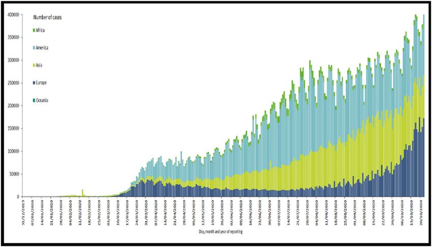
COVID-19 and SARS-CoV-2: A comprehensive review 551
destined to be genetically engineered as a bioweapon, reservoir host for SARS-CoV-2 [29]. However, even with
it should have optimal-binding capability with its the very close resemblance, the RBD sequence of the S is
receptor. In reality, S of the virus has been observed still different in SARS-CoV-2 [30]. Likewise, CoVs from
to have much higher affinity for its receptor than pangolin specie Manis javanica have also been reported
theoretically predicted. This observed high-affinity [15,25] to have analogous genome to SARS-CoV-2, and
binding of SARS-CoV-2 to hACE2 can be explained even in some of them, the RBD sequence too is exactly
hypothetically as the virus naturally select the like that of the S in SARS-CoV-2. However, none of the BB
hACE2 for attachment and the attachment further CoVs from either bat or pangolin has so far been reported
making conditions ideal for its optimal binding to have the same polybasic cleavage site as is present in
with the mentioned receptor [15]. the border of S1 and S2 subunits [31]. As the diversity in
(ii) To bioengineer a virus, the easy way is to borrow a BB CoV species is very high and a majority of them are yet
genetic skeleton of a previously known/encountered to be discovered/studied, it is expected that in future we
pathogenic virus; however, the genome of the SARS- may encounter with such BB CoVs that have a similar
CoV-2 as analyzed by multiple scientists is natural cleavage site [15]. Moreover, mutations in the S1–S2 sub-
that is originated wildly and has quite a similar unit junction are quite possible and expected, so it can be
genome to that of coronaviruses that are normally assumed that this virus has acquired the unique poly-
present in bats and pangolins from ages [15–24]. basic furin cleavage site and its current form of S in either
(iii) The sheer intelligence of the virus for acquiring the an animal host [32]. The second possibility is that its
degree of natural selection to select hACE2 with more ancestor may have been transferred to humans in some
specificity than the same receptor present in other spe- other form long before assuming the current pandemic
cies, attaching to it, and then creating an optimal- form through adaptation and mutations. In the latter
binding environment for its entry into the cells is some- case, the mutations, especially acquiring the cleavage
thing that seems to be the result of mutations that have site, may have occurred initially in human-to-human
happened through the course of years and not possible transmission stages [15]. Scientists still not have arrived
through bioengineering at least with the current to a definite conclusion whether the immediate origin of
resources and technologies [15,17,25,26]. SARS-CoV-2 is pangolin or bat for which extensive animal
studies are required to establish the origin and inter-
Another controversial theory postulated by someone mediate host through which transmission of the virus
is that this virus has escaped from a biosafety level-2 lab has taken place [33].
where such types of highly pathogenic microorganisms Owing to the extensive genetic diversity, widespread
and viruses are kept for the sake of research [10]. The distribution, and the high rate of mutations in their
theory is supported by referring to the already reported genome, the family of the viruses poses a continuous
cases when SARS-CoV was escaped [27]. Escaping from serious threat to human health and even survival. SARS
the lab can be ruled out by the fact that the unique and was the result of zoonotic transfer of the SARS-CoV from
never before reported features in the virus, i.e., specific bat to civet cat and then to humans and MERS was a
RBD sequence and the polybasic cleavage site, could not result of zoonotic transfer of MERS-CoV from dromedary
be achieved by cell culture passages that would at least camels to human. Keeping in view that these two epi-
require an ancestor SARS-CoV-2 and repeated prolonged demics were caused by CoVs, the zoonotic transfer of
passages in cell culture. Moreover, the specific predicted SARS-CoV-2 to humans is not much surprising – an event
O-glycan could not have been produced in cell culture as more likely to happen in the future as well [21,34].
its production is induced by immune response [28].
4 Epidemiology
3.2 Who is the intermediate reservoir host;
bat or pangolin? 4.1 Geographic distribution
The very close resemblance of the genome of SARS-CoV-2 The outbreak started in the Wuhan city of China and
with SARS-CoV and SARS related coronaviruses, SARSr- spread to all its provinces affecting a total of 83,014
CoVs, suggests that bats might have played a role as people with 3,343 recorded deaths. On 19th of March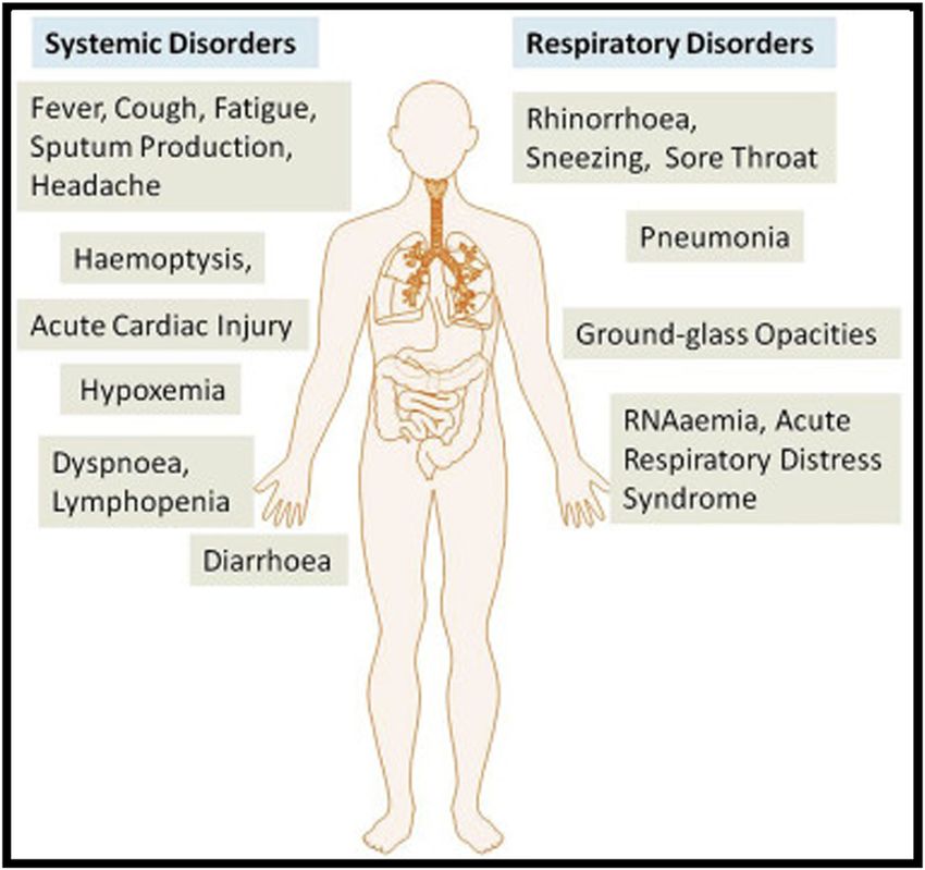
552 Sumaira Naz et al.
2020, China announced that there is no new local case negatives. Globally, the cases have been reduced to
reported since last 36 h, although the imported cases minimum in late October 2020; however, with start of
are still emerging [35,36]. Next on the list was Italy with winter season in many countries, the number of infected
152,271 confirmed cases and 19,468 deaths and Iran with people is on rise these days.
70,029 confirmed cases and 4,357 deaths where the epi-
demic is slowing down although yet continued. Next
badly hit in Europe was Spain, where the number of con-
firmed cases was 161,852 with 16,480 deaths, surpassing 4.2 Case fatality rate (CFR)
Italy. The pandemic continued spreading swiftly around
the world, and as on 12 April 2020, it had affected 210 The CFR is calculated as the number of reported deaths/
countries and territories across the globe. The US has numbers of reported confirmed cases. It is highly variable
started experiencing a dramatic surge in the number of among various countries and quite unpredictable in
confirmed cases since late March and on 12th April 2020, the current scenario. As on 09th April 2020, the Centre
it was the country with the highest number of confirmed for Evidence-Based Medicine estimated the CFR of each
cases (519,453) and deaths (20,283). On 22nd October, 2020, country, from the lowest 0.08% in New Zealand to the
the number of total confirmed cases was 41,696,520 and the highest 13.04% in Algeria. However, the CFR of COVID-19
total number of recorded deaths was 1,137,204 worldwide is affected by many factors like location, intensity of
(All the number of confirmed cases and deaths have been transmission, and age (older than 60 years are more at
taken from the coronavirus resource center map at Johns risk) of the population in the given location along with
Hopkins University and Medicine) [37]. A chart repre- the incidence of comorbidities and poorer health care.
senting continent-wise distribution of COVID-19 cases is Besides, a large number of asymptomatic undiagnosed
represented Figure 3. cases are expected to be present which are therefore
These numbers were high from the mentioned cases not reported, and hence, not included in the determina-
as the number of reported confirmed cases globally as tion of CFR reported [37]. Although the pandemic has
well as country-wise was affected by several factors like taken a lot of lives, still the CFR is lower than that
how many tests have been done both globally and in reported for SARS (10%) and MERS (34%) [39]. The
specific countries, the population of a country, selection case fatality ratio and deaths per 100,000 population
bias (individuals with severe symptoms are tested prefer- worldwide is graphically represented in Figures 4 and 5,
entially), asymptomatic infected individuals, and false respectively.
Figure 3: Continent-wise distribution of COVID-19 cases [38].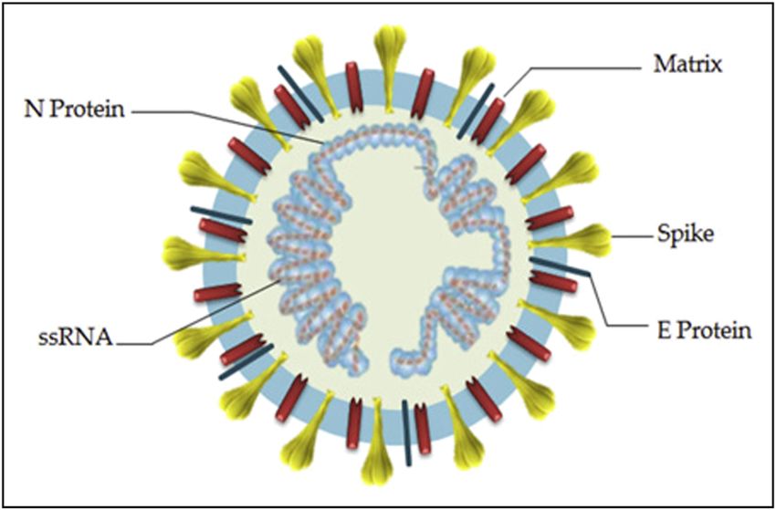
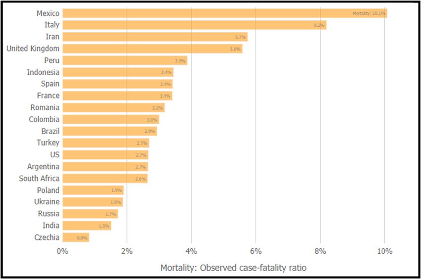
COVID-19 and SARS-CoV-2: A comprehensive review 553 Figure 4: Global case fatality ratio, as of 22 October 2020 [36]. 4.3 Mortality rate 4.4 Transmission Initially, the WHO estimated the mortality rate up to 2%, So far, extensive research has been carried out by the but on 3rd March 2020, the organization declared the scientists and they have thoroughly investigated the overall approximate mortality rate is 3.4%. Mortality route of transmission of the virus, its mode, and rate of rate is observed to be higher in individuals with older its spread so that it is easy to decide who the potential age, from zero for children under nine and 14.8% in transmitter is and how it is transmitted. Human-to- patients aged 80 years or above [39]. human transmission can occur through any of these Figure 5: Global deaths per 100,000 population as of 22 October 2020 [36].
554 Sumaira Naz et al.
average, the infected person may transmit the virus
after 4.6 days of being infected. In other words, the
infected person may transfer the disease to other
people before the incubation period and onset of
symptoms of the disease. The Rο value is considerably
affected by certain other factors, both negatively
and positively. For instance, poor implementation
of countermeasures like isolation, quarantine, and
social distancing, the limited number of populations
tested, and delayed diagnosis could add up to the
value of Rο. Moreover, effective sanitization and dis-
infection measures, social distancing, and isolating
patients and suspects could efficiently reduce the
value of Rο in a given area at a given time. However,
whether these measures could sustainably reduce the
value of Ro is still uncertain [43,44].
(ii) According to China CDC, this is super spreading
(highly contagious) disease associated with many
super spreading events (SSEs). The SSEs have sig-
nificantly contributed to a steady surge in the number
of infected cases and sustaining the epidemic. Besides,
the more symptomatic patients are carrying more oral
and laryngeal load of the infection and are the super-
spreader of the disease [45].
(iii) The stability of the SARS-CoV-2 in transmission media
is also important. A single cough by an infected person
can produce thousands of respiratory droplets, of
Figure 6: Modes of spread of COVID-19 [40]. which the smaller, lighter ones remain in air in the
form of aerosols and the bigger, heavier ones can
land on clothes and other surfaces coming in their
routes: direct transmission, contact transmission and air-
way like doorknobs, worktops, floors, bedsheets, etc.
borne transmissions through aerosols, and may also be
Different studies suggest different viability period for
through medical procedures (Figure 6). Cough, sneeze,
the virus on various surfaces, for example, according
droplet inhalation, and contact with oral, nasal, and
to the reported studies [46,47], the virus is viable up to
eye mucous membranes are the common causes of its
2–3 days on surfaces like stainless steel and plastic,
spread. Viral shedding occurs from respiratory tract,
approximately 24 h on cardboard, up to 4 h on copper,
saliva, feces, and urine [40,41].
and can remain viable in aerosols for about 3 h.
(iv) Environmental and behavioral factors also decide
the degree of transmission. Dense population and
4.5 Factors affecting the transmission confined settings, e.g., health care facilities, eleva-
tors, mass gatherings, event halls, nursing homes,
The impact of the pandemic is highly dependent upon etc., fuel the super spreading of SARS-CoV-2.
various transmission factors which are discussed in the Another important transmission route is fecal-oral
following lines. transmission [48]. Thus, poor hygienic habits are a
(i) The basic reproductive number, i.e., Rο value of big factor in transmitting the infection, while observing
SARS-CoV-2, which reflects the degree of conta- healthy hygienic measures is the key to prevention.
giousness of a virus, is estimated to be 2–4 (median The WHO and the US EPA (Environmental Protection
value 2.76) which means that an infected person Agency) have reported a list of disinfectants, saniti-
could on average transmit the infection further to zers, and their active ingredients for public use
2.76 persons [42]. The median SI (series interval) which could be utilized safely to reduce the chances
has been reported to be 4.6 which means that, on of getting infected [49].COVID-19 and SARS-CoV-2: A comprehensive review 555
4.6 Routes of transmission sputum production (27%), and other typical symptoms
like sore throat, headache, and body aches or shortness
The disease is transmitted through various respiratory of breath that are observed in other respiratory tract
and extra respiratory routes. Some of the known and viral infections as well.
more important ways of transmission are: ‒ Second: About 14% patients may show severe clinical
(i) Direct contact with the infected person, i.e., person symptoms like dyspnea, hypoxia, and respiratory dis-
to person transmission (main mode); tress that may require mechanical ventilation.
(ii) Respiratory droplets; ‒ Third: A third class with lesser proportion of the patients
(iii) Oral-fecal transfer; (5%) who may develop severe pneumonia, respiratory
(iv) Infected blood and body fluids; failure, and from mild to severe acute respiratory dis-
(v) Touching contaminated surfaces; tress syndrome (ARDS) followed by sepsis, septic shock,
(vi) Viral aerosols in a given confined space; and multiorgan dysfunction syndrome [52].
(vii) Sewage waste, air condition systems, and contami-
nated water/food; and Severity and duration of onset of the symptoms, how-
(viii) Transfer from asymptomatic and presympto- ever, depend upon the age of the patient, comorbidities,
matic infected persons as asymptomatic shed- and the immune response of the body [4]. The overall
ding [44,46,48–50]. symptoms (systemic and respiratory disorders) and com-
plications related to the COVID-19 have been shown in
Recently, a study investigating air samples of the the Figure 7 [53].
hospital wards including Intensive Care Unit (ICU) and
general ward reported that the virus can travel up to four
meters (13 feet) from the source (infected patients). The
investigation also reports that a high concentration of the 6 Risk factors
virus can be found on floors of the ward, on bed rails,
computer mouse, and other regularly touched surfaces as Although no one is at zero risk of getting COVID-19 or
well as on the shoe soles of the ICU staff [51]. invulnerable to it, getting sick to different levels, chances
Continued transmission of SARS-CoV-2 suggests that of getting hospitalized, and mortality rate depend upon
tertiary and quaternary transmission routes would also certain risk factors.
be there. Now, a question arises: as compared to the
primary and secondary transmission of the SARS-CoV-2,
during which it was transmitted from an animal to human
and then from human to human, have the rate of transmis-
sion and severity of pathogenicity of SARS-CoV-2 been
affected (increased/decreased) with the continued trans-
mission and after multiple passages in human or not? [43].
To answer this question, further investigational studies are
required.
5 Clinical features
According to China CDC, the range of clinical manifesta-
tions can be broadly divided into three categories:
‒ First: Many of the infected individuals (81%) may
remain asymptomatic with no pneumonia or mildly
symptomatic having minor pneumonia. In case of
mild to moderate pneumonia, the patients may experi- Figure 7: The systemic and respiratory disorders caused by COVID-
ence fever (99%), fatigue (70%), dry cough (59%), 19 infection [53].556 Sumaira Naz et al.
6.1 Age 6.2 Comorbidities and underlying health
conditions
According to the reports by the WHO and China CDC, the
overall rate of getting infected and developing symptoms The older individuals are more at risk because they have
is directly proportional to the increased age. In a study by other underlying health conditions normally associated
China CDC, 87% of the cases reported were in the age with their age. A young person having an underlying
group 30–87 years [52,54]. Another study has revealed health condition will be at the same risk and would be
that a major portion of the hospitalized patients were affected with the same severity as individuals of older age
adults with median age in the range from 49 to 56 years with the same health condition. According to the studies
[55]. US CDC has recently released its report on the survey conducted in China, Italy, and the US, the majority of the
of total hospitalized patients in the US (admitted in individuals who were infected and developed severe
March 2020). The report concludes that the increase in symptoms had at least one underlying condition. In one
the age had a direct effect on the rate of hospitalization; study, carried out in China, CDC reports that 37.6% of
in the age group 0–4 and 5–17 years, it was recorded 0.3 total patients had one or more underlying health condi-
and 0.1%, respectively, the rate increased to 2.5% for the tions, 78% out of them were admitted to ICU and had
age group 18–49 years, 7.4% in the age group 50–64 at least one preexisting comorbidity [62]. Likewise, in
years, and much higher, i.e., 13.8%, in the aged indivi- another report of the Italian national institute of health,
duals (65–74 years). The highest hospitalization rate out of 355 patients who reportedly died due to the COVID-
(17.2%) was observed for individuals of age 85 years 19, only 0.3% had no prior underlying health conditions,
or above [56]. Also, CFR is observed to increase with whereas 99% of them had at least one, 49% had three or
increasing age as reported in two different studies. One more, and 26% had two preexisting comorbidities [36].
was carried out in China and they reported that the CFR is A recent study reported by the US CDC estimated that out
8% for patients having age from 70–79 years and 15% for of all adult hospitalized patients admitted in March in US,
patients with 80 years of age or above. The other study 89% had one or more comorbidity [56].
conducted in Italy reported that 12% CFR is for patients The most prevalent commonly reported underlying
having age from 70 to 79 years and 20% for patients health conditions that are associated with developing
having age about 80 years and above [57]. In the same severe symptoms and mortality are discussed below.
way, another study carried out in the US on 2,449
patients showed that 65% of patients were from the age
group 45 years or above and 80% of all the fatalities were 6.2.1 Hypertension (HT) and cardiovascular
observed for this age group as well [58]. Besides, as diseases (CVD)
shown in a number of surveys, it seems that quite a
smaller percentage of children appear to be symptomatic, Many studies suggest that individuals with HT and CVDs
exhibiting only mild symptoms. In two independent stu- have a higher risk of contracting the infection and devel-
dies in China and South Korea, only 2 and 6% (respec- oping severe symptoms. Although the exact mechanism
tively) of all the reported confirmed cases were those is not understood, two possible reasons are proposed: (i)
having age 20 years or below [52,59]. A study conducted ACE2 – the target receptor is abundantly expressed in the
on much younger individuals from age 0–9 years reported cardiovascular system (CVS). The enzyme has a role in
that 26% of them needed hospitalization, only one of them vasodilation by negatively regulating renin-angiotensin
was admitted to ICU and zero fatality was observed. It system (RAS) and thereby reducing blood pressure. The
was also observed in another study that in the younger damage to CVS by the direct cytopathic effect of the virus
individuals, infants are at higher risk, as in more than is thus possible. (ii) Due to the flooding of CVS with cyto-
half of the hospitalized cases of younger individuals with kines produced as a result of an exaggerated response by
severe to critical symptoms were infants having age below the immune system [63].
1 year [60,61]. HT is at the top of the list of comorbidities that pose
At the endnote, it is also questionable whether the risk for getting infected with the COVID-19 as reported by
younger people do not get infected at faster rate like that several studies [56,64–66]. Nevertheless, some experts
of old age people or they do not develop severe symptoms disagree with listing HT as a major risk factor by ques-
upon infection and thus remains unreported. tioning the sufficiency of data given to prove it. They haveCOVID-19 and SARS-CoV-2: A comprehensive review 557
compared the data in 11 different studies which have immune response is best if given in the very start of the
listed HT as a risk factor and conclude that the data col- infection, but once the infection spreads, the immune
lected so far are based on unadjusted analysis as they are response given by the innate system in the form of
collected in patients of older age group only where the more and more cytokines production causes inflamma-
prevalence of HT is just common and expected comor- tion, which, if becomes uncontrolled, can cause much
bidity in this age group. Whether HT is a risk factor or not harm than good by damaging the body’s uninfected cells
can be decided after a well-adjusted study where the and tissues – a phenomenon called “cytokines storm” [70].
incidence of the COVID-19 in individuals with HT is com- It is assumed by the experts that in 80% of indivi-
pared with those individuals who do not have HT, taking duals with milder or no symptoms of the infection, the
into account the exposure history of both groups [66]. innate response is quick and effective enough to control
Preexisting CVDs account for 9.0% of the reported the infection at the very first step and also quick in acti-
confirmed cases according to a study conducted by vating the acquired immune response. But in the indivi-
China CDC, 52% of which required hospitalization, out duals with immunodeficiencies, both the responses
of whom, 29% were admitted to the ICU. Acute coronary are delayed and the innate immune system continues to
syndrome in COVID-19 patients leads to cardiac insuffi- produce its components (which are amplified steadily)
ciency, ischemia, and critical illness [56,62]. Similarly, in and overstated inflammation that result in widespread
another study, it was observed that 44 and 24% of all the damage to the cells and lead to the damage of healthy
studied patients who developed severe symptoms were tissue and organs, making the patient severe or critically
those with preexisting arrhythmia and heart disease, ill [55,68].
respectively [67].
6.2.3 Other underlying health conditions
6.2.2 Poor immunity
According to a report of China CDC, concluded from a
When a person is infected with SARS-CoV-2, the innate study conducted on quite a large number of patients
immune system is supposed to be the first responder. (122,653), 10.9% had diabetes, 9.2% had chronic lung
It identifies the viral RNA through pattern-recognition diseases, 2.1% were former smokers, and the other 1.3%
receptors (PRRs), and in the first step, produces an array were found to be current smokers [62].
of cytokines (the type-1 interferon is produced specifi-
cally in viral infection) to wall off the virus immediately
and to prevent the spread of infection and viral replica-
tion. The presence of cytokines in the blood activates the 6.3 Gender – has it any role?
second component of innate immunity, i.e., macrophages
and dendritic cells which respond by phagocytizing Multiple studies from Italy, China, and the US showed
infected cells and producing even more cytokines and that men seem to be more affected, hospitalized, and
signal the acquired immune system to switch on, if the became critically ill as compared to women. CFR was
viral infection is still not eliminated. The innate immune also high for men as reported in these studies. In South
system continues its action until the acquired immune Korea, however, more women were found affected than
system gets active, which by the production of specific men but still, the CFR was high for men. Likewise, in
Abs against the virus finally clears the viral load com- Spain, men and women were equally affected according
pletely [68]. This is at least the expected scenario in to the report of the health ministry, but still, men were
recovered patients and in those who get vaccinated for likely to end up in ICU and develop severe symptoms. It is
the infection, although no evidence-based study is yet to be confirmed if male sex is a risk factor or not;
available yet. however, various behavioral differences, for example,
In older patients or those with low immunity result- more men are smokers as compared to women in a given
ing from underlying health condition/s, the immune population, may be most probable reasons. However,
system is compromised and thus the immune response men are considered to be more at risk due to development
is delayed [69]. This gives a better coping and replicating of underlying health conditions like CVD and type-2 dia-
time to the virus and spreading of the infection. Innate betes, which are established risk factors [37,56,71–73].558 Sumaira Naz et al.
7 Diagnosis 7.3.2 Immunoassay test
The test could be either antibody (Ab) or antigen (Ag)-
7.1 Testing criteria
based; it uses ELISA, i.e., enzyme-linked immunosorbent
assay, where specific Ab or Ag are quantified after their
The WHO has urged for testing more and more people
attachment with the Ab or Ag used, depending upon the
around the globe to restrict the pandemic. In a given
type of the assay [62].
location, who should be tested is decided by the govern-
ment, local health authorities, and the doctors. There
are standard criteria and guidelines given by the WHO, 7.3.3 Nonspecific tests
ECDPC (European Centre for Disease Prevention and
Control), and the US CDC which need to be followed in (i) Observing signs and symptoms of the disease like
deciding who should to be tested first. According to their fever, cough, and gastrointestinal symptoms.
guidelines, hospitalized patients should be the first (ii) Computed tomography (CT) scan (of chest) has been
priority, followed by patients with a higher risk of devel- shown to be a very important tool for diagnosis and
oping complications and having comorbidities and then suggesting potential infection. According to a study,
those who work in health care and infrastructure or any most (86%) of the observed patients had ground-
suspected case from the community [74–76]. glass opacities (GGOs) and 64% had consolidation
plus GGOs in the CT scans. These characteristics
were observed in critical patients and patients with
mild symptoms. Although critically ill patients were
7.2 Sampling for testing
older than those with mild symptoms, there were no
significant differences between the CT scans of both
The US CDC has provided a proper guideline for the col-
groups. The specific findings of the CT scan may be
lection of samples and its handling. Commonly, naso-
present even before the onset of symptoms and detec-
pharyngeal and oropharyngeal swabs, expectorated sputum
tion of viral genetic material in the samples from the
of a productive cough (not induced), bronchoalveolar
upper respiratory tract [62,72]. Table 1 presents details
lavage, and feces can be tested with accuracy rate of 32,
about the available diagnostic methods.
72, 95, and 44%, respectively. A small percentage (3%) of
blood samples also exhibited positive results in a study.
To avoid false negatives and to improve the sensitivity of
8 Incubation period
the test, studies have recommend that samples from mul-
tiple sites should be taken and tested [76]. Furthermore,
According to the WHO, the incubation period (the time
if a suspected person tested negative, the test should be
taken in the appearance of symptoms after infection) is
repeated with resampling from multiple sites according
between 1–14 days, and from a study carried out on 181
to the WHO guidelines [77].
confirmed cases of the disease, it was concluded that
mean incubation period was 5.1 days with the possibility
that some (1%) may still develop the symptoms after
7.3 Types of diagnostic tests 14 days of monitoring [78]. Moreover, according to a study,
the duration of incubation is also dependent upon age
The symptoms expressed by COVID-19 patients are non-
specific and cannot be used for an accurate diagnosis.
Molecular techniques are more suitable than syndromic Table 1: Available major diagnostic methods for COVID-19
testing and CT scans for accurate diagnosis because they
can target and identify specific pathogens. Test What is detected Sample type
RT-PCR Viral genome Nasopharyngeal swabs,
saliva, and sputum
7.3.1 Molecular test ELISA Antibody Blood
CT scan Nonspecific characteristics NA
The test extracts the genetic material of the virus (if any) like GGOs and
consolidations are
and uses real-time polymerase chain reaction (rRT-PCR)
observed
for its detection after amplifying its number of copies.COVID-19 and SARS-CoV-2: A comprehensive review 559
and immunity; more the age and weaker the immune virus in the lungs. The virus as discussed earlier enters its
system, the shorter will be the incubation period [79]. target through its spike S; S1 subunit of the S has RBD
that recognizes and attaches to ACE2 membrane, while S2
anchors in the membrane; S2′ site (present directly after
9 Pathogenesis S2) is cleaved by the target cell’s protease, i.e., TMPRSS2
(transmembrane protease, serine-2), which changes the
conformation of S2 and let it ready for fusion with the
9.1 Entry of SARS-CoV-2 in the target target cell membrane [10,85].
receptor
The target receptor as discussed earlier for SARS-CoV-2 is
ACE2 enzyme, whichever cell in any organ throughout 9.3 Replication in the target cells
the body that possesses it will expectedly be the target
cell for the virus. Studies [17,80] show that this virus has Once the virus enters into the cell, its capsid is removed
much more affinity for its target as compared to that of and it releases its RNA in the host cell’s cytoplasm. The
SARS-CoV. A recent study through the release of 3D elec- RNA, on one hand, uses the host cell ribosome and trans-
tron micrograph of the virus has proposed that once lation machinery to produce its two polyproteins that are
SARS-CoV-2 gets access to human’s upper respiratory used for the synthesis of its component proteins and
tract through the mouth or nose, it can stay and replicate enzymes. Structural proteins are also synthesized for
there for a while before going to the lungs because ACE-2 making glycoproteins, spike, and nucleocapsid; to do
is expressed in the mucous membrane of mouth and so, it uses host proteinases and nucleocapsid proteins.
larynx. Although the expression of ACE-2 there is quite On the other hand, the RNA uses the host cell’s RNA-
small than that in the lungs, because of the very high dependent-RNA polymerase to make its copies. The viral
affinity for its receptor, SARS-CoV-2 can still bind and cell components and RNA so formed are finally assembled
enter the ACE-2 present in the mucous membrane of making many numbers of virus particles. They are released
nose and larynx and multiply there before going to the outside of the cell through apoptosis/pyroptosis of the
lungs contrary to SARS-CoV which was observed to repli- cell [86].
cate only in lungs [81]. After the primary replication, the
virus goes to the lungs where it enters its target receptor-
expressing cells. The virus can enter the peripheral blood
theoretically from the lungs and also into the gastroin- 9.4 Immune response to the viral entry and
testinal tract (GIT) and start approaching to the cells that replication
have ACE2 expressed like CVS, kidney, intestine, and
liver. The presence of the viral infection in areas other When the virus particles are released, they attack other
than the lungs is evident from studies where blood and fecal cells and are recognized by PRR present on the antigen-
samples tested were found positive for the presence of the presenting cells to provoke an immune response. The
virus. In some cases, quite a large proportion of individuals’ immune response can be divided into two stages.
fecal test was positive repeatedly after negative results of
the respiratory samples. Moreover, gastrointestinal pro-
blems as reported in some studies and the observed acute 9.4.1 Primary immune response
damage to liver, kidney, and heart also suggest the presence
of infection in these areas [69,82,83]. As ACE2 is also The engagement of the viral PRR leads to the release of
expressed in seminiferous tubules’ lining, damage to testi- antiviral cytokines which are reported to be interleukins
cular tissues is pointed out in a reported study [84]. (IL) of several types like IL-1β, IL-4, and IL-10; interferon
(IFN)-γ inducible protein-10 (IP-10) and monocyte chemo-
attractant protein (MCP)-1 are also released in the very
early response of innate immunity [87]. Based on the
9.2 Entry to the target receptor in the lungs knowledge of what happened in the case of SARS and
MERS [70,86], our basic knowledge of immunity against
Reportedly, type-2 pneumocytes and enterocytes are the viruses, the expected series of events appear to be like in
ACE2 expressing cells, and hence, are entry sites of the the following lines.560 Sumaira Naz et al.
The rapid viral replication and subsequent massive memory Abs. But in worst case scenario, the anti-S-IgG may
cell apoptosis lead to vascular leakage and increased trigger Fc receptors-mediated inflammatory response – a
release of pro-inflammatory cytokines which increase phenomenon known as antibody-dependent enhanced
capillary permeability and vasodilation causing interstitial immunity (ADE) where a NAb is not able to efficiently bind
space and alveolar edema. Surface tension is increased to the virus attached to it and hence only partially coat it.
because of decreased surfactant levels in the alveoli and When these partially coated virions are taken up by macro-
the leakage and damage of type-2 pneumocytes that are phages/monocytes, they replicate there, infecting them,
responsible for surfactant production. This leads to alveolar and cause an abnormal immune response, leading to severe
collapse, causing difficulty in gas exchange, hypoxemia, inflammation and acute lung injury. The anti-S-IgG ADE
and increase work of breathing. On the other hand, the was first observed in SARS, where the patients in whom
inflammatory mediators lead to the release of neutrophils the levels of anti-S-IgG rapidly reached to the peak had
which release ROS, hydrolases, and proteases to destroy more severe lung injury, persistent infection, and higher
the affected cells and when the immune response goes chances of death. A proposed potential therapeutic approach
uncontrolled as occurs in SARS-CoV which happens here to prevent the anti-S-IgG ADE phenomenon is to block the
too, they also start damaging the unaffected pneumocytes FcR and prevent the binding of anti-S-IgG to them, hence
(both type 1 and type 2). This leads to impaired gaseous preventing the inflammatory response [70,72,89].
exchange and decreased surfactant production. Accord- In a nutshell, if the infected person provides a better
ing to a very comprehensive review about the immune immune system’s primary response and control the resul-
response to SARS-CoV-2 [88], the infection causes pyrop- tant inflammation at the earlier phase, the better will
tosis in the macrophages and lymphocytes; their subse- be the outcome because most of the times, secondary
quent pulmonary infiltration along with the debris of the inflammatory response can go wrong and lead to critical
destroyed cells, fluids, and other destroyed immune pro- illness and fatality. However, the uncontrolled cascade of
ducts results in alveolar consolidation, and hence, altered events either in the primary immune response, i.e., the
gaseous exchange. Side by side, downregulation and shed- enhanced viral replication, cell apoptosis and pyroptosis,
ding of ACE2 are expected due to the action of the virus and and ACE2 shedding, or in secondary immune response,
due to the release of pro-inflammatory cytokine, especially i.e., FcR-mediated ADE, can result in an overstated
tumor necrosis factor (TNF)-α. The ACE2 thus released in immune response resulting in cytokines storm. The pre-
the blood is called soluble ACE2 (sACE2) that is suggested sence of cytokines and chemokines in high concentra-
by some authors to have a role in the pathogenicity of the tions has been reported in a study where the critical
virus. The loss of ACE2 function leads to dysfunction of ICU patients had higher levels and more types of cyto-
the RAS, vasodilation, and alveolar edema, thus worsening kines in their blood as compared to non-ICU patients
the inflammation. More likely, most patients with good [60]. The cytokine storm activates an exaggerated immune
innate immune responses can cope with the infection at reaction resulting in more harm than good, damaging the
the first stage, decrease or clear the viral load, and subse- body’s healthy tissues, organs, and systems. This leads to
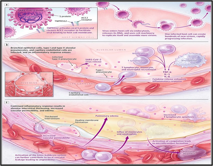
quently recover from the inflammation [60,65,70,88]. ARDS and multiorgan failure and fatality [90–92]. Figure 8
shows the schematic representation of various stages of
pathogenesis caused by SARS-CoV-2 [93].
9.4.2 Secondary immune response
So far, very limited data are available on the humoral
response to SARS-CoV-2, but based on the humoral immune 9.5 Sepsis and septic shock
response (adaptive immunity) given by the body in viral
infection which has been observed previously in case of The cytokines leaked into bloodstream reach to multiple
SARS and MERS, the expected response is the production organs and cause vasodilation in the blood vessels, which
of neutralizing Abs (NAb)-IgM and IgG. A specialized IgG is in turn decrease total peripheral resistance leading to
also formed specifically against the spike S called anti-S- hypotension, low perfusion to different organs, and sub-
IgG. In positive outcomes, that happen mostly; the virus is sequently cause systemic inflammatory response (SIR) fol-
neutralized by the NAb, viral load is decreased, and the lowed by sepsis leading to multiple organ failure and
infection subsides gradually along with the production of septic shock [62,68].COVID-19 and SARS-CoV-2: A comprehensive review 561
Figure 8: Immunopathogenesis of COVID-19 [93]. (a) SARS-CoV-2 viral infection of host airway cells, (b) early-stage COVID-19, (c) late-stage
COVID-19.
9.6 Effects on the kidneys ICU, and the majority (6/9) of these didn’t meet kidney
disease improving global outcome criteria for AKI [100].
At least 10 studies have been conducted so far on the Even though a uniform pattern was not followed in these
effect of COVID-19 on the kidney and its functions and studies and there were limitations, some generalization
development of acute kidney injury (AKI) in the patients can be made:
[65,67,82,94–99]. Out of the 10 studies, one study [100] (i) The overall observed AKI range was 0.5 to 23%;
performed in 116 hospitalized patients, among whom five (ii) The incidence and severity of AKI were directly
patients had chronic kidney disease (CKD), showed that related to mortality rate;
none of the 111 non-CKD patients developed AKI and no (iii) Proteinuria, hematuria, elevated serum creatinine
abnormality was observed in any kidney function. The levels, high blood urea nitrogen, and glomerular
nine out of 10 studies, however, report oppositely with filtration rate less than the normal rate were the
results that AKI occurred in the majority of the studied common observations in almost all these studies
patients. The dichotomy between the results of the one [60,82,96–101].
study with those of the nine other studies may be due to
the reason that some of the nine studies were undertaken While talking about the impact of the infection on
in older patients only (above 60 years), as in some stu- the kidneys, two mechanisms can be suggested for the
dies, a proportion of patients had CKD, while some stu- observed AKI. (i) The SIR impairs blood circulation to the
dies were carried out on critically ill patients admitted in kidneys rendering them unable to perform their function;562 Sumaira Naz et al.
if the infection continues, it can result in kidney failure. COVID-19 which are metabolized in the liver; as observed
(ii) Due to the excessive expression of ACE-2 and TMPRSS2 in a study, that was the reason for the liver injury in
in renal tubules [69,82], they could be equally targeted by patients mainly because of the use of lopinavir/ritonavir
the virus if it reaches to the kidney. The virus replicates antiviral in these patients [111].
there, infects, and destroys the tubular cells.
9.8 Effects on the CVS
9.7 Effects on the liver
As in the kidney, the pathogenic effect on CVS is mediated
At least 12 different studies have reported the impact of either due to the SIR, cytokines storm, and hypoxemia or
COVID-19 on the liver; their findings can be summarized due to the direct cytopathic effect of the virus after the
into these points: attachment to ACE2 that are abundantly expressed in
(i) The prevalence of liver injury was in the range of CVS. Acute myocardial injury, heart damage, and cardiac
14.3 to 53% of the patients studied; arrest have been observed in patients. Moreover, the severity
(ii) In most cases, alanine aminotransferase, and aspar- of the pathogenic effect is more in the patients with preex-
tate aminotransferase, levels were found elevated isting heart diseases wherein they develop severe symptoms
and the abnormal levels were more prevalent in and higher chances of mortality [63].
severe and ICU patients than in the mild and non-
ICU patients;
(iii) A slight to moderate increase in the serum levels of
bilirubin, γ-glutamyl transferase, myoglobin, crea- 9.9 Effect on the GIT
tine kinase, and lactate dehydrogenase was also
observed in most cases; The expression of ACE2 and TMPRSS2 in the glandular
(iv) The derangement of the observed parameters and epithelia of stomach, duodenum, and the enterocytes of
liver injury in almost all studies increased with an ileum and colon makes the GIT equally susceptible to the
increase in the severity of the disease; attack of SARS-CoV-2 as are the lungs [112,113]. The inci-
(v) Of the studied patients, 2–11% were those who had dence of GIT symptoms like diarrhea, abdominal pain,
preexisting liver diseases; and and vomiting observed in many cases is sufficient to point
(vi) The severity and incidence of liver injury were much out the involvement of GIT in the infection [114,115]. The
high (up to 70% reported) in non-survivors than in entry and replication of the virus in GIT are also evident
the survivors [55,65,67,84,88,102–108]. from many studies where the viral RNA could be detected
in feces even after the respiratory samples were found
The mechanism of liver injury is not well-estab- negative for the presence of the virus, showing the poten-
lished, but different reasons could be proposed. tial leakage of the virus from GIT to feces [79,116,117].
ACE2 is moderately expressed in liver and highly
expressed in the cholangiocytes of the bile duct.
Cholangiocytes regenerate the damaged liver cells and
have a role in the immune response of the liver. It is 10 Management and treatment
safe to assume that more damage to liver happens due
to the cytopathic effect of the virus on the cholangiocytes There is no specific recommended treatment for COVID-
and not on the liver itself. In addition, damage to the 19; the patients are given supportive care and manage-
liver could also be due to the sepsis and the cytokines ment based on the stage/severity of their disease.
storm. In a study, postmortem biopsy of a deceased
COVID-19 patient revealed microvascular steatosis along
with lobular and portal inflammation – a presentation 10.1 Clinical management
normally associated with drug-induced injury, but as
the data are very limited, it is also possible that the injury The WHO has provided proper guidelines (criteria) on
was/may be due to the virus infection [84,109,110]. prehospital and clinical management of the disease. Clinical
Besides, similar to the situation in SARS, antibiotics, anti- management is further categorized in different categories
virals, and steroids are widely used for the treatment of based on progress of the disease.COVID-19 and SARS-CoV-2: A comprehensive review 563
(i) Patients with mild symptoms or mild pneumonia do care for the relief of symptoms and different combinational
not necessarily require hospitalization, but should therapies including administration of systemic corticoster-
be isolated either in hospital, home, or any other oids and antiviral drugs. The currently in-practice treatments
setup for containment of the infection and provided for the cure and management of severely affected COVID-19
with antipyretics and monitored for their symptoms. patients are summarized in Table 2, while the chemical
(ii) Severe COVID-19 patients who are hypoxemic and structures along with molecular formulae of them are given
have respiratory distress require supplemental oxygen in Table 3 [118].
and antimicrobial treatment and close monitoring.
(iii) Critical patients with ARDS who are severely hypoxemic
and whose symptoms do not improve require advanced 10.2.1 Corticosteroids
ventilatory support and conservative fluid management.
(iv) Patients with sepsis and septic shock in whom sepsis Glucocorticoids have been widely used in syndromes clo-
is recognized require fluid resuscitation and vaso- sely related to COVID-19, including SARS, MERS, severe
pressors administration. influenza, and community-acquired pneumonia. However,
the evidence to support or discourage the use of glucocorti-
There is a detailed description of management in coids under these conditions is weak owing to the lack
each step as provided by WHO with proper guidelines, of data from sufficiently powered randomized, controlled
but still answer is needed to important questions like trials. Several studies were conducted to establish the effec-
which type of management is required for which specific tiveness of dexamethasone in the COVID-19 patients. It is
patient, the expertise required, and what are the require- likely that the beneficial effect of glucocorticoids in severe
ments, be it diagnostic, supportive, or for treatment of the viral respiratory infections is dependent on a selection of
symptoms in each step. Specific guidelines are also there for the right dose, at the right time, and in the right patient.
the management of children, pregnant and breastfeeding High doses may be more harmful (than helpful), as may
women, and individuals with chronic conditions [62]. such treatment have given at a time when control of viral
replication is paramount and inflammation is minimal. The
greater mortality benefit of dexamethasone in patients with
COVID-19 who are receiving respiratory support and among
10.2 Medications those recruited after the first week of their illness suggests
that at this stage the disease may be dominated by
Unfortunately, there is no specific vaccine or antiviral immunopathological elements, with active viral replica-
drug available for the treatment of COVID-19 yet. Although tion playing a secondary role [119].
few companies have launched their vaccines into market, it
is still early as they have tested them in hurry and no one is
aware about their side effects. Correspondingly, their pro- 10.2.2 Anti-inflammatory drugs
duction on large scale is a challenge to fulfill the need of
such huge population living in different countries, especially Glucocorticoids: In the view of the previous knowledge of
in third world countries that are thickly populated and are the severe outcomes by the use of glucocorticoids in viral
poor. Thus, at present, in terms of patient treatment and infection like influenza and MERS and no beneficial
management, the main focus is the provision of supportive result in case of SARS, with several short and long-term
Table 2: Treatment options for COVID-19
Therapeutic class Treatment options
Antiviral >85% of patients received antiviral agents, including oseltamivir (75 mg every 12 h orally), ganciclovir (0.25 g every
12 h intravenously), and lopinavir/ritonavir tablets (400/100 mg twice daily). Remdesivir is currently under trials at
more than ten medical institutions in Wuhan and has been known to prevent MERS-CoV.
Antimalarial An antimalarial drug, chloroquine phosphate, has been effective in inhibiting the exacerbation of pneumonia due
to its antiviral and anti-inflammatory activities.
Herbal treatments Traditional Chinese Medicines were used most extensively during the previous outbreak of SARS-CoV in China.
They were reported to be used for the treatment of current COVID-19 treatment as well. The herbs used commonly
were Glycyrrhizae Radix Et Rhizoma (Gancao), Astragali Radix (Huangqi), Atractylodis Macrocephalae Rhizoma
(Baizhu), Saposhnikoviae Radix (Fangfeng), and Lonicerae Japonicae Flo.You can also read