CROSSTALK BETWEEN ENVIRONMENTAL SIGNALS AND 3D GENOME ORGANIZATION IN THE REGULATION OF GENE EXPRESSION - The Department of Oncology-Pathology ...
←
→
Page content transcription
If your browser does not render page correctly, please read the page content below
The Department of Oncology-Pathology,
      Karolinska Institutet, Stockholm, Sweden

 CROSSTALK BETWEEN ENVIRONMENTAL
SIGNALS AND 3D GENOME ORGANIZATION IN
 THE REGULATION OF GENE EXPRESSION

           Anna Lewandowska Ronnegren

                   Stockholm 2021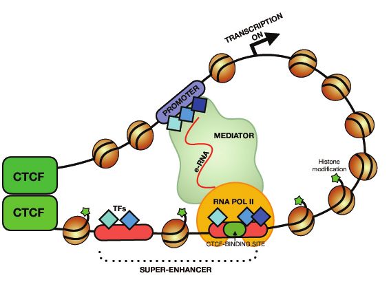
All previously published papers were reproduced with permission from the publisher.
Published by Karolinska Institutet.
Printed by Universitetsservice US-AB, 2021
© Anna Lewandowska Ronnegren, 2021
ISBN 978-91-8016-115-2
CROSSTALK BETWEEN ENVIRONMENTAL SIGNALS AND
3D GENOME ORGANIZATION IN THE REGULATION OF
GENE EXPRESSION

THESIS FOR DOCTORAL DEGREE (Ph.D.)

By

Anna Lewandowska Ronnegren

The thesis will be defended in public at B0317, BioMedicum, Karolinska Institutet, Solna,
12th February 2021, kl 10.00
Zoom meeting ID: 649 2515 1495

Principal Supervisor:                          Opponent:
Associate Professor Anita Göndör               Professor Mattias Mannervik
Karolinska Institutet                          Stockholm University
Department of Oncology-Pathology               Department of Molecular Biosciences

Co-supervisor(s):                              Examination Board:
Professor Thomas Helleday                      Professor Ann-Kristin Östlund Farrants
Karolinska Institutet                          Stockholm University
Department of Oncology-Pathology               Department of Molecular Biosciences

                                               Professor Einar Hallberg
                                               Stockholm University
                                               Department of Biochemistry and Biophysics

                                               Associate Professor Lena Ström
                                               Karolinska Institutet
                                               Department of Cell and Molecular Biology
To my parents, who always believed in me.
Rodzicom, którzy zawsze we mnie wierzyli
POPULAR SCIENCE SUMMARY OF THE THESIS

The nucleus is the “heart“ of the cell. It contains all the accumulated information about our
physical appearances, habits and preferences. Every single detail of our body functions is
written in the form of a genetic code, composed of four elements — four types of
nucleotides. These nucleotides are aligned in a 2 meter long DNA string. However, this
extended molecule has to be tightly packed into the extremely small volume provided by
the nucleus and yet be amenable to differentiate and maintain the numerous cell types of
our bodies in response to internal and external cues. Each human arises from one single
cell, the fertilized oocyte, which during prenatal development generates trillions of cells of
different morphology and function, despite that almost (plasma B cells and T cells the
exception with rearranged Ig and TCR genes) all of them have exactly the same genetic
material. The secret to this lies is in the packaging and spatial organisation of genome and
its dynamic regulation. One could compare it to the piano, which symbolizes the genome,
while the pianist represents the machinery interpreting the different notes generated by both
intrinsic and extrinsic signals to define different cell types. A similar phenomena takes
place in the cell — it is using some part of available genetic material to regulate gene
expression, and hence its morphology and functions, while storing unnecessary (temporary
or permanently) genetic units in transcriptionally repressive nuclear sub-compartments —
often at the nuclear periphery. Understanding the principles behind this genome
organisation is crucial not only for understanding how our organism works, but also for
being able to identify novel therapeutic strategies in case if something goes wrong in the
cell — during cancer development, for example.

This thesis focuses on chromatin fibres movements within the nuclear architecture and how
such dynamic processes set the stage for encounters regulating gene expression. By
exploring “cellular routines” in repositioning certain DNA fragments between active and
inactive parts of the nucleus, new principles underlying cellular choices of usage of certain
DNA fragments were uncovered. Again, using the pianist metaphor, one could say that the
aim of the research is to understand how “the cell” is ”choosing” notes which are going to
be played in certain moment of its life, and how the notes might be changing in time or in
response to a changing environment. These aims are highly relevent for our understanding
of how they can go awry to cause human diseases, such as cancer. By understanding cancer
architectural complexity, developmental choices and/or responses to outside stimuli, we
might learn the habits of the “cancer day-scheme” to fight it by cutting off its necessary
supplies by identifying targets for therapeutic regiments and their timing.
ABSTRACT

The thesis explores the connection between environmental stimuli and gene expression
regulated by the spatial changes in genome organization. In Paper I, by applying state of the
art Circular Chromosome Conformation Capture assay (4C) and Chromatin in situ
Proximity (ChrISP) techniques, we show that transcriptionally active circadian genes meet
in space with repressed lamina-associated domains (LADs), and that these interactions are
under the control of the circadian clock. External time cues thus synchronised circadian
transcriptional   oscillations   by   repositioning   clock-controlled   genes    from   the
transcriptionally permissive sub-compartment of nuclear interior to the transcriptionally
repressive nuclear periphery. These processes relied on the rhythmic formation of
complexes between CTCF and PARP1, two master regulators of the genome, to increase
the amplitude of circadian gene expression.

In Paper II we took an advantage of the novel, ultrasensitive Nodewalk technique to
explore the stochastic nature of MYC interactions with its flanking enhancers. By pushing
the Nodewalk limits of identification of chromatin interactions in the input material
corresponding to less than 8 cells, we could show that MYC is likely screening for
neighbouring interaction partners rather than vice versa. Moreover, we could show that
MYC does not interact with enhancers, once its transcription had been initiated. These
findings suggest that enhancer hubs simultaneously interacting with MYC are likely virtual
consequences of high cell population analyses and that MYC interacts with its enhancers in
a mutually exclusive manner.

Paper III concentrates on the role of a CTCF binding site within the oncogenic super-
enhancer (OSE) in the regulation of MYC gene gating in colon cancer cells. CRISPR
induced mutations in the CTCF binding site within the OSE abrogated WNT-dependent
nuclear export of MYC mRNA, providing genetic evidence to the claim of the OSE-
mediated gating of active MYC alleles to the nuclear pore.This manuscript documents,
moreover, that the communication between OSE and MYC, as well as their repositioning to
the nuclear pore, involves PARP1 to indicate a more general role for the CTCF :PARP1
complex in gene regulation.
In summary, this thesis has uncovered novel principles underlying the roles of stochastic
chromatin interactions and mobility within the 3D nuclear space to regulate gene
expression with a focus on circadian transcriptional regulation and the recently discovered
gene gating phenomenon in humans. These findings contribute to our understanding of
principles in which the nuclear architecture and genome organisation synergize to induce or
maintain the properties of the cell. By extrapolation, such findings might form a platform
for identifying new therapeutic strategies to battle cancer, for example.
LIST OF SCIENTIFIC PAPERS

 I.
      Zhao H*, Sifakis EG*, Sumida N*, Millán-Ariño L*, Scholz BA, Svensson
      JP, Chen X, Ronnegren AL, Mallet de Lima CD, Varnoosfaderani FS, Shi C,
      Loseva O, Yammine S, Israelsson M, Rathje LS, Németi B, Fredlund E,
      Helleday T, Imreh MP, Göndör A.

      PARP1- and CTCF-Mediated Interactions between Active and Repressed
      Chromatin at the Lamina Promote Oscillating Transcription. Mol Cell,
      2015.59(6): p. 984-97.

 II. Noriyuki Sumida, Emmanouil G Sifakis, Narsis A Kiani, Anna
     Lewandowska Ronnegren, Barbara A Scholz, Johanna Vestlund, David
     Gomez-Cabrero, Jesper Tegner, Anita Göndör and Rolf Ohlsson.

      MYC as a driver of stochastic chromatin networks: Implications for the
      fitness of cancer cells. Nucleic Acids Research, 2020 Oct 13, gkaa817.

III. Ilyas Chachoua*, Ilias Tzelepis,* Hao Dai*, Anna Lewandowska
     Ronnegren*, Jia Pei Lim*, Mirco Martino, Felipe Beccaria Casagrande,
     Rashid Mehmood, Anita Göndör.

      A CTCF binding site within the oncogenic super-enhancer coordinates the
      WNT-regulated gating of MYC. Manuscript.
CONTENTS
1 INTRODUCTION ......................................................................................................... 1
1.1 Epigenetic principles ............................................................................................ 1
1.1.1 The primary chromatin fiber .................................................................... 1
1.1.2 3D chromatin interactions ........................................................................ 4
1.2 Nuclear organization ............................................................................................ 5
1.2.1 Spatial separation between active and inactive chromatin states ............ 5
1.2.2 Chromatin movements ............................................................................. 6
1.2.3 Chromatin hubs vs stochastic movements............................................... 7
1.2.4 Contribution of CTCF and PARP1 to chromatin interactions and
mobility .................................................................................................... 8
1.2.5 Technologies exploring 3D chromatin structures ................................... 9
1.2.6 The Nodewalk technique ....................................................................... 11
1.3 The 4th dimension of THE regulation of nuclear functionS - introduction
to the circadian rhythm....................................................................................... 12
1.3.1 The entrainment of circadian rhythm ....................................................... 14
1.3.2 The role of circadian rhythm ................................................................. 15
1.3.3 Circadian organization of the epigenome .............................................. 16
1.3.4 The roles of CTCF and PARP1 in the circadian cycle.......................... 17
1.3.5 Transcriptional activation and repression directed by chromatin
movements ............................................................................................. 17
1.3.6 Connection between enhancers and circadian chromatin
movements ............................................................................................. 18
2 RESEARCH AIMS ..................................................................................................... 23
3 MATERIALS AND METHODS ................................................................................ 25
3.1 CELL CULTURES AND TREATMENTS ........................................................ 25
3.2 MUTATION OF THE OSE-SPECIFIC CTCF BINDING SITE BY
CRISPR .............................................................................................................. 25
3.3 RNA/DNA FISH ANALYSES ........................................................................... 26
3.4 IN SITU PROXIMITY LIGATION ASSAY (ISPLA) ..................................... 27
3.5 CHROMATIN IN SITU PROXIMITY (CHRISP) ............................................ 28
3.6 GRID WIDE-FIELD MICROSCOPY ................................................................ 28
3.7 CHROMATIN NETWORKS AND INTEGRATION ANALYSES ............... 28
3.7.1 Circular chromatin conformation capture sequencing (4C-Seq) ............. 28
3.7.2 Nodewalk .................................................................................................. 29
3.8 IMMUNOPRECIPITATION ANALYSIS ....................................................... 32
3.8.1 ChIP-qPCR ................................................................................................ 32
3.8.2 Co-immunoprecipitation (co-IP) assay ..................................................... 32
3.9 WESTERN BLOT ANALYSIS ........................................................................ 33
3.10 RNA ANLYSES ................................................................................................ 34
3.10.1 siRNA transfection.................................................................................. 34
3.10.2 Pulse labeling of RNA and nuclear RNA export assay.......................... 343.10.3 RT-qPCR analysis of transcription ......................................................... 35
4 RESULTS .................................................................................................................... 37
4.1 PAPER I: PARP1- AND CTCF-MEDIATED INTERACTIONS
BETWEEN ACTIVE AND REPRESSED CHROMATIN AT THE
LAMINA PROMOTE OSCILLATING TRANSCRIPTION .......................... 37
4.1.1 Inter-chromosomal interactome and connection between circadian
loci and repressed domains .................................................................... 37
4.1.2 Role of CTCF and PARP1 interactions in connecting circadian loci
to LADs .................................................................................................. 38
4.1.3 CTCF/PARP1 mediated circadian transcriptional attenuation by
oscillating repositioning of circadian loci to the nuclear periphery ...... 39
4.2 PAPER II: MYC AS A DRIVER OF STOCHASTIC CHROMATIN
NETWORKS: IMPLICATIONS FOR THE FITNESS OF CANCER
CELLS................................................................................................................ 41
4.2.1 MYC-driven chromatin networks as a sum of stochastic interactions ..... 41
4.2.2 Mutually exclusive interactions of MYC with flanking enhancers .......... 42
4.3 PAPER III: A CTCF BINDING SITE WITHIN THE ONCOGENIC
SUPER-ENHANCER COORDINATES THE WNT-REGULATED
GATING OF MYC ............................................................................................ 43
4.3.1 CTCF regulates the WNT-dependent pre-nucleoporin-OSE
interactions ............................................................................................. 43
4.3.2 CTCF coordinates several steps of the WNT-regulated gating of
MYC ....................................................................................................... 44
5 DISCUSSION .............................................................................................................. 45
5.1 Nuclear periphery and its role in the regulation of gene expression ................... 45
5.2 Coordination of enhancer-promoter interactions and sub-nuclear
localisation at the MYC locus............................................................................. 47
5.3 The roles of CTCF and PARP1 in the regulation of chromatin networks and
their sub-nuclear localization ............................................................................. 48
5.4 The dysregulation of chromatin structures in diseases ........................................ 49
6 CONCLUSIONS ......................................................................................................... 51
7 POINTS OF PERSPECTIVE ...................................................................................... 53
8 ACKNOWLEDGEMENTS ........................................................................................ 55
9 REFERENCES ............................................................................................................ 63LIST OF ABBREVIATIONS 3C chromatin conformation capture 3D three dimensional 4C circular chromosome conformation capture 4D four dimensional 5C chromosome conformation capture carbon-copy AHCTF1 AT-Hook containing transcription factor 1 Arntl2 aryl hydrocarbon receptor nuclear translocator-like 2 ATP adenosine triphosphate BAC bacterial artificial chromosome BER base excision repair BMAL1 brain and muscle arnt-like 1 CAB chlorophyll a/b-binding proteins ChIA-PET chromatin interaction analysis by paired-end tag sequencing ChIP chromatin immunoprecipitation ChIP-loop chromatin immunoprecipitation loop ChIP-Seq chromatin immunoprecipitation sequencing ChrISP chromatin in situ proximity CLOCK circadian locomotor output cycles kaput Co-IP co-immunopurification CR circadian rhythm CRY cryptochromes CT chromosome territory CTCF CCCTC-binding factor DAPI 4,6-diamidino-2-phenylindole DBP D-element binding protein DMR differently methylated region DMSO dimetylsulfoxid DNA deoxyribonucleic acid DNAse I deoxyribonuclease I DTT ditiotreitol EDMD Emery-Dreifuss muscular dystrophy EGF epidermal growth factor EnhD enhancer D EU 5-ethynyl uridine FBS fetal Bovine Serum FISH fluorescence in situ hybridization GFP green fluorescent protein GUN5 genomes uncoupled 5 HAT histone acetyltransferase HCEC human colon epithelial cell HCoEpiC colonic epithelial c+B91ell Medium HDAC histone deacetylase HEB human embryoid body HESC human embryonic stem cell HGPS Hutchinson-Gilford progeria syndrome HMT histone methyltransferase eRNA enhancer RNA ICC immunocytochemistry ICR imprinting control region IGF2 insulin-like growth factor 2 IgG immunoglonulin G INM inner nuclear membrane ISPLA in situ proximity ligation assay
kb           kilobase
LAD          lamina-associated domain
LAP2         lamina associated peptide 2
LBR          lamin B receptor
lncRNA       long non-coding RNA
LOCK         large organized chromatin K9 modification
Mb           megabase
MED1         mediator subunit 1
MEF          mouse embryonic fibroblast cell
MLL1         mixed lineage leukaemia 1
MYC          myelocytomatosis viral oncogene
NPC          nuclear pore complex
NUP          nucleoporin
NuRD         nucleosome remodelling and deacetylase
OCT4         octamer-binding transcription factor 4
OSE          oncogenic super-enhancer
PAR          poly ADP ribose
PARD3        partitioning defective 3 homolog
PARG         poly (ADP-ribose) glycohydrolase
PARP1        poly (ADP-ribose) polymerase 1
PARylation   poly (ADP-ribosyl)ation
PBS          phosphate-buffered saline
PC           plastocyanin
PCR          polymerase chain reaction
PER          period
PGE2         prostaglandin E2
PTM          post-translational modification
RBCS         Rubisco small subunit
REV-ERBα/β   reverse erithroblastosis α and β
RNA          ribonucleic acid
RNAPII       RNA polymerase II
RNP          ribonucleoprotein
RORα         retinoic acid receptor-related orphan receptor-α
RORE         ROR/ REV-ERB -binding element
SOX2         sex determining region Y
SCN          suprachiasmatic nuclei
SE           super-enhancer
SNP          single nucleotide polymorphism
SSC          saline sodium citrate
TARDBP       TAR DNA-binding protein 43
TAD          topologically associating domain
TE           typical enhancer
TCF4         transcription factor 4
TF           transcription factor
TGF-β        transforming growth factor- β
TTFL         transcriptional/translational feedback loop
UV           ultraviolet
VAT1L        vesicle amine transport 1 homolog-like protein
1 INTRODUCTION
1.1     EPIGENETIC PRINCIPLES

A single human genome can give rise to not only numerous different cell types and
specialized functions, but also their responses to varying developmental and environmental
cues. This epigenetic adaptability in the cellular interpretation of genetic information
depends on the establishment of cell type-specific gene expression patterns. With a mere
2% of the genome coding for proteins, the remaining 98% of the genome is replete with
regulatory elements that underlie context-specific gene activity(1). Chromatin adaptations
implemented by the epigenetic machinery as well as its 3D mobility are key factors in the
induction and subsequent stable propagation of gene expression pattern without altering the
underlying DNA sequence(2). Moreover, the chromatin fibre is an essential platform on
which transcription factors (TF), singling pathways and other chromatin modifications
converge and in some instances collaborate in response to environmental stimuli(1). Thus,
chromatin states maintain flexible features in response to the appropriate cues and
conditions and yet are stable enough to ensure the propagation of robust phenotypes during
development. Consequently, the loss of this robustness increases the potential for disease
development(1,3–5). However, very little is known about the mechanisms which govern the
transitions between normal and pathological epigenetic plasticity. It has been proposed that
the compartmentalization of active and inactive domains, co-existence of different
epigenetic marks as well as interactions between different chromatin loci or regulatory
elements like promoters and enhancers underlie how chromatin states respond to
environmental cues, similar to the establishment of developmentally stable gene expression
patterns(6,7).

1.1.1    The primary chromatin fiber

The 6 billion bases of coding and non-coding DNA of the diploid genome are wrapped
around approximately 30 million nucleosomes to form chromatin, which is the
physiological and structural form of human genetic information(1,2). Nucleosomes are
formed by wrapping ~145–147 bp of DNA around histone octamer cores separated by a
                                                                                          1
linker region of ca 20-40 bp to define the ‘primary structure’ of chromatin fibres. This simplified picture is, however, compounded by the ever-increasing number of histone variants as well as of post-translational histone modifications (PTMs) and DNA modifications(8), to generate an almost astronomical number of theoretically possible variations in the chromatin primary structure. The PTMs play important roles in establishing equilibriums between different structural states, including the regulation of the chromatin compaction as well as the interactions between nucleosomes and non-histone proteins. Among many distinct histone PTMs, acetylation of lysine residues and methylation of arginine residues are best known. Thus, acetylation of lysine neutralizes the positive charge of the amino acid and is often associated with chromatin decompaction and vice versa, whereas arginine methylation plays important roles in the regulation of gene expression(6,9). These and other PTMs decorate not only the chromatin of gene bodies, but also regulatory elements, such as enhancers. Regular enhancers are typically defined as short (~100–1000 bp) noncoding DNA sequences, composed of concentrated clusters of transcription factor (TF) recognition motifs. Such complexes tend to bend the DNA to generate so-called DNase I hyper-sensitive sites demarcating such regions(10–13). The type of combinatorial PTMs associated with regulatory regions defines their functions. For example, active enhancers are marked with H3K4me1, H3K27ac, H3K122ac and absent or low levels of H3K27me3 and H2K9me2/3 marks. Conversely, enhancers which are poised to become active enhancers are demarcated with both active and repressive marks, such as H3K4me1 and H3K27me3(6,14,15). This picture is complicated, however, by the demonstrations that some of the enhancer regions can be devoid of the typical enhancer- specific chromatin marks(16,17). It has been estimated that each cell type can have anything from 10,000 to 150,000 enhancers with an accumulated number exceeding 1 million enhancers active in all human cells(11,18,19). Accordingly, enhancers are key regulatory elements controlling tissue-specific transcription programs and thus essential for the robust maintenance of a diverse range of phenotypes(20). More recently, a new class of enhancers, the so-called super-enhancers (SE), has been identified. Such regions are often found near genes that control cell states and have cell type-specific functions in response to a diverse range of signaling pathways(11,18,20). SEs are defined not only by the usual enhancer marks, such as H3K4me1, H3K27ac, master TFs, p300, but also by the prominent presence of the Mediator complex (Med1)(10,11). A schematic representation of the loop formation that such super-enhancers can form with targeted promoters is presented in 2
Figure 1. In a comparison to typical enhancers (TE), SE typically spreads from 10 kb to
over several hundreds of kb (while the median size of TEs ranges from 1 kb to 4 kb), often
contain more than one separate region that is bound by multiple TFs (such as OCT4, SOX2
and NANOG in Embryonic Stem Cells), drive targeted gene transcription with a high
precision potentially related to the higher expression of enhancer RNAs (eRNAs) than
typical enhancers. Accordingly, the number of SEs in each cell type is fewer by one to two
orders of magnitude than the corresponding number of TE(10,11,21–23).

Fig.1 Schematic loop formation between promoter and super-enhancer Super-enhancers
are enriched in histones modifications (H3K4me1 and/or H3K27ac) as well as
transcription factors, p300 and the Mediator complex. CTCF can bind to chromatin to
establish domain boundaries, enhancers and promoters. Bi-directionally transcribed
eRNAs play stabilizing roles in loop formation between promoter and super-enhancer by
providing a platform for interactions between trans-acting factors.

                                                                                         3
1.1.2 3D chromatin interactions The enhancer needs to physically interact with the promoter of its target gene to initiate its transcription. This is an essential step to ensure that the correct set of genes are turned on at the correct time during a developmental window(24). Physical enhancer-promoter contacts generally involve collaboration between the Mediator complex at the enhancer and promoters which usually are marked with the H3K4me3 modification. The resulting chromatin loop involves the cohesin complex that promotes the search for promoters with active marks by a loop extrusion principle(17,25,26). The formation of enhancer-gene loops can be further stabilized by enhancer RNAs (eRNAs), which constitute links between enhancers and their target genes(20,27). The CTCF (CCCTC-binding factor) is another important factor, also for SEs, by being able to physically link distal regions to form chromatin loops with a median size ca. 200kb in convergent orientations. The loss of this feature, which promotes highly specific and precise SE-promoter interactions, affects loop domains and transcriptional patterns(21,27–30). Moreover, the CTCF binding site can provide a structural hierarchy necessary for the function of SEs (by mediating and/or facilitating long-range chromatin interactions) (27,31). However, very little is known about the mechanisms underlying the regulation of such chromatin loops – a situation further compounded by the fact that only 7% of distal regulatory elements control the most proximal promoters with enhancer regions located anywhere from 1kb to tens of Mbs away from their target promoters(6,17,25). Moreover, one enhancer can contact more than one promoter(25). It is also important to note that interactions between promoters and enhancers might be established without leading to overt transcriptional initiation. Indeed, for the large subunit of the RNA polymerase complex to trigger transcription it must acquire a serine 2 phosphorylation mark at its large subunit(17,24). Apart from promoter-enhancer interactions, chromatin interactions can involve different types of regulatory elements like promoter-promoter, enhancer-enhancer or insulator- insulator loops(24). The chromatin insulator is a sequence element of DNA that blocks enhancer-promoter interactions in a position-dependent manner to potentially contribute to the spatial arrangement of chromosomes in the nucleus(30,32). However, their ability to antagonize enhancer-gene interactions is position-dependent - the promoter thus remains capable of being activated by other enhancers and the enhancer can activate other promoter(32). The H19 imprinting control region (ICR) functions as a methylation 4
sensitive, CTCF-dependent chromatin insulator and has been shown to physically interact
with a differently methylated region (DMR) with a silencer function at the maternally
inherited Igf2 allele(33,34). The H19 ICR region is inherited unmethylated from the
maternal germline and methylated from the paternal germline to direct parent of origin-
specific expression of the proximal H19 and the more distal Igf2 alleles. Since only the
unmethylated H19 ICR is able to bind CTCF, only the maternal Igf2 allele is insulated from
downstream enhancers. This feature requires the ability of the CTCF-H19 ICR complex to
form chromatin loops(33–38). Conversely, while the paternally methylated H19 ICR allele
is unable to prevent activation of Igf2, it silences the H19 gene.

1.2     NUCLEAR ORGANIZATION

1.2.1   Spatial separation between active and inactive chromatin states

As discussed above, a fundamental property of the genome is its spatial organization at
several, hierarchical levels in cell nucleus. In interphase nuclei, individual chromosomes
occupies a limited space called chromosome territories, which are approximately 2-4 μm in
diameter(7,39). Despite cell type-specific differences in the localization of individual
chromosome territories in individual cells, larger (and gene-poor) chromosomes tend to
locate close to the nuclear periphery while smaller and more gene-rich chromosomes
inhabit a more central position(40). One of the most striking features of the chromosomal
organization is thus the global spatial separation of active and inactive chromatin referred to
as A (active) and B (inactive) compartments, which are further divided into sub-
compartments with distinctive chromatin states, association with nuclear landmarks and
replication timing(40–42). Within nuclear sub-compartments chromatin is organized into
topologically-associated domains (TADs) - areas of highly interacting chromatin separated
from each other by, for example, CTCF boundaries demarcating the TADs(29,40,43). The
constraining of chromatin movements between TADs likely restricts contacts to involve
only neighboring enhancers and promoters(44). TADs can be either A-type (with open,
gene-rich chromatin) or B-type (gene-poor, closed chromatin)(45). Lamina-associated
domains (LADs) are distinct regions of the genome interacting with nuclear lamins (a thin
meshwork of filaments that lines the inner nuclear membrane). LADs encompass 0,1 to

                                                                                             5
10Mb, are AT-rich and contain H3K9me2 and H3K27me3 marks with poor representation of H3K36me3(39,45) to constitute 35-40% of the mammalian genome(29,45,46). Similarly to TAD borders, LAD borders are enriched in CTCF binding sites(45) and approximately 9% of LAD borders contain CTCF within 10kb of the boundary(47). Furthermore, LADs are represented by cell type-specific variants (facultative, fLADs) or cell type-invariant (constitutive, cLADs) LADs(48). These types of LADs differ from each other in gene density and conservation between mouse and human(48). Interestingly LADs overlap to a high degree with Large Organized Chromatin Lysine Modifications (LOCKs), which are regions enriched in the H3K9me2 mark and highly dynamic throughout the differentiation of murine embryonic stem cells (mESC)(45,48). Genes localized within LOCKs are typically silenced and comprise 31% of the genome of differentiated ES cells, but less than 5% in undifferentiated cells(49). Interestingly both LADs and LOCKs might play an important role in the regulation of chromatin movements. 1.2.2 Chromatin movements Although the distribution of the chromosome territories within the nucleus to a high degree depends on their size and gene-richness, their preferred position is not absolute(50). This also translates to the kind and dynamics of chromatin fiber interactions and distinct loci of particular chromosomes manifest different features in terms of mobility and search for interacting partners(51). Whereas most chromatin interactions take place within the chromosome territory, genes and regulatory regions can also interact in trans with inter- chromosomal interactions occurring preferably at the edge of chromosome territories(7). However, particular loci can also loop into the CT of another chromosome or multiple loci can loop out of their CTs(7,38). The mobility of distinct loci depends on their position within nuclear sub-compartments with genes associated with the nuclear periphery or the nucleolus exhibiting significantly lower mobility in comparison to genes within the nucleoplasm(52). Such features may underlie long-range chromatin interactions that have been implicated in the epigenetic regulation of gene expression(51). Experiments with molecular tethering to the nuclear periphery using an inducible system showed that physical repositioning of examined genes to the nuclear periphery can 6
reversibly repress the activity of these genes in human cells(53). Although not a universal
feature, the tethering of some of genomic loci to the periphery is thus generally promoting
silencing of gene activity(54). Conversely, it is generally considered that the mobility of
active chromatin to the site of mRNA export, the nuclear pore, might increase expression
efficiency(55). The so-called ‘gene gating’ phenomena thus states that certain loci can be
juxtaposed to the nuclear pore and facilitate rapid nuclear export of processed mRNAs. In
one such example, it was shown that an oncogenic super-enhancer mediated the
repositioning of active MYC alleles to nuclear pores, which enabled MYC transcripts to
escape the more rapid degradation pathway in the nucleus than in the cytoplasm in colon
cancer cells(55–57). Of note, Paper III provide genetic evidence in support of this scenario
by showing that a single CTCF binding site within the oncogenic super-enhancer
coordinates the WNT-regulated gating of MYC to the nuclear pores. In contrast to short-
range interactions, global mobility of chromatin might depend on the actin/myosin system
to seemingly produce active and directed movements(58). The specificity of the
repositioning of certain loci to the nuclear periphery is likely dependent on a combination
of DNA sequence, trans-acting factors, PTMs and lncRNA(6,59).

1.2.3   Chromatin hubs vs stochastic movements

The nuclear architecture is dynamic in nature and yet stably maintained(7). This may relate
to that the nucleus is considered to be built mainly by self-organizing principles(51,60).
Such a dynamic environment displays a high level of stochastic collisions between
chromatin fibers. Although gene expression itself is a fundamentally stochastic process,
transient chromatin fibre interactions don’t necessarily imply any regulatory function(7,61).
Nonetheless, enhancer interactions both in cis and trans as well as the establishment of the
so-called transcription factories provide well documented examples for the meta-stable
character of such functional chromatin networks(17,62,63). However, the frequency of
spatial meetings between two interacting genomic regions is not universal for all cells in a
cell population(51). Even though chromatin-fiber interactions might display a high level of
noise to appear largely non-functional, they might increase the potential for a functional
outcome to contribute to cellular plasticity as well as establishing/maintaining stochastic
patterns of mono-allelic gene expression(6,7,64,65).
                                                                                           7
1.2.4 Contribution of CTCF and PARP1 to chromatin interactions and mobility As noted above, CTCF plays an important role in the formation of chromatin loops. CTCF is an evolutionary conserved 11-zinc-finger protein with ability to bind various DNA motifs and different regulatory proteins(66–68). Its ability to interact with DNA, proteins as well as RNA is a consequence of its domain organization – the central zinc-finger domain allows CTCF to bind DNA with the use of different zinc-fingers combinations, whereas binding to its protein partners is mediated via interactions with any part of the entire protein, i.e. its central zinc finger domain or the N- and C-terminal parts. For example, N- terminal region has the ability to bind PARP1 protein, as described later in the text, while the unstructured C-terminal region can bind RNA(66,67). The complexity of the CTCF structure translates into the various significant roles CTCF plays in regulation of cellular processes and development. CTCF was initially discovered as a transcriptional repressor of chicken c-myc gene(69) and its important function as an insulator in the regulation of Igf2/H19 imprinting has been well studied(35,70–72). Subsequent research showed that CTCF binds to 40,000-80,000 sites genome wide in mouse and human ESCs to play role in alternative splicing, DNA repair, recombination and mediation of enhancer-promoter interactions(17,66,73–75). Many, but not all, of CTCF binding sites are co-occupied by the cohesin complex, which physically interacts with CTCF as a biochemically stable complex. Together they shape chromatin architecture, although CTCF and the cohesin complex differ in the dynamics of chromatin binding and mechanism of contribution to the chromatin organization(76–81). CTCF, which has been characterized as a master weaver of the genome(67), also binds to a wide range of factors including PARP1. Although PARP1 is mostly known for its role in DNA- damage response, it has been implicated in epigenetic modifications of both histones and DNA as well as maintaining the integrity of constitutive and facultative heterochromatin(82–84). Many of these functions are in partnership with CTCF, as witnessed by the PARP1-mediated poly(ADP-ribosyl)ation of its N-terminal region (82,85). Dynamic PARylation of CTCF in response to cellular signals or environmental cues thus plays an important role in CTCF-dependent chromatin insulation and loop formation(86–88) as well as the regulation of chromatin mobility (Paper I). 8
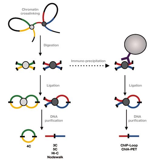
1.2.5   Technologies exploring 3D chromatin structures

The mapping of the 3D structure of the genome has been possible thanks to technological
achievements represented by two main approaches: In situ techniques, such as proximity
ligation and 3D DNA FISH(89) and chromosome conformation capture (3C) and derived
“C” techniques(90). The “C” family of techniques focuses on local and/or genome wide
DNA-DNA contacts and involves restriction digestion of formaldehyde-crosslinked
chromatin followed by intra-molecular ligation to identify proximities between distal
regions (Fig.2). In the original 3C technique, this was performed by PCR, while later
versions of the method rely primarily on high throughput DNA sequencing(91,92). The
parental 3C technique has been extensively used to demonstrate interactions, such as
formation of chromatin loops between two genomic loci(91). However, the drawback of
this technology is that it is based on educated guesses that two particular regions might
interact. To overcome this biased limitation, Circular 3C (4C) in combination with high
throughput sequencing was innovated(38). Strategic placing of the primers within the bait
and formation of circular DNA between bait and interacting sequences allowed the
identification of unknown intra- and inter-chromosomal interactions of a bait(93,94). Next,
the chromosome conformation capture carbon copy (5C) allowed obtaining information on
contacts between multiple genomic loci(95). This was possible by introducing a step where
3C products were incubated with a mix of specific oligos which annealed exactly at one of
the restriction sites covering an entire genomic region of interest(91). However, the “many-
to-many” 5C strategy was subsequently replaced by an “all-to-all” approach represented by
the Hi-C technique. The introduction of biotin-labeled nucleotides for filling restriction
“sticky-ends” (followed by “blunt-ends” ligation) allowed the selection of ligation junctions
by biotin pull-down(89,96). Although the Hi-C technique has been very useful to study
genome wide TAD structures, it is less ideal for the examination of individual loci
interaction pattern due to low resolution and reduced statistical power(91). Moreover,
similarly to almost all of the other 3C-based methods, Hi-C analyses are mostly performed
on the cell-population level with low sensitivity to blur the dynamics of chromatin
encounters. Thus, Hi-C maps are unable to discriminate between stable 3D contacts present
in all cells and an average of stochastic contacts which can differ between particular cells of
a cell population(97). The single-cell Hi-C (which introduces an in-nucleus ligation step)
helped to overcome the problem of the use of cell population, but the technique still suffers
from poor sensitivity and resolution(98). Moreover, the introduction of exponential PCR
                                                                                             9
amplification steps – a feature shared by almost all “C” techniques, is likely to introduce a
bias. This limitation is resolved by including a linear RNA amplification step in the newly
developed Nodewalk technique (Paper II), which will be further discussed in chapter 1.2.6.
Fig.2 Schematic illustration of the “C” family of techniques
10Another common limitation of all the ”C” family techniques is that they do not with few
exceptions (Paper I) provide robust information as to where in the nuclear space the
interactions take place and their frequency of interactions. This generally acknowledged
drawback usually requires validation of identified interactions by microscopy assays, such
as 3D fluorescence in situ hybridization (3D-FISH). Whereas the power of this technique
lies in that it represents a single-cell analysis of gene positioning to provide information of
chromatin dynamics in relation to the nuclear architecture, it is limited by the resolution of
the fluorophores of choice(89). To quantitatively detect chromatin proximities in single
cells with an optimal resolution, chromatin in situ proximity (ChrISP) assay is a method of
choice(99). This technology detects proximities between two different antibodies (against
labeled probes specific to target DNA sequences, or between a DNA sequence and any
other epitope represented by a protein, for example) within 160Å of each other and allows
the spatial visualization of chromatin interactions or higher order chromatin confirmations
in single cells in relation to structural hallmarks(100).
1.2.6 The Nodewalk technique
The Nodewalk method is a member of “C” family techniques which rely on the ligation of
digested DNA to generate covalently linked intra-molecular chromatin complexes.
However, the DNA tagmentation by transposase allows both further fragmentation of
ligated DNA and the incorporation of suitable, tailed primer sequences in Nodewalk,
including the promoter for T7 RNA polymerase, followed by only 5-7 amplification cycles.
This step avoids amplification biases and allows the generation of a template for the in vitro
linear, production of large amounts of RNA from very small amount of input material.
Following the selection of bait, specific primers positioned close to a restriction enzyme
site can be used to prime cDNA synthesis and subsequent high throughput sequencing.
Nodewalk thus combines high resolution with high sensitivity improving it >10,000-fold in
comparison to other “many-to-all” techniques. This in turn allowed the reproducible
identification of chromatin interactions in input material corresponding to less than 8
cells(101) (Paper II). The Nodewalk technique was instrumental in providing key data
11underlying the discovery of the gating of the active MYC gene to the nuclear pore in human
colon cancer cells(56).
1.3 THE 4TH DIMENSION OF THE REGULATION OF NUCLEAR FUNCTIONS
- INTRODUCTION TO THE CIRCADIAN RHYTHM.
Many cellular processes, including chromatin movements or chromatin fiber interactions,
exhibit periodic behavior. These time-dependent events are often regulated by the circadian
clock machinery that likely has evolved to maximize organismal fitness in response to
changing external cues. The circadian clock is a highly conserved system that enables
organisms to adjust to daily changes in the environment, such as food availability and
night-day cycles(102). It controls a variety of physiological processes like hormone
secretion, feeding behavior or sleep-wake cycles. Almost all eukaryotic organisms manifest
behavioral, physiological and metabolic oscillations, with a period of ca 24 hours.
Circadian rhythms display unique properties – although endogenous free-running periods of
approximately 24h, the phase of the rhythm can be entrained or re-set by external time cues.
Moreover, the periodicity of circadian rhythm is stable across a wide range of temperatures
(termed temperature compensations)(103). The mammalian timing system exhibits a
hierarchical architecture with the master pacemaker located in the suprachiasmatic nucleus
(SCN). This controls the phase of oscillations at both the tissue and cellular levels(104) by
providing an output to peripheral tissues (peripheral clocks) following its synchronization
by external time cues, such as light. Peripheral clocks oscillate autonomously and can be
entrained also by food intake(104,105). At the cellular level, the molecular clock machinery
regulates cellular processes via several transcriptional/translational feedback loops
(TTFLs).
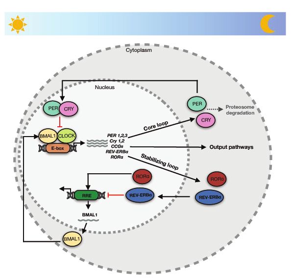
12Fig. 3 Schematic representation of mammalian core circadian feedback loops. The
positive feedback loop (CLOCK/BMAL1) regulates the transcription of negative
feedback loop components – PER and CRY. PER and CRY proteins enter the nucleus to
suppress the transcriptional activity of the CLOCK/BMAL1 complexes. The machinery
is stabilized by antagonizing actions of RORα and Rev-erbα.
In mammals, the positive limb of the core TTFL feedback loop consists of two
transcriptional activators: the circadian locomotor output cycles kaput (CLOCK) and brain
and muscle ARNT-like protein 1 (BMAL1) proteins. They form a heterodimer to promote
the expression of the negative elements (PERIOD (PER) and CRYPTOCHROME (CRY)
homologs). Following a time delay, dimerized complex between PER and CRY translocate
to the nucleus where they inhibit the action of CLOCK/BMAL1
13complexes(102,103,106,107). The core TTFL is, moreover, coupled to other sub-loops which contribute to the functions of the circadian clock. One of these sub-loops comprises transcription factors reverse erythroblastosis virus-α (Rev-erbα) and retinoic acid receptor- related orphan receptor-α (RORα) which bind to RORE in the BMAL1 promoter to either activate (RORα) or repress (Rev-erbα) BMAL1 transcription(102,103,107,108). The principles of mammalian core circadian feedback loops are schematically shown in Figure 3. The core clock machinery is connected to many cellular pathways and has additional layers of regulation that includes PARP1, for example. This topic is further elaborated on in chapter 1.4.1. 1.3.1 The entrainment of circadian rhythm As mentioned in chapter 1.3, the synchronization of the clock to 24h oscillations requires entrainment by external timing cues, also called “synchronizers”, “zeitgebers” or “entraining agents”(107). Circadian rhythm synchronization can be obtained by variety of factors, which relate to the architectural organization of the clock machinery. Among them, light and food are well studied examples of external cues that regulate the activity of the clock components. Following the recognition of light at the retina, the signal reaches the SCN via the retino-hypothalamic tract(109). This “photic entrainment” indirectly regulates light-insensitive peripheral clocks by both humoral and non-humoral pathways(107,110). Food composition and their timing of ingestion, on the other hand, cause periodic changes in the availability of circulating macronutrients, which can reset the circadian clocks specifically in peripheral tissues(103,107). Temporal food restriction can induce circadian rhythmicity of locomotor behavior even in animals with SCN lesions(111). At the molecular level, the feeding process increases blood glucose levels, which can both downregulate the expression of PER1 and PER2, and indirectly regulate CRY stability(112,113). The complexity of the clock machinery is further exemplified by the observation that the CLOCK/BMAL1 binding to DNA can be affected by food restriction- dependent alternations in the cellular redox state(107). Apart from food and light, synchronization of the phase of the circadian rhythm can be obtained by action of other synchronizers, such as arousal stimuli, which include social interactions, exercise, stress or caffeine consumption(107,114). Even though one of the important features of circadian 14
rhythms is their temperature compensation (the period length is maintained in a wide range
of physiological temperatures), peripheral cells and tissues can be synchronized by
temperature fluctuations(107). Not only in vivo models but also in vitro cell cultures or
explants can be synchronized in their circadian rhythms, and many factors can function as
zeitgebers. It was shown that serum shock, for example, can affect the molecular circadian
clock, and various factors in the blood, such as glucose, calcium, EGF, PGE2 or 1α,25-
dihydroxyvitamin D3, can affect rhythmic oscillations(107,115). Finally, chemical
compounds, such as dexametasone (an artificial glucocorticoid) or forskolin (which acts
similarly to the serum shock) can synchronize circadian rhythms by resetting intrinsic
biological processes(116,117).

1.3.2   The role of circadian rhythm

Circadian rhythms (CR) play important roles at both the organismal and cellular levels.
Their existence provide organisms with selective behavioral advantages, allowing for
instance the anticipation of daily food availability and predator avoidance(118). Moreover,
CR allow the physiological adaptation of the organisms to food intake, metabolism and
detoxification, and the circadian clock of plants prepares them to kick-start photosynthesis
in the presence of appearing light energy by ensuring the production of photo-system I and
II components already before the sunrise(118–120). Additionally, temporal separation of
chemically incompatible cellular processes acts in favor for the organisms. For example,
simultaneous photosynthesis and nitrogen fixation in cyanobacteria would cause inefficient
nitrogen fixation due to the poisoning of the nitrogenase by oxygen generated during
photosynthesis(121). It has also been suggested that the circadian clock underlies the
heterogeneity in stem cells populations, to promote developmental potential(122). Studies
with mouse models showed, moreover, that epidermal stem cells in hair-follicle bulge
coexisted as two subpopulations, which differentially expressed core components of the
clock machinery to prevent simultaneous expression of genes responsible for stem cell
dormancy, proliferation or differentiation, such as TGFß and WNT pathway members(123–
125). The DNA damage response is yet another biological process affected by the circadian
rhythm – the ability to repair UV irradiation-induced DNA damages were higher in the late
afternoon and early evening than before dawn(126). Such observations are compounded by
                                                                                         15
the fact that a majority of circadian genes are not only expressed in a tissue-specific manner, but also that different circadian transcripts might accumulate with different phases in different cell populations within a tissue. This heterogeneity is likely the sum of differences both in the spatial distribution of environmental cues and the ability of the cells to respond to these cues(119). Accordingly, the disruption of circadian rhythms by unhealthy lifestyle or shift working, as well as polymorphisms/mutations in the clock genes might contribute to many diseases such as sleep disorders, cardiovascular diseases, diabetes or cancer(127). 1.3.3 Circadian organization of the epigenome Many studies have made the link between distinct epigenetic regulatory layers and the regulation of circadian transcription. One of the earliest reports showed that the phosphorylation of H3 at Serine 10 is light-dependent in SCN neurons(128). Subsequent studies reinforced this observation by showing that epigenetic modifications, such as DNA methylation and PTMs of histones, play roles in rhythmic transcription. For example, both the activating H3K4me3/H3K9ac and the repressive H3K9me3 marks exhibits circadian rhythmicity at regulatory regions(129–132). Consequently, the recruitment of PolII to promoters of circadian genes as well as its regulation at the elongation step display circadian-depended variations(131,133). To sustain circadian dynamics of the epigenome, the molecular clock collaborates with many epigenetic modifiers. These include mixed lineage leukaemia 1 (MLL1, interacting with CLOCK/BMAL1), nucleosome remodeling deacetylase (NuRD, which interacts with and assists the PER complex) and the Polycomb group enzyme KMT6/EZH2, which co-precipitates with CLOCK/BMAL1(131–134). However, the circadian clock also influences the nuclear architecture and the topology of the genome. One of the first indications of this effect were shown in the studies of Chlamydomonas reinhardtii revealing diurnal oscillations of DNA supercoiling in the chloroplasts(135). Similarly, temporal changes in the organization of the albumin D element-binding protein (Dbp) circadian gene and its 3D chromatin fibre interactome could be observed in the mouse. Thus, rhythmic changes in the local chromatin condensation conferred oscillating transcription of genes included in the Dbp circadian interactome in mouse embryonic fibroblasts (MEF)(136,137). 16
1.3.4   The roles of CTCF and PARP1 in the circadian cycle

Since the chromatin fibre serves as a platform for the integration and propagation of many
signaling and metabolic pathways, many cellular factors, which are not directly categorized
as part of the core clock machinery, might be affected by or influenced by the circadian
rhythm. This is exemplified by CTCF and PARP1, which influence chromatin interactions
and mobility, as described in detail in chapter 1.2.4. Thus, PARP1 can influence food
uptake since its loss of function correlates with diet-induced obesity(138,139). Moreover,
Asher et al. showed that ADP-ribosylation of PARP1 oscillates in a rhythmic manner in
synchrony with feeding-fasting cycles in the mouse liver(140,141). These findings provide
an important link between circadian rhythms and metabolism, and have inspired the
formulation of a molecular model. This posits that at the beginning of the light phase
PARP1 binds to and poly(ADP-ribosyl)ates CLOCK. The loss of PARP1 would therefore
cause a phase-shift in the interactions between CLOCK/BMAL1 and PER/CRY complexes.
In line with this reasoning, mice lacking a functional Parp1 gene displayed impaired food
entrainment of peripheral circadian clocks(140,141). CTCF, one of the partners of PARP1,
prominently binds to CLOCK/BMAL1 enhancers implying that these factors join forces to
facilitate long-range chromatin interactions between CLOCK/BMAL1 enhancers and their
target genes(142). But their ability to join forces to effectuate the circadian rhythm does not
end there.

1.3.5   Transcriptional activation and repression directed by chromatin movements

As circadian transcription takes place in the context of the 3D nucleus, mechanisms
allowing genome wide cyclic chromatin transitions must relate to the structural hallmarks
of the nucleus(129). Paper I demonstrated that such events include a collaboration between
PARP1 and CTCF. In short, this novel finding showed that circadian interactions between
CTCF and PARP1 were driving the rhythmic recruitment of a subset of genomic loci to the
repressive nuclear periphery(143). Subsequently, another study implicated that CTCF and
PARP1 influenced the repositioning of the Arntl2 mouse gene in breast cancer cells(144).
Thus, Ha et al. showed that the presence of SNP at the promoter of this gene affected its
interaction with CTCF and PARP1. They hypothesized that this SNP might therefore affect
                                                                                            17
the recruitment/release of the Arntl2 gene to/from the nuclear envelope, thereby altering the timing of Arntl2 expression(144). Correlation between chromatin mobility and changes in transcriptional state has been observed also in Arabidopsis thaliana(145). Feng et al. showed that the chlorophyll a/b-binding proteins (CAB) locus migrates from the nuclear interior to the nuclear periphery during its transcriptional activation, and that this relocation was triggered by light. Similar light-induced repositioning during transcriptional activation has been documented for the Rubisco small subunit (RBCS), plastocyanin (PC) and genomes uncoupled 5 (GUN5) loci(145). 1.3.6 Connection between enhancers and circadian chromatin movements Circadian dynamics altering spatial gene regulation and transcriptional feedback events likely reflect rhythmic enhancer-promoter communications(146). This supposition was borne out in two parallel studies from Mermet et al. and Kim et al., in which both groups highlighted the connections between circadian rhythms and 3D genome folding(147,148). Mermet et al. used the 4C technology to demonstrate that clock-controlled promoter- enhancer interactions act as regulatory layers underlying circadian transcription in mouse liver and kidney. Accordingly, the deletion of intronic enhancer elements in Cry1 or Bmal1 influenced circadian rhythmicity(148). Similarly, Kim et al. showed that circadian gene expression is in mouse liver controlled by rhythmic interactions between promoter and enhancer elements, and that Rev-erbα contributed to rhythmic gene expression by antagonizing the formation of functional loops between target gene promoters and Rev- erbα-regulated enhancer(147,149). It remains to be seen, however, how such rhythmic enhancer-promoter interactions relate to their position within the nuclear architecture and the potential rhythmic mobility of the involved regions between transcriptionally permissive and repressive nuclear environments. This question is in the focus of Paper III, showing that the gating of MYC to nuclear pores required coordination of enhancer- promoter interactions with the recruitment of this complex to nuclear pores by CTCF and PARP1. As the MYC gene is expressed in a circadian manner in many different cell types(150), this finding raises the question whether the rhythmic repositioning of circadian genes to the nuclear periphery might initially involve their anchoring to nuclear pores to increase the amplitude of their cytoplasmic mRNA products, followed by their transient 18
You can also read


