Cystatin-B Negatively Regulates the Malignant Characteristics of Oral Squamous Cell Carcinoma Possibly Via the Epithelium Proliferation/ ...
←
→
Page content transcription
If your browser does not render page correctly, please read the page content below
ORIGINAL RESEARCH
                                                                                                                                            published: 24 August 2021
                                                                                                                                       doi: 10.3389/fonc.2021.707066

                                              Cystatin-B Negatively Regulates the
                                              Malignant Characteristics of Oral
                                              Squamous Cell Carcinoma Possibly
                                              Via the Epithelium Proliferation/
                                              Differentiation Program
                                              Tian-Tian Xu 1, Xiao-Wen Zeng 1, Xin-Hong Wang 2, Lu-Xi Yang 1, Gang Luo 1*
                                              and Ting Yu 1*
                                              1Department of Periodontics, Affiliated Stomatology Hospital of Guangzhou Medical University, Guangzhou Key Laboratory
                                              of Basic and Applied Research of Oral Regenerative Medicine, Guangzhou, China, 2 Department of Oral Pathology and
                                              Medicine, Affiliated Stomatology Hospital of Guangzhou Medical University, Guangzhou Key Laboratory of Basic and Applied
                                              Research of Oral Regenerative Medicine, Guangzhou, China

                                              Disturbance in the proteolytic process is one of the malignant signs of tumors. Proteolysis
                          Edited by:
                          Eva Csosz,          is highly orchestrated by cysteine cathepsin and its inhibitors. Cystatin-B (CSTB) is a
     University of Debrecen, Hungary          general cysteine cathepsin inhibitor that prevents cysteine cathepsin from leaking from
                       Reviewed by:           lysosomes and causing inappropriate proteolysis. Our study found that CSTB was
                       Csongor Kiss,
                                              downregulated in both oral squamous cell carcinoma (OSCC) tissues and cells
     University of Debrecen, Hungary
                       Gergely Nagy,          compared with normal controls. Immunohistochemical analysis showed that CSTB was
     University of Debrecen, Hungary          mainly distributed in the epithelial structure of OSCC tissues, and its expression intensity
                  *Correspondence:            was related to the grade classification. A correlation analysis between CSTB and clinical
                          Gang Luo
                   gangluo@139.com            prognosis was performed using gene expression data and clinical information acquired
                            Ting Yu           from The Cancer Genome Atlas (TCGA) database. Patients with lower expression levels of
                   dent_yu@163.com
                                              CSTB had shorter disease-free survival times and poorer clinicopathological features
                   Specialty section:
                                              (e.g., lymph node metastases, perineural invasion, low degree of differentiation, and
         This article was submitted to        advanced tumor stage). OSCC cell models overexpressing CSTB were constructed to
              Head and Neck Cancer,
                                              assess the effects of CSTB on malignant biological behaviors and upregulation of CSTB
               a section of the journal
                 Frontiers in Oncology        inhibited cell proliferation, migration, and invasion in vitro. Weighted gene correlation
           Received: 08 May 2021              network analysis (WGCNA) and gene set enrichment analysis (GSEA) were performed
           Accepted: 30 July 2021             based on the TCGA data to explore potential mechanisms, and CSTB appeared to
         Published: 24 August 2021
                                              correlate with squamous epithelial proliferation-differentiation processes, such as
                              Citation:
       Xu T-T, Zeng X-W, Wang X-H,
                                              epidermal cell differentiation and keratinization. Moreover, in WGCNA, the gene module
   Yang L-X, Luo G and Yu T (2021)            most associated with CSTB expression (i.e., the brown module) was also the one most
  Cystatin-B Negatively Regulates the
                                              associated with grade classification. Upregulation of CSTB promoted the expression
    Malignant Characteristics of Oral
  Squamous Cell Carcinoma Possibly            levels of markers (LOR, IVL, KRT5/14, and KRT1/10), reflecting a tendency for
      Via the Epithelium Proliferation/       differentiation and keratinization in vitro. Gene expression profile data of the
              Differentiation Program.
            Front. Oncol. 11:707066.
                                              overexpressed CSTB cell line were obtained by RNA sequencing (RNA-seq)
     doi: 10.3389/fonc.2021.707066            technology. By comparing the GSEA enrichment results of RNA-seq data (from the

Frontiers in Oncology | www.frontiersin.org                                         1                                      August 2021 | Volume 11 | Article 707066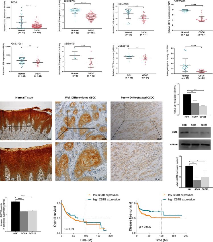
Xu et al.                                                                                                                                 Cystatin-B in OSCC

                                              OSCC models overexpressing CSTB) and existing public database data, three gene sets
                                              (i.e., apical junction, G2/M checkpoint, etc.) and six pathways (e.g., NOTCH signaling
                                              pathway, glycosaminoglycan degradation, mismatch repair, etc.) were enriched in the
                                              data from both sources. Overall, our study shows that CSTB is downregulated in OSCC
                                              and might regulate the malignant characteristics of OSCC via the epithelial proliferation/
                                              differentiation program.
                                              Keywords: oral squamous cell carcinoma, head and neck squamous cell carcinoma, cysteine cathepsin, cystatin-B,
                                              epithelial proliferation/differentiation, WGCNA, GSEA

INTRODUCTION                                                                         carcinoma, and esophageal squamous cell carcinoma (ESCC)
                                                                                     (32, 33)], in which the expression, in which the expression of
As the sixth most common cancer in the world, head and neck                          CSTB is changed in different directions. For instance, CSTB was
squamous cell carcinoma (HNSCC) continues to rise yearly, and                        increased in HCC (25–28), epithelial ovarian tumors (29, 30), and
oral squamous cell carcinoma (OSCC) accounts for 90% of                              breast cancer (31), while decreased in laryngeal squamous cell
HNSCC cases in the region (1–4). The survival of OSCC                                carcinoma and ESCC (32, 33). Some studies have indicated that
patients, especially those with distant metastases, remains low,                     increased CSTB is related to a poorer prognosis in bladder cancer
at approximately 39% (1, 4, 5). Moreover, due to the special                         (34) and a higher risk of tumor metastasis in HCC (27). However,
anatomical location of the oral cavity, OSCC patients often                          most of the existing studies on the correlation between CSTB and
experience great psychological stress and compromised quality                        tumors just provide observational evidence and whether CSTB
of life (6). To date, the potential pathogenesis, including the                      has a causal role in cancers (including OSCC) is unclear. Only a
progression mechanism, of OSCC is not fully understood. It is                        few studies found that CSTB may participate in tumors by
generally accepted that the initiation and development of OSCC is                    regulating cell apoptosis and oxidative stress (31). Only one
a complex process requiring the accumulation of genomic                              noninterventional study based on human OSCC tissue
alterations, which is modified by individual genetic                                  specimens reported that low expression of CSTB in the invasive
predisposition and environmental carcinogenic risk factors (7–                       tumor front was correlated with local tumor recurrence. In
9). It is necessary and challenging for clinical oncology and                        addition, CSTB-specific peptides in saliva were associated with
precision medicine science to understand the carcinogenic and                        lymph node metastasis (35). However, the single study reached a
progression mechanisms of OSCC, to identify biomarkers for                           conclusion based only on OSCC specimens but lacked a
early screening and to establish an accurate prognostic evaluation                   nontumor (normal control) group. Moreover, there is a lack of
system, all of which would contribute to the reduction in the                        in-depth and comprehensive mechanistic studies on the role of
incidence and improvement of both the survival rate and living                       CSTB in the progression of OSCC. Based on the above evidence,
quality of tumor-bearing patients (10–12).                                           we hypothesized that CSTB may play a role in OSCC. Our
    Proteolysis has a vital role in the normal life activities of                    research aimed to identify the role of CSTB in OSCC and to
organisms. Disorders between the enzymatic reaction and the                          explore the relevant mechanisms.
inhibitory reaction of the proteolytic cascade process are one of
the malignant characteristics of tumors, and they also lead to
tumor invasion and metastasis (13–16). Numerous evidences
have indicated that cysteine cathepsins play a crucial role in                       MATERIALS AND METHODS
these processes (17–20). Cysteine cathepsin can degrade the
proteins of the extracellular matrix (ECM) and reshape the                           Cell Line Acquisition and Culture
tumor microenvironment by regulating a variety of cytokines                          The human tongue squamous cell carcinoma cell lines SCC9 and
and thus participate in tumor growth, invasion, angiogenesis, and                    SCC25 (ATCC, ATCC ® CRL-1629 ™ and CRL-1628 ™ ,
metastasis (21). Compared with the fully elucidated role of the                      Manassas, VA, USA) and the normal oral epithelial
cysteine protease family in tumors, research on its inhibitors in                    keratinocyte line HOK (AULU, Guangdong, China) were
tumors is relatively limited. Most studies only focus on certain                     purchased and cultured in Dulbecco’s modified Eagle’s
types of inhibitors and are not in-depth enough. As an inhibitor                     medium (DMEM)/F12 (Gibco, Waltham, MA, USA) and
of cysteine cathepsin, cystatin-B (coded by Cstb gene) is                            DMEM, respectively. The complete cell culture medium
considered a general cysteine cathepsin inhibitor in mammalian                       contained 10% fetal bovine serum (FBS, Gibco) and
cells, preventing cysteine cathepsin from leaking from lysosomes                     1% penicillin/streptomycin (Gibco), while the medium for
and causing inappropriate proteolysis (18, 22–24). CSTB plays a                      carcinoma cells was supplemented with an additional 400-ng/
bimodal role in cancer. Recent studies have associated CSTB with                     ml hydrocortisone (MedChemExpress, Monmouth Junction, NJ,
various cancers [e.g., hepatocellular carcinoma (HCC) (25–28),                       USA). The serum-free cell culture medium for carcinoma cells
epithelial ovarian tumors (29, 30), breast cancer (31), laryngeal                    was prepared as DMEM/F12 containing 1% penicillin/
squamous cell carcinoma, and esophageal squamous cell                                streptomycin and 400 ng/ml hydrocortisone for a number of

Frontiers in Oncology | www.frontiersin.org                                      2                                   August 2021 | Volume 11 | Article 707066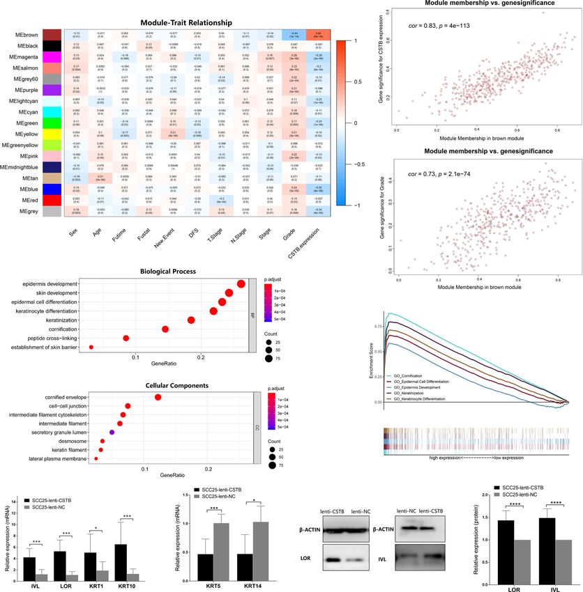
Xu et al.                                                                                                                     Cystatin-B in OSCC

subsequent experiments (e.g., enzyme-linked immunosorbent                  was normalized to that of the internal GAPDH control. The
assay, wound-healing assay, cell migration, and invasion                   primer sequences used are listed in the Table S1. The relative
assays). The cells were cultured in a 37°C, 5% CO2 incubator.              expression of targeted genes was calculated using the 2−△△Ct
                                                                           method (37). Each test was repeated at least three times.
Clinical Patient Specimen Collection
and Immunohistochemistry Staining                                          Western Blotting Analysis
for Cystatin-B                                                             Total protein was isolated from cell samples using precooled cell
The studies involving human participants were approved by the              lysis buffer (Cell Signaling Technology, Danvers, MA, USA) with
Ethics Committee of the Stomatology Hospital of Guangzhou                  protease inhibitor (RayBiotech, Guangzhou, China) and quantified
Medical University. Twenty-three primary OSCC specimens and                using a BCA protein assay kit (Beyotime, Shanghai, China). Equal
fifteen normal oral tissues were obtained from the Department of            protein extracts (20 µg) were separated by SDS-PAGE and
Periodontics and Oral Mucosal Diseases, Stomatology Hospital of            transferred to polyvinylidene fluoride membranes (Millipore,
Guangzhou Medical University. All OSCC specimens were from                 Burlington, MA, USA). After successive incubations with the
samples pathologically diagnosed as OSCC. Normal specimens were            primary antibody and the secondary antibody, the target protein
excess tissues that needed to be removed due to tooth extractions or       was visualized by chemiluminescence using an ECL kit (Beyotime).
oral surgeries, which were also examined as normal oral mucosal            The following antibodies were used in the Western blot assay: anti-
epithelium by pathology. The pathological grading (36) of tissue           loricrin polyclonal antibody (1:1,000, 55439-1-AF, Proteintech,
sections was classified blindly by a single pathologist (Xin-Hong           Wuhan, China), anti-involucrin polyclonal antibody (1:1,000,
Wang). Immunohistochemistry (IHC) staining for cystatin-B was              55328-1-AP, Proteintech), anti-cystatin-B monoclonal antibody
performed in the above tissues (Materials and Methods S1.1). After         (1:1,000, 66712-1-Ig, Proteintech), anti-beta-actin monoclonal
staining, five fields of view were randomly selected to take images          antibody (1:10,000, 66009-1-Ig, Proteintech), anti-GAPDH
under an upright optical microscope. The integrated optical density        antibody (1:10,000, EPR16891, Abcam, Cambridge, UK), goat
(IOD) value of each point on the image was collected by ImageJ and         anti-mouse IgG (H+L) antibody (HRP) (1:10,000, SA00001-1,
then the average density (%, IOD/target distribution area) was             Proteintech), and goat anti-rabbit IgG H&L (HRP) antibody
calculated, representing the expression level of CSTB in certain           (1:10,000, ab205718, Abcam). GAPDH or beta-actin served as the
specimens. Immunostaining was analyzed by a researcher who was             internal controls to calculate the relative expression of the
blinded to the pathological grade of the samples. Correlation              targeted proteins.
analysis between CSTB expression and the degree of pathological
differentiation was conducted.                                             Enzyme-Linked Immunosorbent Assay
                                                                           Cells were inoculated at a density of 1 × 10⁶ in complete medium,
                                                                           which was replaced by serum-free medium after 2 days of culture.
Upregulation of CSTB in OSCC Cell Lines
                                                                           After another 2 days, the supernatant was collected, centrifuged
by Lentiviral Transfection                                                 (4°C, 2,000 rpm, 10 min), and stored at −80°C. The protein levels
The commercial recombinant lentivirus (OBIO, Shanghai,
                                                                           of CSTB in cell supernatants were examined by an enzyme-linked
China) named lenti-CSTB was utilized to overexpress CSTB,
                                                                           immunosorbent assay (ELISA) kit (R&D Systems, DCYB00)
and an empty carrier lentivirus named lenti-NC was used as a
                                                                           according to the manufacturer’s instructions. Each test was
negative control. SCC9 and SCC25 cells were infected with the
                                                                           repeated at least three times.
above lentivirus (MOI = 30) and screened with puromycin
(3 µg/ml) for 15 days. Quantitative reverse transcription-
                                                                           Cell Proliferation Assay
polymerase chain reaction (qRT-PCR) and Western blotting
                                                                           Transfected SCC9 and SCC25 cells were inoculated with
were used to validate the overexpression of CSTB at the
                                                                           complete medium in 96-well plates at a density of 3,000 cells
mRNA and protein levels, respectively. After successful
                                                                           per well. Cell proliferation was detected using a Cell Counting
transfection, the cell lines (i.e., SCC9/25-lenti-CSTB and SCC9/
                                                                           Kit-8 (Dojindo, Kumamoto, Japan) at 1, 2, 3, and 5 days
25-lenti-NC) were cultured with complete medium containing
                                                                           according to the manufacturer’s instructions. Each test was
puromycin (1 µg/ml) for subsequent analysis.
                                                                           repeated at least three times.

RNA Isolation and Quantitative                                             Colony Formation Assay
Real-Time PCR                                                              Three thousand transfected SCC9 cells and SCC25 cells were
Total RNA was extracted from the cells using an RNA extraction             seeded respectively in six-well plates and cultured for 10 days in
kit (AG21017, Accurate Biology, Hunan, China). Extracted RNA               complete medium. Afterward, the colonies were fixed with 4%
was analyzed for quantity and quality by measuring A260/A280               paraformaldehyde and stained with crystal violet. The colonies
with a spectrophotometer (NanoDrop 2000, Thermo Fisher                     that contained more than 50 cells were counted. Each test was
Scientific, Waltham, MA, USA). RNA integrity was confirmed                   repeated at least three times.
by 1.5% agarose gel electrophoresis. For qRT-PCR, 1 mg of total
RNA was used to synthesize cDNA (AG11706, Accurate Biology,                Wound-Healing Assay
China). qRT-PCR was performed using a SYBR Green qPCR kit                  For the wound-healing assay, transfected SCC9 and SCC25 cells
(AG11718, Accurate Biology, China). Relative mRNA expression               were first incubated and cultured with complete medium. A scratch

Frontiers in Oncology | www.frontiersin.org                            3                                 August 2021 | Volume 11 | Article 707066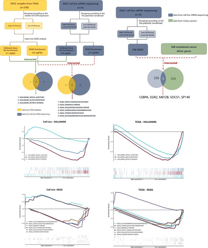
Xu et al.                                                                                                                      Cystatin-B in OSCC

was made with a sterile pipette tip after a confluent monolayer of          protein-coding genes (PCGs) was performed, the top 25% (5,716
cells was formed. Afterward, the cells were washed with phosphate-         genes) of which were selected for WGCNA. The WGCNA package
buffered saline (PBS) and cultured with serum-free medium. Images          in R Studio software was applied to construct a gene coexpression
were taken at 0, 24, and 36 h postwounding. The wound-healing              network. The soft threshold power (b = 6) was selected to ensure a
areas were assessed by ImageJ to calculate the wound-healing rate.         scale-free network. The adjacency matrix was transformed into a
Wound-healing rate% = [Areat0 − Areat1]/Areat0 × 100% (Areat0 is           topological overlap matrix (ToM), and the corresponding
the area of the wound measured immediately after scratching, and           dissimilarity was calculated. The module eigengenes were
Areat1 is the area of the wound measured t1 hours after scratching).       calculated to identify modules that were significantly associated
In our research, the area of the wound was measured at 0, 24, and          with the clinical feature information. In this process, the mRNA
36 h. Each test was repeated at least three times.                         expression level of Cstb was also regarded as a feature and was
                                                                           inputted into the correlation analysis, aiming to identify the
Cell Migration and Invasion Assays                                         functional modules related to its expression. Modules with a
Cells were seeded at a density of 1 × 10⁵ (a density of 5 × 10⁴ for        high correlation coefficient were considered candidate modules
transfected SCC9 cells for the migration assay) in serum-free              related to clinical features and were selected for subsequent
DMEM/F12 medium in the upper wells of Transwell chambers                   analysis. Finally, Gene Ontology (GO) and Kyoto Encyclopedia
(8 mm pore size, Corning, New York, NY, USA), while the lower              of Genes and Genomes (KEGG) enrichment analyses were
wells were filled with complete medium containing 10% FBS.                  performed to reveal the functions of the genes in the target
Chambers for the invasion assay were coated with 100 µl of                 modules. p-Value ≤0.05 and q-value ≤0.05 were considered
Matrigel (200 mg/µl, Corning) and incubated for 2 h at 37°C.               statistically significant. The detailed method for WGCNA is
Cells in the upper layer were removed with a swab after 24 h of            shown in the Materials and Methods S1.2.
culture, and cells on the bottom membrane were fixed with 4%
paraformaldehyde and stained with crystal violet. The results of           mRNA Sequencing of the Transfected
each group were photographed at five randomized visual fields,               Cell Line
and the experiments were repeated three times.                             The total RNA of transfected SCC25 cells (lenti-CSTB/NC-SCC25)
                                                                           was extracted for further RNA-seq analysis (n = 3). High-
Bioinformatics Analysis for Identifying                                    throughput sequencing was conducted by SEQHEALTH
                                                                           (Wuhan, China). The raw reads of samples were obtained. The
CSTB Expression
                                                                           clean reads were mapped to the reference genome of Homo sapiens
Five expression microarray series containing OSCC tumor and
                                                                           (Homo_sapiens, GRCh38) using STRA software (version 2.5.3a)
normal samples were downloaded from the Gene Expression
                                                                           with default parameters. The reads mapped to the exon regions of
Omnibus (GEO) database (https://www.ncbi.nlm.nih.gov/geo/)
                                                                           each gene were counted by FeatureCounts (Subread-1.5.1;
(Table S2). One expression microarray series containing OSCC
                                                                           Bioconductor), and then the reads per kilobase of exon model
tumor and oral leucoplakia samples was also downloaded from
                                                                           per million mapped reads (RPKM) were calculated. Genes
GEO (Table S2). The Cancer Genome Atlas (TCGA) OSCC
                                                                           differentially expressed between groups were identified using the
mRNA normalized count data and clinicopathological
                                                                           edgeR package. A p-value cutoff of 0.05 and fold-change cutoff of
information of 329 OSCC tissues and 32 matched normal oral
                                                                           1.5 were used to judge the statistical significance of gene expression
mucosal epithelial tissues were downloaded from the Genomic
                                                                           differences. Afterwards, the differentially expressed genes (DEGs)
Data Commons Data Portal (https://cancergenome.nih.gov/).
                                                                           were compared with the mutational cancer driver gene set
The Cstb expression levels of both the OSCC and control
                                                                           published in IntOGen (https://www.intogen.org/search#driver-
groups in all datasets were extracted and compared using R
                                                                           genes:table) (38). Detailed methods for mRNA sequencing are
Studio software.
                                                                           shown in the Materials and Methods S1.3. Details of the mutational
                                                                           cancer driver genes are shown in the Supplementary Data Sheet 1.
Survival Analysis and Clinicopathological
Correlation Analysis                                                       Gene Set Enrichment Analysis
TCGA patients with clinicopathological information were used               First, samples obtained from TCGA were divided into high- and
for clinicopathological correlation analysis. Among the patients,          low-expression groups based on the median expression level of
291 OSCC patients with complete clinicopathological and                    Cstb. A similar analysis was also performed based on the RNA-
survival data were selected for survival analysis. Next, these             seq data acquired from mRNA Sequencing of the Transfected Cell
291 patients were divided into high- and low-expression                    Line. KEGG and GO analyses were used to explore potential
groups based on the median expression level of Cstb for                    cancer-related biological pathways, while Hallmark GSEA was
weighted gene correlation network analysis (WGCNA) and                     used to correlate the expression level of Cstb with the biological
gene set enrichment analysis (GSEA).                                       functions of oncogenes. p-Value ≤0.05 and FDR ≤25% were
                                                                           considered statistically significant.
Weighted Gene Coexpression
Network Analysis                                                           Statistical Analysis
The gene expression data and sample clinical information were              The data were analyzed using data statistics software (GraphPad
downloaded from TCGA. First, variance analysis of 22,862                   Prism 7.0 and SPSS Statistics 16.0). The data are presented as the

Frontiers in Oncology | www.frontiersin.org                            4                                  August 2021 | Volume 11 | Article 707066
Xu et al.                                                                                                                                            Cystatin-B in OSCC

means and standard deviations. Student’s t-test was performed                           of CSTB in the epithelial layer of normal oral mucosal epithelial
for two independent samples, while analysis of variance                                 tissues. The staining of CSTB was lighter in OSCC and showed
(ANOVA) was used for multiple independent samples, and                                  preferential localizations in well-differentiated structures (i.e.,
post-hoc comparisons were made. The correlation between                                 cancer nests and keratinized pearls). There was little staining of
CSTB expression and the degree of pathological differentiation                          CSTB in either the tumor stroma in OSCC or subepithelial tissues
in IHC analysis was determined using Fisher’s exact test. Logistic                      in control tissues (Figure 2I). Correlation analysis revealed that
regression analysis was performed to analyze the relationship                           the levels of CSTB were positively correlated with the degree of
between the expression of CSTB and the clinicopathological                              tissue differentiation (p < 0.05, Table 1). For subcellular
characteristics of OSCC patients. The correlation between                               localization, positive CSTB staining was mainly distributed in
genes in WGCNA was analyzed using Pearson’s correlation.                                the cytoplasm and occasionally distributed in the nucleus in both
p-Value
Xu et al.                                                                                                                                              Cystatin-B in OSCC

       A                                      B                                         C                                      D

       E                                      F                                         G                                      H

                                                                                                                                   J

       I

                                                                                                                                   K

                                                                                                                                   L

       M                                      N                                                      O

 FIGURE 2 | CSTB is downregulated in OSCC and is associated with poor prognosis. Relative expression of CSTB mRNA in OSCC tissues (n = 329) and matched
 normal tissues (n = 32) from the TCGA database. (B–F) Relative expression of CSTB mRNA in OSCC tissues and normal tissues from the GEO database.
 The sample size of each group is shown in the figures. (G) Relative expression of CSTB mRNA in OSCC tissues (n = 34) and oral leucoplakia (OPL) tissues (n = 15)
 from the GEO database. (H) Average optical density of CSTB in normal (n = 15) and OSCC (n = 23) tissues in IHC analysis. (I) IHC staining of CSTB in normal and
 OSCC tissues. Magnification at ×100 (upper panel) and ×200 (lower panel); scale bars, 200 mm. (J) The relative expression of CSTB mRNA in the oral epithelial
 keratinocyte line and OSCC cell lines was quantified by RT-qPCR. (K, L) CSTB protein expression in the oral epithelial keratinocyte line and OSCC cell lines was
 identified by Western blot analysis (K) and quantitatively analyzed (L). (M) The content of CSTB in the culture supernatant in the oral epithelial keratinocyte line and
 OSCC cell lines was quantified by ELISA. (N, O) Kaplan-Meier survival curves of overall survival and disease-free survival in the high and low CSTB expression
 groups. Error bars represent the standard deviation. *p < 0.05, **p < 0.01, ***p < 0.001, ****p < 0.0001.

Frontiers in Oncology | www.frontiersin.org                                         6                                         August 2021 | Volume 11 | Article 707066
Xu et al. Cystatin-B in OSCC
TABLE 1 | Association between CSTB expression and tissue differentiation. highly synergistic with CSTB. In WGCNA, the top 25% variant
Degree of Low CSTB High CSTB Sum p-
PCGs (a total of 5,716 genes) were selected as the input in the
differentiationa expression (n)b expression (n)b valuec analysis. Thirteen outlier samples were removed, and the
remaining 278 samples were clustered with clinical features and
Well 5 10 15 0.027
Cstb expression (Figure S1). A soft threshold power of b = 6 was
Moderate to poor 7 1 8
Sum 12 11 23
selected to establish a ToM and further construct a scale-free
a
network (Figure S2). The clustering tree was divided into 18
Grade 1 was regarded as well differentiation. Grades 2 and 3 were regarded as moderate
modules using dynamic shearing, and the modules were merged
~ poor differentiation.
b
Patients were divided into two groups (low and high CSTB expression) based on the according to the coefficient of dissimilarity ofXu et al. Cystatin-B in OSCC
A B C D
E F G H
I J
K
M
L
N
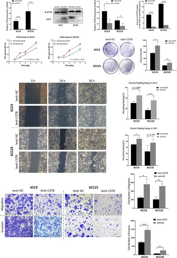
FIGURE 3 | Upregulation of CSTB inhibits the proliferation, migration, invasion and adhesion of OSCC cell lines. (A) Relative expression of CSTB mRNA in OSCC
cell lines after lentivirus transfection. (B, C) CSTB protein expression in OSCC cell lines after lentivirus transfection was identified by Western blot analysis (B) and
quantitatively analyzed (C). (D) The content of CSTB in the culture supernatant of OSCC cell lines after lentivirus transfection was quantified by ELISA. (E, F) CCK8
assays for transfected OSCC cell lines. (G, H) Colony formation assays for transfected OSCC cell lines. (G) The representative staining images of each group; (H) the
number of clones formed in each group. (I–K) Wound-healing assays for transfected OSCC cell lines. Representative images in (I) and statistical results in (J, K) at 0,
24, and 36 h in each group. (L–N) Transwell assays for transfected OSCC cell lines. Representative images for cell migration and invasion of each group are shown
in (L). Statistical results for cell migration and invasion are shown in (M, N), respectively. Error bars represent the standard deviation. *p < 0.05, **p < 0.01,
***p < 0.001, ****p < 0.0001.
Frontiers in Oncology | www.frontiersin.org 8 August 2021 | Volume 11 | Article 707066Xu et al. Cystatin-B in OSCC
A B
C
D
F
E
G H I J
FIGURE 4 | CSTB may regulate epithelial cell differentiation and keratinization in OSCC. (A) Heatmap of the correlation between module eigengenes (vertical axis) and
clinical features (horizontal axis). The correlation coefficient (upper row) and p-value (lower row) are presented in each block. The color of each block represents the
correlation coefficient according to the legend axis on the right. (B) Scatter diagram for the correlation between module membership and the gene significance of CSTB
expression in the brown module. Cor, correlation coefficient; p, p-value. (C) Scatter diagram for the correlation between module membership and gene significance of
grade classification in the brown module. Cor, correlation coefficient; p, p-value. (D, E) GO enrichment analysis of the brown module genes. (F) Squamous epithelial
keratinization- and differentiation-related pathways with statistically significant differences in GSEA using KEGG enrichment analysis. (G, H) Relative mRNA expression of
cell differentiation and keratinization-related markers in OSCC cell lines after lentivirus transfection. (I, J) Protein expression of cell differentiation and keratinization-related
markers in OSCC cell lines after lentivirus transfection. Error bars represent the standard deviation. *p < 0.05, **p < 0.01, ***p < 0.001, ****p < 0.0001.
Finally, cell differentiation/keratinization-related markers overexpressing CSTB (Figures 4G–J). It was found that
(i.e., IVL, LOR, KRT1, KRT5, KRT10, and KRT14) were upregulation of CSTB promoted the expression of these
validated at the protein and gene levels in OSCC models markers, reflecting a tendency for differentiation and
Frontiers in Oncology | www.frontiersin.org 9 August 2021 | Volume 11 | Article 707066Xu et al. Cystatin-B in OSCC
keratinization. In summary, CSTB might regulate the epithelial (32, 59) and laryngeal squamous cell carcinoma (33) (another
proliferation/differentiation program in OSCC. certain kind of HNSCC). To systematically compare the
expression of CSTB in common tumor types, the GEPIA
RNA Sequencing Derived Pathways/Genes database (60) was used to show the difference in CSTB
of Interest Associated With CSTB in OSCC expression between the tumor and normal groups (Figure S4):
In addition to keratinization- and differentiation-related (1). CSTB was downregulated in HNSCC, which was consistent
pathways, other pathways of interest that CSTB may with the findings of our study and a previous study on HNSCC
participate in were also identified. A comparison was made (i.e., laryngeal squamous cell carcinoma) (33) (2). CSTB was
between the GSEA enrichment results of RNA-seq data (from upregulated in most carcinomas (i.e., malignant tumors of
the OSCC models overexpressing CSTB) and existing public epithelial origin), except for HNSCC and ESCC. This radically
database data (Supplementary Data Sheets 3–4). Three gene different trend of expression may be partly explained by the
sets (i.e., apical junction, G2/M checkpoint and mitotic spindle) different histological origins of these epithelial-derived
and six pathways (i.e., glycosaminoglycan degradation, tumors. Among these carcinomas, HNSCC and ESCC share
homologous recombination, mismatch repair, NOTCH a common histological origin (i.e., stratified squamous
signaling pathway, nucleotide excision repair, and steroid epithelium), while other carcinomas originate from simple
biosynthesis) were enriched in the data from both sources columnar epithelium (e.g., cholangiocarcinoma, colon
(Figures 5A, C–F). These nine pathways/gene sets were named adenocarcinoma, cervical squamous cell carcinoma, and
as shared pathways/gene sets. In the GSEA results for the cell line uterine corpus endometrial carcinoma) or glandular epithelium
RNA-seq data, the core enrichment genes in the above shared (e.g., pancreatic adenocarcinoma, rectum adenocarcinoma, and
pathways/gene sets were used to compare with 256 DEGs in stomach adenocarcinoma). In addition, the pleiotropic roles of
order to obtain crossover genes (Table 3, No. 1–12), suggesting CSTB in tumors may also contribute to this inconsistent trend.
that overexpression of CSTB may be involved in the above For instance, knockdown of CSTB in an epithelial ovarian cancer
shared pathways by regulating these 12 crossover genes. cell line inhibited cell proliferation and promoted apoptosis (29),
In addition to exploration at the pathway level, RNA-seq data whereas the opposite result was found in a gastric cancer cell line
were used for comparisons with the existing recognized (61) (3). The same expression trend of CSTB in HNSCC and
mutational cancer driver genes. Among the 256 DEGs, five ESCC could be attributed to the similarity of these two tumors.
genes (i.e., CEBPA, EGR2, MEF2B, SOCS1, and SP140) were First, they are both located in the proximal digestive tract and
recognized as mutated cancer driver genes (Figure 5B). originate from stratified squamous epithelium, sharing some
Overexpression of CSTB can upregulate EGR2 and MEF2B similar histological characteristics under physiological and
while downregulating CEBPA, SOCS1, and SP140 in OSCC pathological (i.e., appearance of keratin pearls in tumor status)
cell lines (Table 3, No. 13–17). conditions (62). Second, the histological distributions of CSTB
in HNSCC and ESCC are also consistent, that is, it is located in
the epithelial structure of carcinoma tissues (59) (Figure 2I).
Under physiological conditions, CSTB is located in the epithelial
DISCUSSION layer of both normal oral cavity tissues and esophageal
tissues (Figures 2I and S5). Third, similarities in genomic
The roles of CSTB, a cysteine cathepsin inhibitor associated with characterization between ESCC and HNSCC may also lead to
various cancers, have been poorly understood, and few studies have the same expression trend of CSTB (7, 63–67). There are some
focused on its association with HNSCC. The present study focused groups of gene sets that mutate in both ESCC and HNSCC,
on OSCC and explored the regulatory roles of CSTB in vivo and in which indicates that common mechanisms can regulate the
vitro with both experimental and bioinformatic data. The findings initiation of squamous cell carcinoma (SCC) across different
supported our hypothesis and showed that CSTB was tissues (7, 8, 62, 64, 68–72).
downregulated in OSCC compared with normal tissues, and this As the expression trend of CSTB differs from tumor to tumor,
downregulation was related to worse clinical outcomes or signs of the relationship between CSTB and prognosis is also inconsistent in
tumor malignancy. The promoting role of CSTB in the OSCC different tumor types. Elevated levels of CSTB in tumor samples
proliferation-differentiation program was unprecedentedly were related to a higher risk of recurrence and advanced tumor
proposed. In addition, some relevant mechanisms by which CSTB stages in bladder cancer (34) and were linked to a higher risk of
might participate in OSCC were suggested at both the pathway and tumor metastasis in HCC (27). In our study on OSCC, CSTB
gene levels. expression was inversely related to the risk of aggressive tumor
In OSCC, we found that CSTB was downregulated both in characteristics (i.e., lymph node metastasis, perineural infiltration,
vivo and in vitro, both at the protein and gene levels. CSTB has and lymphovascular invasion). These characteristics favor tumor
been reported to have abnormal expression in various types of recurrence and metastasis (73) and pose great challenges to the
tumors, in which the variation trend of CSTB was inconsistent tumor-free goal of radical surgery (73–79). Enhanced cancer cell
between different tumors. For instance, CSTB seems to be migration and the capacity of stromal infiltration are essential steps
upregulated in HCC (25–28), epithelial ovarian tumors (29, of OSCC invasion and metastasis, as is the proliferation of cancer
30), and breast cancer (31), but downregulated in ESCC cells in metastatic foci (80). These three abilities of OSCC cells were
Frontiers in Oncology | www.frontiersin.org 10 August 2021 | Volume 11 | Article 707066Xu et al. Cystatin-B in OSCC
A B
C D
E F
FIGURE 5 | CSTB may participate in OSCC through other tumor-related pathways. (A) The process and results of GSEA based on data from the TCGA database/
cell lines mRNA sequencing. (B) Comparison between mutational cancer driver genes and DEGs obtained from cell line mRNA sequencing. (C, D) GSEA using
Hallmark gene sets. Gene sets with statistically significant differences based on data from the TCGA database/cell line mRNA sequencing. (E, F) GSEA using KEGG
enrichment analysis. Pathways with both statistically significant differences based on data from the TCGA database/cell line mRNA sequencing.
Frontiers in Oncology | www.frontiersin.org 11 August 2021 | Volume 11 | Article 707066Xu et al. Cystatin-B in OSCC
TABLE 3 | Highlighted genes out of 256 differentially expressed genes.
Items No. Gene Fold p- Enriched pathway/ Description in NCBI Reported in
change Value gene sets HNSCC
Enriched in KEGG pathways/Hallmark 1 MAML3 1.84Xu et al. Cystatin-B in OSCC increased, which is usually not seen in healthy epidermis (92) NOTCH signaling pathway by regulating these two genes, but (i.e., normal skin). The regulatory mechanisms of CSTB in additional validation is required. squamous epithelial keratinization and differentiation An interesting phenomenon was found in our research: the programs are not yet clear. There might be at least two content of extracellular/intracellular CSTB showed a consistent possible mechanisms, i.e., by affecting the formation of change trend in the in vitro experiment (Figures 2M, D). CSTB cornified cell envelopes (CE) or by regulating the NOTCH is regarded as a protease inhibitor that is localized in the signaling pathway. First, the terminal differentiation of intracellular region (18, 23, 24). However, many studies have squamous epithelium is a process accompanied by the shown that CSTB and its changes can be detected in the body formation of CE (95), which is mediated and regulated by fluids of cancer patients [e.g., serum (28), ascites (109), and urine TGM1 [transglutaminase-1 (TGase-1)] (96, 97). In our study, (34)] and are related to the clinical characteristics or prognosis. the CE was enriched as a cell component in GO analysis in the Given the consistency of the anatomical location, saliva as a body brown module in WGCNA (Figure 4E) and TGM1 was a fluid may be more representative for reflecting the disease state of significant hub gene in the brown module (module OSCC. A study of OSCC emphasized the potential of CSTB- membership = 0.85, module eigengene = 0.65). Given that the specific peptides in saliva to reflect the status of lymph node brown module was a gene module containing genes coexpressed metastasis in tumor-bearing patients (35). Although our study with CSTB, these results suggested that CSTB might regulate the found differences in the content of CSTB in normal/OSCC cell TGM1-mediated CE formation process and ultimately regulate culture supernatants, whether CSTB could be used to specifically the terminal differentiation of squamous epithelium. Second, identify tumor patients from the normal population is worthy of NOTCH is mostly regarded as a tumor suppressor in HNSCC, further research. Furthermore, a study showed the presence of and loss-of-function mutations in NOTCH1 are common events CSTB in oral acquired enamel pellicles (110, 111). Future in HNSCC. The NOTCH signaling pathway plays an important research may consider the changes in CSTB in plaque as a role in the normal functioning of squamous epithelium novel research direction in terms of the potential of CSTB as a development and differentiation (7, 62, 67, 98–100), acting as a biomarker, given that plaque could be regarded as a more stable promoter of keratinocyte differentiation (101, 102). Conditional form reflecting the biochemical composition of saliva. Other deletion of NOTCH1 in the mouse epidermis can lead to basal nontumor studies have identified certain extracellular effects of hyperplasia and basal cell carcinoma (103). Our GSEA results CSTB. CSTB in mouse synaptosomes can be secreted into based on cell line RNA-seq data were consistent with this cerebral spinal fluid in a depolarization-controlled manner and downregulation trend of NOTCH signaling. The NOTCH is involved in synaptic plasticity (84, 112). HIV-infected signaling pathway became more active after CSTB microglia secret more CSTB, participating in the neurotoxicity overexpression, which was also in line with the differentiation- induced by cathepsin B (113). These studies suggest that promoting effect of CSTB that we have observed. However, this pathological signals (tumor, virus, and abnormal nerve signals) alteration in the NOTCH signaling pathway showed an opposite may cause CSTB to translocate and participate in extracellular trend (i.e., more active in the low CSTB expression group) based biological processes, while its specific extracellular role in tumors on patient RNA-seq data from TCGA. This could be explained needs to be clarified by more tracer studies. by the contextual and bimodal role of NOTCH in cancers, Other roles of CSTB in the context of OSCC were explored at including HNSCC (64, 67, 104–106). NOTCH can act as an both the pathway and gene levels in our study, which might oncogene and tumor suppressor gene in different cell provide a comprehensive perspective for further elucidating its populations within the same tumor (103), which was a possible mechanism. First, pathways related to the DNA damage repair reason for the difference in NOTCH signals in the data from two mechanism (G2/M checkpoint, mitotic spindle, homologous sources (TCGA data and cell line RNA seq data). The cell line recombination, mismatch repair, and nucleotide excision RNA-seq data came from a single type of tumor cell (OSCC cell repair) were more active in the low CSTB expression group. line), while the tissue sequencing data of TCGA were composed This finding indicated that downregulation of CSTB might act as of a variety of cells including tumor cells, stromal cells, immune a predisposing factor of DNA damage in OSCC, thereby cells, etc. Another possible reason for the different NOTCH activating the pathways related to DNA damage repair signals is that the involved NOTCH paralogues and ligands vary pathways. Many studies have emphasized the protective effect in different disease contexts, resulting in different biological of CSTB against oxidative stress (31, 114, 115). CSTB deficiency outcomes (107). For instance, in GSEA, NOTCH1, and increases the sensitivity of cells to oxidative stress in cerebellar NOTCH3 were enriched in the high CSTB expression group granule neurons (114) or breast cancer primary cells (31). Given based on cell line RNA-seq data, while NOTCH1 and NOTCH4 that oxidative stress is a common factor that causes DNA were enriched in the low CSTB expression group based on damage (116, 117), we speculate that the effect of CSTB on the TCGA data (Supplementary Data Sheet 4). Studies have DNA damage repair mechanism in OSCC might be related to identified a bimodal role for NOTCH3 in HNSCC (39), while the loss of the protective role of CSTB in oxidative stress. Second, MAML3 is essential for the operation of NOTCH signaling the identification of all the mutated genes capable of driving (108). In the RNA-seq data, we found that overexpression of tumors is a landmark achievement towards tumor research (38). CSTB could upregulate the expression of NOTCH3 and MAML3 To explore the correlation between CSTB and the known cancer in vitro (Table 3), suggesting that CSTB may be involved in the driver genes, we compared the DEGs obtained by cell line mRNA Frontiers in Oncology | www.frontiersin.org 13 August 2021 | Volume 11 | Article 707066
Xu et al.                                                                                                                       Cystatin-B in OSCC

sequencing with the cancer driver gene set. As a result, five genes,           differentiation process of OSCC, but it is not the only factor
CEBPA, EGR2, MEF2B, SOCS1, and SP140, were identified.                         determining the degree of tumor differentiation.
CEBPA was reported to be mutated in HNSCC (51) and to play                        In conclusion, our study identified the modulatory role of
an important role in regulating the epidermal differentiation                 CSTB in the malignant characteristics of OSCC for the first time
program (51, 52, 118–121). SP140 is an immune-related gene                    and proposed some new relevant mechanisms, namely, CSTB
mutated in OSCC cell lines (57, 58, 122, 123). SOCS1, a negative              may participate in OSCC by promoting the differentiation
regulator of cytokine signaling pathways (55, 124–126), was                   process of squamous epithelium. Our research provides a new
reported to regulate epigenetic modification in head and neck                  perspective for interpreting the role of CSTB in tumors,
cancer (56, 125, 127). Cumulative studies have suggested the role             especially in SCC. The specific role of CSTB in OSCC and its
of CSTB in immunomodulation, especially the relationship                      fine regulation of the squamous epithelial differentiation
between CSTB and macrophages (128, 129). The lack of CSTB                     program still need to be explored in-depth by functional
could transform macrophages to a proinflammatory phenotype                     experiments and mechanistic research.
by regulating related cytokines such as IL-10 (128). Meanwhile,
some immune-related pathways (e.g., antigen processing and
presentation and primary immunodeficiency) were enriched by
GSEA based on cell RNA-seq data in our study (Supplementary                   DATA AVAILABILITY STATEMENT
Data Sheet 3). Both EGR2 and MEF2B are regulators of cell
                                                                              The original contributions presented in the study are included in
transcription (130, 131), and the former was reported to be
                                                                              the article/Supplementary Material. Further inquiries can be
involved in the epithelial-mesenchymal transition (EMT)
                                                                              directed to the corresponding authors.
pathway (131, 132), while the latter was differentially expressed
between primary tumors and nodal metastasis tumor in HNSCC
(53). Our experiments did not identify genes that are mutated
due to changes in the expression level of CSTB, although changes              ETHICS STATEMENT
in the expression level of the transcriptome are a common
consequence of gene mutations. However, our aim was to                        Studies involving human participants were reviewed and
interpret the possible role of CSTB in cancer at the genetic                  approved by the Ethics Committee of the Stomatology
level, and the results still provide innovative insights while                Hospital of Guangzhou Medical University (LCYJ2021013).
exploring the functions of CSTB in OSCC.                                      The patients/participants provided their written informed
    There were some limitations in our study. First, the                      consent to participate in this study. No potentially identifiable
correlations between Cstb and clinical characteristics found                  human images or data are presented in this study.
through data mining methods have not been confirmed by
additional independent clinical data. The findings should be
validated at the protein levels in tissues and body fluid (e.g., saliva
and blood). Second, the regulatory role of CSTB in the
                                                                              AUTHOR CONTRIBUTIONS
epithelium differentiation phenotype was mainly recognized                    TX contributed to the conception and design of the study. TX
through bioinformatic analysis and in vitro experiments and                   and LY performed the experiments. TX, XZ, XW, and LY
needs in vivo confirmation in future studies. Third, the regulated             performed the statistical analysis. TX wrote the first draft of
pathways/genes identified by GSEA provided a relatively                        the manuscript. TX, TY, and GL critically revised the
comprehensive perspective in clarifying the role of CSTB in                   manuscript. All authors contributed to the article and
OSCC. However, the relevant pathways/genes lacked targeted                    approved the submitted version.
validation, so they should be interpreted with caution. Fourth,
the sample size of OSCC tissue for IHC was relatively small,
which was why we used Fisher’s exact test. In the subgroup
analysis, the subgroup satisfying both high CSTB expression and               FUNDING
moderate/poor differentiation included only one sample, which
may be partly due to our relatively small overall sample size.                This study was supported by the Health Committee of
Given the relationship of CSTB and differentiation in our study               Guangdong Province (Grant No. B2020209), Guangzhou
on OSCC, samples that meet both the phenotypes of high CSTB                   Medical University (Grant No. C195015024), and the National
expression and low differentiation were relatively rare, which                Natural Science Foundation of China (Grant No. 81700985).
may be another reason for the small sample size of this subgroup.
However, this state [i.e., high CSTB expression and poor tissue
differentiation) is not uncommon in some other tumor types                    SUPPLEMENTARY MATERIAL
(e.g., HCC (27) and bladder cancer (34)], where the degree of
tumor differentiation and CSTB expression show a negative                     The Supplementary Material for this article can be found online
correlation. This finding may further indicate that, compared                  at: https://www.frontiersin.org/articles/10.3389/fonc.2021.
with other tumor types, CSTB has a special additional role in the             707066/full#supplementary-material

Frontiers in Oncology | www.frontiersin.org                              14                                August 2021 | Volume 11 | Article 707066
Xu et al. Cystatin-B in OSCC
REFERENCES Myoclonus Epilepsy, EPM1, Mutations. Europ J Hum Genet (2005) 13:208–
15. doi: 10.1038/sj.ejhg.5201300
1. Johnson DE, Burtness B, Leemans CR, Lui VWY, Bauman JE, Grandis JR. 23. Turk V, Stoka V, Turk D. Cystatins: Biochemical and Structural Properties,
Head and Neck Squamous Cell Carcinoma. Nat Rev Dis Primers (2020) 6:92. and Medical Relevance. Front Biosci (2008) 13:5406–20. doi: 10.2741/3089
doi: 10.1038/s41572-020-00224-3 24. Kos J, Werle B, Lah T, Brunner N. Cysteine Proteinases and Their Inhibitors
2. Bray F, Ferlay J, Soerjomataram I, Siegel RL, Torre LA, Jemal A. Global in Extracellular Fluids: Markers for Diagnosis and Prognosis in Cancer. Int J
Cancer Statistics 2018: GLOBOCAN Estimates of Incidence and Mortality Biol Markers (2000) 15:84–9. doi: 10.1177/172460080001500116
Worldwide for 36 Cancers in 185 Countries. CA Cancer J Clin (2018) 25. Zhou X, Wang X, Huang K, Liao X, Yang C, Yu T, et al. Investigation of the
68:394–424. doi: 10.3322/caac.21492 Clinical Significance and Prospective Molecular Mechanisms of Cystatin
3. Ferlay J, Colombet M, Soerjomataram I, Mathers C, Parkin DM, Piñeros M, Genes in Patients With Hepatitis B Virus−Related Hepatocellular
et al. Estimating the Global Cancer Incidence and Mortality in 2018: Carcinoma. Oncol Rep (2019) 42:189–201. doi: 10.3892/or.2019.7154
GLOBOCAN Sources and Methods. Int J Cancer (2019) 144:1941–53. 26. Liu GM, Zeng HD, Zhang CY, Xu JW. Identification of a Six-Gene Signature
doi: 10.1002/ijc.31937 Predicting Overall Survival for Hepatocellular Carcinoma. Cancer Cell Int
4. Chi AC, Day TA, Neville BW. Oral Cavity and Oropharyngeal Squamous Cell (2019) 19:138. doi: 10.1186/s12935-019-0858-2
Carcinoma–An Update. CA Cancer J Clin (2015) 65:401–21. doi: 10.3322/ 27. Lin YY, Chen ZW, Lin ZP, Lin LB, Yang XM, Xu LY, et al. Tissue Levels of
caac.21293 Stefin A and Stefin B in Hepatocellular Carcinoma. Anat Rec (Hoboken)
5. National Cancer Institute (NCI) Website. Available at: https://www.cancer.gov. (2016) 299:428–38. doi: 10.1002/ar.23311
6. Osazuwa-Peters N, Simpson MC, Zhao L, Boakye EA, Olomukoro SI, 28. Lee MJ, Yu GR, Park SH, Cho BH, Ahn JS, Park HJ, et al. Identification of
Deshields T, et al. Suicide Risk Among Cancer Survivors: Head and Neck Cystatin B as a Potential Serum Marker in Hepatocellular Carcinoma. Clin
Versus Other Cancers. Cancer (2018) 124:4072–9. doi: 10.1002/cncr.31675 Cancer Res (2008) 14:1080–9. doi: 10.1158/1078-0432.Ccr-07-1615
7. Dotto GP, Rustgi AK. Squamous Cell Cancers: A Unified Perspective on 29. Guan W, Wang X, Lin Q, Zhang J, Ren W, Xu G. Transforming
Biology and Genetics. Cancer Cell (2016) 29:622–37. doi: 10.1016/ Growth Factor−b/Mir−143−3p/Cystatin B Axis Is a Therapeutic Target in
j.ccell.2016.04.004 Human Ovarian Cancer. Int J Oncol (2019) 55:267–76. doi: 10.3892/ijo.
8. Leemans CR, Snijders PJF, Brakenhoff RH. The Molecular Landscape of 2019.4815
Head and Neck Cancer. Nat Rev Cancer (2018) 18:269–82. doi: 10.1038/ 30. Wang X, Gui L, Zhang Y, Zhang J, Shi J, Xu G. Cystatin B Is a Progression
nrc.2018.11 Marker of Human Epithelial Ovarian Tumors Mediated by the TGF-b Signaling
9. Choi S, Myers JN. Molecular Pathogenesis of Oral Squamous Cell Carcinoma: Pathway. Int J Oncol (2014) 44:1099–106. doi: 10.3892/ijo.2014.2261
Implications for Therapy. J Dent Res (2008) 87:14–32. doi: 10.1177/ 31. Butinar M, Prebanda MT, Rajković J, Jerič B, Stoka V, Peters C, et al. Stefin B
154405910808700104 Deficiency Reduces Tumor Growth via Sensitization of Tumor Cells to
10. Thomson PJ. Perspectives on Oral Squamous Cell Carcinoma Prevention— Oxidative Stress in a Breast Cancer Model. Oncogene (2014) 33:3392–400.
Proliferation, Position, Progression and Prediction. J Oral Pathol Med (2018) doi: 10.1038/onc.2013.314
47:803–7. doi: 10.1111/jop.12733 32. Zhang J, Wang K, Zhang J, Liu SS, Dai L, Zhang JY. Using Proteomic
11. Pai SI, Westra WH. Molecular Pathology of Head and Neck Cancer: Approach to Identify Tumor-Associated Proteins as Biomarkers in Human
Implications for Diagnosis, Prognosis, and Treatment. Annu Rev Pathol Esophageal Squamous Cell Carcinoma. J Proteome Res (2011) 10(6):2863–
(2009) 4:49–70. doi: 10.1146/annurev.pathol.4.110807.092158 72. doi: 10.1021/pr200141c
12. Umelo IA, Costanza B, Castronovo V. Innovative Methods for Biomarker 33. Colombo J, Fachel AA, De Freitas Calmon M, Cury PM, Fukuyama EE,
Discovery in the Evaluation and Development of Cancer Precision Tajara EH, et al. Gene Expression Profiling Reveals Molecular Marker
Therapies. Cancer Metastasis Rev (2018) 37:125–45. doi: 10.1007/s10555- Candidates of Laryngeal Squamous Cell Carcinoma. Oncol Rep (2009)
017-9710-0 21:649–63.
13. Ji K, Mayernik L, Moin K, Sloane BF. Acidosis and Proteolysis in the Tumor 34. Feldman AS, Banyard J, Wu CL, McDougal WS, Zetter BR. Cystatin B as a
Microenvironment. Cancer Metastasis Rev (2019) 38:103–12. doi: 10.1007/ Tissue and Urinary Biomarker of Bladder Cancer Recurrence and Disease
s10555-019-09796-3 Progression. Clin Cancer Res (2009) 15:1024–31. doi: 10.1158/1078-
14. Osuala KO, Ji K, Mattingly RR, Sloane BF. Breast Cancer: Proteolysis and 0432.Ccr-08-1143
Migration. Adv Exp Med Biol (2019) 1152:401–11. doi: 10.1007/978-3-030- 35. Carnielli CM, Macedo CCS, De Rossi T, Granato DC, Rivera C, Domingues
20301-6_21 RR, et al. Combining Discovery and Targeted Proteomics Reveals a
15. Ni J, Zhang L. Cancer Cachexia: Definition, Staging, and Emerging Prognostic Signature in Oral Cancer. Nat Commun (2018) 9:3598.
Treatments. Cancer Manag Res (2020) 12:5597–605. doi: 10.2147/cmar. doi: 10.1038/s41467-018-05696-2
S261585 36. EI-Naggar AK, Chan JKC, G randis JR, Takata T, Slootweg PJ. WHO
16. Li X, Song Y. Proteolysis-Targeting Chimera (PROTAC) for Targeted Classification of Head and Neck Tumours. 4th edition. IA RC: Lyon (2017).
Protein Degradation and Cancer Therapy. J Hematol Oncol (2020) 13:50. 37. Livak KJ, Schmittgen TD. Analysis of Relative Gene Expression Data Using
doi: 10.1186/s13045-020-00885-3 Real-Time Quantitative PCR and the 2(-Delta Delta C(T)) Method. Methods
17. Rudziń ska M, Parodi A, Soond SM, Vinarov AZ, Korolev DO, Morozov AO, (2001) 25:402–8. doi: 10.1006/meth.2001.1262
et al. The Role of Cysteine Cathepsins in Cancer Progression and Drug 38. Martinez-Jimenez F, Muinos F, Sentis I, Deu-Pons J, Reyes-Salazar I,
Resistance. Int J Mol Sci (2019) 20(14):3602. doi: 10.3390/ijms20143602 Arnedo-Pac C, et al. A Compendium of Mutational Cancer Driver Genes.
18. Pogorzelska A, Ż ołnowska B, Bartoszewski R. Cysteine Cathepsins as a Nat Rev Cancer (2020) 20:555–72. doi: 10.1038/s41568-020-0290-x
Prospective Target for Anticancer Therapies-Current Progress and 39. Fukusumi T, Califano JA. The NOTCH Pathway in Head and Neck
Prospects. Biochimie (2018) 151:85–106. doi: 10.1016/j.biochi.2018.05.023 Squamous Cell Carcinoma. J Dent Res (2018) 97(6):645–53. doi: 10.1177/
19. Shamsi A, Bano B. Journey of Cystatins From Being Mere Thiol Protease 0022034518760297
Inhibitors to at Heart of Many Pathological Conditions. Int J Biol Macromol 40. Shah PA, Huang C, Li Q, Kazi SA, Byers LA, Wang J, et al. NOTCH1
(2017) 102:674–93. doi: 10.1016/j.ijbiomac.2017.04.071 Signaling in Head and Neck Squamous Cell Carcinoma. Cells (2020) 9
20. Olson OC, Joyce JA. Cysteine Cathepsin Proteases: Regulators of Cancer (12):2677. doi: 10.3390/cells9122677
Progression and Therapeutic Response. Nat Rev Cancer (2015) 15:712–29. 41. Zhao YY, Yu GT, Xiao T, Hu J. The Notch Signaling Pathway in Head and
doi: 10.1038/nrc4027 Neck Squamous Cell Carcinoma: A Meta-Analysis. Adv Clin Exp Med (2017)
21. Breznik B, Mitrović A, Lah TT, Kos J. Cystatins in Cancer Progression: More 26(5):881–7. doi: 10.17219/acem/64000
Than Just Cathepsin Inhibitors. Biochimie (2019) 166:233–50. doi: 10.1016/ 42. Xu X, Tassone B, Ostano P, Katarkar A, Proust T, Joseph JM, et al. HSD17B7
j.biochi.2019.05.002 Gene in Self-Renewal and Oncogenicity of Keratinocytes From Black Versus
22. Alakurtti K, Weber E, Rinne R, Theil G, de Haan GJ, Lindhout D, et al. Loss White Populations. EMBO Mol Med (2021) 13:e14133. doi: 10.15252/
of Lysosomal Association of Cystatin B Proteins Representing Progressive emmm.202114133
Frontiers in Oncology | www.frontiersin.org 15 August 2021 | Volume 11 | Article 707066Xu et al. Cystatin-B in OSCC
43. Tilston-Lünel AM, Haley KE, Schlecht NF, Wang Y, Chatterton ALD, Cancer (1998) 79:175–8. doi: 10.1002/(sici)1097-0215(19980417)79:23.0.co;2-9
Dependent Manner in Mammalian Cells. J Mol Cell Biol (2016) 8:439–55. 60. Tang Z, Li C, Kang B, Gao G, Li C, Zhang Z. GEPIA: A Web Server for
doi: 10.1093/jmcb/mjw020 Cancer and Normal Gene Expression Profiling and Interactive Analyses.
44. Son SH, Park J, Jung MJ, Lee SK, Kim H, Kim KR, et al. Transforming Nucleic Acids Res (2017) 45:W98–W102. doi: 10.1093/nar/gkx247
Growth Factor-b-Regulated Fractalkine as a Marker of Erosive Bone 61. Zhang J, Shi Z, Huang J, Zou X. CSTB Downregulation Promotes Cell
Invasion in Oral Squamous Cell Carcinoma. Eur J Oral Sci (2021) 129: Proliferation and Migration and Suppresses Apoptosis in Gastric Cancer
e12750. doi: 10.1111/eos.12750 SGC-7901 Cell Line. Oncol Res (2016) 24:487–94. doi: 10.3727/
45. Merhi M, Raza A, Inchakalody VP, Nashwan AJJ, Allahverdi N, 096504016x14685034103752
Krishnankutty R, et al. Squamous Cell Carcinomas of the Head and Neck 62. Sanchez-Danes A, Blanpain C. Deciphering the Cells of Origin of Squamous
Cancer Response to Programmed Cell Death Protein-1 Targeting and Cell Carcinomas. Nat Rev Cancer (2018) 18:549–61. doi: 10.1038/s41568-
Differential Expression of Immunological Markers: A Case Report. Front 018-0024-5
Immunol (2018) 9:1769. doi: 10.3389/fimmu.2018.01769 63. Grandis JR. Comprehensive Genomic Characterization of Head and Neck
46. Gyanchandani R, Ortega Alves MV, Myers JN, Kim S. A Proangiogenic Squamous Cell Carcinomas. Clin Cancer Res (2017) 23. doi: 10.1158/1557-
Signature Is Revealed in FGF-Mediated Bevacizumab-Resistant Head and 3265.AACRAHNS17-IA09
Neck Squamous Cell Carcinoma. Mol Cancer Res (2013) 11:1585–96. 64. Agrawal N, Frederick MJ, Pickering CR, Bettegowda C, Chang K, Li RJ, et al.
doi: 10.1158/1541-7786.Mcr-13-0358 Exome Sequencing of Head and Neck Squamous Cell Carcinoma Reveals
47. Khanal S, Strickley JD, Ha T, Demehri S, Ghim SJ, Jenson AB, et al. Human Inactivating Mutations in NOTCH1. Science (2011) 333:1154–7.
Papillomavirus-Positivity Is Associated With EREG Down-Regulation and doi: 10.1126/science.1206923
Promoter Hypermethylation in Head and Neck Squamous Cell Carcinoma. 65. Gao YB, Chen ZL, Li JG, Hu XD, Shi XJ, Sun ZM, et al. Genetic Landscape of
Exp Mol Pathol (2020) 117:104549. doi: 10.1016/j.yexmp.2020.104549 Esophageal Squamous Cell Carcinoma. Nat Genet (2014) 46:1097–102. doi:
48. Xu Y, Xu F, Lv Y, Wang S, Li J, Zhou C, et al. A ceRNA-Associated Risk 10.1038/ng.3076
Model Predicts the Poor Prognosis for Head and Neck Squamous Cell 66. Lin DC, Hao JJ, Nagata Y, Xu L, Shang L, Meng X, et al. Genomic and
Carcinoma Patients. Sci Rep (2021) 11:6374. doi: 10.1038/s41598-021- Molecular Characterization of Esophageal Squamous Cell Carcinoma. Nat
86048-x Genet (2014) 46:467–73. doi: 10.1038/ng.2935
49. Shaikh I, Ansari A, Ayachit G, Gandhi M, Sharma P, Bhairappanavar S, et al. 67. Stransky N, Egloff AM, Tward AD, Kostic AD, Cibulskis K, Sivachenko A,
Differential Gene Expression Analysis of HNSCC Tumors Deciphered et al. The Mutational Landscape of Head and Neck Squamous Cell
Tobacco Dependent and Independent Molecular Signatures. Oncotarget Carcinoma. Science (2011) 333(6046):1157–60. doi: 10.1126/
(2019) 10:6168–83. doi: 10.18632/oncotarget.27249 science.1208130
50. Wang L, Jia Y, Jiang Z, Gao W, Wang B. FSCN1 Is Upregulated by SNAI2 and 68. Cancer Genome Atlas Research Network, Analysis Working Group: Asan
Promotes Epithelial to Mesenchymal Transition in Head and Neck Squamous University, BC Cancer Agency, Brigham and Women’s Hospital, Broad
Cell Carcinoma. Cell Biol Int (2017) 41:833–41. doi: 10.1002/cbin.10786 Institute, Brown University, et al. Integrated Genomic Characterization of
51. Gaykalova DA, Mambo E, Choudhary A, Houghton J, Buddavarapu K, Oesophageal Carcinoma. Nature (2017) 541(7636):169–75. doi: 10.1038/
Sanford T, et al. Novel Insight Into Mutational Landscape of Head and Neck nature20805
Squamous Cell Carcinoma. PloS One (2014) 9:e93102. doi: 10.1371/ 69. Cancer Genome Atlas Network, Lawrence MS, Sougnez C, Lichtenstein L,
journal.pone.0093102 Cibulskis K, Lander E, et al. Comprehensive Genomic Characterization of
52. Bennett KL, Hackanson B, Smith LT, Morrison CD, Lang JC, Schuller DE, Head and Neck Squamous Cell Carcinomas. Nature (2015) 517:576–82.
et al. Tumor Suppressor Activity of CCAAT/enhancer Binding Protein doi: 10.1038/nature14129
Alpha Is Epigenetically Down-Regulated in Head and Neck Squamous 70. Chen YP, Wang YQ, Lv JW, Li YQ, Chua MLK, Le QT, et al. Identification
Cell Carcinoma. Cancer Res (2007) 67:4657–64. doi: 10.1158/0008- and Validation of Novel Microenvironment-Based Immune Molecular
5472.Can-06-4793 Subgroups of Head and Neck Squamous Cell Carcinoma: Implications for
53. Liu CJ, Liu TY, Kuo LT, Cheng HW, Chu TH, Chang KW, et al. Differential Immunotherapy. Ann Oncol (2019) 30:68–75. doi: 10.1093/annonc/mdy470
Gene Expression Signature Between Primary and Metastatic Head and Neck 71. Song Y, Li L, Ou Y, Gao Z, Li E, Li X, et al. Identification of Genomic
Squamous Cell Carcinoma. J Pathol (2008) 214:489–97. doi: 10.1002/ Alterations in Oesophageal Squamous Cell Cancer. Nature (2014) 509:91–5.
path.2306 doi: 10.1038/nature13176
54. Liu CJ, Lin SC, Chen YJ, Chang KM, Chang KW. Array-Comparative 72. Pickering CR, Zhang J, Yoo SY, Bengtsson L, Moorthy S, Neskey DM, et al.
Genomic Hybridization to Detect Genomewide Changes in Microdissected Integrative Genomic Characterization of Oral Squamous Cell Carcinoma
Primary and Metastatic Oral Squamous Cell Carcinomas. Mol Carcinog Identifies Frequent Somatic Drivers. Cancer Discovery (2013) 3:770–81.
(2006) 45:721–31. doi: 10.1002/mc.20213 doi: 10.1158/2159-8290.CD-12-0537
55. Yao Y, Yan Z, Lian S, Wei L, Zhou C, Feng D, et al. Prognostic Value of Novel 73. Bakst RL, Glastonbury CM, Parvathaneni U, Katabi N, Hu KS, Yom SS.
Immune-Related Genomic Biomarkers Identified in Head and Neck Squamous Perineural Invasion and Perineural Tumor Spread in Head and Neck
Cell Carcinoma. J Immunother Cancer (2020) 8(2):e000444. doi: 10.1136/jitc- Cancer. Perineural Invasion Perineural Tumor Spread Head (2019)
2019-000444 103:1109–24. doi: 10.1016/j.ijrobp.2018.12.009
56. Lee TL, Yeh J, Van Waes C, Chen Z. Epigenetic Modification of SOCS-1 74. Mupparapu M, Shanti RM. Evaluation and Staging of Oral Cancer. Dent Clin
Differentially Regulates STAT3 Activation in Response to Interleukin-6 North Am (2018) 62:47–58. doi: 10.1016/j.cden.2017.08.003
Receptor and Epidermal Growth Factor Receptor Signaling Through JAK 75. Obinu A, Gavini E, Rassu G, Maestri M, Bonferoni MC, Giunchedi P.
and/or MEK in Head and Neck Squamous Cell Carcinomas. Mol Cancer Lymph Node Metastases: Importance of Detection and Treatment Strategies.
Ther (2006) 5:8–19. doi: 10.1158/1535-7163.Mct-05-0069 Expert Opin Drug Deliv (2018) 15:459–67. doi: 10.1080/17425247.2018.
57. Mo BY, Li GS, Huang SN, Wei ZX, Su YS, Dai WB, et al. Laryngeal 1446937
Squamous Cell Carcinoma: Potential Molecular Mechanism and Prognostic 76. Schmitd LB, Scanlon CS, D'Silva NJ. Perineural Invasion in Head and Neck
Signature Based on Immune-Related Genes. Med Sci Monit (2020) 26: Cancer. J Dent Res (2018) 97:742–50. doi: 10.1177/0022034518756297
e928185. doi: 10.12659/MSM.928185 77. Binmadi NO, Basile JR. Perineural Invasion in Oral Squamous Cell
58. Song Y, Pan Y, Liu J. The Relevance Between the Immune Response-Related Carcinoma: A Discussion of Significance and Review of the Literature.
Gene Module and Clinical Traits in Head and Neck Squamous Cell Carcinoma. Oral Oncol (2011) 47:1005–10. doi: 10.1016/j.oraloncology.2011.08.002
Cancer Manag Res (2019) 11:7455–72. doi: 10.2147/cmar.S201177 78. Chinn SB, Spector ME, Bellile EL, McHugh JB, Gernon TJ, Bradford CR,
59. Shiraishi T, Mori M, Tanaka S, Sugimachi K, Akiyoshi T. Identification of et al. Impact of Perineural Invasion in the Pathologically N0 Neck in Oral
Cystatin B in Human Esophageal Carcinoma, Using Differential Displays in Cavity Squamous Cell Carcinoma. Otolaryngol Head Neck Surg (2013)
Which the Gene Expression Is Related to Lymph-Node Metastasis. Int J 149:893–9. doi: 10.1177/0194599813506867
Frontiers in Oncology | www.frontiersin.org 16 August 2021 | Volume 11 | Article 707066You can also read