CYTOMORPHOLOGICAL ASPECTS OF SOME OTO-RHINO-LARINGOLOGY (O.R.L.) CANCERS IN THE CANINE AND FELINE
←
→
Page content transcription
If your browser does not render page correctly, please read the page content below
THE PUBLISHING HOUSE                                                                                                 MEDICINE
    OF THE ROMANIAN ACADEMY                                                                                          Research article

CYTOMORPHOLOGICAL ASPECTS OF SOME OTO-RHINO-LARINGOLOGY (O.R.L.)
              CANCERS IN THE CANINE AND FELINE

                                EMILIA BALINT1, NICOLAE MANOLESCU2 and D. LASTOFKA3

                            1
                             Veterinary Faculty of Medicine, Comparative Medicine Institute, Romania
                           Corresponding author: N. Manolescu, E-mail: manolescunicolae@yahoo.com

                                                     Received October 15, 2013

               In the canine and feline species from Romania, cancers located in oto-rhino-laringeal area have a
               dominant oto-rhino-laringologic distribution lacking noteworthy in the larynx area. These have a low-
               to average frequency, not exceeding 2 – 3%. We analyzed a number of 17 clinical cases of which
               14 in canine species and 3 in feline species. In these 17 clinical cases, after clinical and paraclinical
               examination which showed the presence of a tumor in ENT area, a cytomorphologic examination was
               performed by fine needle puncture or by saline lavage. The interpretation was accomplished under the
               microscope with a magnification ranging between 600 X and 1,000–2,000 X. We observed that
               cavum, nasal fossa, sinuses and middle ear malignant neoplasms evolve generally very fast and the
               therapy is ineffective. While both non-Hodgkin malignant lymphoma, and lymphoepithelioma can be
               identified in the humans in these anatomical areas, these cancers could not be identified in the
               investigated canine and feline. Also, the presence of tumoral virus Ebstein-Barr could not be
               identified. Between canine and feline investigated the sarcoma/carcinoma ratio was equal, but tumors
               of nervous origin were identified only in the canine species. Clinical, all these have the following
               common elements: areal distorsion implying specific symptomatology for the co-interested
               anatomical area; mucosa ulceration; development of an exuberant epithelium with a sessile or
               pediculate cauliflower-like appearance. In conclusion, the malignant neo-formation frequency is
               lower within general malignancy but the high frequency of some inflammatory and cancer-mimicking
               aspects in veterinary clinic requires correct positive and differential diagnosis. In order to properly
               deal with these demands, a bio-puncture from the affected area, and a cytomorphological examination
               must be performed.
               Key words: canine, feline, cytomorphology, ENT cancers.

                  INTRODUCTION1                                        average frequency, not exceeding 2–3% (Emilia
                                                                       Balint, N. Manolescu, 2010). Their distribution on
    Cancers in some anatomical Oto-Rhino-                              morphological forms in Romania shows a quasi –
Laringology areas in the canine and feline from                        equality among epithelial and connective tissue
Romania have a dominant Oto-Rhino-Laringology                          cancers. The rarest cancers have a nervous origin.
distribution lacking noteworthy in the larynx area.                    Scientific data show the same aspects regarding
(Balint Emilia, N. Manolescu, 2010). Among                             these neoplasias incidence, which do not exceed
these, the highest incidence is noticed in the naso-                   2–3 % from the total cancers in the two species
otic area and the lowest incidence in the sinusal                      (Stephan I. Withrow and David M. Vail, 2007).
and “cavum” areas. Reported to the total malign                        The same authors show the morphological O.R.L
neoplasias in the two species we conclude that for                     cancers distribution, noticing that cancers with
Romania, the O.R.L. cancers have a low-to                              conjunctive origin are dominant in the canine,

Proc. Rom. Acad., Series B, 2014, 16(1), p. 11–23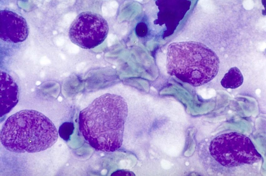
12                                                                         Emilia Balint, Nicolae Manolescu and D. Lastofka

while epithelial cancers are dominant in the feline.                 – Squamos carcinoma – 1 case;
Regarding the above facts, it is to be noted (according              – Undifferentiated vegetant carcinoma –
to Sarafoleanu Dorin, 1987) that generally in the                        1 case;
humans the incidence of malignant tumours is                         – Sensory cell carcinoma – 1 case.
lower in the Europeans comparative to Asians                         Regarding these cases evolution, and therapeutic
where the incidence is higher. The similitude                    results, there must be noted that cavum, nasal
between humans and cats is to be taken into                      fossa, sinuses and middle ear malignant neoplasms
account as well, regarding the incidence of the                  evolve generally very fast and the therapy is
cytomorphological aspects of O.R.L. cancer                       ineffective.
                                                                     Only the extern ear cancers have a long
dominated by the epithelial malignant tumors.
                                                                 evolution, similar to carcinoma neoplasms of the
                                                                 tegument. These can really benefit from surgery
          MATERIALS AND METHODS                                  followed by chemotherapy.
                                                                     From comparative oncology point of view, the
    In the 17 clinical cases (14 canine and 3 feline), after     following aspects can be highlighted:
clinical general,paraclinical and radiological examination           • While both non-Hodgkin malignant lymphoma,
(which revealed the presence of an O.R.L. neo-formation), the    and lymphoepithelioma can be identified in the
cytomorphologic examination was performed by fine needle         humans in these anatomical areas, these cancers
puncture (for cavum and external and middle ear) or by saline
                                                                 could not be identified in the investigated canine
lavage for nasal cavity and sinuses. The harvested lavage was
centrifuged at 1000 rotations/minute for 5 minutes. Smears       and feline.
from sediment, stained with classical panoptic technique, were       • The following differences were noted in the
made. The interpretation of both lavage and bio-puncture         investigated canine and feline:
under the microscope with a magnification ranging between            – A sensitive sarcoma/carcinoma equal report;
600 X and 1,000–2,000 X, was performed.                              – Tumours of nervous origin were identified
                                                                 only in the canine species;
          RESULTS AND DISCUSSIONS                                    Carcinomas and non-Hodgkin lymphomas
                                                                 reach the highest frequency in humans.
    Our casuistry, investigated from cytomor-                        The diagnosis assessment is very important for
phologically point of view showed the following                  the clinical practice and this can be accomplished
aspects:                                                         only by fine needle bio-puncture followed by
    • 7 cases consisted of nasal cavities neoplasms              cytomorphological evaluation, thus leading to a
as:                                                              correct differential diagnosis between an
    – Vegetant adenocarcinoma – 2 cases;                         inflammation and a benign or malignant tumour.
    – Estesiocarcinoma – 1 case;                                     Clinically, all these have a common denominator:
    – Fibrosarcoma – 2 cases;                                        • areal distorsion implying specific sympto-
    – Massive undifferentiated carcinoma – 1 case;               matology for the co-interested anatomical area;
    – Squamos carcinoma – 1 case.                                    • mucosa ulceration;
    • One neoplasm was identified in the cavum, as                   • development of an exuberant epithelium with
an invading malignant melanoma within adjacent                   a sessile or pediculate cauliflower-like appearance.
vertebral bone structures.
    • Two neoplasms in the fronto-ethmoidal sinuses,
                                                                           CONCLUDING REMARKS
consisting of:
    • Vegetant adenocarcinoma – 1 case;                             1. The malignant neo-formation frequency is
    • Osteoclastic osteosarcoma – 1 case.                        lower within general malignancy.
    The last site of the investigated neoplasms                     2. The high frequency of some inflammatory
(7 cases) was the external pavilion of the ear, or               and cancer-mimicking aspects in veterinary clinic
the middle ear. These neoformations showed the                   requires correct positive and differential diagnosis.
cytomorphological aspects of:
                                                                    3. In order to properly deal with these demands,
    – Rhabdomyosarcoma – 1 case;
                                                                 a bio-puncture from the affected area, and a
    – Osteoclastic osteosarcoma – 2 cases;
                                                                 cytomorphological examination must be performed.
    – Fibrosarcoma – 1 case;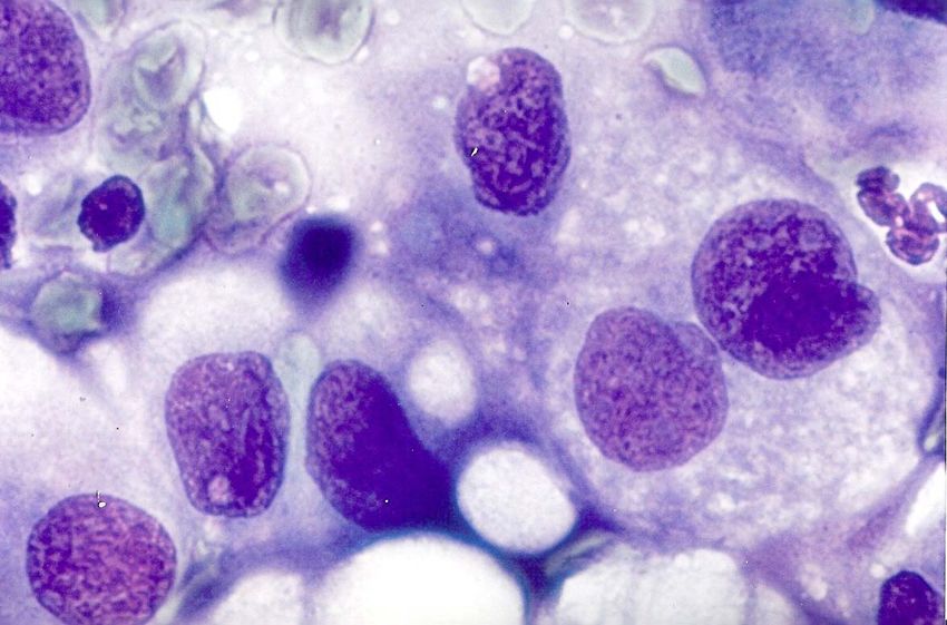
Cancers in the canine and feline                            13

                                   Figure 1. Cavum tumor.

                                   Figure 2. Nose tumor.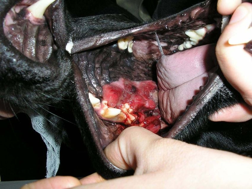
14                          Emilia Balint, Nicolae Manolescu and D. Lastofka

     Figure 3. Ear tumor.

     Figure 4. Ear tumor.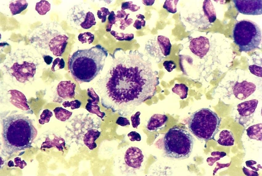
Cancers in the canine and feline 15
Figure 5. Nose tumor.
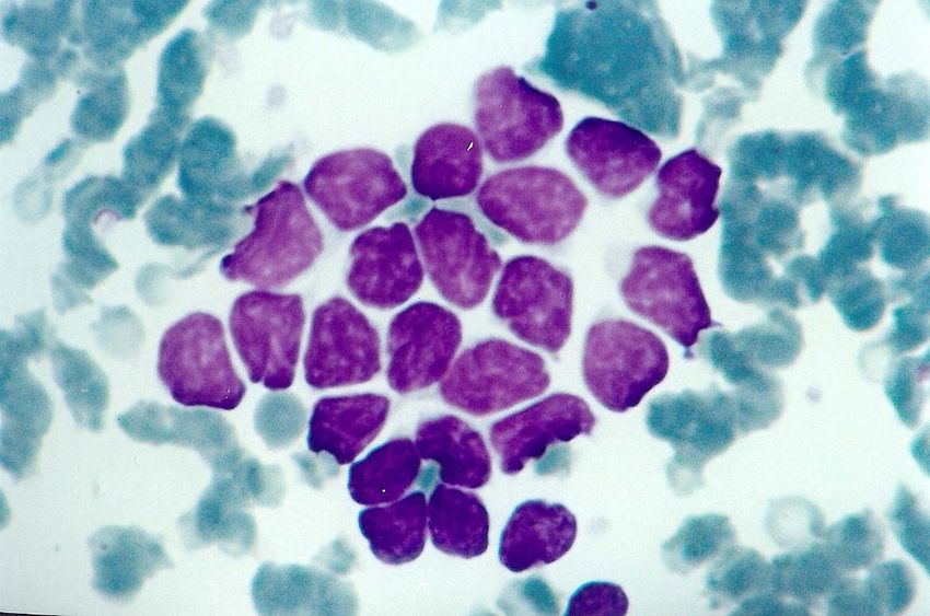
Figure 6. Nasal vegetative-like adenocarcinoma: We can notice a tumor strand made of epithelial undifferentiated cells that
proliferate under the shape of adenoid-like vegetations – glove finger shape.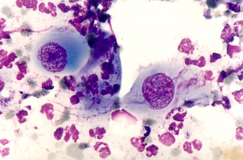
16                                                                                Emilia Balint, Nicolae Manolescu and D. Lastofka

Figure 7A. Estesiocarcinoma. These images are mainly represented by mixt origin (neuro-epithelial) cells, presenting themselves as
large elements, having broad and slightly basophil cypoplasm; the nuclei are atypical, are situated in the vecinity of the peripheric
                   area of the cell, have lax chromatin and allow the 2 or 3 basophil nucleolus to be noticed.

Figure 7B. Estesiocarcinoma. These images are mainly represented by mixt origin ( neuro-epithelial ) cells, presenting themselves as
large elements, having broad and slightly basophil cypoplasm; the nuclei are atypical, are situated in the vecinity of the peripheric
                   area of the cell, have lax chromatin and allow the 2 or 3 basophil nucleolus to be noticed.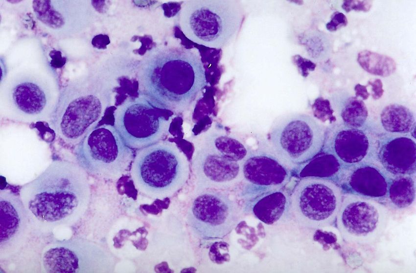
Cancers in the canine and feline                                                                                                     17

Figure 7C. Estesiocarcinoma. These images are mainly represented by mixt origin (neuro-epithelial) cells, presenting themselves as
large elements, having broad and slightly basophil cypoplasm; the nuclei are atypical, are situated in the vecinity of the peripheric
                 area of the cell, have lax chromatin and allow the 2 or 3 basophil nucleolus to be noticed.

Figure 8. Giant cell fibrosarcoma: A cellular proliferation is noticed, of mesenchymal origin, with fusiform and large sized cells, that
 are over 40 microns; the cells are highly atypical, having a central nucleus in which the base chromatide prevails, the cytoplasm is
                                  elongated, basophil, has extensions and shows numerous vacuoles.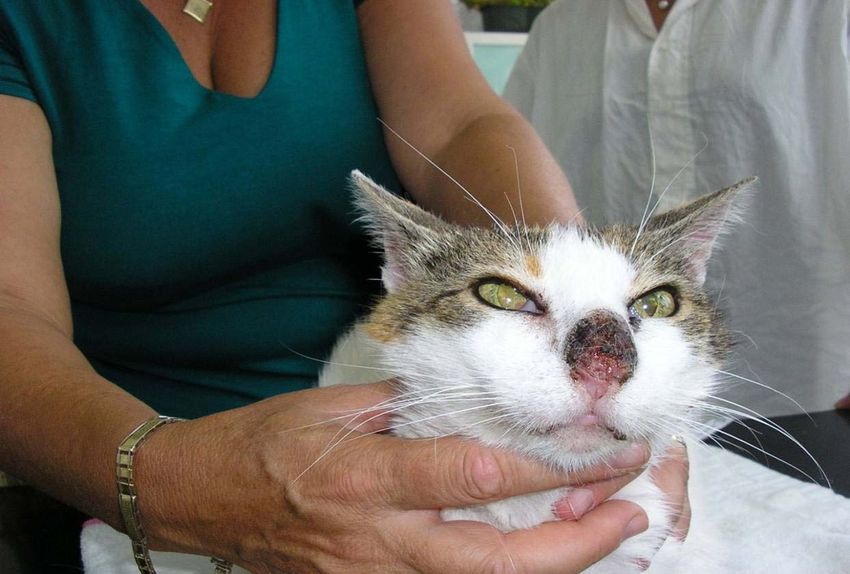
18                                                                                     Emilia Balint, Nicolae Manolescu and D. Lastofka

 Figure 9A. Squamos carcinoma. The image show an epithlial proliferation with medium sized (aprox. 15-18 microns) squamous cells; the
   nucleus in relatively round and central; the cytoplasm is variable, turning from acid to base and becoming amphoteric in some places.

 Figure 9B. Squamos cell carcinoma. The image show an epithlial proliferation with medium sized (aprox. 15-18 microns) squamous cells;
  the nucleus in relatively round and central; the cytoplasm is variable, turning from acid to base and becoming amphoteric in some places.
Cancers in the canine and feline                                                                                                   19

Figure 10A. Cavum-malignant melanoma: We can see a proliferation of cell of nervous origin; the melanoma cells hold a high
degree of cellular atipism and various shapes of cytomorphological aspects; among these we can notice either cells that have their
nucleus shielded by a massive presence of melanoma granules of big sizes, compared to other cells present that contain small
                                          granules strictly inside the cytoplasm.

Figure 10B. Malignant melanoma. We can see a proliferation of cell of nervous origin; the melanoma cells hold a high degree of
cellular atipism and various shapes of cytomorphological aspects; among these we can notice either cells that have their nucleus
shielded by a massive presence of melanoma granules of big sizes, compared to other cells present that contain small granules strictly
                                                    inside the cytoplasm.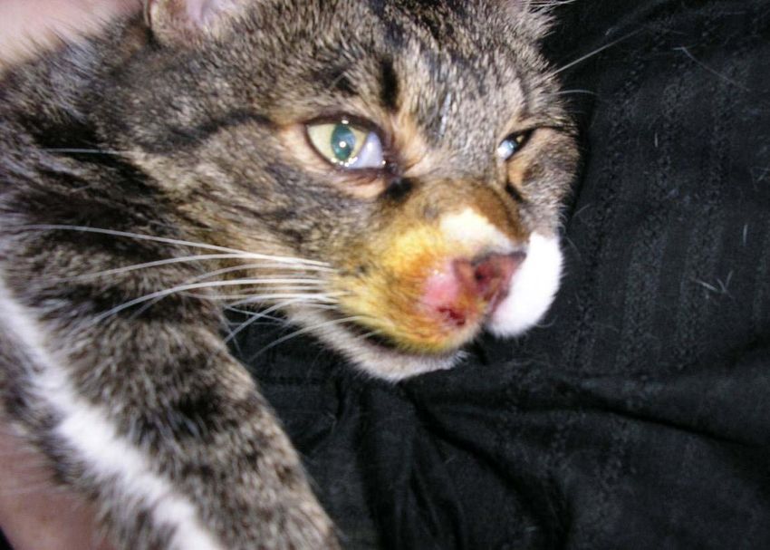
20 Emilia Balint, Nicolae Manolescu and D. Lastofka
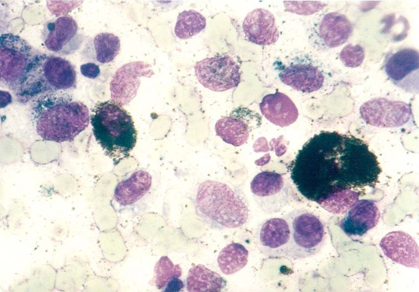
Figure 11A. Sinuses-osteoclastic osteosarcoma. The image shows a cellular proliferation of mesenchymal origin, mainly osteoblastic,
accompanied by giant multnucleated cells that characterize the osteoclast.
Figure 11B. Osteoclastic osteosarcoma. The image shows a cellular proliferation of mesenchymal origin, mainly osteoblastic,
accompanied by giant multnucleated cells that characterize the osteoclast.Cancers in the canine and feline 21
Figure 12A. Medium ear-sensory cell carcinoma. Cellular proliferation of mixed neuro-epithelial cells that characterise the mid ear
sensory carcinoma the cells have a large size (aprox. 40 microns) and have a dense nucleus at the base pole of the cell; at the apical
pole, these cells show a large amount of microvilli that are specific to this cellular type.
Figure 12B. Sensory cell carcinoma. Cellular proliferation of mixed neuro-epithelial cells that characterise the mid ear sensory
carcinoma the cells have a large size (aprox. 40 microns) and have a dense nucleus at the base pole of the cell; at the apical pole,
these cells show a large amount of microvilli that are specific to this cellular type.22 Emilia Balint, Nicolae Manolescu and D. Lastofka
Figure 13A. Rhabdomyosarcoma. The presence of mesenchymal origin cells that characterise the rhabdomyoscrcoma through the
high degree of cellular atypism, along with frequent giant multi-nucleated cells; under these conditions, we have to proceed to a clear
diferentiation between the giant cells present in rhabodmyosarcoma, that always show cytoplasmatic extensions, usually present in a
large number (Fig. 19 show 3 cytoplasmatic extensions of the giant multi-nucleated cells); these images are totally different from
those of the giant cells present in the case of the osteoclastic sarcoma (Figs. 15 and 16), which are round giant cells, that show no
extensions and sometimes are accopanied by a brush-like edge.
Figure 13B. Rhabdomyosarcoma. The presence of mesenchymal origin cells that characterise the rhabdomyoscrcoma through the
high degree of cellular atypism, along with frequent giant multi-nucleated cells; under these conditions, we have to proceed to a clear
diferentiation between the giant cells present in rhabodmyosarcoma, that always show cytoplasmatic extensions, usually present in a
large number (Fig. 19 show 3 cytoplasmatic extensions of the giant multi-nucleated cells); these images are totally different from
those of the giant cells present in the case of the osteoclastic sarcoma (Figs. 15 and 16), which are round giant cells, that show no
extensions and sometimes are accopanied by a brush-like edge.Cancers in the canine and feline 23
REFERENCES 16. Tromblee TC, Jones JC, Etue AE, et al. Association between
clinical characteristics, computed tomography characteristics
1. Dorin Sarafoleanu, Mihai Lazareanu, Breviar clinic de and histologic diagnosis for cats with sinonasal disease. Vet
oto-rino-laringologie. Editura Academiei Romane, Radiol Ultrasound 2006; 47(3):241-248.
Bucuresti, 1987. 17. Theon AP, Madewell BR, Searn VI, et al. Prognostic
2. Stephen I. Withrow, David M. Vail, Small Animal Clinical factors associated with radiotherapy of squamous cell
Oncology, 4th edition, Ed. Saunders Elsevier, 2007. carcinoma of the nasal plane in cats. JAVMA
3. Research Contract –PRECLIN – 2007 – 2010 National Au- 1995;206(7):991-996.
thority for Scientific Research of Romania – N. Manolescu. 18. Thrall DE, Adams WM. Radiotherapy of squamous cell
4. N. Manolescu, Emilia Balint, Atlas de oncocitomorfologie carcinomas of the canine nasal plane. Vet Radiol
la canide si feline, Editura Curtea Veche, Bucuresti, 2010. Ultrasound 1982;23(5):193-195.
5. Meuten Dj. Tumors in Domestic Animals, 4th ed. Ames: 19. Lacelles BD, Parry AT, Stidworthy MF, et al. Squamous
Blackwell Publishing; 2002. cell carcinoma of the nasal planum in 17 dogs. Vet Rec
6. Withrow SJ, Vail Dm. Withrow and MacEwen’s Small 2000;147:473-476.
Animal Clinical Oncology, 4th ed. Philadelphia: WB 20. Andrade AL, Fernandes MAE, Biazzona L, et al. Clinical
Saunders; 2006. trial with strontium-90 low radiation for treatment of third
7. MacMillan R, Withrow SJ, Gillette El. Surgery and eyelid neoplasms in dogs. Proc Genes Dogs Cancer 3rd
regional irradiation for treatment of canine tonsillar
Annu Canine Cancer Conf 2003.
squamous cell carcinoma: retrospective review of eight
21. Theon AP, VanVechten MK, Madewell BR. Intratumoral
cases. JAAHA1982:311-314.
8. Brooks MB, Matus RE, Leifer CE, et al. Chemotherapy administration of carboplatin for treatment of squamous
versus chemotherapy plus radiotherapy in the treatment of cell carcinomas of the nasal plane in cats. AM J Vet Res
tonsillar squamous cell carcinoma in the dog. J Vet Inter 1996;57(2):205-210.
Med 1988:2:206-211. 22. Lasa Se, Dernell Ws, Lafferty MH, et al. Use of radiation
9. Bertone ER, Snyder LA, Moore AS. Environmental and and a slow-release cisplatin formulation for treatment of
lifestyle risk factors for oral squamous cell carcinoma in canine nasal tumors. Vet Radiol Ultrasound
domestic cats, J Vet Inter Med 2003; 17(4):557-562. 2004;45(6):577-581.
10. Snyder LA, Bertone ER, Jakowski RM et al. p53 23. LeBlanc AK, LaDue TA, Turrel JM, et al. Unexpected
expression and environmental tobacco smoke exposure in toxicity following use of gemcitabine as a radiosenzitizer
feline oral squamous cell carcinoma Vet Pathol 2004; in head and neck carcinomas: a veterinary radiation
41:209-214. therapy oncology group pilot study. Vet Radiol
11. Zur G. Bilateral ear canal neoplasia in three dogs. Vet Ultrasound 2005; 45(5):466-470.
Dermatol 2005; 16(4):276-280. 24. Langova V, Mutasers AJ, Phillips B, et al. Treatment of
12. Lana SE, Ogilvie GK, Withrow Sj, et al. Feline Cutaneous eight dogs with nasal tumors with alternating doses of
squamous cell carcinoma of the nasal planum and the doxorubicin and carboplatin in conjunction with oral
pinnae: 61 cases. JAAHA 1997;33(4)329-332. piroxicam. Aust Vet J 2004;82(11):676-680.
13. Goodfellow M, Hayes A, Murphy S, et al. A retrospective 25. Murphy S, Hayes A, Adams V, et al. Role of carboplatin
study of (90) Strontium plesio therapy for feline squamous
in multimodality treatment of canine tonsillar squamous
cell carcinoma of the nasal planum. J Feline Med Surg.
cell carcinoma- a case series of five dogs. J Small Animal
14. Postorino Reves NC, Turrel HM, Withrow SJ. Oral
Pract 2006; 47(4):216-220.
squamous cell carcinoma in the cat. JAAHA 1993;
25(9):438-441. 26. De Vos JP, Burm AGO, Focker BP. Results from the
15. Schoenborn WC, Wisner ER, Kass PP, et al. Retrospective treatment of advanced stage squamous cell carcinoma of
assessment of computer tomographic imaging of feline the nasal planum in cats, using a combination of
sinonasal disease in 26 cats. Vet Radiol Ultrasound intralesional carboplatin and superficial radiotherapy: a
2003;44(2):185-195. pilot study. Vet Comp Oncol 2004;2(2)75-81.You can also read