Cytotoxicity and osteogenic potential of experimental medication with calcium hydroxide and activated charcoal
←
→
Page content transcription
If your browser does not render page correctly, please read the page content below
Research, Society and Development, v. 10, n. 5, e26010514671, 2021
             (CC BY 4.0) | ISSN 2525-3409 | DOI: http://dx.doi.org/10.33448/rsd-v10i5.14671

Cytotoxicity and osteogenic potential of experimental medication with calcium
hydroxide and activated charcoal
Citotoxicidade e potencial osteogênico de medicação experimental com hidróxido de cálcio e carvão
ativado
Citotoxicidad y potencial osteogénico de la medicación experimental con hidróxido de calcio y
carbón activado

                                                    Received: 04/05/2021 | Reviewed: 04/15/2021 | Accept: 04/20/2021 | Published: 06/05/2021

                                                                                           Gabriela SumieYaguinuma Gonçalves
                                                                                             ORCID: https://orcid.org/0000-0001-5120-3172
                                                                                    São Paulo State University Júlio de Mesquita Filho, Brazil
                                                                                                            E-mail: gabisumie@hotmail.com
                                                                                                                      Danielle Gregorio
                                                                                             ORCID: https://orcid.org/0000-0002-0098-624X
                                                                                                  Universidade Pitágoras UNOPAR, Brazil
                                                                                                       E-mail: daanigregorio@hotmail.com
                                                                                                            Isabelly Ribeiro Custódio
                                                                                              ORCID: https://orcid.org/0000-0002-0859-4822
                                                                                                   Universidade Pitágoras UNOPAR, Brazil
                                                                                                              E-mail: isabelly.rc@gmail.com
                                                                                                                  Luciana Prado Maia
                                                                                              ORCID: https://orcid.org/0000-0001-5697-2587
                                                                                                   Universidade Pitágoras UNOPAR, Brazil
                                                                                                          E-mail: lucianapmaia@gmail.com
                                                                                                                            Bruno Piazza
                                                                                              ORCID: https://orcid.org/0000-0001-8975-1545
                                                                                                     Universidade do Oeste Paulista, Brazil
                                                                                                            E-mail: brunopiazza@unoeste.br
                                                                                                               Graziela Garrido Mori
                                                                                              ORCID: https://orcid.org/0000-0002-8690-5294
                                                                                                     Universidade do Oeste Paulista, Brazil
                                                                                                                 E-mail: graziela@unoeste.br

      Abstract
      Objectives: The aim of this study was to evaluate the cytotoxicity and osteogenic potential of experimental
      medications containing Calcium Hydroxide (CH) and Activated charcoal (AC). Methodology: Osteoblastic (MC3T3)
      and fibroblastic (L929) cells were cultivated in 96 well plates (1 x 10⁴ cells/well) and, after 24 h, treated with extracts,
      according to experimental groups [(Experimental groups: C - Control; CH - CH paste; CH+10%AC - experimental
      paste 1 (CH paste + 10% AC); CH+5%AC - experimental paste 2 (CH paste + 5% AC)]. Cytotoxicity and potential
      osteogenic was performed by MTT and alkaline phosphatase activity, respectively, after 1, 3 and 7 days. Results: For
      intergroups comparisons, ANOVA 2 factors were used, followed by Tukey test (p < 0.05). There was no difference
      among the pastes for cytotoxicity in both cells (p > 0.05). For osteogenic potential, it was found that all experimental
      groups stimulated mineralization in relation to control group, except for experimental paste 2 at 7 days. Conclusion:
      The addition of AC to the CH paste does not modify toxicity and osteogenic properties. Add 10% of AC appears to be
      more effective than 5%.
      Keywords: Alkaline phosphatase; Calcium hydroxide; Charcoal; Endodontics.

      Resumo
      Objetivos: O objetivo deste estudo foi avaliar a citotoxicidade e o potencial osteogênico de medicamentos
      experimentais contendo Hidróxido de Cálcio (HC) e Carvão Ativado (CA). Metodologia: Células osteoblásticas
      (MC3T3) e fibroblásticas (L929) foram cultivadas em placas de 96 poços (1 x 10⁴ células / poço) e, após 24 h,
      tratadas com extratos, de acordo com os grupos experimentais [(Grupos experimentais: C – Controle; CH - pasta HC;
      HC + 10% CA - pasta experimental 1 (pasta HC + 10% CA); HC + 5% CA - pasta experimental 2 (pasta HC + 5%
      CA)]. Citotoxicidade e potencial osteogênico foram realizados por Atividade do MTT e da fosfatase alcalina,
      respectivamente, após 1, 3 e 7 dias. Resultados: Para as comparações intergrupos, foram utilizados os fatores ANOVA
      2, seguido do teste de Tukey (p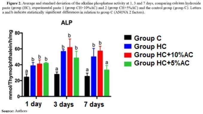
Research, Society and Development, v. 10, n. 5, e26010514671, 2021
                 (CC BY 4.0) | ISSN 2525-3409 | DOI: http://dx.doi.org/10.33448/rsd-v10i5.14671

         (p> 0,05). Para o potencial osteogênico, verificou-se que todos os grupos experimentais estimularam a mineralização
         em relação ao grupo controle, exceto a pasta experimental 2 aos 7 dias. Conclusão: A adição de CA na pasta HC não
         altera a toxicidade e as propriedades. A adição de 10% de CA parece ser mais eficaz do que 5%.
         Palavras-chave: Fosfatase alcalina; Hidróxido de cálcio; Carvão vegetal; Endodontia.

         Resumen
         Objetivos: El objetivo de este estudio fue evaluar la citotoxicidad y el potencial osteogénico de medicamentos
         experimentales que contienen Hidróxido de calcio (HC) y Carbón activado CA. Metodología: Se cultivaron células
         osteoblásticas (MC3T3) y fibroblásticas (L929) en placas de 96 pocillos (1 x 10⁴ células / pocillo) y, a las 24 h, se
         trataron con extractos, según grupos experimentales [(Grupos experimentales: C - Control; HC - pasta HC; HC + 10%
         CA - pasta experimental 1 (pasta HC + 10% CA); HC + 5% CA - pasta experimental 2 (pasta HC + 5% CA)]. La
         citotoxicidad y potencial osteogénico fue realizada por Actividad de MTT y fosfatasa alcalina, respectivamente,
         después de 1, 3 y 7 días. Resultados: Para las comparaciones intergrupales se utilizaron factores ANOVA 2, seguido
         de la prueba de Tukey (p  0.05).
         Para el potencial osteogénico, se encontró que todos los grupos experimentales estimularon la mineralización en
         relación al grupo control, excepto la pasta experimental 2 a los 7 días. Conclusión: La adición de CA a la pasta HC no
         modifica la toxicidad y propiedades Agregar un 10% de AC parece ser más efectivo que un 5%.
         Palabras clave: Fosfatasa alcalina; Hidróxido de calcio; Carbón vegetal; Endodoncia.

1. Introduction
         Calcium hydroxide (CH) is considered one of the gold standard materials in dentistry (Carvalho, et al., 2020; Reddy,
et al., 2020; Victorino, et al., 2021) and has many adequate properties, including biocompatibility, antimicrobial action, high
pH, osteogenic potential, capacity of diffusion in dentin, and stimulation apical and periapical tissues repair. These properties
were justified and widely described by its mechanism of action (Estrella, et al., 1995) as an ionic dissociation and release of
hydroxyl ions that determine the strong alkalinity, as well, the strong antibacterial action and anti-inflammatory action. The
dissolution of dentin mineral is prevented by alkaline pH and it inactivate the lactic acid and also activates alkaline
phosphatases enzyme (Estrella, et al., 1995; Reddy et al., 2020). These aspects justify its wide use as medication in endodontic
(Mohammadi & Dummer, 2011; Hilton, et al., 2013; Labban, et al., 2014; Pereira, et al., 2019). Nevertheless, some
disadvantages are observed, such as solubility in contact with oral environment (Hilton, et al., 2013), and limited or no action
against some microorganisms present in secondary and/or persistent endodontic infections, especially on Enterococcus faecalis
(E. faecalis) and Candida albicans (C. albicans) (Mohammadi & Dummer, 2011).
         Different materials were added in CH pastes to improve their characteristics, as non-steroidal anti-inflammatory and
antibiotic agents (da Silva, et al., 2020;), chlorhexidine (Souza-Filho, et al., 2008; Saatchi, et al., 2014), dichloroisocyanurate
(Chan, et al.,2020), zinc oxide (Tanomaru-Filho, et al., 2015) and iodoform nanoparticles (Garrocho-Rangel, et al., 2021) and
activated charcoal (AC) (Elfaramawy, 2021). The mechanism of action consists in adsorption capacity of different toxic
substances (Ballini, et al., 2019; Gao, et al., 2019). It is also capable of removing a wide variety of organic, inorganic and
metal contaminants, reduce the products to be absorbed, besides it has stability and a high surface area (Sugumaran, et al.,
2012; Ballini, et al., 2019). AC has antimicrobial action on E. Faecalis (Kim, et al., 2009) and C. Albicans (Saravanan, et al.,
2016) and adequate biological properties (Illingworth, et al., 2012).
           Considering the limited action of CH in persistent endodontic infections caused by E. faecalis and C. albicans, the
addition of AC in CH pastes for intracanal usage could be indicated. However, since the literature in this field is very incipient,
different concentrations of experimental pastes were idealized. In addition, analysis of the interaction between materials and
cells is an important criterion for the evaluation of new dental materials and in vitro tests are the first step in this process.
Therefore, before clinical indications, it is necessary to verify some actions of experimental pastes, as the cytotoxicity and the
bioactivity, whereas these aspects can interfere on apical and periapical tissues repair (Mohammadi & Dummer, 2011;
Oliveira, et al., 2013).
                                                                2
Research, Society and Development, v. 10, n. 5, e26010514671, 2021
(CC BY 4.0) | ISSN 2525-3409 | DOI: http://dx.doi.org/10.33448/rsd-v10i5.14671
The choice of cells for this kind of analysis is also important, and they should mimic to the maximum the clinical
situation in which the material is used. Since an intracanal medications comes in contact with the periapical tissues, its effects
on fibroblasts and osteoblasts should be evaluated using cell lines that should have physiological and adhesive properties
similar to those of human fibroblasts and. osteoblasts. Several established and immortalized strains of fibroblasts and.
osteoblasts are commercially available, among which we can mention the L929 and MC3T3-E, respectively.
Therefore, the aim of present study was to evaluate the cytotoxicity and the osteogenic potential from two
experimental medications containing CH and AC, testing the hypothesis that the addition of AC in different concentrations
does not change the properties of conventional CH paste, considering the cytotoxicity and osteogenic potential.
2. Methodology
Cell cultures
Two 2 cells cultures was used: Mouse pre-osteoblastic cell line MC3T3-E1 subclone 14 (American Type Culture
Collection, VA), cultivated in growth media (GM) Alpha-Minimum Essential Media (α-MEM) (Gibco, USA) supplemented
with 10% fetal bovine serum (FBS), (Sigma Life Science, USA) and 1% antibiotic and antimycotic (Gibco, USA); L929
fibroblastic cells (Mouse conjunctive tissue - ATCC CCL-1 NCTC) (Adolfo Luiz Institute - SP, Brazil) cultivated in GM
Minimum Essential Media (MEM) (Gibco, Glasgow, UK) supplemented with 10% FBS and 1% antibiotic and antimycotic.
Both cultures were kept at 37°C, in a humidified atmosphere containing 5% CO 2 and 95% air. After subconfluence, cultures
were subcultured and plated in 96-well culture plates at a cell density of 1x10⁴ cells/well L929 were cultivate in GM and
MC3T3 cultured in osteogenic medium (OM), which was GM supplemented with 5 µg/mL ascorbic acid (Sigma-Aldrich,
Brazil) and 7 mmol/L b-glycerophosphate (Sigma-Aldrich, MO).
Experimental medications
The medias, GM for L929 and OM for MC3T3 were exposed during 24h to experimental groups: Group C: Negative
control (culture media); Grupo CH: CH standard paste, containing 1 g of pure CH (Biodinâmica, Lobato, PR, Brazil) and 1 mL
of saline solution (Samtec, Lagoinha, SP, Brazil), representing the CH paste commonly used in endodontic treatments, Group
CH+10%AC: Experimental gr, composed by 0.9 g of pure CH (Biodinâmica), 0.1 g of powdered AC (Natuvita, Palhoça, SC,
Brazil) (CH + 10% AC) and 1 mL of saline solution (Samtec); and Group CH+5%AC: Experimental paste 2, composed of 0.95
g of pure CH (Biodinâmica), 0.05 g of AC (Natuvita) and 1 ml of saline solution (Samtec) (CH + 5% AC). As a positive
control, 35% hydrogen peroxide (Synth, Diadema, SP, Brazil), used only for methodology validation. Ratio extraction was
established at 0.2 g/mL according to ISO 10993-12 (ISO E. Biological evaluation of medical devices-Part 12: Sample
Preparation and reference materials. 2012). The extraction media was collected and filtered with sterile 0.22 µm pore diameter
filters, and different concentrations (undiluted, 1/2 and ¼) were prepared for cytotoxicity experiments, for osteogenic potential
evaluation, only pure extracts (100%) were used. All experiments were carried out in triplicate, according to the ISO 10993-5
(ISO, Standardization IOf. ISO 10993-5: Biological evaluation of medical devices-Part 5: Tests for in vitro cytotoxicity. ISO
Geneva; 2009).
Cytotoxicity test
The cytotoxicity test was performed with fibroblastic (5th passage) and osteoblastic cells (20th passage) at 1, 3 and 7
days, and the cell viability was evaluated by the colorimetric method Thiazolyl blue tetrazolium bromide (MTT). This assay is
dependent on the reduction of MTT (Sigma, St. Louis, MO, USA) in formazan, by mitochondrial viable cells dehydrogenase,
3Research, Society and Development, v. 10, n. 5, e26010514671, 2021
(CC BY 4.0) | ISSN 2525-3409 | DOI: http://dx.doi.org/10.33448/rsd-v10i5.14671
which can be quantified by spectrophotometry (Mosmann, 1983). It is a quantitative method used to evaluate cytotoxicity,
proliferation and activation of viable cells with high precision. Aliquots of MTT at 5 mg/ml in phosphate buffered saline
solution (PBS; Gibco) were prepared and the primary cultures were then incubated with 10% solution in culture media for 4
hours at 37oC, in a humidified atmosphere containing 5% CO2.
After this period, the entire solution from was removed and 100 µL of dimethyl sulfoxide solution (DMSO) was
added to each well under stirring for 5 minutes for complete solubilization of the precipitate formed. The colorimetric
measurement was performed on a spectrophotometer (SpectraCount - Packard Instrument Company, USA) using a wavelength
of 570 nm.
Osteogenic potential test
To evaluate the osteogenic potential, alkaline phosphatase activity (ALP) was performed in osteoblastic cell cultures
on days 1, 3 and 7 using the biochemical method. ALP activity was quantitatively assessed by releasing thymolphthalein by
hydrolysis of the thymolphthalein monophosphate substrate, using a commercial kit (Labtest Diagnostic SA, Belo Horizonte,
MG, Brazil), following the manufacturer's instructions.
Blank, standard and tests tubes were used. Initially, proteins were extracted from each well with 0.1% sodium lauryl
sulfate (Sigma, Aldrich, USA) for 30 min. In all tubes, 5 µL of substrate and 50 µL of 0.3 mmol/mL diethanolamine buffer
with pH 10.1 were added. In standard tube, 5 µL of standard solution was added. The tubes were kept at 37ºC for 2 min. Then,
5 µL of cell lysate from each well was added to each test tube and kept at 37º C for 10 min. After this period, 200 µL of
reagent color (Na2CO3 0.09 µmol/mL and NaOH 0.25 µmol/mL) were added to all tubes (blank, standard and tests) and
absorbance was measured in a spectrophotometer using a wavelength of 590 nm (µQuanti, BioTek Instruments, Inc.,
Winooski, VT, USA).
ALP activity, in µmol of thymolphthalein/mL, was calculated from the standard tube measurement and normalized by
total protein content determined by modified Lowry method (de Oliva, et al., 2009). For this, the proteins extracted with 0.1%
sodium lauryl sulfate (ThermoFisher Scientific) were mixed with Lowry's solution (ThermoFisher Scientific) (1:1) for 20 min
at room temperature. The extract was diluted in Folin and Ciocalteau phenol reagent (ThermoFisher Scientific) for 30 min. The
absorbance was read in a spectrophotometer using a wavelength of 680 nm.
Statistical analysis
Cell viability values were expressed in percentage in relation to group C (negative control). ALP was calculated from
the standard tube measurement and normalized by the total protein content. Normality of errors of the data was verified by the
tests of Shapiro-Wilk. The significance level was set at 5%. The significance of differences among groups were assessed by
two-way analysis of variance test (ANOVA two-way), followed by post-hoc Tukey test. GraphPad Prism 7 (GraphPad
Software).
3. Results
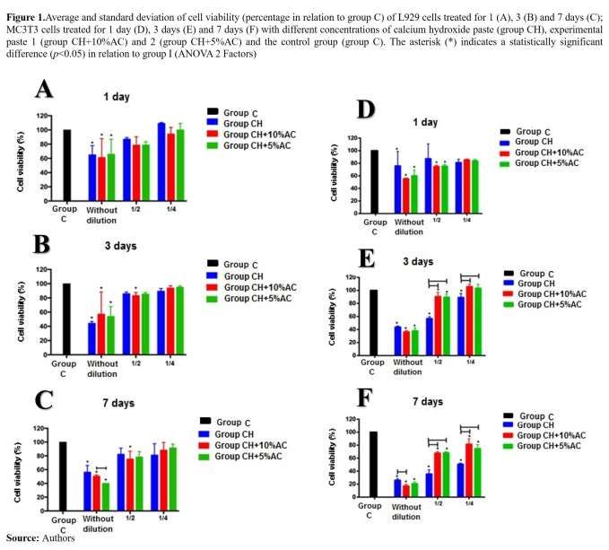
After evaluate the fibroblast cells, no difference was observed among three pastes, after 1 (Figure 1A) and 3 days
(Figure 1B) of treatment, regardless of the dilution (without dilution, 1/2 and 1/4); however, after 7 days (Figure 1C), group
CH+5%AC significantly reduced cell viability when compared to group CH+10%AC (pResearch, Society and Development, v. 10, n. 5, e26010514671, 2021
(CC BY 4.0) | ISSN 2525-3409 | DOI: http://dx.doi.org/10.33448/rsd-v10i5.14671
and C after 3 (Figure 1B)- and 7-days treatment (pResearch, Society and Development, v. 10, n. 5, e26010514671, 2021
(CC BY 4.0) | ISSN 2525-3409 | DOI: http://dx.doi.org/10.33448/rsd-v10i5.14671
4. Discussion
The hypothesis of the study was confirmed, since the addition of activated charcoal to the conventional calcium
hydroxide paste did not significantly change its properties. The cell lines used in the present study was determinant to verify
similarly action to the adjacent tooth tissues as would be occur in in vivo studies.
Regarding osteogenic potential, it was found that experimental paste 1 (CH + 10% AC) was similar to CH paste,
unlike experimental paste 2 (CH + 5% AC), which had its potential reduced at 7 days.
The analysis of biological properties is important to endodontic medications, due those medications must reduce the
pathogenic microorganisms without damage the apical and periapical cells and tissues (Correa, et al., 2009; Carvalho, et al.,
2018; da Silva, et al., 2020). Cytotoxicity tests are performed to assess biocompatibility of materials and understand
metabolism and repair process (Correa, et al., 2009; Lim, et al., 2017). MTT assay is based on the ability of living cells to
convert yellow salt tetrazolium for lilac formazan crystals, being a simple, fast, accurate and with high reproducibility test
(Correa, et al., 2009; Carvalho, et al., 2018). In present study was observed CH pastes are not cytotoxic, and literature agree,
being the most used intracanal medications in endodontics (Desai & Chandler, 2009; Mohammadi & Dummer, 2011;
Althumairy, et al., 2014). Even investigating different substances, da Silva, et al. (2020) showed that CH pastes with 5%
sodium diclofenac, ibuprofen or amoxicillin presented low cytotoxicity in 1, 2, 3 and 7 days and different concentrations,
corroborating the results of this study, which obtained low cytotoxicity results, and that can be justified by the presence of
calcium hydroxide in its compositions on both studies. The test periods were chosen to understand the action of the
components in initial analyzes, and long periods with cell culture are not recommended due to the limitations involving this
type of study (Silva, et al., 2020).
Calcium hydroxide paste dissociates into calcium ions and hydroxyl ions when it fills the main root canal; these ions
diffuse through the dentinal tubules, and the concentrations are different in root canal and apical and periapical tissues
(Mohammadi & Dummer, 2011). In view of this, assessing cell viability in contact with different concentrations becomes
6Research, Society and Development, v. 10, n. 5, e26010514671, 2021
(CC BY 4.0) | ISSN 2525-3409 | DOI: http://dx.doi.org/10.33448/rsd-v10i5.14671
necessary. Additionally, studies have already shown that cell proliferation can be altered over time (Correa, et al., 2009; Pires,
et al., 2016), which makes it pertinent to evaluate different time points. In present study, differences were found in relation to
cellular viability at different experimental times and concentrations (dilutions), demonstrating that the cytotoxicity is directly
proportional to the time of exposure of cells to experimental pastes.
Correa, et al. (2009) observed that the pure extract (without dilution) of endodontic sealers containing calcium
hydroxide associated with oily vehicles significantly reduces the cell viability of THP-1 monocytes significantly after 24 hours
of treatment (Correa, et al., 2009). The authors demonstrated that the paste calcium hydroxide only stop significantly reducing
cell viability at dilutions below 1%; a fact that corroborates the findings of our study, in which the pure extracts of the three
pastes significantly reduced the cell viability of fibroblasts and osteoblastic cells at all times evaluated.
In this context, Labban, et al. (2014) suggest that the ability of calcium hydroxide to promote proliferation or prevent
cell death is concentration-dependent, so that its dissolution is shown as an alternative to possible negative effects and favours
pulp healing (Labban, et al., 2014). This property of preventing cell death was also observed in the present study, since CH
paste did not reduce cell viability. Otherwise, adding AC has not increased the cytotoxicity of the conventional CH paste
already used in dental clinic.
Another important point to be discussed refers to the evaluation of cell bioactivity through ALP, which allows to
verify the potential to induce formation of mineralized tissue. The alkaline phosphatase is a biochemical marker linked to the
membrane, which is secreted by osteoblasts (Modareszadeh, et al., 2012). Chen et al. (2016) should that CH has osteogenic
potential using the ALP staining assay as in our study (Chen, et al., 2016). The bioactive of CH+10%AC was higher than
CH+5%AC; we believe the characteristics of AC can improve the osteogenic potential, since that the ALP was bigger in
experimental paste with more AC.
It is important to emphasized that this is an initial study, and cytotoxicity and osteogenic potential are only some of
the wide ranges of properties expected in a biomaterial in development. The present results encourage further experimental
studies to evaluate properties such as tooth discoloration, antimicrobial actions, anti-inflammatory effect and biocompatibility
in animals, in addition to clinical trials, in order to prove the effectiveness of these news experimental medications in dental
clinical practice. These results suggest a dose-dependent cytotoxic effect of all evaluated pastes; however, the addition of AC
to the CH paste does not modify toxicity-related properties. Regarding bioactivity, experimental paste 1 (CH+10%AC) showed
greater activity than paste 2 (CH+5%AC).
Despite of the limitations of the present study, the aspects investigated influence on apical and periapical tissues
repair, and they are fundamental to clinical therapy. The results of the present study support the idea of new experimental
medications, and to future clinical indication, it is essential to analyse antimicrobial action of new medication, mainly, for use
in teeth with pulp necrosis and apical periodontitis. Thus, studies analysing the direct contact with microbial biofilms, effect on
biofilm present in dentinal tubules must be made. After that, in vivo studies must be made to confirm the use of new
medication.
5. Final Considerations
The addition of AC to the CH paste does not modify toxicity and the osteogenic properties. Add 10% of AC appears
to be more effective than 5%. Thus, studies analysing the direct contact with microbial biofilms, effect on biofilm present in
dentinal tubules in different concentrations must be made.
7Research, Society and Development, v. 10, n. 5, e26010514671, 2021
(CC BY 4.0) | ISSN 2525-3409 | DOI: http://dx.doi.org/10.33448/rsd-v10i5.14671
Acknowledgments
The authors thank São Paulo Research Foundation (FAPESP) - Process 2018/16356-2 and Coordination for the
Improvement of Higher Education Personnel - Brazil (CAPES), for the financial support.
References
Althumairy, R. I., Teixeira, F. B., & Diogenes, A. (2014). Effect of dentin conditioning with intracanal medicaments on survival of stem cells of apical papilla.
J. Endod, 40(4):521-5. https://doi.org/10.1016/j.joen.2013.11.008
Ballini, A., Cantore, S., Saini, R., Pettini, F., Fotopoulou, E. A., Saini, S. R., Georgakopoulos, I. P., Dipalma, G., Gargiu lo Isacco, C., & Inchingolo, F. (2019).
Effect of activated charcoal probiotic toothpaste containing Lactobacillus paracasei and xylitol on dental caries: a randomized and controlled clinical trial.
Journal of biological regulators and homeostatic agents, 33(3), 977–981.
Carvalho, G. A. O., de Almeida, R. R., Câmara, J. V. F., & Pierote, J. J. A. (2020). Hidróxido de cálcio versus hibridização em capeamentos pulpares: revisão
de literatura. Research, Society and Development, 9(7), e244974069-e244974069. http://dx.doi.org/10.33448/rsd-v9i7.4069
Carvalho, N. C., Guedes, S., Albuquerque-Júnior, R., de Albuquerque, D. S., de Souza Araújo, A. A., Paranhos, L. R., Camargo, S., & Ribeiro, M. (2018).
Analysis of Aloe vera cytotoxicity and genotoxicity associated with endodontic medication and laser photobiomodulation. Journal of photochemistry and
photobiology. B, Biology, 178, 348–354. https://doi.org/10.1016/j.jphotobiol.2017.11.027
Chan, W., Chowdhury, N. R., Sharma, G., Zilm, P., & Rossi-Fedele, G. (2020). Comparison of the biocidal efficacy of sodium dichloroisocyanurate and
calcium hydroxide as intracanal medicaments over a 7-day contact time: an ex vivo study. Journal of Endodontics, 46(9), 1273-1278.
https://doi.org/10.1016/j.joen.2020.05.011
Chen, L., Zheng, L., Jiang, J., Gui, J., Zhang, L., Huang, Y., Chen, X., Ji, J. & Fan, Y. (2016). Calcium Hydroxide–induced Proliferation, Migration,
Osteogenic Differentiation, and Mineralization via the Mitogen-activated Protein Kinase Pathway in Human Dental Pulp Stem Cells. J. Endod, 42(9):1355-61.
https://doi.org/10.1016/j.joen.2016.04.025
Correa, G. T. B., et al (2009). Cytotoxicity evaluation of two root canal sealers and a commercial calcium hydroxide paste on THP1 cell line by Trypan Blue
assay. Journal of Applied Oral Science, 17(5), 457-461. https://dx.doi.org/10.1590/S1678-77572009000500020
da Silva, G. F., Cesário, F., Garcia, A., Weckwerth, P. H., Duarte, M., de Oliveira, R. C., & Vivan, R. R. (2020). Effect of association of non-steroidal anti-
inflammatory and antibiotic agents with calcium hydroxide pastes on their cytotoxicity and biocompatibility. Clinical oral investigations, 24(2), 757–763.
https://doi.org/10.1007/s00784-019-02923-y
de Oliva, M. A., Maximiano, W. M., de Castro, L. M., da Silva, P. E., Jr, Fernandes, R. R., Ciancaglini, P., Beloti, M. M., Nanci, A., Rosa, A. L., & de
Oliveira, P. T. (2009). Treatment with a growth factor-protein mixture inhibits formation of mineralized nodules in osteogenic cell cultures grown on titanium.
The journal of histochemistry and cytochemistry: official journal of the Histochemistry Society, 57(3), 265–276. https://doi.org/10.1369/jhc.2008.952713
Desai S, Chandler N. (2009). Calcium hydroxide-based root canal sealers: a review. J Endod.35(4):475-80. 10.1016/j.joen.2008.11.026.
Elfaramawy, M. (2021). The Effect Of The Addition Of Activated Charcoal To Different Formulations Of Calcium Hydroxide On The ir Effect On The
Fracture Resistance Of Endodontically Treated Teeth. Egyptian Dental Journal, 67(2), 1629-1623. 10.21608/edj.2021.48721.1325
Estrela, C., Sydney, G. B., Bammann, L. L., & Felippe Junior, O. (1995). Mechanism of the action of calcium and hydroxy ions of calcium hydroxide on tissue
and bacteria. Braz Dent J, 6(2): 85-90. http://143.107.206.201/bdj/t0262.html
Gao, Y., Wang, G., Li, Y., Lv, C., & Wang, Z. (2019). Effects of oral activated charcoal on hyperphosphatemia and vascular ca lcification in Chinese patients
with stage 3-4 chronic kidney disease. Journal of nephrology, 32(2), 265–272. https://doi.org/10.1007/s40620-018-00571-1
Garrocho-Rangel, A., Escobar-García, D. M., Gutiérrez-Sánchez, M., Herrera-Badillo, D., Carranco-Rodríguez, F., Flores-Arriaga, J. C., & Pozos-Guillén, A.
(2021). Calcium hydroxide/iodoform nanoparticles as an intracanal filling medication: synthesis, characterization, and in vitro study using a bovine primary
tooth model. Odontology, 1-9. https://doi.org/10.1007/s10266-021-00591-7
Giongo, M, Santos, R. A. M. dos, Maciel, S. M, Fracasso, M. L. C, & Victorino, F. R. (2017). Analysis of pH and release of calcium of association between
melaleuca alternifolia oil and calcium hydroxide. Revista de Odontologia da UNESP, 46(2), 104-108. Epub March 13, 2017.https://doi.org/10.1590/1807-
2577.07816
Hilton, T. J., Ferracane, J. L., Mancl, L., & Northwest Practice-based Research Collaborative in Evidence-based Dentistry (NWP) (2013). Comparison of
CaOH with MTA for direct pulp capping: a PBRN randomized clinical trial. Journal of dental research, 92(7), 16S–22S.
https://doi.org/10.1177/0022034513484336
Illingworth, J. M., Rand, B., Williams, P. T. (2012). Novel activated carbon fibre matting from biomass fibre waste. Proceedings of the Institution of Civil
Engineers-Waste and Resource Management, 165(3):123-132. https://doi.org/10.1680/warm.12.00001
ISO E. Biological evaluation of medical devices-Part 12: Sample Preparation and reference materials. 2012.
ISO, Standardization IOf. ISO 10993-5: Biological evaluation of medical devices-Part 5: Tests for in vitro cytotoxicity. ISO Geneva; 2009.
Kim, H. C., Park, S.J., Lee, C. G., Kim, S. B., Kim, K. W. (2009). Bacterial attachment to iron-impregnated granular activated carbon. Colloids Surf. B:
Biointerfaces, 74(1):196-201. https://doi.org/10.1016/j.colsurfb.2009.07.018
8Research, Society and Development, v. 10, n. 5, e26010514671, 2021
(CC BY 4.0) | ISSN 2525-3409 | DOI: http://dx.doi.org/10.33448/rsd-v10i5.14671
Labban, N., Yassen, G. H., Windsor, L. J., & Platt, J. A. (2014). The direct cytotoxic effects of medicaments used in endodontic regeneration on hum an dental
pulp cells. Dental traumatology, 30(6), 429–434. https://doi.org/10.1111/edt.12108
Lim, M.J., Jang, H.J., Yu, M.K., Lee, K.W., Min, K.S. (2017). Removal efficacy and cytotoxicity of a calcium hydroxide paste using N-2-methyl-pyrrolidone
as a vehicle. Restor Dent Endod, 42(4):290-300. https://doi.org/10.5395/rde.2017.42.4.290
Modareszadeh, M. R., Di Fiore, P. M., Tipton, D. A., & Salamat, N. (2012). Cytotoxicity and alkaline phosphatase activity evaluation of endosequence root
repair material. Journal of endodontics, 38(8), 1101–1105. https://doi.org/10.1016/j.joen.2012.04.014
Mohammadi, Z., & Dummer, P. M. (2011). Properties and applications of calcium hydroxide in endodontics and dental traumatology. International
endodontic journal, 44(8), 697–730. https://doi.org/10.1111/j.1365-2591.2011.01886.x
Mosmann T. (1983). Rapid colorimetric assay for cellular growth and survival: application to proliferation and cytotoxicity assays. Journal of immunological
methods, 65(1-2), 55–63. https://doi.org/10.1016/0022-1759(83)90303-4
Oliveira, I. R., Andrade, T. L., Jacobovitz, M., & Pandolfelli, V. C. (2013). Bioactivity of calcium aluminate endodontic cement. Journal of endodontics,
39(6), 774–778. https://doi.org/10.1016/j.joen.2013.01.013
Pereira, T. C., da Silva Munhoz Vasconcelos, L. R., Graeff, M., Ribeiro, M., Duarte, M., & de Andrade, F. B. (2019). Intratubular decontamination ability and
physicochemical properties of calcium hydroxide pastes. Clinical oral investigations, 23(3), 1253–1262. https://doi.org/10.1007/s00784-018-2549-0
Pires, C. W., Botton, G., Cadoná, F. C., Machado, A. K., Azzolin, V. F., da Cruz, I. B., Sagrillo, M. R., & Praetzel, J. R. (2016). Induction of cytotoxicity,
oxidative stress and genotoxicity by root filling pastes used in primary teeth. International endodontic journal, 49(8), 737–745.
https://doi.org/10.1111/iej.12502
Reddy, S., Prakash, V., Subbiya, A., & Mitthra, S. (2020). 100 Years of Calcium Hydroxide in Dentistry: A Review of Literature. Indian Journal of Forensic
Medicine & Toxicology, 14(4), 1203.
Saatchi, M., Shokraneh, A., Navaei, H., Maracy, M. R., & Shojaei, H. (2014). Antibacterial effect of calcium hydroxide combined with chlorhexidine on
Enterococcus faecalis: a systematic review and meta-analysis. Journal of Applied Oral Science, 22(5), 356-365. http://dx.doi.org/10.1590/1678-775720140032
Saravanan, A., Kumar, P.S., Devi, G.K., Arumugam, T. (2016). Synthesis and characterization of metallic nanoparticles impregn ated onto activated carbon
using leaf extract of Mukia maderasapatna: Evaluation of antimicrobial activities. Microb. Pathog, 97:198-203. https://doi.org/10.1016/j.micpath.2016.06.019
Souza-Filho, F. J. de, et al (2008). Antimicrobial effect and pH of chlorhexidine gel and calcium hydroxide alone and associated with other materials.
Brazilian Dental Journal, 19(1), 28-33. https://doi.org/10.1590/S0103-64402008000100005
Sugumaran, P., Susan, V. P., Ravichandran, P., & Seshadri, S. (2012). Production and characterization of activated carbon from banana empty fruit bunch and
Delonix regia fruit pod. J Sustainable Energy & Environment, 3(3):125-32.
Tanomaru-Filho, M., Guerreiro-Tanomaru, J. M., Faria, G., Aguiar, A. S., & Leonardo, R. T. (2015). Antimicrobial activity and ph of calcium hydroxide and
zinc oxide nanoparticles intracanal medication and association with chlorhexidine. The journal of contemporary dental practice, 16(8), 624-629. DOI:
10.5005/jp-journals-10024-1732
Victorino, F. R., Rocha, I. S., de Oliveira Lazarin, R., Seron, M. A., Sivieri-Araujo, G., & Almeida, R. S. (2021). Maxillary Canine with two roots and two
canals: A case report. Research, Society and Development, 10(2). DOI: http://dx.doi.org/10.33448/rsd-v10i2.12599
9You can also read