Dental Anthropology A Publication of the Dental Anthropology Association
←
→
Page content transcription
If your browser does not render page correctly, please read the page content below
Volume 33, Issue 02, 2020                                  ISSN 1096-9411

                               Dental
                            Anthropology
                 A Publication of the Dental Anthropology Association

Dental Anthropology                                          2020 │ Volume 33 │ Issue 02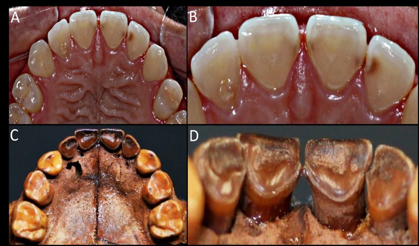
2

                      Dental Anthropology
                                         Volume 33, Issue 02, 2020
          Dental Anthropology is the Official Publication of the Dental Anthropology Association.

                                            Editor: Marin A. Pilloud
                                       Editor Emeritus: G. Richard Scott

                                        Editorial Board (2019-2022)
                         Heather J.H. Edgar                  Alistair R. Evans
                         Scott D. Haddow                     Nicholas P. Herrmann
                         Jaime M. Ullinger                   Cathy M. Willermet

                             Officers of the Dental Anthropology Association
                          Daniel Antoine (British Museum) President (2019-2022)
                Marin A. Pilloud (University of Nevada, Reno) President-Elect (2019-2022)
                Amelia Hubbard (Wright State University) Secretary-Treasurer (2017-2021)
             Katie Zejdlik (Western Carolina University) Executive Board Member (2018-2021)
                Heather J.H. Edgar (University of New Mexico) Past President (2019-2022)
                                        Contact for Manuscripts
                                            Dr. Marin A. Pilloud
                                     Department of Anthropology
                                       University of Nevada, Reno
                                   E-mail address: mpilloud@unr.edu
                                 Website: journal.dentalanthropology.org

                                      Address for Book Reviews
                                         Dr. Daniel H. Temple
                               Department of Sociology and Anthropology
                                      George Mason University
                                  E-mail address: dtemple3@gmu.edu

                                                     Published at
                                               University of Nevada, Reno
                                                 Reno, Nevada 89557
                                 The University of Nevada, Reno is an EEO/AA/Title IX/Section 504/ADA employer

                                                    Editorial Assistant
                                                    Rebecca L. George
                                                   Production Assistant
                                                     Daniel E. Ehrlich

Dental Anthropology                                                                                              2020 │ Volume 33 │ Issue 02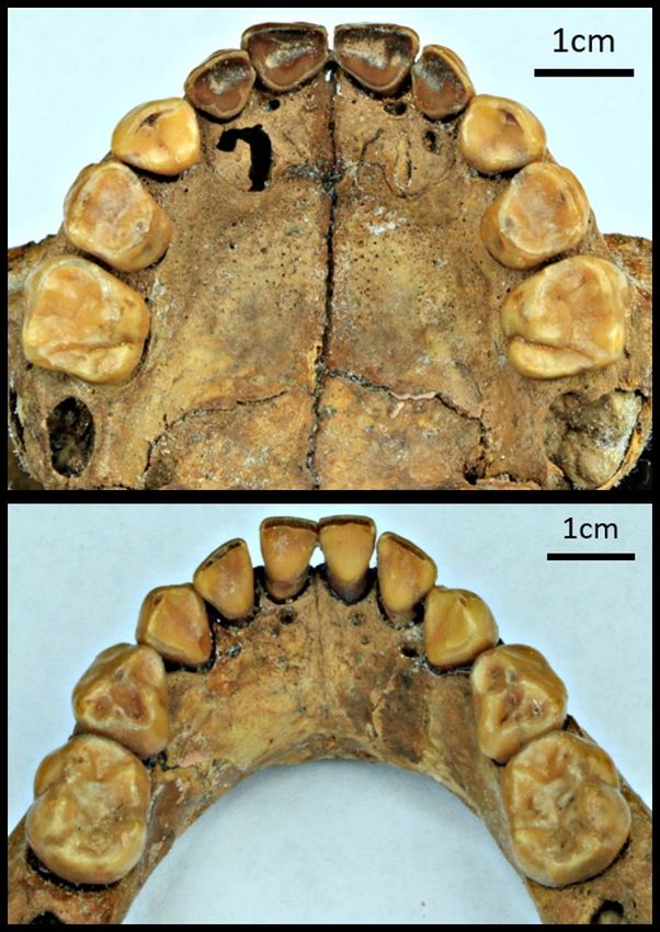
3

        Dental Corrosion in Preindustrial Societies: A Case Study of a
        Child from “Pedra do Cachorro” Dating to 1,470 BP, Northeastern
        Brazil

        Rodrigo Elias Oliveira1*, Ana Solari2, Sergio Francisco S.M. Silva2, Gabriela Martin2,
        Caio Belem Soares2, and Andre Strauss1
        1
            Universidade de São Paulo, Brazil
        2
            Universidade Federal de Pernambuco, Brazil

        Keywords: dental wear, paleopathology, gastroesophageal reflux, dental corrosion, dental erosion

        ABSTRACT Reflux, frequent vomiting, and the high intake of acidic beverages in industrial societies
        result in a relatively elevated frequency of dental corrosion. In the past, however, chemical dental wear
        was rather rare. Here we present and analyze a child from the Fifth Century CE that evidenced a
        growth pattern which was below that expected for an infant of its age. Furthermore, the child also had a
        peculiar pattern of dental erosion. This 3-year-old child dated to 1470±30BP from the archaeological site
        of Pedra do Cachorro (northeastern Brazil) had its bones and teeth analyzed macroscopically, radio-
        graphically (X-ray and tomography), and microscopically (SEM). Harris lines, linear enamel hypoplasia,
        and the poor linear growth presented by this sub-adult suggest malnutrition or some other physiologi-
        cal stress. The unique pattern of chemical wear on the lingual surfaces of upper incisors was compatible
        with dental corrosion, reinforcing the diagnosis of frequent vomiting possibly caused by an undefined
        gastric disorder, which could have been a factor in the early death of this child.

        Built from the hardest tissues found in the human        food bolus being forced against these surfaces by
        body (enamel and dentin) teeth are commonly pre-         the tongue, cheeks, and lips (Grippo et al., 2012).
        served in the archaeological record. Dental wear            These two processes are strongly associated
        has long been studied as an important source of          with the masticatory cycles responsible for the for-
        data regarding a broad spectrum of past and pre-         mation of occlusal wear. The severity of occlusal
        sent human activities (d’Incau et al., 2012; Deter,      wear increases during the lifetime of an individual
        2009; Smith, 1984; Turner and Machado, 1983).            and can therefore be used as a proxy for estimating
        Dental wear is the result of the non-carious loss of     age-at-death (Prince et al., 2008). As abrasion is a
        dental tissues due to the additive effects of me-        direct result of the size and roughness of the in-
        chanical and chemical processes (Brace and               gested particles; the severity of occlusal wear can
        Molnar, 1967; Molnar, 2008; Oliveira and Neves,          also be used in reconstructing dietary habits
        2015; Van’t Spijker et al., 2009).                       (Grippo et al., 2012; Scheid and Weiss, 2012).
           It is used to classify tooth surface loss due to         Deviations from the typical erosion patterns
        attrition, abrasion, abfraction, and corrosion, de-      resulting from attrition/abrasion masticatory oc-
        pending on the nature of the wasting process. In         clusal wear are indicative of pathological condi-
        this sense, “attrition” is a type of dental wear         tions or paramasticatory habits. Some non-
        caused by tooth-to-tooth friction that occurs during     physiological activities can also change dental
        chewing, clenching, and deglutition. This type of        structure. Among others, parafunctional habits
        tooth surface lesion affects the occlusal/incisal are-
        as, as well as the proximal surfaces (Smith, 1984).
                                                                   *Correspondence to:
           “Abrasion” is the result of friction between teeth
        and exogenous agents such as food (e.g. fruits,            Rodrigo Elias Oliveira
        leaves, vegetables, shells, and bones) and exoge-          Instituto de Biociências & Faculdade de
        nous particles in the food bolus (e.g. sand, stone,        Odontologia
        and charcoal). During mastication, lingual and             Universidade de São Paulo
        buccal/facial surfaces can be worn down by the             eliaso@alumni.usp.br

Dental Anthropology                                                                              2020 │ Volume 33 │ Issue 02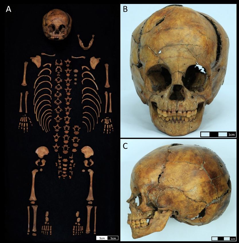
4

        such as clenching and grinding of teeth (bruxism)        “dental erosion”) is more common in industrial
        are very common in modern societies, and might           societies (Robb et al., 1991a, 1991b). The elevated
        be directly associated to psychosocial problems          consumption of liquids with a pH below 3.0, such
        (Carlsson et al., 2003; Manfredini and Lobbezoo,         as carbonated beverages and citrus juice is most
        2009; Pavone, 1985). However, the most common            likely a major cause of the exogenous corrosion
        parafunctional activity observed in preindustrial        (Eccles and Jenkins, 1974; Honório et al., 2008; Jä-
        and traditional societies is the use of teeth as tear-   rvinen et al., 1991; Lussi et al., 2011).
        ing and grabbing tools, also called Lingual Surface          Among the “endogenous corrosions”, gas-
        Attrition of the Maxillary Anterior Teeth                troesophageal reflux disease (GERD) is a potential
        (LSAMAT) when these lesions are present on up-           cause, given that it brings up extremely acidic gas-
        per incisors (Irish and Turner, 1987; Larsen et al.,     tric fluids to the mouth, and therefore in direct con-
        1998; Waters-Rist et al., 2010).                         tact with dentition (Bartlett et al., 2013; Gud-
           “Abfraction” is a less frequent type of mechani-      mundsson et al., 1995). Similarly, dental corrosion
        cal dental wear observed on archaeological skele-        can be associated with bulimia and other eating
        tons. Although the precise etiology is still a matter    disorders that involve systemic and recurrent vom-
        for debate, abfraction is broadly considered to re-      iting (Gudmundsson et al., 1995; Lazarchik and
        flect stress concentration on the cervical region of     Filler, 2000; Moazzez et al., 2004).
        teeth, as a result of excessive cyclic loading               There are many case reports on dental corrosion
        (Lanigan and Bartlett, 2013; Lucas and Omar, 2012;       in the archaeological literature, with most of them
        Oliveira, 2014). Most likely this excessive loading is   presenting dietary erosive wear with or without
        a consequence of parafunctional use of the mastica-      associated attrition as shown by Coupal and
        tory apparatus.                                          Soltysiak (2017). However, it is very rare to see cas-
           While mechanical wear is commonly reported in         es in which the dental corrosion was caused by
        archaeological contexts, chemical wearing is rare in     frequent regurgitation (Robb et al, 1991a; Coupal
        preindustrial      societies.      “Corrosion”      or   and Soltysiak, 2017).
        “biocorrosion” are the terms used to define the              In this article, we describe the case of a 3-year-
        chemical dissolution of teeth surfaces. Corrosion/       old child radiometrically dated to 1,470 ± 30 BP,
        biocorrosion can be divided into four (4) separate       that was exhumed from an archaeological site lo-
        categories: exogenous, endogenous, proteolysis           cated in northeastern Brazil. This child presents a
        (degradation of the small amount of enamel pro-          unique pattern of chemical wear that was compati-
        tein in the caries process), and electrochemical (as     ble with dental corrosion. We then compared this
        the result of piezoelectric effects only on dentin,      observation, against a broader characterization of
        not on enamel) (Grippo et al., 2012). Nevertheless,      oral health, including caries, periapical lesions,
        in an archaeological context we usually find, and        dental calculus, and periodontal bone resorption
        therefore discuss, exogenous and endogenous cor-         (Guatelli-Steinberg et al., 2004; Hillson, 2008;
        rosion.                                                  Oliveira and Neves, 2015). Osteological markers of
           Corrosion happens when the dental surface is          physiological imperilments, such as linear enamel
        exposed to an acidic agent capable of creating a         hypoplasia (LEH) and transverse radiopaque lines
        microenvironment with a constant pH of below 4.0         (Harris lines) were also considered.
        (Dong et al., 1999; Hillson, 2008; Järvinen et al.,
        1991; Scheid and Weiss, 2012). The solubilization of     Burial 2 from Pedra do Cachorro
        hydroxyapatite, the mineral structure of enamel,         The skeleton analyzed in this study – Burial 2 –
        dentin, cementum, and bone, occurs when the local        was uncovered in 2015 at the Pedra do Cachorro
        pH is 5.5 or below, whereas the critical pH for sol-     archaeological site, located in the Parque Nacional
        ubilization of fluorapatite is 4.5 or below (Ekstrand    do Catimbau, Pernambuco, Brazil (Figure 1). This
        and Oliveby, 1999). Microbial biocorrosion or            site is located in the sheltered area formed along
        simply dental caries is the most common human            the side of a large sandstone outcrop. The region
        pathological condition observed on archaeological        presents an important archaeological record for the
        skeletons. Dental caries is caused by the dissolu-       presence of prehistoric foraging groups, dating
        tion of the tooth surface due to the lactic acid pro-    from 6,000 years before the present, onwards. Be-
        duced by cariogenic bacteria (Larsen, 2008;              tween 2015 and 2016, four field campaigns were
        Morimoto et al., 2014).                                  undertaken at the site, resulting in the excavation
           In contrast to the archaeological record, exoge-      of a 68 m² area. Two other burials, directly dated to
        nous or endogenous corrosion (formerly known as          760 ± 30 and 3,560 ± 30 years BP respectively, were

Dental Anthropology                                                                               2020 │ Volume 33 │ Issue 02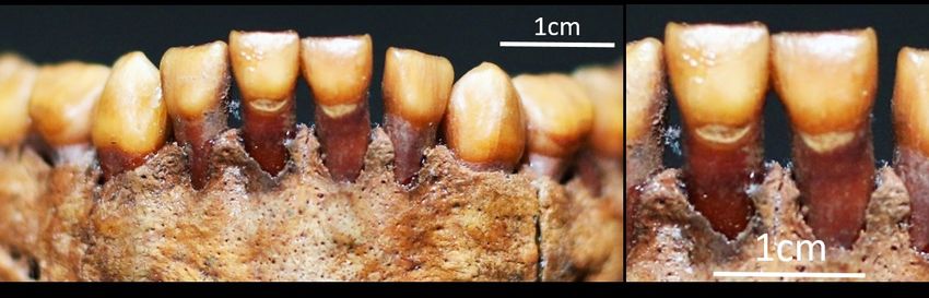
5

           Figure 1. Location of the archaeological site of Pedra do Cachorro, Buíque – Pernambuco, Brazil.

        also found in Pedra do Cachorro but they are not           pit was filled with loose red-brown sediment
        part of the present article (Solari et al., 2015,;2016).   whose current pH was determined to be 6.64 - 7.15
           Burial 2 contains the skeleton of a young child         (Silva et al., 2019). The reddish color of the external
        found within an oval pit (35 cm width; 92 cm long;         surface of the human bones most likely resulted
        20 cm deep) surrounded by sandstone blocks. The            from long exposure to the burial sediment as no
        burial did not contain grave goods. A rib fragment         evidence for ochre was identified (Figure 3). Char-
        from Burial 2 was directly dated to 1,470 ± 30 BP          coal fragments were found amidst the human
        (Beta 447238). The bone distribution indicates that        bones and surrounding sediments. One charcoal
        the body was deposited in a prone position with            piece was dated to 2,100 ± 30 years BP, indicating
        flexed legs (Figure 2) (Solari et al., 2016). The burial   that it was not contemporaneous with Burial 2.

      Figure 2. Burial 2 of Pedra do Cachorro: photograph of the exhumation (A), burial sketch (B), graphic re-
      construction of the original burial position (C).

Dental Anthropology                                                                                  2020 │ Volume 33 │ Issue 02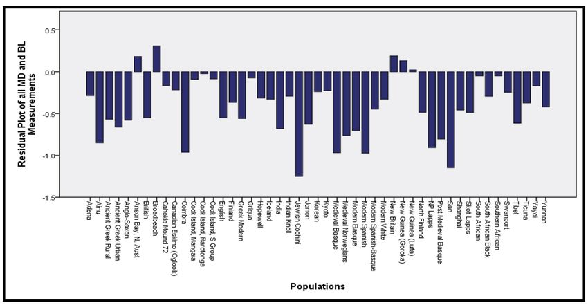
6

        This observation was compatible with the lack of      of its root and crown (Figure 4). The resulting ma-
        any macroscopic signs of thermal modification on      turity index was 15.0 indicating an estimated age-
        the bones.                                            at-death of 3.3 years for a sub-adult of indetermi-
           Age-at-death was estimated using two different     nate sex (Table 2).
        methods: linear measurements of long bones and           Based on the greater sciatic notch morphology
        dental development. The length of the limb bones      (Cunninghan et al., 2016; Schutkowski, 1993; Ub-
        and clavicle indicated an age-at-death of between     elaker, 1989), the skeleton was interpreted to be
        1.5 and 3 years, respectively (Table 1). Dental de-   that of a female – although caution is required giv-
        velopment was assessed using radiographic imag-       en that the application of this method to the skele-
        es generated with “Cone Beam Computed Tomog-          tal remains of young children is associated with
        raphy” (CBCT). Following Demirjian et al. (1973)      high levels of uncertainty (Mays and Cox, 2000).
        each remaining permanent tooth in the mandible
        was scored according to the incremental formation

         Figure 3. The complete child skeleton, showcasing its excellent preservation (A), anterior (B), and lat-
         eral (C) view of the cranium from Burial 2.

Dental Anthropology                                                                           2020 │ Volume 33 │ Issue 02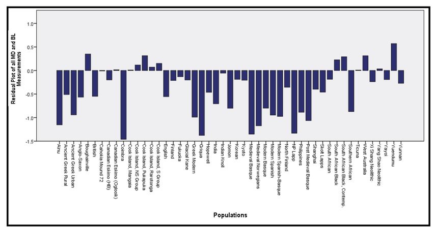
7

                                       Table 1. Age estimation based on bone length of Burial 2.

                                           Maximum Length             Estimated Age
                      Bone                                                                           References
                                                (mm)                      (years)
                    Femur L                     147.2                       1.5                    Maresh, 1970
                    Femur R                     146.7                       1.5                    Maresh, 1970
                     Tibia L                    121.9                       1.5             Gindhart, 1973; Maresh, 1970
                                                                                                  Gindhart, 1973
                     Tibia R                       120.7                     1.5
                                                                                                   Maresh, 1970
                   Fibula L                        118.9                     1.5                   Maresh, 1970
                   Fibula R                        120.4                     1.5                   Maresh, 1970
                  Humerus L                        111.7                     1.5                   Maresh, 1970
                  Humerus R                        110.9                     1.5                   Maresh, 1970
                    Ulna L                         100.9                     1.5                   Maresh, 1970
                    Ulna R                          99.5                     1.5                   Maresh, 1970
                                                                                                  Gindhart, 1973
                    Radius L                       92.2                      1.5
                                                                                                   Maresh, 1970
                                                                                                  Gindhart, 1973
                   Radius R                        91.3                      1.5
                                                                                                   Maresh, 1970
                   Clavicle L                      66.0                    2 to 3             Black and Scheuer, 1996
                   Clavicle R                      64.9                    2 to 3             Black and Scheuer, 1996

           Table 2. Age estimation based on the maturity, based     Health Indicators
           on the specimen being a female individual (Demirjian     The fully erupted deciduous dentition lacks any
           et al., 1973).                                           signs of periapical lesions, linear enamel hypo-
                                                                    plasia (LEH), dental calculus, or periodontal bone
           Developing Teeth     Demirjian’s dental score            resorption under macroscopic observation (for
            First molar (M1)              D/8                       methods used to analyze all pathological condi-
               Canine (C)                D/3.8                      tions of the dentition, see Oliveira and Neves,
           Lateral incisor (I2)          D/3.2                      2015). The only pathological conditions in the de-
           Central incisor (I1)           D/0                       ciduous dentition were superficial dental caries
               Maturity index: M1 + C + I2 + I1 = 15.0              lesions in the buccal surface of the cementoenamel

     Figure 4. Panoramic reconstruction of the maxillae and the mandible (A). The coronal views (CBCT) of the
     developing mandibular teeth: left first molar - LM1 (B); left lateral incisor - LI2 (C); left central incisor - LI1 (D);
     X-ray image from left canine - LC1 (E).

Dental Anthropology                                                                                  2020 │ Volume 33 │ Issue 02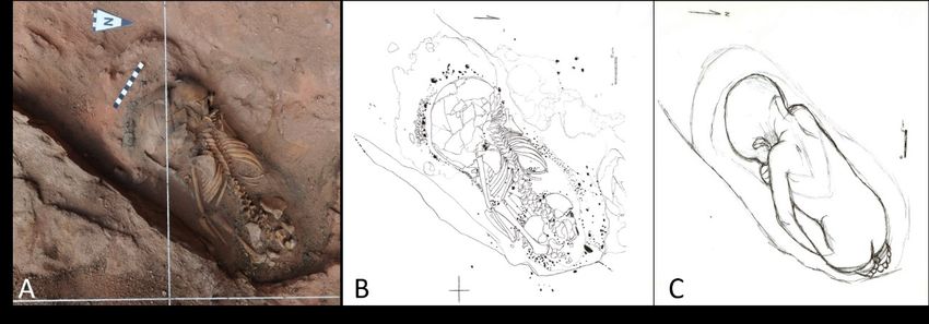
8

       Figure 5. Buccal view from mandibular teeth from Burial 2 with two dental caries in CEJ of right and left
       central incisors.

        junction of the lower central incisors (Figure 5).     molars presented moderate to intense levels of
        The irregular border and rough surface of these        thinning – with dentin observed under a thin layer
        cavities, despite being located in the ce-             of enamel on the lingual portion of both dm¹.
        mentoenamel junction, distinguish them from ab-           In addition to this pattern of differential corro-
        fraction lesions (Nascimento et al., 2016). The per-   sion, the lower and upper dentition – incisors in-
        manent canine (LC1) presents linear enamel hypo-       cluded – also presented normal occlusal wear re-
        plasia (Figure 6) and an analysis of the long bones    sulting from attrition and/or abrasion compatible
        using radiographic images revealed the existence       with Degree 2 on Molnar’s scale (Molnar, 1971;
        of Harris lines along the femora and tibiae (Mays,     Smith, 1984) (Figure 9). The symmetrical occlusal
        1995) (Figure 7).                                      dental wear pattern was observed between tooth
                                                               rows indicating normal masticatory cycles.
                                                                  A scanning electron microscopy (SEM) was
                                                               used to observe microwear of the lingual surface of
                                                               maxillary incisors. The rdi1 and rdi2 were fixed on
                                                               aluminum stubs with silver-containing glue
                                                               (Electron Microscopy Sciences/SDP - Colloidal
                                                               Silver Liquid) and sputter-coated with gold
                                                               (Balzers SCD050 – Bal-Tec/Leica Microsystems).
                                                               Teeth were examined under Sigma VP microscope
                                                               (Carl Zeiss NTS Ltd) with 50X to 600X magnifica-
                                                               tion. It was possible to observe on both specimens
                                                               some light cross-hatched scratches resulting from
                                                               masticatory abrasion (Figure 10). However, the
                                                               whole analyzed surface presented wide smooth
                                                               areas with exposed dentinal tubules, indicating an
                                                               erosion process (Figure 11).
        Figure 6. Permanent canine of Burial 2 presenting
        linear enamel hypoplasia (red arrow).                 Discussion
                                                              Changes to bones and teeth can occur for many
        Dental Wear                                           reasons and diagenesis is one of them. The enamel
        The upper incisors showed a unique pattern of non and dentine loss observed in the upper incisors of
        -incisal wear that is better described as dental cor- the subadult could be the result of dissolution in
        rosion. The severe loss of mineralized tissue result- low-pH solutions from the burial sediment in con-
        ed in the exposure of dentin on the lingual surface tact with teeth. Nevertheless, Burial 2 did not have
        (classified as IIIb on the Eccles modified index for a low pH; rather, pH was neutral at 6.64 - 7.15
        dental erosion; Eccles, 1979; Eccles and Jenkins, (Silva et al., 2019).
        1974) and the presence of a very thin enamel out-        The unique pattern of dental wear found on
        line on the lingual surface (Figure 8). The enamel Burial 2 of Pedra do Cachorro was clearly not relat-
        of the lingual surface of the deciduous canine and ed to the most common processes of occlusal attri-

Dental Anthropology                                                                            2020 │ Volume 33 │ Issue 02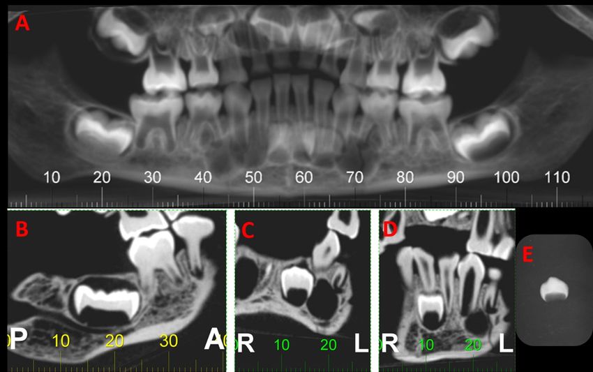
9

        Figure 7. X-ray images of the proximal extremity of femora (A) and distal extremity of tibiae (B) show-
        ing the location of Harris lines (white arrows).

     Figure 8. Detail of maxillary teeth: buccal/labial view (A) and lingual view (D) showing acid erosion on all inci-
     sors. It is possible to observe the convexities on the cervical third and the concavities on the incisal third of the
     lingual surface of upper incisor crowns. The rdi1: metalized sample (B); SEM view: 47x magnification (C); rdi2:
     metalized sample (E); SEM view: 49x magnification (F).

Dental Anthropology                                                                           2020 │ Volume 33 │ Issue 02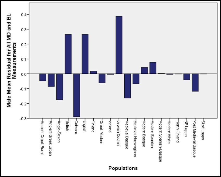
10

Figure 9. Maxillae and mandible from Burial 2. It is possible to observe physiological dental wear on the occlusal surfaces.

Figure 10. SEM of the lingual surface exposing a cross-hatched wear pattern of dental abrasion. Top row: drawing of
rdi1 shows the location of SEM analysis (A); rdi1 SEM view: 200x magnification (B); rdi1 SEM view: 400x magnification
(C). Bottom row: drawing of rdi2 shows the location of SEM analysis (D); rdi2 SEM view: 200x magnification (E); rdi2
SEM view: 400x magnification (F).

Dental Anthropology                                                                           2020 │ Volume 33 │ Issue 02
11

  Figure 11. SEM of the lingual surface showing dentine with exposed dentinal tubules, indicative of dental corrosion.
  Top row: drawing of rdi1 shows the location of SEM analysis (A); rdi1 SEM view: 400x magnification (B); rdi1 SEM view:
  600x magnification (C). Bottom row: drawing of rdi2 shows the location of SEM analysis (D); rdi2 SEM view: 400x mag-
  nification (E); rdi2 SEM view: 600x magnification (F).

        tion resulting from masticatory cycles. Parafunc-      expected in a normal 3-year-old child. The child’s
        tional habits provide alternative mechanisms capa-     deciduous teeth could have been exposed for a
        ble of generating distinct patterns of dental wear.    short period of time to a cariogenic diet, with or
        Bruxism, for example, results in considerable loss     without breastfeeding, that lasted until the third
        of mineral material. However, the abrasion angles      year of life. This was common in other precolonial
        observed in the dentition of Burial 2 were not com-    societies (Da-Gloria et al., 2017; Iida et al., 2007;
        patible with the limited movements of the tem-         Kaplan, 1996; Spielmann, 1989).
        poromandibular joint involved in bruxism (Brace           It is important to note, as mentioned above, that
        and Molnar, 1967; Molnar, 1971). Alternatively,        sex determination is imprecise, and for some au-
        injuries or malformation of the temporomandibu-        thors, it is impossible to be sure of sex when ana-
        lar joint could result in abnormal wearing of teeth.   lyzing sub-adult skeletons. Nevertheless, if we esti-
        However, for Burial 2 of Pedra do Cachorro nor-        mate age-at-death of this skeleton then, our analy-
        mal masticatory movement was indicated both by         sis suggests that if the skeleton was a girl then age-
        the perfect positioning of the upper and lower den-    at-death would have been 3.2 years, while in the
        tition, and by the presence of occlusal wear pattern   case of a boy, it would have been 3.5 years
        compatible with children with a mixed fed/             (Demirjian et al., 1973). Even if we considered that
        weaned diet, or those who had started exclusively      Burial 2 was a girl, her long bone length was that
        masticatory functions earlier in life (Martinez-       of a 1.5 years-old. Therefore the child had a low
        Maza et al., 2016; Moynihan, 2005; Warren et al.,      height for her age.
        2002). Additionally, the presence of a few carious        The frequent use of teeth as tools for creating
        lesions and no periodontal bone resorption is to be    artifacts from vegetable fibers, leather, or bones is

Dental Anthropology                                                                             2020 │ Volume 33 │ Issue 02
12
another parafunctional mechanism capable of gen- and attrition may have contributed to the dental
erating wear patterns not related to the masticatory wear noted on the occlusal surface, but the evi-
cycle. However, once again the angles of the wear dence present on the lingual surface of the maxil-
facets, the macroscopic non-flat surface of superior lary incisors shows an acidic corrosion context sim-
incisors, and the absence of complementary or sim- ilar to that from clinical cases of regurgitation le-
ilar wear on the mandibular incisors described for sions as seen in Figure 12 (Grippo et al., 2012; Lani-
Burial 2 of Pedra do Cachorro were not consistent gan and Bartlett, 2013; Lussi et al., 2011). In both
with this usage (see Figure 10 and 11). It is also cases, the lingual surfaces presented tissue loss
important to observe that Burial 2 was that of a near the gingival margins where tooth-to-tooth
young child, and therefore, less likely to participate contact does not occur (Robb et al., 1991b).
in these kind of socio-cultural activities (e.g. In addition, the SEM views strongly suggest a
Oliveira, 2014; Larsen et al., 1998; Molnar, 1971). dental erosion scenario. Parallel scratches observed
We consider that recurrent episodes of vomiting in attrition or LSAMAT cases are totally absent on
or chronic reflux were the best candidates in ex- rdi1 and rdi2 (see Figures 10 and 11) (Kieser et al.
plaining the pattern of corrosion observed for Buri- 2001). The microscopic images show a combination
al 2. The direction of the flow of gastric fluids into of light abrasive wear due to a normal chewing
the mouth resulting from these conditions process (see Figure 10), and most of the dentine
(posterior-anterior) are known to cause a strong surface with exposed dentinal tubules caused by a
and moderate/mild demineralization of the lin- corrosive process on the maxillary incisors of Buri-
gual surface of the anterior and posterior maxillary al 2 (see Figure 11) (Kieser et al. 2001).
dentition, respectively (Bartlett et al., 2013; Laz- Gastric disorder leading to systemic vomiting or
archik and Filler, 2000). The buccal surface of max- chronic reflux can be caused by a broad range of
illary teeth is partially protected by the oral muco- specific conditions such as gastrointestinal inflam-
sa, whereas mandibular teeth are protected by the matory diseases, anatomical abnormalities, malig-
cheek and tongue during vomiting, protecting nant tumors, intracranial hypertension, central
these dental surfaces from gastric fluids, as seen in nervous system infection, metabolic diseases, and
Burial 2 (Linnett and Seow, 2001). In fact, abrasion toxic food intake (Katz et al., 2013; Nebel et al.,
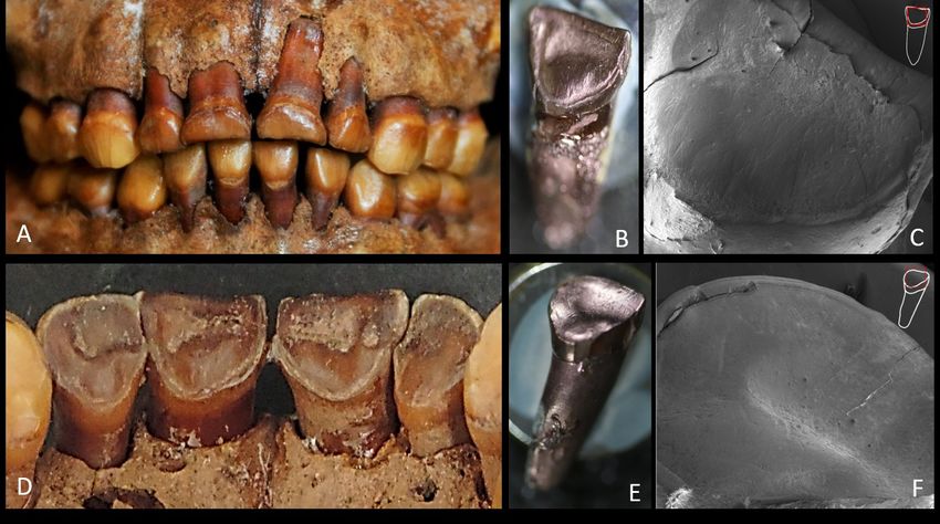
Figure 12. Comparison of the clinical case photos (above) and the archaeological case study photos
(below). The upper anterior teeth of a 27-year-old female patient with lingual wear due to dental corro-
sion by GERD (A and B). Upper anterior teeth of the Burial 2 show very similar lesions along the lin-
gual surface (C and D).
Dental Anthropology 2020 │ Volume 33 │ Issue 0213
1976; Rudolph et al., 2001; Vakil et al., 2006; van Coupal, I., & Sołtysiak, A. (2017). Dental erosion in
Herwaarden et al., 2000; Vandenplas et al., 2009). archaeological human remains: A critical re-
For Burial 2 of Pedra do Cachorro the presence of view of literature and proposal of a differential
LEH, Harris lines, and relatively short limbs seem diagnosis protocol. Archives of Oral Biology,
to indicate that the pathological condition leading 84, 50-57.
to vomiting/reflux was associated with an overall Cunninghan, C., Scheuer, L., Black, S., & Liver-
scenario of malnutrition and physiological stress sidge, H.M. (2016). Skeletal development and
(Guatelli-Steinberg et al., 2004; Oliveira and Neves, ageing, in: Cunningham, C., Scheuer, L., Black,
2015; Umapathy et al., 2013; Mays, 1995). These S., Liversidge, H.M. (Eds.), Development Juvenile
chronic disorders could be associated with the Osteology. Academic Press - Elsevier, London,
premature death of this child (Deaton, 2008; Kiel- UK, pp. 5–18.
mann and McCord, 1978; Maitland et al., 2006; d’Incau, E., Couture, C., & Maureille, B. (2012). Hu-
Onis, 2010; Rice et al., 2000; van den Broeck, 1995). man tooth wear in the past and the present:
It is interesting to note that standard osteological tribological mechanisms, scoring systems, den-
markers of metabolic distress during early child- tal and skeletal compensations. Archives of Oral
hood such as cribra orbitalia and porotic hyperosto- Biology, 57, 214–29.
sis were not observed on this individual. Finally, Da-Gloria, P., Oliveira, R.E., & Neves, W.A. (2017).
our study supports the notion that physical illness- Dental caries at Lapa do Santo, central-eastern
es such as gastric disorder could have been respon- Brazil: An Early Holocene archaeological site.
sible for cases of dental corrosion in ancient human Anais da Academia Brasileira de Ciências, 89, 307–
remains. 316.
Deaton, A. (2008). Height, health, and inequality:
Acknowledgments the distribution of adult heights in India. Amer-
Rodrigo Oliveira and André Strauss wish to ex- ican Economic Review, 98, 468–474.
press their gratitude to the Department of Archae- Demirjian A., Goldstein H., & Tanner J.M. (1973).
ology of UFPE for their kind invitation to analyze A new system of dental age assessment. Hu-
the bones of Burial 2 from Pedra do Cachorro-PE. man Biology, 45(2):211-27.
Ana Solari and Sérgio F.S.M. da Silva are grateful Deter, C.A. (2009). Gradients of occlusal wear in
for the financial support of the INCT-INAPAS. hunter-gatherers and agriculturalists. American
Ana Solari also thanks the financial support of Journal Physical Anthropology, 138, 247–54.
CAPES-PNPD. André Strauss was funded by Dong, Y.M., Pearce, E.I., Yue, L., Larsen, M.J., Gao,
FAPESP (2017/16451-2). The authors would like to X.J., & Wang, J.D. (1999). Plaque pH and asso-
thank Humberto Faria for the beautiful photos of ciated parameters in relation to caries. Caries
the skeleton presented in this article and Márcio V. Research, 33, 428–36.
Cruz for the vital technical support in microscopic Dori, I., & Moggi-Cecchi, J. (2014). An enigmatic
analyses. enamel alteration on the anterior maxillary
teeth in a prehistoric North Italian population.
REFERENCES American Journal Physical Anthropology, 154, 609
Bartlett, D.W., Lussi, A., West, N.X., Bouchard, P., –614.
Sanz, M., & Bourgeois, D. (2013). Prevalence of Eccles, J.D. (1979). Dental erosion of nonindustrial
tooth wear on buccal and lingual surfaces and origin. A clinical survey and classification.
possible risk factors in young European adults. Journal of Prosthetic Dentistry, 42(6), 649–653.
Journal of Dentistry, 41, 1007–1013. Eccles, J.D., & Jenkins, W.G. (1974). Dental erosion
Black, S., & Scheuer, L. (1996). Age changes in the and diet. Journal of Dentistry, 2, 153–159.
clavicle : from the early neonatal period to Ekstrand, J., & Oliveby, A. (1999). Fluoride in the
skeletal maturity. International Journal of Osteo- oral environment. Acta Odontologica Scandica,
archaeology, 6, 425–434. 57, 330–3.
Brace, C.L., & Molnar, S. (1967). Experimental stud- Gindhart, & P.S. (1973). Growth standards for the
ies in human tooth wear: I. American Journal tibia and radius in children aged one month
Physical Anthropology, 27, 361–8. through eighteen years. American Journal Physi-
Carlsson, G.E., Egermark, I., & Magnusson, T. cal Anthropology, 39, 41–48.
(2003). Predictors of bruxism, other oral para- Grippo, J.O., Simring, M., & Coleman, T.A. (2012).
functions, and tooth wear over a 20-year follow Abfraction, abrasion, biocorrosion, and the
-up period. Journal of Orofacial Pain, 17, 50–57. enigma of noncarious cervical lesions: A 20-
Dental Anthropology 2020 │ Volume 33 │ Issue 0214
year perspective. Journal of Esthetic and Restora- land population. Archives of Oral Biology, 58,
tive Dentistry, 24, 10–23. 1450–1456.
Guatelli-Steinberg, D., Larsen, C.S., & Hutchinson. Larsen, C.S., Teaford, M.F., & Sandford, M.K.
D.L. (2004). Prevalence and the duration of (1998). Teeth as tools at Tutu: Extramasticatory
linear enamel hypoplasia: a comparative study behavior in prehistoric St. Thomas, U.S. Virgin
of Neandertals and Inuit foragers. Journal of Islands, in: Lukacs, J.R. (Ed.), Human Dental
Human Evolution, 47, 65-84. Development, Morphology, and Pathology: A trib-
Gudmundsson, K., Kristleifsson, G., Theodors, & ute to Albert A. Dahlberg. University of Oregon
A., Holbrook, W.P. (1995). Tooth erosion, gas- Anthropological Papers, Oregon - USA, pp.
troesophageal reflux, and salivary buffer ca- 401–420.
pacity. Oral Surgery, Oral Medicine, Oral Pathol- Larsen, M.J. (2008). Erosion of the Teeth, in: Fejer-
ogy, and Oral Radiology, 79, 185–189. skov, O., Kidd, E.A.M., Nyvad, B., Baelum, V.
Hillson, S. (2008). The Current State of Dental De- (Eds.), Dental Caries: The Disease and its Clinical
cay, in: Irish, J.D., Nelson, G.C. (Eds.), Tech- Management. Blackwell Munksgaard Publish-
nique and Application in Dental Anthropology. ing Ltda, Oxford - United Kingdom, pp. 233–
Cambridge University Press, Cambridge, Unit- 248.
ed Kingdom, pp. 111–135. Lazarchik, D.A., & Filler, S.J. (2000). Dental ero-
Honório, H.M., Rios, D., Santos, C.F., Magalhães, sion: predominant oral lesion in gastroesopha-
A.C., Buzalaf, M.A.R., & Machado, M.A.A.M. geal reflux disease. American Journal of Gastro-
(2008). Effects of erosive, cariogenic or com- enterology, 95, S33-8.
bined erosive/cariogenic challenges on human Linnett, V., & Seow, K. (2001). Dental erosion in
enamel: an in situ/ex vivo study. Caries Re- children: A literature review. Pediatric Dentis-
search, 42, 454–9. try, 23, 37–43.
Iida, H., Auinger, P., Billings, R.J., & Weitzman, M. Lucas, P.W., & Omar, R. (2012). Damaged! A new
(2007). Association between infant breastfeed- overview of dental wear. Archives of Oral
ing and early childhood caries in the United Biology, 57, 211–3.
States. Pediatrics, 120, e944-52. Lussi, A., Schlueter, N., Rakhmatullina, & E.,
Irish, J.D., & Turner, C.G. More Lingual Surface Ganss, C. (2011). Dental erosion: An overview
Attrition of the Maxillary Anterior Teeth in with emphasis on chemical and histopathologi-
American Indians: Prehistoric Panamanians. cal aspects. Caries Research, 45 Suppl 1, 2–12.
(1987). American Journal of Physical Anthro- Maitland, K., Berkley, J.A., Shebbe, M., Peshu, N.,
pology, 73, 209-213. English, M., & Newton, C.R.J.C. (2006). Chil-
Järvinen, V.K., Rytömaa, I.I., & Heinonen, O.P. dren with severe malnutrition: Can those at
(1991). Risk factors in dental erosion. Journal of highest risk of death be identified with the
Dentistry Research, 70, 942–947. WHO protocol? PLoS Medicine, 3, 2431–2439.
Kaplan, H. (1996). A theory of fertility and parental Manfredini, D., & Lobbezoo, F. (2009). Role of psy-
investment in traditional and modern human chosocial factors in the etiology of bruxism.
societies. American Journal Physical Anthropolo- Journal of Orofacial Pain, 23, 153–166.
gy, 101, 91–135. Maresh, M.M. (1970). Measurements from roent-
Katz, P.O., Gerson, L.B., & Vela, M.F. (2013). genograms, in: McCammon, R.W. (Ed.), Hu-
Guidelines for the diagnosis and management man and Growth Development. Springfield IL:
of gastroesophageal reflux disease. American Thomas, Charles C, pp. 157–200.
Journal of Gastroenterology, 108, 308–28; quiz Martinez-Maza, C., Freidline, S.E., Strauss, A., &
329. Nieto-Diaz, M. (2016). Bone growth dynamics
Kielmann, A.A., & McCord, C. (1978). Weight-for- of the facial skeleton and mandible in Gorilla
age as index of risk of death in children. Lancet, gorilla and Pan troglodytes. Evolutionary
1247–1250. Biology, 43, 60–80.
Kieser, J.A., Dennison, K.J., Kaidonis, J.A., Huang, Mays, S. (1995) - The Relationship between Harris
D., & Herbison, P.G.P., Tayles, N.G. (2001). Lines and other Aspects of Skeletal Develop-
Patterns of Dental Wear in the Early Maori ment in Adults and Juveniles. Journal of Ar-
Dentition. International Journal of Osteoar- chaeological Science, 22, 511–520.
chaeology, 11, 206-217. Mays, S., & Cox, M. (2000). Sex determination in
Lanigan, L.T., & Bartlett, D.W. (2013). Tooth wear skeletal remains, in: Cox, M., Mays, S., Human
with an erosive component in a Mediaeval Ice- Osteology in Archaeology and Forensic Science.
Dental Anthropology 2020 │ Volume 33 │ Issue 0215
Cambridge: Cambridge University Press, pp. tooth wear in ancient British populations. Ar-
117-130. chives of Oral Biology, 36, 595–602.
Moazzez, R., Bartlett, D., & Anggiansah, A. (2004). Robb, N.D., Cruwys, E., & Smith, B.G.N. (1991b). Is
Dental erosion, gastro-esophageal reflux dis- “Lingual Surface Attrition of the Maxillary
ease and saliva: How are they related? Journal Teeth (LSAMAT)” Caused by Dental Erosion?
of Dentistry, 32, 489–494. American Journal Physical Anthropology, 85, 345-
Molnar, P. (2008). Dental wear and oral pathology: 351
possible evidence and consequences of habitu- Rudolph, C.D., Mazur, L.J., Liptak, G.S., Baker,
al use of teeth in a Swedish Neolithic sample. R.D., Boyle, J.T., Colletti, R.B., Gerson, W.T., &
American Journal Physical Anthropology, 136, 423 Werlin, S.L. (2001). Guidelines for evaluation
–31. and treatment of gastroesophageal reflux in
Molnar, S. (1971). Human tooth function and cul- infants and children: Recommendations of the
tural variability. American Journal Physical An- North American Society for Pediatric Gastro-
thropology, 34, 175–190. enterology and Nutrition. Journal of Pediatric
Morimoto, S., Sesma, N., Agra, C.M., Guedes- Gastroenterology and Nutrition, 32, S1–S31.
Pinto, A.C., & Hojo, K.Y. (2014). Dental Scheid, R.C., & Weiss, G. (2012). Dental anomalies,
Erosion: etiology, mechanisms and in: Scheid, R.C., Weiss, G. (Eds.), Woelfel’s Den-
implications. Journal of Biodentistry and tal Anatomy. Lippincott Williams & Wilkins,
Biomaterials, 4, 6–23. Philadelphia - USA, pp. 323–344.
Moynihan, P.J. (2005). The role of diet and nutri- Schutkowski, H. (1993). Sex determination of infant
tion in the etiology and prevention of oral dis- and juvenile skeletons: I. Morphognostic fea-
eases. Bulletin of World Health Organization, 83, tures. American Journal Physical Anthropology,
694–9. 90, 199–205.
Nascimento, M.M., Dilbone, D.A., Pereira, P.N., Smith, B.H. (1984). Patterns of molar wear in
Duarte, W.R., Geraldeli, S., & Delgado, A.J. hunter-gatherers and agriculturalists. American
(2016). Abfraction lesions: etiology, diagnosis, Journal Physical Anthropology, 63, 39–56.
and treatment options. Clinical, Cosmetic and Silva, S.F.S.M., Ghetti, N.C., & Solari, A. (2019).
Investigational Dentistry, 8, 79-87. Medição de Ph em Sedimentos Associados aos
Nebel, O.T., Fornes, M.F., & Castell, D.O. (1976). Ossos de Esqueleto de Criança, Sepultamento
Symptomatic gastroesophageal reflux: Inci- 2, Sítio Pedra Do Cachorro, Buíque – Pe, Brasil.
dence and precipitating factors. The American CLIO – Arqueológica, 2, 1-6. (In press).
Journal of Digestive Diseases, 21, 953–956. Solari, A., Silva, S.F.M.S., & Mello, S. (2015). Estudo
Oliveira, R.E. (2014). Prevalencia de patologías orales de caso sobre indicadores bioarqueológicos de
en los oasis de San Pedro de Atacama, first. ed. práticas mortuárias complexas em esqueleto
Publicia - OmniScriptum GmbH & CO - humano coletado no abrigo Pedra do
Saabrücken, Deutschland, Saarbrücken, Deuts- Cachorro, Buíque, PE. CLIO – Arqueológica, 30,
hland. 99–119.
Oliveira, R.E., & Neves, W.A. (2015). Oral health in Solari, A., Alves-Pereira, A., Sá Espinola, C.,
prehistoric San Pedro de Atacama oases, Martin, G., Pacheco da Costa, I., & Serafim
Northern Chile. HOMO - Journal of Comparative Monteiro da Silva, S.F. (2016). Escavações
Human Biology, 66, 492–507. arqueológicas no abrigo funerário Pedra Do
Onis, M. (2010). Measuring nutritional status in Cachorro, Buíque – Pe. CLIO – Arqueológica, 31,
relation to mortality. World Health Organization: 105–135.
WHO, 10, 1271–1274. Spielmann, K.A. (1989). A Review: Dietary re-
Pavone, B.W. (1985). Bruxism and its effect on the strictions on hunter-gatherer women and the
natural teeth. Journal of Prosthetic Dentistry, 53, implications for fertility and infant mortality.
692–696. Human Ecology, 17, 321–345.
Rice, A.L., Sacco, L., Hyder, A., & Black, R.E. Turner, C.G., & Machado, L.M.C. (1983). A New
(2000). Malnutrition as an underlying cause of Dental Wear Pattern and Evidence for High
childhood deaths associated with infectious Carbohydrate Consumption in a Brazilian Ar-
diseases in developing countries. Bulletin of the chaic Skeletal Population. American Journal
World Health Organization, 78, 1207–1221. Physical Anthropology, 61:125-130.
Robb, N.D., Cruwys, E., & Smith, B.G.N. (1991a). Ubelaker, D.H. (1989). Human Skeletal Remains: Ex-
Regurgitation erosion as a possible cause pf cavation, Analysis and Interpretation, Second ed.
Dental Anthropology 2020 │ Volume 33 │ Issue 0216
Taraxacum, Washington, DC - USA.
Umapathy, T., Jayam, C., Yogish, P., Yogish, A., &
Bandlapalli, A. (2013). Linear Enamel Hypo-
plasia. Journal of Indian Academy of Oral Medi-
cine and Radiology, 25(2), 153-156.
Vakil, N., Van Zanten, S.V., Kahrilas, P., Dent, J.,
Jones, R., Bianchi, L.K., & Cesario, K.B. (2006).
The Montreal definition and classification of
gastroesophageal reflux disease: A global evi-
dence-based consensus. American Journal of
Gastroenterology, 101, 1900–1920.
Van’t Spijker, A., Rodriguez, J.M., Kreulen, C.M.,
Bronkhorst, E.M., Bartlett, D.W., & Creugers,
N.H.J. (2009). Prevalence of tooth wear in
adults. Int. J. Prosthodont. 22, 35–42.
van den Broeck, J. (1995). Malnutrition and mortal-
ity. Journal of the Royal Society of Medicine, 88,
487–90.
van Herwaarden, M.A., Samsom, M., & Smout,
A.J.P.M. (2000). Excess gastroesophageal reflux
in patients with hiatus hernia is caused by
mechanisms other than transient LES relaxa-
tions. Gastroenterology, 119, 1439–1446.
Vandenplas, Y., Rudolph, C.D., Lorenzo, C., Has-
sall, E., Liptak, G., Mazur, L., Sondheimer, J.,
Staiano, A., Thomson, M., Veereman-Wauters,
G., & Wenzl, T.G. (2009). Pediatric gas-
troesophageal reflux clinical practice guide-
lines: joint recommendations of the North
American Society for Pediatric Gastroenterolo-
gy, Hepatology, and Nutrition (NASPGHAN)
and the European Society for Pediatric Gastro-
enterology, Hepatology, and Nutrition
(ESPGHAN). Journal of Pediatric Gastroenterolo-
gy and Nutrition, 49, 498–547.
Warren, J.J., Yonezu, T., Bishara, S.E., & Ortho, D.
(2002). Tooth wear patterns in the deciduous
dentition. American Journal of Orthodontics and
Dentofacial Orthopedics, 122, 614–618.
Waters-Rist, A., Bazaliiskii, V.I., Weber, A., Gori-
unova, O.I., & Katzenberg, M.A. (2010). Activi-
ty-induced dental modification in Holocene
Siberian hunter-fisher-gatherers. American Jour-
nal Physical Anthropology, 143, 266–78.
Dental Anthropology 2020 │ Volume 33 │ Issue 0217
A Dental Metric Study of Medieval, Post Medieval, and Modern
Basque Populations from Northern Spain
Diana Malarchik1*, Marin A. Pilloud2, and G. Richard Scott2
1
University of California, Davis
2
University of Nevada, Reno
Keywords: odontometrics, Basques, dental variation, biodistance
ABSTRACT Basque population history has been examined through classic genetic markers, mtDNA, Y
chromosome haplogroups, craniometrics, and recently dental morphology. Dental morphological data
show Basques have a classic European dental pattern but fall as an outlier among European popula-
tions. Expanding on that work, Basque tooth size was examined to further evaluate the affinities of the
Basque population. Mesiodistal and buccolingual maximum crown measurements were taken from me-
dieval and post medieval skeletons from the Catedral de Santa María in Vitoria, Spain, along with living
samples of modern Basques, Spanish, and Spanish Basques from dental students at the Universidad del
País Vasco. A dental metric examination affirms the outlier status of Basques, as they exhibit smaller
crown areas than neighboring populations. In biodistance analyses Basque populations group with lin-
guistically and geographically distant populations. Even with gene flow from Spain, France, and North
Africa, Basque individuals still demonstrate a unique pattern coincident with their ancient origins.
The Basque Country, Euskalherria, is located in the phisms show low levels of diversity, suggesting
southwestern corner of France and north central that this population has been evolving in the re-
Spain. The population of the region is well known gion for millennia (Alonso et al., 2005; Hurles et al.,
for its unique language, as “the sole surviving pre- 1999).
Indo European language of Western Eu- More recently, data on Basque dental morpholo-
rope” (Trask, 1997:35). Many anthropological ap- gy was investigated to explore the population his-
proaches have been taken to better understand the tory of this group. Typically, European popula-
place of Basques in European history, from linguis- tions are classified by morphologically simple teeth
tic to archaeological research, and more recently where trait absence is more common than trait pres-
investigating genetic haplotypes. Early research ence (Scott and Turner, 1997). Scott and colleagues
explored Basque blood groups, finding that (2013) found that Basque samples, both historic
Basques had high frequencies of the blood type O and living, have high rates of hypocone and hy-
allele (ca. 75%), low rates of blood type B allele (ca. poconulid reduction on UM2 and LM2, respective-
3%), and the world’s highest frequencies of the ly. There is also an extremely high frequency of
negative allele (“r” or “cde”) in the Rhesus blood double rooted lower canines, a classic European
group system (ca. 50%) (Roychoudhury and Nei, trait (Scott et al., 2013; Scott and Turner, 1997).
1988). These frequencies set them apart from other These findings place Basque groups into the over-
Western Europeans (Alberdi et al., 1957; Chalmers all category of Western Europe, within the
et al., 1948; van der Heide et al., 1952). These unu- ‘Eurodont’ dental pattern (as coined by that study).
sual blood types were interpreted by Cavalli- There is no single trait that separates the Basques
Sforza (2000) as a possible link to the first wave of
people coming into Europe during the Paleolithic
and served as the stimulus for many genetic stud- *Correspondence to:
ies to examine the origins and affinities of the Diana Malarchik
Basque population. Analyses of mitochondrial Department of Anthropology
DNA (mtDNA) show unique haplogroups suggest- University of California, Davis
ing in situ evolution with minimal gene flow dmalarchik@ucdavis.edu
(Alzualde et al., 2005; Alzualde et al., 2006; Mar-
tinez-Cruz et al., 2012). Y chromosome polymor-
Dental Anthropology 2020 │ Volume 33 │ Issue 0218
from other European groups. It is rather the accu- one of the authors (GRS) following Moorrees
mulation of slight but consistent differences that (1957). Measurements were taken on the left side of
create their outlier status (Scott et al., 2013). the dental arcade. The right antimere was substi-
To further explore dental variation among tuted in cases of antemortem or postmortem tooth
Basque populations we evaluate here dental met- loss, gross carious lesions, excessive wear, or any
rics. The goal of the present study is to determine other condition that would make the left side un-
if the unique population history of the Basques is observable. Teeth with large carious lesions, exces-
evident in tooth crown size throughout time. If sive dental calculus, or marked occlusal wear were
preceding studies are any indicator, it is expected omitted from analysis. Table 1 is a summary of
that Basques will show tooth size patterns like material available for study in this analysis.
those of the other Western Eurasian groups, with Along with maximum mesiodistal (MD) and
slight differences reflecting their long-term occu- buccolingual (BL) crown measurements, two addi-
pancy in Western Europe along with relative geo- tional measurements were calculated. Tooth size
graphic isolation. It is further expected that these as the product of the maximum crown dimensions
patterns will be evident from Medieval to modern was also analyzed (TS=MDxBL) as was total crown
times. area for each tooth type. Crown area was defined
as the sum of TS (ΣTS) for all teeth in a single tooth
Materials and Methods class, with the exception of the third molar.
The skeletal remains examined in this study were To analyze sexual dimorphism, the male mean
collected from the Catedral de Santa María in Vito- was divided by the female mean of each measure-
ria-Gazteiz, Alava, País Vasco, Spain. These re- ment for each tooth, and then multiplied by 100
mains date from the 11th to the 19th century, and (Garn et al., 1967b; Harris, 1997). Sexual dimor-
were also the subject of studies on dental morphol- phism was also examined through a multivariate
ogy (Scott et al., 2013), oral health (Hopkinson, analysis of variance (MANOVA) and a Student’s t-
2009), craniometry (Janzen, 2011), dental chipping test. Statistical significance was measured using the
(Scott and Winn, 2010), and taphonomy Bonferroni correction. Principal components analy-
(Hopkinson et al., 2009). Sex was estimated by one sis (PCA) and discriminant function analysis
of the authors (GRS) based on skull and pelvic (DFA) were used to explore differences between
morphology (Buikstra and Ubelaker, 1994). populations.
Additionally, dental casts were collected from Three major benefits of using a PCA in the
living people by Alberto Anta at the University of study of human tooth size variation include: (1)
the Basque Country from students who were en- reducing data on inter-correlated variables into
rolled in the dental school at that time. For these compound variables; (2) extracting the major de-
individuals, sex and cultural identification velopmental fields controlling tooth size; and (3)
(Basque, Spanish-Basque, or Spanish) were record- providing statistically independent measures for
ed at time of casting. between group comparisons (Harris, 1997). The
Maximum crown measurements were taken by extracted components were then used in Euclidean
Table 1. Male and female samples by time period.
Time Period Population Location of Collection Male (n) Female (n)
Medieval Basque Catedral de Santa Ma- 65 28
(1100-1350) ria
Catedral de Santa Ma-
Post Medieval Basque ria 90 126
(1400-1850)
Dental Casts; Universi-
Modern Spanish dad del País Vasco, 8 48
(2005) Spanish-Basque Dental School 13 39
Basque 8 28
Total 184 269
Dental Anthropology 2020 │ Volume 33 │ Issue 0219
distance analysis in which Ward’s dendrograms Basque. The degree of sexual dimorphism ((male
were created. mean/ female mean)*100) is in line with other
Tooth apportionment was used to create residu- odontometric studies (Moorrees 1957; Keiser 1990)
al scores, where the expected variation (PCA on that show males with teeth on average 2-4% larger
the sum of the dental arcades) was subtracted from than females, with canines slightly more dimorphic
the observed variation (PCA for all individual at 4-6% (Table 3). The modern Basque sample was
tooth measurements). These are used to view a the only sample to vary, with males not exhibiting
group’s variation in the entire dentition or by mor- larger teeth than the females, although this is most
phogenetic fields, depending on research questions likely due to the over representation of females in
and available data sets (Harris, 1997). the sample (see Table 1).
The use of residual scores shows each group’s First, temporal variation was examined. A cross
variation from their predicted overall tooth size. -validated stepwise DFA classified individuals into
These residual scores can be visualized through one of three groups (medieval, post medieval, or
bar graphs or the scores can be subjected to further modern) with an accuracy of 46.4%, which is
statistical analysis to show population grouping. slightly better than random chance (Table 4). Medi-
The axis on which the scores are plotted represents eval and modern samples have the highest per-
the expected size of the dentition for each sample; centages of correct classification, both around 70%,
negative scores, as indicated by bars plotting be- while the post medieval was the hardest to classify
low the expected line, show teeth that are smaller with a rate of 27%. Poor classification of the post
than expected, while positive scores show teeth medieval group was expected, as this transitional
that are larger than expected. Analysis using resid- group most likely represents the median between
ual scores allows published mean scores to be the medieval and modern groups, thus allowing
used, expanding sample sizes in comparative anal- for incorrect classifications to occur more frequent-
yses (Harris and Rathbun, 1991). ly.
While PCA emphasizes variation within popu- Crown areas were used to examine differences
lations, discriminant function analysis between temporal periods and population. Plotting
examines variation by maximizing differences be- anterior and posterior crown areas for all popula-
tween groups and minimizing variation in a tions collected, there is a clear shift in tooth size as
group (Kachigan, 1986). Raw data are required to time increases. Although among males, there is a
run a discriminant function; therefore, this shift from the expected, as the modern Basque
method was only used to examine variation for populations have slightly smaller teeth than medi-
samples collected as part of this study. Using these eval Basques (Figure 1). This is mostly likely due to
samples, a stepwise DFA was used to compare the poor representation of males in the modern
Basque temporal periods. All analyses were con- sample, where males were underrepresented in the
ducted in SPSS version 22 (IBM Corp., 2013). dental school population when the casts were col-
To explore population variation, eighty-two lected. When looking at females, post medieval
comparative samples of summary statistics of den- Basques show larger tooth size for both premolars
tal metrics were assembled from published sources and molars when compared to medieval Basque
(Table 2). These samples cover multiple temporal samples (Figure 2). Premolars show an increase in
periods and geographic areas and were divided size of 5.7% while molars increased by 6.6%. Mod-
into five regions (Western Eurasia, Sino America, ern Basques exhibit larger teeth than the post me-
Sahul Pacific, Sunda Pacific, and Sub Saharan Afri- dieval samples at 8.3% in premolars and 1% in mo-
ca) for comparisons described by Scott and Turner lars.
(1997). To examine Basque variation, analyses fo- Examining Basque tooth crown apportionment
cused on: (1) temporal variation within the Basque along with other Western Eurasian populations
samples; (2) Basque variation viewed on a conti- from the published literature, residual factors for
nental level comparing Basque samples to Western all dental arcades were used to make bar graphs
Eurasian groups; and (3) Basque variation in a following the methods of Harris and Rathbun
global context. (1991). As tooth crown measurements are sexually
dimorphic, males and females were analyzed sepa-
Results rately. Examining all measurements for male Euro-
Dental metrics were evaluated for sexual dimor- pean samples, medieval Basques show scores of
phism within the five samples: medieval, post me- disproportionally small teeth, with post medieval
dieval, modern Basque, Spanish, and Spanish Basques and medieval Norwegians falling interme-
Dental Anthropology 2020 │ Volume 33 │ Issue 0220
Table 2. Published comparative samples used in analyses by region.
Region Population Citation
Western Eurasia Anglo-Saxon Lavelle 1968
Bedouin Rosenzweig and Zilberman 1969
British Lavelle 1968
Caucasus Kieser et al. 1985
Circassian (Israel) Koyoumdjisky-Kaye et al. 1977
Coimbra Galera and Cunha 1993
Druse Koyoumdjisky-Kaye et al. 1977
English Lavelle 1968
Finland Alvesalo 1985
Iceland Axelsson and Kirveskari 1983
Jewish Cochini Rosenzweig and Zilberman 1967
Medieval Norwegians Beyer-Olsen and Alexandersen 1995
Modern Greek Zorba et al. 2011
Modern White Axelsson and Kirveskari 1983
North Finland Kirveskari et al. 1977
NP Lapp Kirveskari 1977
Pashtun Sakai et al. 1971
Rural Ancient Greek Henneberg 1998
Skolt Lapps Kirveskari 1977
South African Whites Kieser et al. 1985e
Tristan da Cunha Thomsen 1955
Urban Ancient Greek Henneberg 1998
Sino America Adena Sciulli 1979
Ainu Brace and Nagai 1982
Aleut Moorrees 1957
Cahokia Mound 72 Thompson 2013
Canadian Eskimo (Iglooik) Mayhall 1979
Canadian Eskimo (HB) Mayhall 1979
Chinese Bronze Brace 1976
East Greenland Eskimo Pedersen 1949
Glacial Kane Sciulli 1979
Fukuoka Brace and Nagai 1982
Highland Beach Iscan 1989
Hopewell Sciulli 1979
Indian Knoll Perzigian 1976
Jomon Brace and Nagai 1982
Kansas Schultz Mound Phenice 1969
Korean Brace and Nagai 1976
Kyoto Brace and Nagai 1982
Lengua Kieser et al. 1985e
Pecos Nelson 1938
Shanghai Brace and Nagai 1982
St. Lawrence Island Eskimo Scott and Gillispie 2002
Tennessee (A) Hinton et al. 1980
Tennessee (M) Hinton et al. 1980
Tennessee (W) Hinton et al. 1980
Tibet Sharma 1983
Ticuna Harris and Nweeia 1980b
Xi Shang Neolithic Brace, Shao, Zhang 1984
Yayoi Brace and Nagai 1982
Yunnan Brace and Nagai 1982
Dental Anthropology 2020 │ Volume 33 │ Issue 0221
Table 2. Published comparative samples used in analyses by region (cont’d).
Region Population Citation
Sahul Pacific Australian Aborigine Campbell 1925
Broadbeach Smith et al. 1981
Bougainville (Solomon Islands) Bailit et al. 1968
Walbiri, Australia Barrett et al. 1963,64; Brace 1980
Western Australia Freedman and Lofgren 1981
Sunda Pacific Cook Island, Mangaia Yamada et al. 1988
Cook Island, NS Group Yamada et al. 1988
Cook Island, Pukapuka Yamada et al. 1988
Cook Island, Rarotonga Yamada et al. 1988
Cook Island, S Group Yamada et al. 1988
Javanese Bronze Brace 1976
Java Brace 1980
India (Chalcolithic) Lukacs 1985
India Acharya and Prabhu 2011
India Walimbe 1985
Philippines Potter et al. 1981
South-east Java Taverne 1980
Tajik Sakai et al. 1971
Thai Brace 1976
Thai Bronze Brace 1976
Yang Shao Neolithic Brace, Shao, Zhang 1984
Sub Saharan Africa Bantu Shaw 1931
Griqua Kieser 1985
San van Reenan 1982
San Drennan 1929
South African Black, Contemp. Kieser et al. 1987
South African Black Kieser et al. 1987
South African Jacobsen 1982
Southern African van Reenan 1982
Teso Barnes 1969
Table 3. Sexual dimorphism separated by temporal period
UI1MD UI2MD UCMD UP1MD UP2MD UM1MD UM2MD
Medieval 105.23 105.39 104.00 103.66 103.64 103.86 106.09
Post Medieval 100.65 103.54 103.26 101.29 102.57 101.44 102.32
Spanish 102.79 104.51 102.39 101.71 103.35 102.29 101.26
Spanish-Basque 103.54 103.95 104.94 106.74 102.89 105.45 105.95
Basque 105.32 102.95 102.46 99.49 95.94 95.78 97.56
UI1BL UI2BL UCBL UP1BL UP2BL UM1BL UM2BL
Medieval 103.21 104.73 102.54 102.61 103.97 103.30 105.78
Post Medieval 104.60 106.96 103.82 101.24 101.43 102.57 102.97
Spanish 105.88 103.77 103.04 100.68 102.82 101.73 104.15
Spanish-Basque 111.28 115.41 112.71 106.27 107.18 104.27 108.62
Basque 100.43 106.32 104.92 99.18 97.60 100.10 101.38
LI1MD LI2MD LCMD LP1MD LP2MD LM1MD LM2MD
Medieval 97.15 101.33 102.02 103.46 104.94 105.25 104.84
Post Medieval 100.87 102.08 102.70 102.13 100.64 104.55 103.82
Spanish 102.99 104.47 102.55 100.60 100.55 100.16 102.38
Spanish-Basque 101.01 103.32 106.08 105.62 104.61 104.09 105.44
Basque 98.75 102.67 99.94 101.68 85.40 98.63 96.10
LI1BL LI2BL LCBL LP1BL LP2BL LM1BL LM2BL
Medieval 101.56 101.57 103.89 102.50 102.69 101.87 102.42
Post Medieval 103.11 102.47 107.93 102.21 101.35 103.37 104.23
Spanish 104.25 98.66 102.38 100.49 99.43 102.80 103.35
Spanish-Basque 110.68 103.49 107.82 110.29 106.93 106.86 108.19
Basque 103.33 99.63 97.08 98.06 99.64 97.40 97.58
Dental Anthropology 2020 │ Volume 33 │ Issue 02You can also read