Discovery of a New Analgesic Peptide, Leptucin, from the Iranian Scorpion, Hemiscorpius lepturus - MDPI
←
→
Page content transcription
If your browser does not render page correctly, please read the page content below
Article

Discovery of a New Analgesic Peptide, Leptucin,
from the Iranian Scorpion, Hemiscorpius lepturus
Sedigheh Bagheri‐Ziari 1, Delavar Shahbazzadeh 1, Soroush Sardari 2, Jean‐Marc Sabatier 3
and Kamran Pooshang Bagheri 1,*

                                           1 Venom and Biotherapeutics Molecules Laboratory, Medical Biotechnology Department,
                                             Biotechnology Research Center, Pasteur Institute of Iran, Tehran 1316943551, Iran;
                                             sebagheri2000@gmail.com (S.B.‐Z.); shahbazzadeh@yahoo.com (D.S.)
                                           2 Drug Design and Bioinformatics Unit, Medical Biotechnology Department, Biotechnology Research Center,

                                             Pasteur Institute of Iran, Tehran 1316943551, Iran; ssardari@hotmail.com
                                           3 Institute of NeuroPhysiopathology (INP), Faculté de Pharmacie, Université d’Aix‐Marseille, UMR 7051,

                                             27 Bd Jean Moulin, CEDEX 05, 13385 Marseille, France; sabatier.jm1@gmail.com
                                           * Correspondence: k_bagheri@pasteur.ac.ir

                                           Abstract: Hemiscorpius lepturus scorpion stings do not induce considerable pain based on epidemi‐
                                           ological surveys conducted in the southwest part of Iran. Accordingly, this study was aimed to
                                           identify the analgesic molecule in H. lepturus venom by analyzing a cDNA library of the scorpion
                                           venom gland looking for sequences having homology with known animal venom analgesic pep‐
                                           tides. The analgesic molecule is a cysteine rich peptide of 55 amino acids. the synthetic peptide
                                           was deprotected and refolded. RP‐HPLC, Ellman’s, and DLS assays confirmed the refolding accu‐
Citation: Bagheri‐Ziari, S.;               racy. Circular dichroism (CD) showed helix and beta sheet contents. This peptide, called leptucin,
Shahbazzadeh, D.; Sardari, S.;             demonstrated 95% analgesic activity at the dose of 0.48 mg/kg in hot plate assay. Leptucin at the
Sabatier, J.‐M.; Pooshang Bagheri, K.      doses of 0.32, 0.48, and 0.64 mg/kg showed 100% activity in thermal tail flick test. No hemolysis or
Discovery of a New Analgesic
                                           cytotoxicity was observed at 8 and 16 μg. Histopathology evaluations indicated no hepatotoxicity,
Peptide, Leptucin, from the Iranian
                                           nephrotoxicity, and cardiotoxicity. We thus report that leptucin is the analgesic agent of H. lep‐
Scorpion, Hemiscorpius lepturus.
                                           turus venom. Regarding the high in vivo efficacy of leptucin and the fact it shows no observable
Molecules 2021, 26, 2580.
                                           toxicity, it could be suggested as a drug lead in a preclinical study of acute pain as well as the
https://doi.org/10.3390/
molecules26092580                          study of its mechanism of action.

Academic Editor: Rosanna Di Paola          Keywords: leptucin; analgesic peptide; Hemiscorpius lepturus; scorpion

Received: 29 March 2021
Accepted: 26 April 2021
Published: 28 April 2021                   1. Introduction
                                                 Pain is the most well‐known unpleasant experience which protects a human from
Publisher’s Note: MDPI stays neu‐
                                           injuries [1,2]. Injuries cause acute or chronic pain. Acute pain occurs immediately after
tral with regard to jurisdictional
                                           an injury and is usually severe in nature, whereas chronic pain continues beyond the
claims in published maps and insti‐
tutional affiliations.
                                           normal healing process. It can start as acute pain but lasts for more than 3 months. The
                                           main causative agents for acute pain include surgery, traumas, bone fractures, dental
                                           work, burns or cuts, and for chronic pain lower back pain, cancer, and arthritis [3].
                                                 An analgesic is a drug that relieves pain [4]. They are classified into non‐opioid an‐
Copyright: © 2021 by the authors.          algesics (paracetamol, ibuprofen, COX‐2 inhibitors, NSAIDs, diclofenac, etc.) and opioid
Licensee MDPI, Basel, Switzerland.         analgesics (morphine, oxycodone, etc.) [5,6]. Among analgesic drugs, NSAIDs are rou‐
This article is an open access article     tinely used for acute pain, but in the case of contraindication and severe pain, opioids
distributed under the terms and            are used [5]. Among opioids, morphine is the gold standard for pain relief, but their use
conditions of the Creative Commons         is limited due to a number of side effects, including respiratory depression, constipation,
Attribution     (CC      BY)     license   intolerance, dependence, and addiction [7]. Thus, the design or discovery of a new mol‐
(http://creativecommons.org/licenses       ecule with a lesser complication is necessary. Since peptide molecules are more targeted
/by/4.0/).                                 and have fewer side effects than chemical small molecules, analgesic peptides could be a

Molecules 2021, 26, 2580. https://doi.org/10.3390/molecules26092580                                               www.mdpi.com/journal/molecules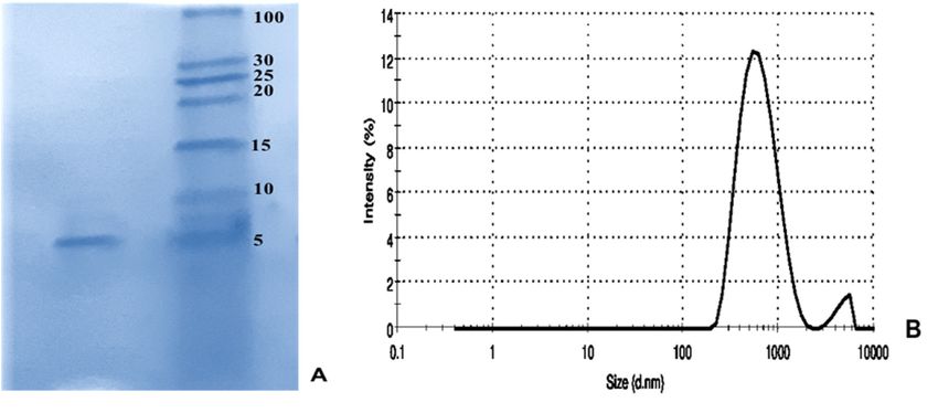
Molecules 2021, 26, 2580                                                                                         2 of 17

                           new revolution in pain‐relief [8–10].
                                Venomous animals are important sources for discovery or development of new
                           drug leads [11–21]. Approved peptide drugs from the venom of venomous animals in‐
                           clude captopril (isolated from the venom of Bothrops jararaca, a pit viper, to control blood
                           pressure), eptifibatide and tirofiban (isolated from viperid snake venom as an antiplate‐
                           let drug), lepirudin and bivalirudin (isolated from leech venom as anticoagulants), zi‐
                           conotide (isolated from cone snails venom to suppress chronic pain), exenatide (isolated
                           from lizard venom to treatment of type 2 diabetes) [15].
                                Venomous creatures have high potential for discovery or development of peptide
                           drug leads [22–24]. Among the venomous animals, scorpions are also an important
                           source for discovery of potential peptide or protein molecules with different pharmaco‐
                           logical activities [24–27].
                                Some peptides in scorpion venom make scorpion stings very painful [28–30], but
                           the stings of the Iranian scorpion, Hemiscorpius lepturus (H. lepturus) do not cause consid‐
                           erable pain based on local evidence, epidemiological studies, and clinical reports [31–35].
                           To our best of knowledge, no study has been conducted on the cause of the painless
                           stings of this scorpion.
                                H. lepturus belongs to the hemiscorpidae family, distributed in Iran, Iraq, Pakistan,
                           and Yemen [36]. To date, some peptides with pharmacological or toxicological activities
                           have been discovered from the venom of H. lepturus venom i.e., hemicalcin [37], hemo‐
                           toxin [36], hemilipin [38], PLD1 [31], HL2153 and HL2155 [39].
                                In the present study, we identified in the cDNA library of the H. lepturus scorpion
                           venom gland a peptide homologue to a known analgesic peptide from sea anemone. The
                           peptide was synthetized, refolded, and analyzed for its structure and analgesic activity
                           as well as its toxicity.

                           2. Materials and Methods
                           2.1. Materials, Cells, and Animals
                                3‐(4,5‐Dimethyl‐2‐thiazolyl)‐2,5‐diphenyl‐2H‐tetrazolium bromide (MTT) was pur‐
                           chased from Sigma (Saint Louis, MO, USA) and isopropanol, triflouroacetic acid (TFA),
                           and acetonitrile (ACN) were purchased from Merck (Darmstadt, Germany). Morphine
                           sulfate was obtained from Darou Pakhsh Pharmaceuticals (Tehran, Iran). DMEM and fe‐
                           tal bovine serum (FBS) were purchased from Gibco, Life Technologies (Grand Island,
                           NY, USA). Human Embryo Kidney (HEK293, NCBI code C497) was obtained from Pas‐
                           teur Institute of Iran (Tehran, Iran).
                                NMRI mice (Male, 25 g) were purchased from the Pasteur Institute of Iran. The an‐
                           imals were allowed to adapt for a week in standard conditions with a dark/light cycle of
                           12 h. The room temperature was 22 ± 1 °C and the relative humidity was adjusted at 50 ±
                           5%. The animals received a standard pellet diet and fresh tap water. All experiments
                           were approved by the Ethical Committee of the Pasteur Institute of Iran (code number
                           IR.PII.REC.1394.86).

                           2.2. Bioinformatics Analyses
                           2.2.1. Data Mining and ORF Determination
                                In this study, we used the data from a cDNA library of the venom gland of H. lep‐
                           turus scorpion, which had been generated by Illumina RNA sequencing in 2017 [31], in
                           order to find the analgesic molecule. The relevant keywords like analgesic, anti‐pain,
                           and pain relieving were searched in the annotated library. Then the corresponding DNA
                           sequence was translated to ORF using ORF finder (https://www.ncbi.nlm.nih.gov/
                           orffinder/) (accessed on 16 March 2020).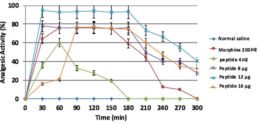
Molecules 2021, 26, 2580                                                                                            3 of 17

                           2.2.2. Similarity Analysis and Determination of the Mature Chain
                                The similarity of ORF was determined against non‐redundant and reference se‐
                           quences using the ‘BLASTP’ server (http://blast.ncbi. nlm.nih.gov/Blast.cgi) (accessed on
                           17 March 2020). The mature chain and signal peptide of its similar sequences were
                           checked in NCBI and UniProt. Then, for accurate determination of signal peptide and
                           mature chain, the identified ORF for H. lepturus analgesic peptide was multiple aligned
                           against the similar sequences using ‘COBALT’ server [40]. To control the accuracy of
                           alignment, the evolutionary signature of aligned peptides was determined too. Then the
                           molecular weight of the mature chain was calculated using the ‘Protparam’ server
                           (http://web.expasy.org/protparam) (accessed on 18 March 2020).

                           2.2.3. Prediction of 3D Structure of the Peptide
                               The 3D structure of the peptide was predicted using the Iterative Threading As‐
                           sembly Refinement (I‐TASSER) server at Michigan University [41]. A model with the
                           lowest root mean square deviation (RMSD) was selected. This model was viewed and
                           analyzed by using ‘UCSF Chimera’ software package [42]. Structural alignment with the
                           analgesic peptide of a sea anemone, was performed and visualized using the UCSF
                           Chimera software. Based on 3D structure analysis, disulfide bonds between cysteine
                           amino acids were predicted.

                           2.3. Peptide Synthesis
                                 The peptide was synthesized by an external facility (Biomatik Co., Cambridge, ON,
                           Canada) using the solid phase method and Fmoc (9‐fluorenyl‐methoxycarbonyl) chemis‐
                           try. In order to avoid oxidizing the sulfur moieties of cysteine, all cysteines in peptides
                           were protected with acetamidomethyl (ACM) groups. The peptide was amidated at the
                           C‐terminal. The peptide was purified up to 95% by reverse phase‐high performance liq‐
                           uid chromatography (RP‐HPLC). The molecular weight of the peptide was also con‐
                           firmed using the mass spectrometry method.

                           2.4. Deprotection of the Synthetic Peptide
                                In order to refold the peptide and examine its activity, protected groups must be
                           removed. Deprotection was performed based on the molar ratio method [43]. First, 1 mg
                           of the synthesized peptide was dissolved in degassed distilled water. Then 100 μL of the
                           peptide solution was added to 900 μL of mercury acetate solution (0.1% in degassed distilled
                           water) and incubated at RT for 60 min. Then, 10 μL of 2‐mercaptoethanol (10 mM) was add‐
                           ed to the solution, incubated for 30 min under nitrogen gas, and centrifuged at 10,625× g for 5
                           min. The supernatant was collected and freeze‐dried at −55 °C for 24 h in a freeze dryer sys‐
                           tem (alpha 1–2 LD plus; Martin Christ Gefriertrocknungsanlagen GmbH, Osterode am Harz,
                           Germany).The abovementioned method was performed in four pH protocols. The peptide
                           was dissolved in two pH conditions (i.e., 4 and 7) and the deprotection of peptides was sub‐
                           sequently performed in two pH conditions (i.e., 4 and 7).

                           2.4.1. Quality Control of the Deprotected Peptide by SDS‐PAGE
                                To compare the yield of peptide in the supernatant and pellet in four pH protocols,
                           SDS‐PAGE was performed using 15% polyacrylamide gel. The best protocol of deprotec‐
                           tion method was considered for further evaluation in RP‐HPLC.

                           2.4.2. Evaluation of the Accuracy of the Deprotection Method by RP‐HPLC
                                RP‐HPLC was carried out on a Knauer system (Knauer Wissenschaftliche Gerate
                           Co., Berlin, Germany), with C18 column (Beckman‐Coulter, Brea, CA, USA) 100 Å, 250 ×
                           4.6 mm). One hundred μL of peptide solution was injected into the injector and a linear
                           gradient of acetonitrile was applied from 0 to 90% (absolute acetonitrile containing 0.1%
                           TFA) at a flow rate of 1 mL/min for 90 min. After that, the retention time of the depro‐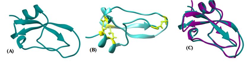
Molecules 2021, 26, 2580                                                                                        4 of 17

                           tected peptide was compared to the protected one. Any alteration in the retention time
                           of the deprotected peptide indicates that the peptide is deprotected. The deprotected
                           peptide was collected and lyophilized as mentioned above.

                           2.4.3. Determination of the Purity and Yield of the Deprotected Peptide
                                The purity of the deprotected peptide was determined by using chromgate software
                           package. The yield was determined by calculation of the difference between the amount
                           of peptide before and after deprotection. The concentration of the peptide was measured
                           by UV estimation at 280 nm in a spectrophotometer. Yield percent was calculated using
                           the following formula [30]:
                                                                peptide amount after deprotection
                                            Yield percent ൌ ൬                                      ൰ ൈ 100         (1)
                                                                peptide amount before deprotection

                           2.5. Refolding of the Deprotected Peptide
                                 At first, ammonium acetate solution (20 mM, pH 6.5) prepared in 900 μL degassed
                           water for injection (WFI). Then, the different amounts of reduced glutathione (GSH) and
                           oxidized glutathione (GSSG) were added to each vial to reach a ratio of GSH/GSSG as
                           10:1 (3.07 mg: 0.612 mg) and 2:1 (0.614 mg: 0.612 mg). Ultimately, two amounts of depro‐
                           tected peptide (40 and 80 μg in 50 and 100 μL, respectively) were added to the separate
                           solutions. The final volume was adjusted to 1 mL by ammonium acetate solution (20
                           mM, pH 6.5).Then the solutions were stirred at 22 °C for 4 h [44]. All steps were per‐
                           formed under nitrogen gas to ovoid oxidizing the cysteine amino acid residues.
                                 To determine the optimal refolding conditions, RP‐HPLC was performed for the
                           sample and negative control (peptide‐free buffer) as well. The results were compared to
                           negative control, and the peak corresponding to the refolded peptide was collected and
                           lyophilized. SDS‐PAGE was then performed to verify the existence of peptide. Then, the
                           resulting peptide was subjected to Ellman assay to further confirmation of peptide re‐
                           folding. For the activity assays, the sample was dialyzed against normal saline in a dial‐
                           ysis tube (MWCO: 3000 Da) for 24 h.

                           2.5.1. Determination of the Purity and Yield of the Refolded Peptide
                                The purity of the refolded peptide was determined by using chromgate software
                           package. The yield was determined by calculation of the difference between the amount
                           of peptide before and after refolding. The concentration of the peptide was measured by
                           UV estimation at 280 nm in a spectrophotometer. Yield percent was calculated as de‐
                           tailed above [30].

                           2.5.2. Confirmation of Refolding by Ellman’s Assay
                                This assay was performed to confirm that there is no free thiol residue in the surface
                           of refolded peptide. No reaction is expected for the refolded peptide. This assay was per‐
                           formed based on the protocol developed by Shahangian, et al. [45].
                                Briefly, refolded peptide (5 mM) incubated with a reaction mixture (8M urea, 100
                           mM sodium‐phosphate, pH 8.0, and 1 mM 5,5′‐dithiobis‐(2‐nitrobenzoic acid) (DTNB)) at
                           room temperature. The optical density (OD) was monitored at 412 nm after 1 h. The
                           Ellman’s reagent solution without refolded peptide was used as negative control. The num‐
                           ber of free thiols was calculated using a molar extinction coefficient of 13,600 M−1 cm−1.
Molecules 2021, 26, 2580                                                                                          5 of 17

                           2.6. Dynamic Light‐Scattering (DLS)
                                To assess the homogeneity of the refolded purified peptide (100 μg in 500 μL wa‐
                           ter/TFA 0.05%), DLS analysis was performed at RT and a scattering angle of 90° using a
                           Zetasizer Nano‐ZS instrument (ZEN 3600, Malvern Co., Malvern, UK,). A typical protein
                           refractive index of 1.45 was used.

                           2.7. Circular Dichroism
                                The secondary structure of the refolded peptide was studied by circular dichroism
                           (CD) spectroscopy using a spectropolarimeter (JASCO J‐810, JASCO International Co.,
                           Ltd., Tokyo, Japan). At first, the refolded peptide (200 μg) was resuspended in 300 μL
                           water/TFA 0.05%. The sample was placed into a 0.1 cm path‐length quartz Jasco cell at
                           25 °C. The spectra were recorded in the range of 190–260 nm with averaging three scans.
                           The CD spectra of peptide free buffer are also recorded as control similarly. The percent
                           of alpha helix, beta sheet, turn, and coil conformations was calculated using the Spectra
                           ManagerTM II software package.

                           2.8. The Evaluation of Analgesic Activity for the Refolded Leptucin
                           2.8.1. Hot Plate Test
                                This assay was performed to investigate the central analgesic effect [46]. Briefly, the
                           mice were first placed on 55 °C hot plate and pain responses (hind paw withdrawal, lick‐
                           ing and/or shaking) were recorded. Mice with normal pain responses were selected and
                           randomly divided into six groups (four different doses of peptide, negative control, and
                           positive control); six mice in each group.
                                The peptide at the doses of 0.16, 0.32, 0.48, and 0.64 mg/kg (4, 8, 12 and 16
                           μg/mouse) were administrated intraperitoneally. Normal saline and morphine (10
                           mg/kg) were used as negative and positive control, respectively.
                                Reference to FDA guidance for testing analgesics, all analgesics has characteristics
                           that create a challenge for clinical trial design. Pain is a subjective phenomenon and of‐
                           ten fluctuates over time. Pain intensity changes over a relatively short period of time,
                           which can present challenges in designing a trial. Pain intensity can be measured by
                           numerical rating scales, visual analog scales, or categorical scales [47]
                           (http://www.fda.gov/Drugs/GuidanceComplianceRegulatoryInformation/Guidances/def
                           ault.htm) (accessed on 18 February 2021).
                                Thus, we followed FDA guidance as a realistic approach to recording pain respons‐
                           es. Accordingly, the peptide efficacy was determined as the percent of activity instead of
                           recording latency.
                                After 30 min, the pain responses in each group were recorded. A cut‐off time was
                           set at 40 s. The time periods to recording pain responses were determined at thirty‐
                           minute intervals up to 300 min after administration. Analgesic activity was calculated at
                           each time point [48].

                           2.8.2. Tail Flick Test
                                 This test was performed using a tail flick apparatus (Pooya Armaghan Co., Tehran,
                           Iran) to evaluate central analgesic effect [49,50]. The mice were placed horizontally in the
                           mice enclosure, their tails (5 cm from the tip) exposed to the heat radiant, and the tail re‐
                           traction time was recorded. The baseline latencies of the radiant heat were set to 2.5–3.5 s
                           to avoid tissue damage and the cut‐off latency was adjusted to 10 s. The mice were ran‐
                           domly divided into 6 groups; four doses of peptide, negative control, and positive con‐
                           trol. Normal saline and morphine (10 mg/kg) were used as negative and positive control,
                           respectively. The peptide at the doses of 0.16, 0.32, 0.48, and 0.64 mg/kg (4, 8, 12 and 16
                           μg/mouse) were administrated intraperitoneally. Then, the latency of the tail reaction in
                           each group was measured before the injection of the peptide or negative and positive
                           controls. Thirty minutes after injection, the tail retraction time was recorded.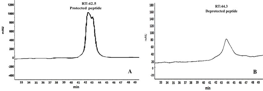
Molecules 2021, 26, 2580 6 of 17
2.9. Toxicity Tests for Leptucin
2.9.1. MTT Test
In order to determine the cytotoxic effect of peptide, MTT assay was done on HEK‐
293 cell line. The cells (2 × 10⁴) were plated in 96‐well plates in DMEM medium, supple‐
mented with 10% FBS and then incubated in the atmosphere containing 5% CO2 and
95% air at 37 °C for overnight. The peptide, at the concentrations ranging from 16 to 0.25
μg/mL, was prepared in DMEM medium, added to each well, and incubated for 24 h. A
10 μL solution of freshly prepared MTT (5 mg/mL in PBS) was added to each well and
incubated for an additional 4 h. Isopropanol (100 μL) was then added and the plates
were shaken gently to facilitate formazan solubilization. The absorbance was measured
at 570 nm by an ELISA reader (ELx808, BioTek, Winooski, VT, USA), and the viability
was then evaluated [51].
2.9.2. In Vitro Hemolysis Assay
This assay was performed according to Memar et al.’s protocol [52]. Briefly, human
blood from a healthy volunteer was collected, centrifuged at 664× g for 5 min, and
washed with PBS three times.
Two doses of peptide (8 μg and 16 μg) were prepared in 100 μL of PBS (1×). One
hundred μL of washed RBCs suspension (2%) was added to each well of a 96‐well mi‐
croplate (Nunc, Sigma Co., St. Louis, MO, USA). Phosphate buffer saline and Triton X‐
100 (0.1%) was used as negative and positive control, respectively. The microplate was
incubated at 37 °C for 2 h and centrifuged at 1664× g for 10 min. The supernatant was
transferred to a new plate and OD was measured at 540 nm in a microplate spectropho‐
tometer (EPOCH, BioTek, Winooski, VT, USA). The experiment was carried out in tripli‐
cate. The degree of hemolysis was determined as the following formula:
[(OD sample − OD neg control)/(OD pos control − OD neg control)] × 100 (2)
2.9.3. In Vivo Hemolysis Assay
Three doses of peptide, 0.32, 0.48, and 0.64 mg/kg (8, 12, and 16 μg/mouse), were in‐
jected intraperitoneally and the blood samples were collected after 24 and 48 h. The
blood samples were centrifuged at 1180× g for 5 min, and the absorbance of supernatant
was measured at 540 nm by a microplate spectrophotometer (EPOCH, BioTek, Winoo‐
ski, VT, USA) [18]. Normal saline was used as negative control.
2.9.4. Histopathological Study
Two doses of peptide, 0.32 and 0.64 mg/kg (8 and 16 μg/mouse), were injected in‐
traperitoneally, and the mice were sacrificed after 48 h of peptide administration. Liver,
kidney, and heart were collected and fixed in 10% buffered formalin solution. After 4
days, the organ sections were routinely processed in an automated tissue processor and
embedded in paraffin wax. The tissue sections were prepared and stained with hema‐
toxylin and eosin (H&E) for pathological studies [53].
2.9.5. Determination of Lethal Dose 50
Lethality due to leptucin was examined on BALB/c mice (male, 20–30 g) using rou‐
tine method. Leptucin at the doses of 0.8, 1.6, 2.4, 3.2, and 4 mg/kg were prepared in 100
μL sterile PBS (1) and injected intraperitoneally into each group (6 mice/group). Sterile
PBS (1) was used as negative control.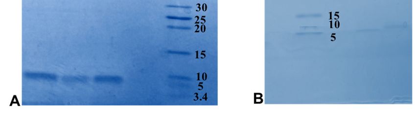
Molecules 2021, 26, 2580 7 of 17
2.10. Motor Coordination Test (Rotarod Test)
The animals are placed on textured drums (1¼ inch diameter) to avoid slipping.
When an animal drops onto the individual sensing platforms, the result is recorded. Five
mice were tested at a rate of 4 rpm.
Naive mice were trained until they could remain on the rotarod for 5 min. Animals
that failed to meet this criterion within three trials were discarded.
Mice were intraperitoneally pretreated with either normal saline or leptucin at the
doses of 0.32, 0.48, and 0.64 mg/kg (8, 12, and 16 μg/mouse). Thirty minutes after the in‐
jection of peptide, mice were placed on the rotarod for 5 min. If a mouse fell from the
rotarod during this time period, it was scored as motor impaired. The data was subject‐
ed to ANOVA followed by Student’s t‐test [54].
2.11. Data Analysis
Data obtained from the two pain models tests are expressed as mean ± SD. Normal‐
ity was assessed using the Kolmogorov‐Smirnov test. The significance of the effect was
tested by one‐way analysis of variance (ANOVA) followed by Tukey’s post hoc analysis.
In all cases, the results were considered significant where p < 0.05. Effect dose of 50%
(ED50) was measured by linear regression assay. LD50 was determined within 72 h fol‐
lowing i.p. administration of leptucin; six mice in each group.
3. Results
3.1. Identification of Mature Chain and Sequence Analysis
According to the data mining analyses, an 859 bp sequence corresponding to the
annotation of analgesic peptide was concluded in the cDNA library. Analysis of the raw se‐
quence in ORF finder server showed a 231 bp sequence encrypted as a peptide of 76 amino
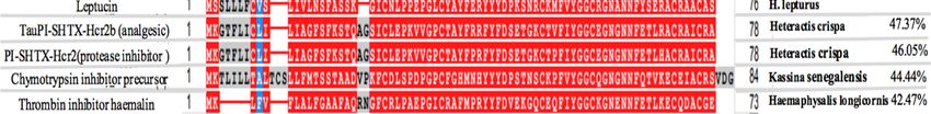
acids (Figure 1). Analysis of sequence similarity by blastP showed that the H. lepturus anal‐
gesic peptide was similar to different peptides with identity ranging from 47.37 to 42.47%
(Figure 2). TauPI‐stichotoxin‐Hcr2b (UniProt P0DMJ5, AltName: APHC1), an analgesic pep‐
tide from the sea anemone; Heteractis crispa, had the highest similarity (Identity 47%) to H.
lepturus analgesic peptide (Figure 2). The obtained result reconfirmed the accuracy of annota‐
tion analysis of cDNA library of H. lepturus venom gland.
Multiple alignment analyses showed a conserved signature in the selected similar
peptides as [C (6XGX) C (8XYXD5X) C (2XFXYGG) C (XGNXNNF5X) C (3X) C (2X)]. In
all selected similar peptides, the signal peptide consisted of 22 amino acids, except for
one, which were 15 amino acids.
The sequence comparison between H. lepturus analgesic peptide and similar pep‐
tides led to determination of the mature chain in H. Lepturus analgesic peptide. The ma‐
ture chain is a cysteine‐rich peptide which contains 55 amino acids (Figure 1), hereafter
designated as leptucin and its theoretical molecular weight was calculated as 6262.09 Da.
Figure 1. The amino acid and ORF sequence of leptucin; the H. lepturus analgesic peptide. The area of the signal peptide
and mature chain are colored gray and black respectively.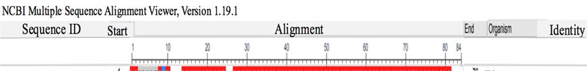
Molecules 2021, 26, 2580                                                                                                        8 of 17

      Figure 2. Multiple alignment of leptucin with selected similar peptides by COBALT server. Leptucin’s ORF was com‐
      pared to its closest similar peptides. As shown in the figure, that was similar to different peptides with identity ranging
      from 47.37 to 42.47%. Also, analysis showed a conserved signature sequence in the selected similar sequences as [C
      (6XGX) C (8XYXD5X) C (2XFXYGG) C (XGNXNNF5X) C (3X) C (2X)].

                                 3.2. Prediction of 3D Structure of the Peptide
                                      The predicted structure of leptucin contains two β sheets and one α helix (Figure
                                 3A). Disulfide bonds between cysteine amino acids were determined by UCSF Chimera
                                 software packages (Figure 3B). Structural alignment of leptucin with TauPI‐stichotoxin‐
                                 Hcr2b [55] by UCSF Chimera software package showed a significant homology (RMSD =
                                 1.085) as depicted in Figure 3C.

      Figure 3. (A) Prediction of the 3D structure of leptucin. Analysis of the result showed that the peptide comprises alpha
      helices (25.45%), beta sheets (25.45%), and random coil (49.09%). (B) Based on 3D structure, disulfide bonds between cys‐
      teine amino acids were predicted in leptucin as C1‐C6*C2‐C4*C3‐C5. (C) Superimposition of the structure of TauPI‐
      stichotoxin‐Hcr2b with the predicted structure of leptucin.

                                 3.3. Deprotection Analysis
                                      Based on the molar ratio deprotection method, the peptide dissolved in pH 4 and
                                 after that added to deprotection solution (pH 4, protocol 1). SDS‐PAGE was performed
                                 on the supernatant and pellet to control the efficiency of the protocols. The results
                                 showed that the peptide was degraded and no band was seen. It was speculated that
                                 deprotected peptide would be degraded in this pH. In order to overcome this issue, dif‐
                                 ferent protocols were used as detailed in Table 1.
                                      SDS‐PAGE was finally performed on the supernatants and pellets of all 3 protocols.
                                 The SDS‐PAGE results showed that the band density in the supernatant of protocol 2 and 4
                                 is approximately the same and was greater than protocol 3 (Figure 4A,B). There was a thin
                                 deprotected peptide band in the pellet of protocol 4, but no band was seen in the pellet of
                                 protocol 2. According to the results, protocol 2 was considered the optimal protocol.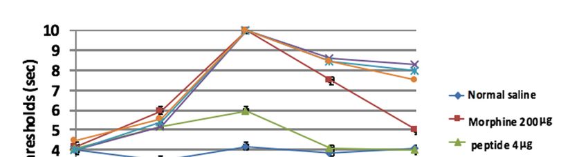
Molecules 2021, 26, 2580                                                                                                9 of 17

                           Table 1. Different pH in the molar ratio deprotection method.

                                          Dissolution                                           pH
                                         Deprotection                           4                           7
                                                             4              Protocol 1                  Protocol 3
                                       pH
                                                             7              Protocol 2                  Protocol 4

                           Figure 4. SDS‐PAGE performed on the pellets and supernatants. (A) SDS‐PAGE on the superna‐
                           tant showed that the peptide destroyed in protocol 1. The result showed that the peptide amount
                           obtained in protocol 2 (peptide in pH 4, deprotection in pH 7) and 4 (peptide in pH 7, deprotection
                           in pH 4) were more than protocol 3 (peptide in pH 7, deprotection in pH 7). (B) SDS‐PAGE on the
                           pellets showed that peptide band did not exist in protocol 1 (peptide in pH 4, deprotection in pH
                           4), but in protocol 3 (peptide in pH 7, deprotection in pH 7) and protocol 4 (peptide in pH 7,
                           deprotection in pH 4) there were a little deprotected peptide.

                           3.4. The Accuracy of Deprotection
                                To confirm the accuracy of deprotection, RP‐HPLC was performed. One hundred
                           μL of the peptide (in supernatant), collected from the best deprotection protocol, was in‐
                           jected onto a C18 column. RP‐HPLC analysis showed that the retention time of the
                           deprotected peptide (Figure 5A) had shifted 1.8 min to the right (Figure 5B) in compari‐
                           son to the protected peptide.
                                One major peak was seen in the chromatogram and its purity calculated as 92% by
                           chromgate software (Figure 5B). A total of 114 μg deprotected peptide recovered from
                           300 μg protected one, indicating the yield as 38%.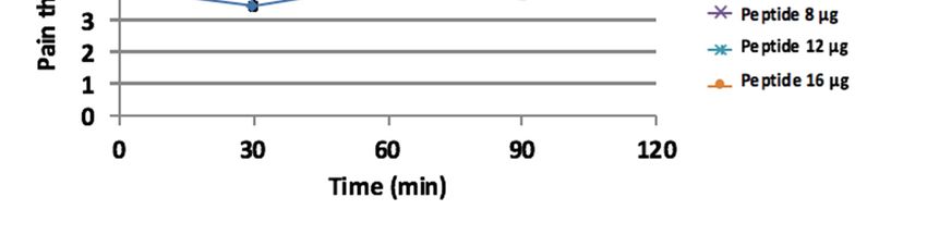
Molecules 2021, 26, 2580                                                                                                 10 of 17

      Figure 5. Comparison of the retention time between deprotected and protected in RP‐HPLC. ACM protected (A) and
      deprotected (B). The retention time after deprotection shifted 1.8 min to the right.

                              3.5. Refolding Analysis
                                   After refolding of the deprotected peptide in the different ratio of GSH/GSSG, RP‐
                              HPLC was performed. The results show that there isn’t any peak of refolded peptide at
                              the amount of 40 μg in the ratios of GSH2/GSSG1 and GSH10/GSSG1 in comparison to
                              negative control. After changing the amount of peptide to 80 μg, in a ratio of
                              GSH2/GSSG1 a sharp peak was observed. The amount of 80 μg peptide in a ratio of
                              GSH10/GSSG1 concluded two peaks (Figure S1). The refolding of 80 μg peptide in the
                              ratio of GSH2/GSSG1 concluded the best result in which just one peak was observed
                              (Figure 6).
                                   Analysis of the RP‐HPLC results showed that the retention time of the refolded
                              peptide had shifted 3.4 min to the right in comparison to the deprotected peptide.
                                   The purity of the refolded peptide was calculated as 90% by chromgate software. A
                              total of 108.3 μg refolded peptide recovered from 114 μg deprotected one. Based on the
                              calculation, the yield of refolding was reported as 95%.

                              Figure 6. RP‐HPLC for the refolding peptide. The refolding of 80 μg peptide in the ratio
                              GSH2/GSSG1 concluded the best result in which just one peak was observed.
Molecules 2021, 26, 2580 11 of 17
3.6. Refolding Confirmation
The refolded peptide was shown by SDS‐PAGE (Figure 7A). According to the re‐
sult, obtained from Ellman’s assay, no free thiol residue was detected in the refolded
peptide indicating all cysteine residues participated in the formation of disulfide bridges
in the peptide. The homogeneity of refolded peptide was analyzed using DLS. The result
showed that the homogeneity in the refolded peptide was 95.6% (Figure 7B).
Figure 7. Refolding confirmation. (A) SDS‐PAGE for the refolded leptucin, (B) Homogeneity eval‐
uation of the refolded peptide using dynamic light scattering.
3.7. Circular Dichroism
The CD spectrum of leptucin showed a negative band in the 208–222 nm region,
which is characteristic of an α-helical structure. Also, one broad negative band around
217 nm and a shoulder around 195 nm were observed corresponding to a β-sheet struc‐
ture (Figure 8). The proportions of α helices, β strands, turns, and random coils were cal‐
culated to be equal to 20.4, 43.1, 11.6, and 25%, respectively.
Figure 8. The CD spectra of leptucin. Analysis of the results showed that the peptide comprises
alpha helices (20.4 %), beta sheets (43.1%), turn (11.6 %), and random coil (25%).
3.8. Activity Assays
3.8.1. Hot Plate Assay
The mice were treated with peptide at the amount of 0.16, 0.32, 0.48, and 0.64 mg/kg
(4, 8, 12, and 16 μg/mouse) or morphine at 10 mg/kg (200 μg/mouse). The results showed
a significant increase in the analgesic effect in comparison to negative control (p <
0.0001), with the exception of 0.16 mg/kg. The maximum analgesic effect (activity 95%)
for the peptide was obtained at the dose of 0.48 mg/kg (12 μg/mouse) after 30 min and
continued up to the time point of 180 min. No differences were detected between pep‐Molecules 2021, 26, 2580 12 of 17
tide‐treated groups of 0.32 and 0.64 mg/kg (p > 0.05) (Figure 9). The ED50 value was equal
to 0.19 mg/kg (4.9 μg/mouse).
Figure 9. Hot plate assays. The maximum analgesic activity was 95% at 0.48 mg/kg (12 μg/mouse) after 30 min and con‐
tinued up to the time point of 180 min. Data is expressed as mean ± SD. All groups had n = 6.
3.8.2. Tail Flick Assay
Based on statistical analyses by using ANOVA, the peptide significantly increased
the pain threshold at the doses of 0.16, 0.32, 0.48, and 0.64 mg/kg (4, 8, 12, and 16
μg/mouse), and there were significant differences between the test and control groups (p
< 0.0001). The maximum analgesic effect of peptide was obtained at the doses of 0.32,
0.48, and 0.64 mg/kg after 60 min (p > 0.05). The latency of pain threshold continued at
the doses of 0.32, 0.48, and 0.64 mg/kg for 120 min but did not last for morphine (Figure
10). The ED50 value of leptucin was equal to 0.17 mg/kg (4.3 μg/mouse).
Figure 10. Tail‐flick assay. The mice were treated by leptucin or morphine. The maximum analge‐
sic activity of leptucin was obtained after 60 min at the doses of 0.32, 0.48, and 0.64 mg/kg (8, 12,
and 16 μg/mouse). This result was similar to morphine (p > 0.05). Data are expressed as mean ±
SD. All groups had n = 6.Molecules 2021, 26, 2580 13 of 17
3.9. Toxicity Assays
The analgesic peptide showed no toxicity on HEK293 cells at all examined concen‐
trations (Figure S2). No toxicity was seen on human RBCs at the doses of 8 and 16 μg
(Table S1). The results obtained from the in vivo hemolysis assay indicated no toxicity
after 24 and 48 h at all examined doses (Table S2). Histopathology evaluation of liver,
kidney, and heart showed that administration of analgesic peptide induces no hepato‐
toxicity (Figure S3), nephrotoxicity (Figure S4) and cardiotoxicity (Figure S5) up to 0.64
mg/kg (16 μg/mouse). LD50 value of leptucin was >4 mg/kg (100 μg/mouse) (Table S3).
3.10. Influence of Leptucin on Motor Coordination
In comparison to control group, leptucin had no effect on motor coordination (p >
0.05) which means that the peptide did not cause motor deficits in mice.
4. Discussion
NSAIDs and morphine are the most common analgesic in acute pain [5], but their
use is limited due to many side effects [7]. Concerning this issue, the design or discovery
of a new molecule with lesser complication is necessary [8,9].
Using a screening strategy and bioinformatics analysis, we identified a sequence of
an analgesic peptide in the annotated cDNA library of H. lepturus. Using multiple
alignment analyses, the mature chain of peptide was determined and designated as Lep‐
tucin, which is a cysteine‐rich peptide. Moreover, for the first time, an evolutionary sig‐
nature was also determined [i.e., C (6XGX) C (8XYXD5X) C (2XFXYGG) C
(XGNXNNF5X) C (3X) C (2X)] in the peptide sequence referring to its considerable simi‐
larity with other peptides in the Kunits‐type cysteine‐rich peptide family.
3D prediction showed that the peptide comprises α helices (25.45%), β sheets
(25.45%), and random coil (49.09%) where the amounts of α helices, β strands, turns, and
random coil were calculated to be equal to 20.4, 43.1, 11.6, and 25% respectively. The re‐
sults obtained from 3D prediction and CD analysis thus did not match.
Preliminary analysis based on structural prediction and observation of structural
similarity indicated that refolding is necessary for analgesic activity of leptucin.
In all deprotection methods, from the earliest established by Veber on cysteine‐rich
peptides [56], and then the modified Veber et al.’s protocol (Becker et al.’s protocol) [57],
to the method of Eisapoor et al., which was the best method of deprotection [43], it’s a
common point. In these methods, the removal of the ACM group with mercury acetate
had been performed in pH 4 [43,56,57].
In this study, we used the molar ratio protocol for deprotection of leptucin because
of its high yield, rapid and cost‐effective advantages [43]. Our result showed that the
peptide is destroyed in pH 4 after one day. Thus, to overcome this problem, we exam‐
ined four pH protocols as mentioned in Table 1. Although deprotection could be per‐
formed immediately without peptide degradation, but in terms of stability it is better to
reach an optimized protocol to avoid degradation during solubilization and storage.
According to the result, the peptide was seen in the collected supernatant in proto‐
col 2. This finding is not in agreement with Becker et al., Veber et al., and Eisapoor et
al.’s protocols as they had found the peptide in the precipitate. This finding can be so in‐
teresting in terms of industrial parameters. Being soluble in supernatant is cost‐effective
since the steps and the time for deprotection are decreased.
Among refolding methods, the methods based on the implementation of reduced
and oxidized glutathione and guanidine chloride induce correct refolding in comparison
to the other methods [44,58]. According to a 2006 study by Edward et al., guanidine is
denaturing and can disrupt the three‐dimensional structure of peptides. Thus, we pre‐
ferred to use reduced and oxidized glutathione for refolding [59]. Refolding plays an
important role in the stability and activity of the cysteine‐rich peptides [60].Molecules 2021, 26, 2580 14 of 17
The refolding of cysteine‐rich peptides depends on the peptide concentration and
the ratio of GSH and GSSG. Different ratios of GSH and GSSG are used to refold differ‐
ent peptides [60,61]. In this study, a ratio of GSH10:GSSG1 and a minimum ratio of 2:1
with 40 and 80 μg of peptide were considered. The results showed that the ratio of 2:1
with 80 μg of peptide is the optimal molar ratio. The refolding was quite economical in
terms of consuming reagents. The refolded peptide in this ratio showed one peak and
shifted to the right in comparison to the deprotected peptide as shown in RP‐HPLC. Us‐
ing a ratio of GSH2: GSSG1, our study is in agreement with Lopez‐Vera et al., and
Buczek, et al. [42,58]. Based on Ellman’s result, it can be concluded that there is no free
thiol moiety in the surface structure of leptucin and the refolding is done correctly.
Based on the results obtained from DLS, the refolded peptide is mono‐dispersed and
homogenous [62].
Analgesic effects of leptucin were assessed using two well‐known models of pain
including hot plate and tail flick assays. Leptucin, at the doses of 0.32 and 0.48 mg/kg (8
and 12 μg/mouse) demonstrated maximum analgesic effect in tail‐flick and hot plate as‐
says, respectively. Analysis of the data obtained from tail flick and hot plate assays
showed that leptucin has about 31.25‐ and 20.83‐fold greater activity than morphine.
Thus, it can be deduced that leptucin has more specificity towards the responsible chan‐
nels for pain. This issue demonstrates that the peptide can exert less or no toxicity in
comparison to morphine. The primary pathological evaluations and survival assay
showed no toxicity in terms of acute toxicity. This fact may raise the hope for using this
peptide for relieving acute pain as well as chronic pains, however, comprehensive toxici‐
ty studies should be performed.
According to the hot plate assay, the maximum effect of leptucin (activity 95%) was
observed 30 min after administration and its maximum activity was constant for 150 min
whereas in morphine group, the maximum effect (76.2%) was constant for 90.
As shown in the tail‐flick assay, the maximum activity (100%) for leptucin was ob‐
tained after 60 min. The activity decreased slowly to 86% and was constant up to 120
min. In the morphine group, the maximum activity of 100% was seen after 60 min, but
the activity decreased slowly to 0% up to 120 min.
Morphine is generally accepted to act at supraspinal sites or directly on spinal opi‐
oid receptors [63]. Thus, analgesic activity of leptucin in both of hot plate and tail‐flick
tests suggests that leptucin may act at supraspinal sites and directly on spinal opioid re‐
ceptors too. Tail flick and hot plate assays are the most common tests of nociception. The
nociceptive experience is short‐lasting in acute pain models [2]. Therefore, it is suggested
that Leptucin has an analgesic effect in an acute pain model.
Based on the analgesic activity and the sequence similarity, it would be interesting
to test leptucin activity on ion channels of nociception TRPV1 [55].
Our result showed that leptucin at the dose of 8 and 16 μg had no toxicity on hu‐
man red blood cells. In vivo hemolysis assay on mice demonstrated no hemolysis too.
Also, the peptide had no toxicity on HEK293 cell line by MTT assay up to 16 μg.
Moreover, histopathological sections of liver, kidney, and heart demonstrated that Lep‐
tucin had no toxicity up to 0.64 mg/kg.
5. Conclusions
We discovered that leptucin is the analgesic agent of H. lepturus venom. Optimiza‐
tion of pH in deprotection steps led to obtaining the deprotected peptide in the superna‐
tant. This point is important in terms of cost‐effectiveness in the laboratory and industri‐
al scale. According to the results gathered in Ellman’s test, DLS, and spectropolarimetry,
the peptide was refolded successfully.
Regarding the high in vivo efficacy of leptucin and showing no observed toxicity, it
could be suggested as a drug lead. Further studies should be performed to decipher
more details about its action mechanism. Since peptide molecules can target specificMolecules 2021, 26, 2580 15 of 17
channel and have fewer side effects than chemical small molecules, leptucin would be a
new hope in pain‐relief.
Supplementary Materials: Figure S1: RP‐HPLC for the refolding peptide., Figure S2: MTT assay
for leptucin., Figure S3: The histopathology evaluation of liver., Figure S4: The histopathology
evaluation of kidney., Figure S5: The histopathology evaluation of heart. Table S1: In vitro hemol‐
ysis assay for leptucin., Table S2: In vivo hemolysis for leptucin., Table S3: Determination of LD50
for leptucin.
Author Contributions: S.B‐Z. contributed to practical assays, bioinformatics analyses, and to writ‐
ing the manuscript. D.S. and J.‐M.S. served as advisor and contributed to the revision of the manu‐
script. S.S. contributed to CD assay and analyses, and revision of the manuscript. K.P.B. super‐
vised the project and contributed to experimental design, all bioinformatics assays and analyses,
writing, revision, and redaction of the manuscript. The idea for discovery of an analgesic peptide
from H. lepturus venom belongs to K.P.B. and D.S. All authors have read and agreed to the pub‐
lished version of the manuscript.
Funding: This paper is the part of the PhD thesis of Sedigheh Bagheri‐Ziari; approved and funded
by Pasteur Institute of Iran. The APC was funded by Jean‐Marc Sabatier.
Institutional Review Board Statement: The study was conducted according to the guidelines of
the Declaration of Helsinki, and approved by the Institutional Ethics Committee of Pasteur Insti‐
tute of Iran (protocol code IR.PII.REC.1394.86, 22/06/2018). All experiments were approved by the
Ethical Committee of the Pasteur Institute of Iran (code number IR.PII.REC.1394.86).
Informed Consent Statement: Not applicable.
Data Availability Statement: Not applicable.
Conflicts of Interest: The authors declare no conflict of interest.
Sample availability statement: Not applicable.
References
1. Shilpi, J.A.; Uddin, S.J. Analgesic and antipyretic natural products. Medicinal Natural Products. Dis. Focused Approach 2020, 55,
435.
2. Feng, J.; Lepetre‐Mouelhi, S.; Gautier, A.; Mura, S.; Cailleau, C.; Coudore, F.; Hamon, M.; Couvreur, P. A new painkiller na‐
nomedicine to bypass the blood‐brain barrier and the use of morphine. Sci. Adv. 2019, 5, eaau5148, doi:10.1126/sciadv.aau5148.
3. Abd‐Elsayed, A.; Deer, T.R. Different Types of Pain. In Pain; Abd‐Elsayed, A., Ed.; Springer: Berlin/Heidelberg, Germany,
2019; doi:10.1007/978‐3‐319‐99124‐5_3.
4. Hena, S.; Znad, H. Membrane bioreactor for pharmaceuticals and personal care products removal from wastewater. In Com‐
prehensive Analyti‐Cal Chemistry; Elsevier: Amsterdam, The Netherlands, 2018; Volume 81, pp. 201–256..
5. Munir, M.A.; Enany, N.; Zhang, J.‐M. Nonopioid Analgesics. Anesthesiol. Clin. 2007, 25, 761–774,
doi:10.1016/j.anclin.2007.07.007.
6. Kumar, M.; Shete, A.; Akbar, Z. A review on analgesic: From natural sources. Int. J. Pharm. Biol. Arch. 2010, 1, 95–100.
7. Cui, J.M.; Zhao, L.; Wang, Z.J.; Ma, M.T.; Wang, Y.; Luo, K.Y.; Wang, L.Q.; Wei, S.; Zhang, X.H.; Han, C.Z.; et al. MEL endo‐
morphins act as po‐tent inflammatory analgesics with the inhibition of activated non‐neuronal cells and modulation of pro‐
inflammatory cytokines. Neuropharmacology 2020, 168, 107992.
8. Bruno, B.J.; Miller, G.D.; Lim, C.S. Basics and recent advances in peptide and protein drug delivery. Ther. Deliv. 2013, 4, 1443–
1467, doi:10.4155/tde.13.104.
9. Lau, J.L.; Dunn, M.K. Therapeutic peptides: Historical perspectives, current development trends, and future directions. Bioor‐
ganic Med. Chem. 2018, 26, 2700–2707, doi:10.1016/j.bmc.2017.06.052.
10. Maatuf, Y.; Geron, M.; Priel, A. The Role of Toxins in the Pursuit for Novel Analgesics. Toxins 2019, 11, 131,
doi:10.3390/toxins11020131.
11. Newman, D.J.; Cragg, G.M. Natural Products as Sources of New Drugs from 1981 to 2014. J. Nat. Prod. 2016, 79, 629–661,
doi:10.1021/acs.jnatprod.5b01055.
12. Sable, R.; Parajuli, P.; Jois, S. Peptides, Peptidomimetics, and Polypeptides from Marine Sources: A Wealth of Natural Sources
for Pharmaceutical Applications. Mar. Drugs 2017, 15, 124, doi:10.3390/md15040124.
13. Escoubas, P.; King, G.F. Venomics as a drug discovery platform. Expert Rev. Proteom. 2009, 6, 221–224, doi:10.1586/epr.09.45.
14. Moghadasi, Z.; Shahbazzadeh, D.; Jamili, S.; Mosaffa, N.; Bagheri, K.P. Significant Anticancer Activity of a Venom Fraction
Derived from the Persian Gulf Sea Anemone, Stichodactyla haddoni. Iran. J. Pharm Res. 2020, 19, 402–420.
15. Pennington, M.W.; Czerwinski, A.; Norton, R.S. Peptide therapeutics from venom: Current status and potential. Bioorganic
Med. Chem. 2018, 26, 2738–2758, doi:10.1016/j.bmc.2017.09.029.Molecules 2021, 26, 2580 16 of 17
16. Zarghami, V.; Ghorbani, M.; Bagheri, K.P.; Shokrgozar, M.A. Melittin antimicrobial peptide thin layer on bone implant chi‐
tosan‐antibiotic coatings and their bactericidal properties. Mater. Chem. Phys. 2021, 263, 124432,
doi:10.1016/j.matchemphys.2021.124432.
17. Harvey, A.L. Toxins and drug discovery. Toxicon 2014, 92, 193–200, doi:10.1016/j.toxicon.2014.10.020.
18. Pashaei, F.; Bevalian, P.; Akbari, R.; Bagheri, K.P. Single dose eradication of extensively drug resistant Acinetobacter spp. In a
mouse model of burn infection by melittin antimicrobial peptide. Microb. Pathog. 2019, 127, 60–69,
doi:10.1016/j.micpath.2018.11.055.
19. Khozani, R.S.; Shahbazzadeh, D.; Harzandi, N.; Feizabadi, M.M.; Bagheri, K.P. Kinetics study of antimicrobial peptide, melit‐
tin, in simultaneous bio‐film degradation and eradication of potent biofilm producing MDR Pseudomonas aeruginosa isolates.
Int. J. Pept. Res. Ther. 2019, 25, 329–338.
20. Dezfuli, H.T.; Shahbazzadeh, D.; Eidi, A.; Bagheri, K.P.; Pakravan, N.; Amini, S.; Aghasadeghi, M.R.; Mahdavi, M. Induction
of IFN‐γ cytokine re‐sponse against hepatitis B surface antigen using melittin. Gastroenterol. Hepatol. Bed. Bench. 2014, 7, 108–
117.
21. Akbari, R.; Hakemi‐Vala, M.; Pashaie, F.; Bevalian, P.; Hashemi, A.; Bagheri, K.P. Highly Synergistic Effects of Melittin with
Conventional Antibiotics against Multidrug‐Resistant Isolates of Acinetobacter baumannii and Pseudomonas aeruginosa. Microb.
Drug Resist. 2019, 25, 193–202, doi:10.1089/mdr.2018.0016.
22. Utkin, Y.N. Animal venom studies: Current benefits and future developments. World J. Biol. Chem. 2015, 6, 28–33,
doi:10.4331/wjbc.v6.i2.28.
23. Ortiz, E.; Gurrola, G.B.; Schwartz, E.F.; Possani, L.D. Scorpion venom components as potential candidates for drug develop‐
ment. Toxicon 2015, 93, 125–135, doi:10.1016/j.toxicon.2014.11.233.
24. Kazemi, S.M.; Sabatier, J.‐M. Venoms of Iranian Scorpions (Arachnida, Scorpiones) and Their Potential for Drug Discovery.
Molecules 2019, 24, 2670, doi:10.3390/molecules24142670.
25. Chippaux, J.‐P.; Goyffon, M. Epidemiology of scorpionism: A global appraisal. Acta Trop. 2008, 107, 71–79,
doi:10.1016/j.actatropica.2008.05.021.
26. Zabihollahi, R.; Bagheri, K.P.; Keshavarz, Z.; Motevalli, F.; Bahramali, G.; Siadat, S.D.; Momen, S.B.; Shahbazzadeh, D.;
Aghasadeghi, M.R. Venom Components of Iranian Scorpion Hemiscorpius lepturus Inhibit the Growth and Replication of
Human Immunodeficiency Virus 1 (HIV‐1). Iran. Biomed. J. 2016, 20, 259–265.
27. Ahmadi, S.; Knerr, J.M.; Argemi, L.; Bordon, K.C.F.; Pucca, M.B.; Cerni, F.A.; Arantes, E.C.; Çalışkan, F.; Laustsen, A.H. Scor‐
pion Venom: Detriments and Benefits. Biomedicines 2020, 8, 118, doi:10.3390/biomedicines8050118.
28. Yang, S.; Yang, F.; Zhang, B.; Lee, B.H.; Li, B.; Luo, L.; Zheng, J.; Lai, R. A bimodal activation mechanism underlies scorpion
toxin–induced pain. Sci. Adv. 2017, 3, e1700810, doi:10.1126/sciadv.1700810.
29. Hakim, A.; Jiang, W.; Luo, L.; Li, B.; Yang, S.; Song, Y.; Lai, R. Scorpion Toxin, BmP01, Induces Pain by Targeting TRPV1
Channel. Toxins 2015, 7, 3671–3687, doi:10.3390/toxins7093671.
30. Rowe, A.H.; Xiao, Y.; Scales, J.; Linse, K.D.; Rowe, M.P.; Cummins, T.R.; Zakon, H.H. Isolation and Characterization of CvIV4:
A Pain Inducing α‐ Scorpion Toxin. PLoS ONE 2011, 6, e23520, doi:10.1371/journal.pone.0023520.
31. Torabi, E.; Behdani, M.; Chafi, M.H.; Moazzami, R.; Sabatier, J.; Khalaj, V.; Shahbazzadeh, D.; Bagheri, K.P. Characteristics and
Lethality of a Novel Recombinant Dermonecrotic Venom Phospholipase D from Hemiscorpius lepturus. Toxins 2017, 9, 102,
doi:10.3390/toxins9030102.
32. Sanaei‐Zadeh, H. Painless Stings of Yellow Iranian Scorpions. Iran. Red Crescent Med, J. 2017, 19, 42645, doi:10.5812/ircmj.42645.
33. Dehghani, R.; Arani, M.G. Scorpion sting prevention and treatment in ancient Iran. J. Tradit. Complement. Med. 2015, 5, 75–80,
doi:10.1016/j.jtcme.2014.11.007.
34. Pipelzadeh, M.H.; Jalali, A.; Taraz, M.; Pourabbas, R.; Zaremirakabadi, A. An epidemiological and a clinical study on scorpi‐
onism by the Iranian scorpion Hemiscorpius lepturus. Toxicon 2007, 50, 984–992, doi:10.1016/j.toxicon.2007.07.018.
35. Dehghani, R.; Kamiabi, F.; Mohammadi, M. Scorpionism by Hemiscorpius spp. in Iran: A review. J. Venom. Anim. Toxins Incl.
Trop. Dis. 2018, 24, 1–10, doi:10.1186/s40409‐018‐0145‐z.
36. Pourkhalili, K.; Kim, E.; Mashayekhy, N.R.; Kamyab, M.; Hoseiny, S.M.; Evazy, R.; Mirakabadi, A.Z.; Seyedian, R. Cardiotoxic
and Arrhythmogenic Effects of Hemiscorpius lepturus Scorpion Venom in Rats. J. Arthropod‐Borne Dis. 2015, 9, 215–225.
37. Shahbazzadeh, D.; Srairi‐Abid, N.; Feng, W.; Ram, N.; Borchani, L.; Ronjat, M.; Akbari, A.; Pessah, I.N.; De Waard, M.; El
Ayeb, M. Hemicalcin, a new toxin from the Iranian scorpion Hemiscorpius lepturus which is active on ryanodine‐sensitive
Ca2+ channels. Biochem. J. 2007, 404, 89–96, doi:10.1042/bj20061404.
38. Jridi, I.; Catacchio, I.; Majdoub, H.; Shahbazeddah, D.; Ayeb, M.; Frassanito, M.A.; Ribatti, D.; Vacca, A.; Borchani, L. Hemili‐
pin, a novel Hemiscor‐pius lepturus venom heterodimeric phospholipase A2, which inhibits angiogenesis in vitro and in vivo.
Toxicon 2015, 105, 34–44.
39. Maleki, M.; Dounighi, N.M. Purification and characterization of a novel type of neurotoxic peptides from the venom of the
Iranian scorpion Hemiscorpius lepturus. Iran. J. Basic Med. Sci. 2020, 23, 195–201.
40. Papadopoulos, J.S.; Agarwala, R. COBALT: Constraint‐based alignment tool for multiple protein sequences. Bioinformatics
2007, 23, 1073–1079, doi:10.1093/bioinformatics/btm076.
41. Roy, A.; Kucukural, A.; Zhang, Y. I‐TASSER: A unified platform for automated protein structure and function prediction. Nat.
Protoc. 2010, 5, 725–738, doi:10.1038/nprot.2010.5.Molecules 2021, 26, 2580 17 of 17
42. Huang, C.C.; Meng, E.C.; Morris, J.H.; Pettersen, E.F.; Ferrin, T.E. Enhancing UCSF Chimera through web services. Nucleic
Acids Res. 2014, 42, W478–W484, doi:10.1093/nar/gku377.
43. Eisapoor, S.S.; Jamili, S.; Shahbazzadeh, D.; Mostafavi, P.G.; Bagheri, K.P. A New, High Yield, Rapid, and Cost‐Effective Pro‐
tocol to Deprotection of Cysteine‐Rich Conopeptide, Omega‐Conotoxin MVIIA. Chem. Biol. Drug Des. 2016, 87, 687–693,
doi:10.1111/cbdd.12702.
44. Lopez‐Vera, E.; Walewska, A.; Olivera, A.B.M.; Bulaj, G. Role of Hydroxyprolines in the in Vitro Oxidative Folding and Bio‐
logical Activity of Conotoxins. Biochemistry 2008, 47, 1741–1751, doi:10.1021/bi701934m.
45. Shahangian, S.S.; Sajedi, R.; Hasannia, S.; Jalili, S.; Mohammadi, M.; Taghdir, M.; Shali, A.; Mansouri, K.; Sariri, R. A confor‐
mation‐based phage‐display panning to screen neutralizing anti‐VEGF VHHs with VEGFR2 mimicry behavior. Int. J. Biol.
Macromol. 2015, 77, 222–234.
46. Liu, X.; Yao, G.; Wang, K.; Liu, Y.; Wan, X.; Jiang, H. Structural and Functional Characterization of Conotoxins from Conus
achatinus Targeting NMDAR. Mar. Drugs 2020, 18, 135, doi:10.3390/md18030135.
47. Guidance for Industry. Analgesic Indications: Developing Drug and Biological Products. Office of Communications, Division
of Drug Information, Center for Drug Evaluation and Research, Food and Drug Administration. 02/06/2014. Available online:
http://www.fda.gov/Drugs/GuidanceComplianceRegulatoryInformation/Guidances/default.htm (accessed on 1 December
2020).
48. Moghadasi, Z.; Jamili, S.; Shahbazadeh, D.; Bagheri, K.P. Toxicity and Potential Pharmacological Activities in the Persian Gulf
Venomous Sea Anemone, Stichodactyla haddoni. Iran. J. Pharm. Res. 2018, 17, 940–955.
49. Carlsson, K.‐H.; Jurna, I. Depression by flupirtine, a novel analgesic agent, of motor and sensory responses of the nociceptive
system in the rat spinal cord. Eur. J. Pharmacol. 1987, 143, 89–99, doi:10.1016/0014‐2999(87)90738‐2.
50. Regalado, A.I.; Mancebo, B.; Paixão, A.; López, Y.; Merino, N.; Sánchez, L.M. Antinociceptive Activity of Methanol Extract of
Tabebuia hypoleuca (C. Wright ex Sauvalle) Urb. Stems. Med. Princ. Pract. 2017, 26, 368–374.
51. Aghazadeh, H.; Memariani, H.; Ranjbar, R.; Bagheri, K. The activity and action mechanism of novel short selective LL‐37‐
derived anticancer peptides against clinical isolates of Escherichia coli. Chem. Biol. Drug Des. 2019, 93, 75–83.
52. Memar, B.; Jamili, S.; Shahbazzadeh, D.; Bagheri, K.P. The first report on coagulation and phospholipase A2 activities of Per‐
sian Gulf lionfish, Pterois russelli, an Iranian venomous fish. Toxicon 2016, 113, 25–31, doi:10.1016/j.toxicon.2016.02.004.
53. Treuting, P.M.; Dintzis, S.M. Comparative Anatomy and Histology. In A Mouse and Human Atlas; Elsevier: London, UK, 2012;
p. 461, ISBN: 978‐0‐12‐381361‐9.
54. Bansinath, M.; Bose, A.C.; Hema, S.; Guruswami, M.N. Interaction of metamizol with some hypnotics in rats. Arch. Int. Phar‐
macodyn. Ther. 1982, 260, 14–27.
55. Nikolaev, M.V.; Dorofeeva, N.A.; Komarova, M.S.; Korolkova, Y.V.; Andreev, Y.A.; Mosharova, I.V.; Grishin, E.V.; Tikhonov,
D.B.; Kozlov, S.A. TRPV1 activation power can switch an action mode for its polypeptide ligands. PLoS ONE 2017, 12,
e0177077, doi:10.1371/journal.pone.0177077.
56. Veber, D.; Milkowski, J.; Varga, S.; Denkewalter, R.; Hirschmann, R. Acetamidomethyl. A Novel Thiol Protecting Group for
Cysteine. J. Am. Chem. Soc. 1972, 94, 5456–5461, doi:10.1021/ja00770a600.
57. Becker, S.; Atherton, E.; Gordon, R.D. Synthesis and characterization of mu‐conotoxin IIIa. JBIC J. Biol. Inorg. Chem. 1989, 185,
79–84, doi:10.1111/j.1432‐1033.1989.tb15084.x.
58. Steiner, A.M.; Bulaj, G. Optimization of oxidative folding methods for cysteine‐rich peptides: A study of conotoxins contain‐
ing three disulfide bridges. J. Pept. Sci. 2010, 17, 1–7, doi:10.1002/psc.1283.
59. O’Brien, E.P.; Dima, R.I.; Brooks, B.; Thirumalai, D. Interactions between Hydrophobic and Ionic Solutes in Aqueous Guani‐
dinium Chloride and Urea Solutions: Lessons for Protein Denaturation Mechanism. J. Am. Chem. Soc. 2007, 129, 7346–7353,
doi:10.1021/ja069232+.
60. Annis, I.; Hargittai, B.; Barany, G. [10] Disulfide bond formation in peptides. Branched Chain Amino Acids Part B 1997, 289, 198–
221, doi:10.1016/s0076‐6879(97)89049‐0.
61. Buczek, P.; Buczek, O.; Bulaj, G. Total chemical synthesis and oxidative folding of delta‐conotoxin PVIA containing an N‐
terminal propeptide. Biopolymers 2005, 80, 50–57.
62. Fan, X.; Zhao, F.; Wang, X.; Wu, G. Doxorubicin‐triggered self‐assembly of native amphiphilic peptides into spherical nano‐
particles. Oncotarget 2016, 7, 58445–58458, doi:10.18632/oncotarget.11213.
63. Ummenhofer, W.C.; Arends, R.H.; Shen, D.D.; Bernards, C.M. Comparative Spinal Distribution and Clearance Kinetics of In‐
trathecally Administered Morphine, Fentanyl, Alfentanil, and Sufentanil. Anesthesiology 2000, 92, 739–753,
doi:10.1097/00000542‐200003000‐00018.You can also read