Disparate bone anabolic cues activate bone formation by regulating the rapid lysosomal degradation of sclerostin protein - eLife
←
→
Page content transcription
If your browser does not render page correctly, please read the page content below
RESEARCH ARTICLE

                                   Disparate bone anabolic cues activate
                                   bone formation by regulating the rapid
                                   lysosomal degradation of sclerostin
                                   protein
                                   Nicole R Gould1*, Katrina M Williams1, Humberto C Joca2, Olivia M Torre1,
                                   James S Lyons1, Jenna M Leser1, Manasa P Srikanth3, Marcus Hughes1,
                                   Ramzi J Khairallah4, Ricardo A Feldman3, Christopher W Ward1*,
                                   Joseph P Stains1*
                                   1
                                    Department of Orthopaedics, University of Maryland School of Medicine,
                                   Baltimore, United States; 2Center for Biomedical Engineering and Technology,
                                   University of Maryland School of Medicine, Baltimore, United States; 3Department
                                   of Microbiology and Immunology, University of Maryland School of Medicine,
                                   Baltimore, United States; 4Myologica, LLC, New Market, United States

                                   Abstract The downregulation of sclerostin in osteocytes mediates bone formation in response
                                   to mechanical cues and parathyroid hormone (PTH). To date, the regulation of sclerostin has been
                                   attributed exclusively to the transcriptional downregulation of the Sost gene hours after
                                   stimulation. Using mouse models and rodent cell lines, we describe the rapid, minute-scale post-
                                   translational degradation of sclerostin protein by the lysosome following mechanical load and PTH.
                                   We present a model, integrating both new and established mechanically and hormonally activated
                                   effectors into the regulated degradation of sclerostin by lysosomes. Using a mouse forelimb
*For correspondence:
                                   mechanical loading model, we find transient inhibition of lysosomal degradation or the upstream
ngould@som.umaryland.edu
(NRG);
                                   mechano-signaling pathway controlling sclerostin abundance impairs subsequent load-induced
ward@som.umaryland.edu             bone formation by preventing sclerostin degradation. We also link dysfunctional lysosomes to
(CWW);                             aberrant sclerostin regulation using human Gaucher disease iPSCs. These results reveal how bone
jstains@som.umaryland.edu (JPS)    anabolic cues post-translationally regulate sclerostin abundance in osteocytes to regulate bone
                                   formation.
Competing interest: See
page 21
Funding: See page 22
Received: 27 October 2020          Introduction
Accepted: 26 March 2021            The osteocyte-derived protein, sclerostin, is a fundamentally important inhibitor of bone formation,
Published: 29 March 2021           with decreases in abundance mediating mechanically and hormonally induced bone formation. Scle-
                                   rostin (gene name Sost) is a secreted 27 kDa glycoprotein that inhibits the differentiation and activity
Reviewing editor: Subburaman
Mohan, Loma Linda University,
                                   of bone-forming osteoblasts by antagonizing the Wnt/b-catenin signaling pathway (Baron and Gori,
United States                      2018; Drake and Khosla, 2017). Genetic deletion of the Sost gene in mice results in extraordinarily
                                   high bone mass (Li et al., 2008). In humans, mutations in the SOST gene underlie high bone mass
   Copyright Gould et al. This
                                   and bone overgrowth in patients with sclerosteosis and van Buchem disease (Balemans et al., 2002;
article is distributed under the
                                   Balemans et al., 2001; Appelman-Dijkstra et al., 1993). Accordingly, regulating sclerostin bioavail-
terms of the Creative Commons
Attribution License, which         ability has tremendous therapeutic potential for conditions of low bone mass, such as osteoporosis.
permits unrestricted use and       Indeed, targeting sclerostin protein with neutralizing antibodies is incredibly effective at increasing
redistribution provided that the   bone mass, and Romosozumab, a humanized monoclonal antibody targeting sclerostin, has been
original author and source are     FDA approved to treat osteoporosis in post-menopausal women at a high risk for fracture
credited.                          (McClung, 2017; Bandeira et al., 2017). Despite this critical role for sclerostin in skeletal

Gould et al. eLife 2021;10:e64393. DOI: https://doi.org/10.7554/eLife.64393                                                         1 of 26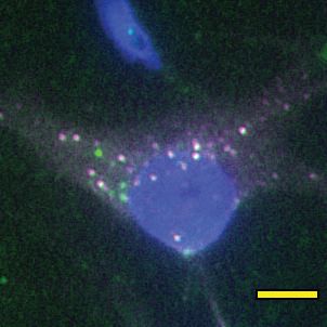
Research article                                                                                                       Cell Biology

                                   homeostasis and its therapeutic potential, there are substantial gaps with respect to the molecular
                                   control of this key regulatory protein.
                                       In response to bone mechanical loading, osteocytes sense and respond to fluid shear stress (FSS)
                                   in the lacunar canalicular network by ultimately decreasing sclerostin protein abundance, de-repres-
                                   sing Wnt/b-catenin signaling, and unleashing osteoblast differentiation and bone formation. When
                                   administered intermittently, parathyroid hormone (PTH) causes net bone formation in part by
                                   decreasing sclerostin (Keller and Kneissel, 2005; Bellido et al., 2005). This has been exploited in
                                   the clinic through the established osteoanabolic drug, teriparatide (PTH, amino acids 1–34). Despite
                                   their clinical application, little is known about how mechanical load and PTH, two disparate bone
                                   anabolic signals, directly regulate sclerostin protein. To date, the regulation of sclerostin protein
                                   abundance has been attributed to the transcriptional downregulation of the Sost gene that occurs
                                   on an hour timescale after mechanical load or PTH exposure (Sebastian and Loots, 2017;
                                   Wein, 2018; Bonnet et al., 2012; Bonnet et al., 2009; Meakin et al., 2014).
                                       Using a recently established osteocyte-like cell line, Ocy454 cells, which is one of the few cell lines
                                   that reliably express detectable sclerostin protein (Wein et al., 2015; Spatz et al., 2015), we previ-
                                   ously described a mechano-transduction pathway that regulates osteocyte sclerostin protein abun-
                                   dance in response to FSS in vitro (Figure 1A; Lyons et al., 2017; Williams et al., 2020). Using this
                                   in vitro model, we found that osteocyte mechano-signaling required a subset of detyrosinated
                                   microtubules, which transduce load signals to activate NADPH oxidase 2 (NOX2), which produces
                                   reactive oxygen species (ROS) signals. These ROS signals then elicit a transient receptor potential
                                   vanilloid 4 (TRPV4)-dependent primary calcium (Ca2+) influx. Calcium/calmodulin-dependent kinase II
                                   (CaMKII) is activated in response to this primary Ca2+ influx and is required for reduction of osteo-
                                   cyte sclerostin protein abundance (Figure 1A). While these discoveries integrated with and
                                   extended several established models of the osteocyte mechanical response (Thompson et al., 2012;
                                   Schaffler et al., 2014; Geoghegan et al., 2019; Baik et al., 2013), we found the loss of sclerostin
                                   protein was surprisingly rapid (minute scale) and was likely wholly distinct from the well-characterized
                                   transcriptional regulation of the Sost gene, which occurs on the hour timescale (Sebastian and
                                   Loots, 2017; Wein, 2018; Bonnet et al., 2012; Bonnet et al., 2009; Meakin et al., 2014). Despite
                                   its physiologic significance, little is known about the post-translational control of sclerostin protein.
                                   Additionally, given the in vitro nature of our prior work on this pathway, the contribution of this
                                   mechano-transduction pathway to in vivo bone mechano-responsiveness remained unresolved.
                                   Here, we examined the kinetics of sclerostin protein degradation in vitro and in vivo, the mechanism
                                   regulating the rapid decline in osteocyte sclerostin protein, its relevance in vivo in bone physiology
                                   during ulnar load, and its dysfunction in skeletal disease. We also extended the observation of the
                                   FSS-induced rapid degradation of sclerostin protein to PTH, another clinically relevant bone anabolic
                                   signal.

                                   Results
                                   Sclerostin protein is rapidly lost after mechanical load in vitro and in
                                   vivo
                                   Our prior work showed sclerostin protein disappearance on a minute timescale following FSS in
                                   Ocy454 cells, an effect that required activation of CaMKII (Figure 1A; Lyons et al., 2017;
                                   Williams et al., 2020). To more precisely characterize the dynamics of mechanically stimulated scle-
                                   rostin protein loss in osteocytes, we examined two sclerostin expressing cell lines: Ocy454 osteo-
                                   cytes and UMR106 osteosarcoma cells. Consistent with our previous work, we observed the
                                   mechano-activated increase in CaMKII phosphorylation and decrease in sclerostin protein abun-
                                   dance in both Ocy454 osteocytes (Figure 1B) and UMR106 osteosarcoma cells (Figure 1C) within 5
                                   min of the acute application of FSS. To establish the relevance of a load-induced loss of sclerostin
                                   protein in vivo, we examined if this rapid loss of sclerostin abundance, as determined by western
                                   blot or sclerostin immunofluorescence in sectioned bone, occurred following an acute bout of ulnar
                                   loading. Sclerostin protein abundance was assessed in 16 week old female or 17 week old male
                                   mice subjected to a single bout of ulnar load. Ulnae were harvested 5 min post-load, a time frame
                                   consistent with in vitro observations, and osteocyte-enriched cortical bone lysates were profiled by
                                   western blotting or ulnae were sectioned to evaluate sclerostin-positive osteocytes by

Gould et al. eLife 2021;10:e64393. DOI: https://doi.org/10.7554/eLife.64393                                                            2 of 26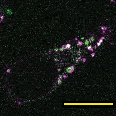
Research article Cell Biology
A.
deTyr Sclerostin
FSS NOX2 ROS TRPV4 Ca2+ CaMKII
Microtubules Protein Loss
B. FSS
1min C. FSS
1min D.
Load
90sec
5min 5min 5min
Collect Collect Collect
FSS - + FSS - + Load - +
Sclerostin Sclerostin Sclerostin
55kDa 55kDa
GAPDH Total Total
Protein 35kDa Protein 35kDa
pCaMKII
pCaMKII pCaMKII
Total CaMKII
Ocy454 Total CaMKII Total CaMKII
p=0.0519 UMR106 Bone
1.5 2.0
Relative Sclerostin
Relative pCaMKII
1.5 4
Relative Sclerostin
*
Relative pCaMKII
* 2.5 6
Relative Sclerostin
*
Relative pCaMKII
1.5 * **
1.0 3 2.0
1.0
1.0 4
2 1.5
0.5
0.5 0.5 1.0
G. 1 2
0.0 0.0 0.5
FSS - + FSS - + 0.0 0
FSS - + FSS - + 0.0 0
Load - + Load - +
E. Time Post FSS (min) F. Time Post FSS (min) G. Time Post FSS (min)
- 1 5 10 15 20 - 1 5 10 15 20 - 1 5 10 15 20
Sclerostin Sclerostin Myc-Sclerostin
β-Actin α-Tubulin β-Actin
Ocy454 UMR106 Ocy454
Relative Sclerostin
Relative Sclerostin
Relative Sclerostin
1.0 1.0 1.0
0.5 0.5 0.5
0.0 0.0 0.0
- 1 5 10 15 20 - 1 5 10 15 20 - 1 5 10 15 20
Time Post FSS (min) Time Post FSS (min) Time Post FSS (min)
Figure 1. Sclerostin protein is rapidly degraded after mechanical stimulus in vitro and in vivo. (A) FSS causes the rapid loss of sclerostin protein through
a number of molecular mediators. (B) Ocy454 cells (n = 3–4) or (C) UMR106 cells (n = 3) were exposed to 1 min of FSS at 4 dynes/cm2 and lysed 5 min
post-flow. Western blots were probed for sclerostin, GAPDH, pCaMKII, and total CaMKII. (D) Sixteen week old female C57Bl/6 mice were ulnar loaded
(1800 me, 90 s, 2 Hz), cortical osteocyte-enriched lysates isolated 5 min post-load, and western blots probed for sclerostin (n = 10 mice), pCaMKII, and
total CaMKII (n = 5 mice). Sclerostin abundance relative to the loading control or pCaMKII relative to total CaMKII was quantified. (E) Ocy454 cells with
endogenous sclerostin (n = 2), (F) UMR106 cells with endogenous sclerostin (n = 4), or (G) Ocy454 cells transfected with Myc-tagged sclerostin (n = 1)
were subjected to 5 min of FSS at 4 dynes/cm2 and lysed at the indicated times post-flow. Western blots were probed for sclerostin and b-actin. A
representative time course is shown for each. Sclerostin abundance relative to the loading control was quantified. For each antibody, western blots are
from a single gel and exposure; a vertical black line indicates removal of irrelevant lanes. Graphs depict mean ± SD. *pResearch article Cell Biology
(Figure 1—figure supplement 1A), establishing that rapid sclerostin protein loss also occurs in vivo
following a single bout of mechanical stimulus.
A temporal assessment of sclerostin protein abundance following FSS in Ocy454 cells (Figure 1E)
and UMR106 cells (Figure 1F) revealed a decrease in sclerostin protein as early as 1 min post-FSS,
with the nadir of protein expression occurring at 10 min, followed by a subsequent rebound. This
rapid regulation strongly suggested post-translational control of sclerostin abundance. Consistent
with the possibility of post-translational control of sclerostin, we recently reported that a single 5
min bout of FSS, as used here, does not decrease Sost mRNA levels despite a decrease in sclerostin
protein (Williams et al., 2020). To confirm the post-translational control of sclerostin, Ocy454 cells
were transfected with either a myc- or GFP-tagged sclerostin expression vector under control of a
CMV promoter, which is not controlled by the same transcriptional elements that regulate Sost tran-
scription. Like endogenous sclerostin, overexpressed sclerostin protein was rapidly and transiently
decreased by FSS (Figure 1G, Figure 1—figure supplement 1B). The regulated degradation did
not extend to all proteins as pro-collagen type 1a1 abundance was unchanged following FSS in
Ocy454 cells (Figure 1—figure supplement 1C), supporting some level of specificity for the regu-
lated degradation of sclerostin.
PTH1-34 treatment also causes rapid sclerostin protein loss
Like mechanical loading, intermittent administration of PTH stimulates bone remodeling and results
in net bone formation (Compston, 2007). Both chronic and intermittent PTH treatment decrease
osteocyte Sost gene expression and protein abundance hours after exposure Sebastian and Loots,
2017; however, rapid loss of sclerostin protein has not been reported. In Ocy454 cells, sclerostin
protein abundance decreased after 30 min of PTH treatment (Figure 1—figure supplement 1D),
with no change in pro-collagen type 1a1 (Figure 1—figure supplement 1E). Examining the post-
translational regulation of sclerostin protein abundance in GFP-sclerostin transfected Ocy454 cells,
we show PTH treatment caused sclerostin loss over a 30 min time period (Figure 2A), with no
change in Sost mRNA levels after this 30 min treatment (Figure 2—figure supplement 1). To con-
firm that rapid regulation occurs in an intact bone with endogenous sclerostin, dissected tibiae were
A. PTH Exposure (min) B. PTH
30min
C. PTH Exposure (min)
10 20 30 5 10 20
Collect
PTH - + - + - + PTH - + PTH - + - + - +
Sclerostin Sclerostin pCaMKII
`-Actin `-Actin Total CaMKII
Ocy454 Bone 4 Ocy454
Relative pCaMKII
**
Relative Sclerostin
1.5
Relative Sclerostin
1.5
p=0.08
** 3
**
1.0
1.0 2
0.5
0.5 1
0.0 0
- + - + - + 0.0 PTH - + - + - +
10 20 30 PTH - + 5 10 20
Figure 2. Sclerostin protein is rapidly degraded after PTH exposure in vitro and ex vivo. (A) Ocy454 cells transfected with GFP-sclerostin were treated
with vehicle or PTH (1–34) (10 nM) for the indicated time and were lysed. Western blots were probed for sclerostin and b-actin (n = 2–3). (B) Dissected
tibiae flushed of marrow were treated with vehicle or PTH (1–34) (10 nM) for 30 min ex vivo and homogenized. Western blots were probed for sclerostin
and b-actin (n = 6 mice). (C) Ocy454 cells were treated with vehicle or PTH (1–34) (10 nM) for the indicated time and were lysed. Western blots were
probed for pCaMKII and total CaMKII (n = 6–8). Graphs depict mean ± SD. *pResearch article Cell Biology
treated with PTH ex vivo, which decreased sclerostin protein abundance in the same minute-scale
timeframe (Figure 2B).
Given that both FSS and PTH treatment rapidly and post-translationally regulate sclerostin abun-
dance, we next wanted to examine common signal transducers between the two stimuli. Like FSS,
PTH may also regulate CaMKII activation in bone cells (Quinn et al., 2000; Williams et al., 2016).
As with FSS (Lyons et al., 2017; Williams et al., 2020), PTH exposure increased phospho-CaMKII
abundance (Figure 2C), representing a possible intersection point in the signaling cascades used by
these two anabolic stimuli.
In total, while the kinetics sclerostin downregulation following PTH treatment were slower than
FSS, the loss of sclerostin protein was still relatively rapid and occurred with overexpressed protein,
supporting that both mechanical load and PTH regulates post-translational control of sclerostin, pos-
sibly through CaMKII activation.
Sclerostin is degraded through the lysosome after exposure to bone
anabolic stimuli
Next, we examined the mechanism by which sclerostin protein was so rapidly reduced. The post-
translational loss of sclerostin protein shortly after exposure to a bone anabolic stimulus likely occurs
through either rapid protein degradation or protein secretion. Accordingly, we interrogated the
route of rapid sclerostin loss after anabolic stimuli by blocking protein degradation or protein secre-
tion pathways using different pharmacological inhibitors. In these degradation assays, cells were
treated with cycloheximide (CHX) to prevent de novo protein synthesis, as well as either the lyso-
some inhibitor, bafilomycin A1, the proteasome inhibitor, MG-132, or the secretion inhibitor, brefel-
din A. Subsequently, the cells were exposed to 5 min of FSS or 30 min of PTH, and sclerostin protein
abundance monitored by western blot. In Ocy454 cells transfected with GFP-sclerostin, inhibition of
lysosomal function with bafilomycin A1 prevented both FSS- and PTH-induced degradation of sclero-
stin protein (Figure 3A,B), whereas inhibition of proteasomal degradation with MG-132 or secretion
with brefeldin A had no effect on the FSS-induced decrease in sclerostin (Figure 3A). Likewise, inhi-
bition of lysosomal activity with bafilomycin A1 or leupeptin prevented FSS-induced sclerostin degra-
dation in UMR106 osteosarcoma cells (Figure 3—figure supplement 1A). Notably, in the absence
of a stimulus, the half-life of sclerostin protein is about 3 hr (Figure 3—figure supplement 1B). This
is in stark contrast to the minute-scale degradation observed after stimulation with FSS or PTH, con-
firming that the kinetics of sclerostin degradation are altered in response to bone anabolic cues.
In support of this notion of lysosomal degradation of sclerostin, many secreted N-linked man-
nose-6-phosphate and GlcNAC-modified glycoproteins, like sclerostin (Brunkow et al., 2001), are
targeted to the lysosome to regulate abundance (Braulke and Bonifacino, 2009). Typically, these
proteins contain conserved lysosomal signal sequences, particularly the Asn-X-Ser/Thr sites that are
subjected to N-linked glycosylation (Braulke and Bonifacino, 2009). In silico analysis of the sclero-
stin protein amino acid sequence identified at least two putative sites for N-linked glycosylation
(Asn-X-Ser/Thr) and three Tyr-X-X-F lysosome targeting motifs that are conserved across species
(Figure 3C and Figure 3—figure supplement 1C).
Sclerostin protein co-localizes with lysosomal markers
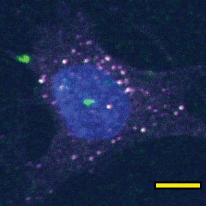
To validate that sclerostin can be targeted to the lysosome, we examined the sub-cellular distribu-
tion of sclerostin and lysosomes in cultured Ocy454 cells. Both endogenous and exogenous sclero-
stin were found in distinct puncta in Ocy454 cells (Figure 4A). In live Ocy454 cells, GFP-sclerostin
was co-localized with acidic vesicles identified by LysoTracker and co-localized with lysosomes
labeled by siR-lysosome (Figure 4B). In 3D projections of Z-stacks, it is clear that sclerostin co-local-
izes with lysosomes identified by siR-lysosome, but not all sclerostin is contained in lysosomes and
not all lysosomes contain sclerostin (Figure 4—figure supplement 1). In fixed cells, endogenous
sclerostin co-localized with p62/sequestosome-1 protein, an autophagy cargo adapter protein that
shuttles proteins for lysosomal degradation (Figure 4C). That sclerostin is found localized with lyso-
somal adapter proteins and is degraded by the lysosome is consistent with the recent finding that
sclerostin is found in extracellular vesicles positive for the lysosome-associated protein LAMP1
(Morrell et al., 2018). Interestingly, sclerostin was also co-detected with Rab27a in Ocy454 cells
(Figure 4C). Rab27a is a small GTPase that is a master regulator of the trafficking, docking, and
Gould et al. eLife 2021;10:e64393. DOI: https://doi.org/10.7554/eLife.64393 5 of 26Research article Cell Biology
ly
te
ia
ed
in
m
m
5 min
30
Im
0 hrs 4 hrs
1.5
Relative Sclerostin
Collection **
A. Treat CHX FSS with CHX **
and Inhibitor and Inhibitors **
1.0
Time After
DMSO Baf A1 Bref A MG-132
FSS Start: - 5 35 - 5 35 - 5 35 - 5 35
Sclerostin 0.5
DMSO Brefeldin A
`-Actin Bafilomycin A1 MG-132
Ocy454
0.0
0 5 10 15 20 25 30 35
B. DMSO Baf A1 1.2 Time After FSS Start (min)
Relative Sclerostin
**
****
PTH - + - + 1.0
**
Sclerostin
0.8
`-Actin Lysosomal Signal
0.6 Sequences
Ocy454
0.4 YXXø
PTH - + - + Partially Conserved
or Inverted YXXø
C. DMSO Baf A1
NXS/T
1
Mouse MQP... EYPEPP... NNQTM... HYTRFL... DFRCIP... HNQSE...NAY211
42 50 82 129 172
1
Human MQL...42EYPEPP...52NNKTM...84HFTRYV...131DFRCIP...174HNQSE...NAY213
1
Rat MQL...42EYPEPP...52NNQTM...84HYTRFV...131DFRCIP...174HNQSE...NAY213
1
Cow MQL...42EYPEPL...51NNKTM...83HFTRYV...130DFRCIP...173HNQSE...NAY212
1
Chicken MQI...40ENTETP...52NNNTM...87RITRYV...134DYRCIP...177HNQSE...NAY216
Figure 3. Sclerostin is rapidly degraded by the lysosome following bone anabolic stimuli. (A) Ocy454 cells transfected with GFP-sclerostin were treated
with cycloheximide (150 mg/mL) to prevent new protein synthesis and either DMSO (0.1%), bafilomycin A1 (100 nM) to inhibit lysosomal degradation,
brefeldin A (2 mm) to inhibit secretion, or MG-132 (10 mm) to inhibit the proteasome 4 hr prior to FSS. Cells were subjected to 5 min of FSS at 4 dynes/
cm2 and lysed immediately after the end of FSS or 30 min after the conclusion of FSS. Western blots were probed for sclerostin and b-actin. Time
courses show mean ± SEM (n = 3–6 independent experiments/group). (B) Ocy454 cells transfected with GFP-sclerostin were pre-treated with DMSO
(0.1%) or bafilomycin A1 (100 nM) to inhibit lysosomal degradation for 30 min prior to the addition of vehicle or PTH (1–34) (10 nM) for an additional 30
min (n = 3). Sclerostin abundance relative to the loading control was quantified. Graph depicts mean ± SD. *pResearch article Cell Biology
A. B. GFP-Sclerostin Lysosome Dye Merge C. Sclerostin Lysosome Marker Merge
LysoTracker
Endogenous
Sclerostin
p62
SiR-Lysosome
GFP-Sclerostin
Rab27a
Ocy454 Ocy454 Ocy454
D. FSS E. PTH F. Magic Red
- + - + DAPI
p62 p62 8000
*
Fluorescene Intensity
No Flow
b-Actin b-Actin
LC3-I LC3-I 6000
LC3-II LC3-II
Ocy454 Ocy454
3 1.5 * 2.0 1.5 ** 4000
* *
Relative p62
Relative p62
LC3-II/LC3-I
LC3-II/LC3-I
1.5
2 1.0 1.0 2000
1.0
Flow
1 0.5 0.5 0
0.5 FSS - +
0 0.0 0.0 0.0
FSS - + FSS - + PTH - + PTH - +
4 1.5 ** **
G. * H.
Relative p62
*
Relative p62
DMSO KN-93 3 GFP T268A
FSS - + - + PTH - + - + 1.0
p62 2 p62
a-Tubulin a-Tubulin 0.5
1
0 0.0
FSS - + - + PTH - + - +
DMSO KN-93 GFP T268A
Figure 4. Sclerostin co-localizes with lysosomal markers, and both FSS and PTH induce lysosome activation through CaMKII. (A) Endogenous sclerostin
(top) and GFP-tagged sclerostin (bottom) form discrete puncta in Ocy454 cells. (B) Ocy454 cells were transfected with GFP-sclerostin, and lysosomes
were visualized with Lysotracker (1 mM, 1 hr) or siR-Lysosome (1 mM, 4 hr). Scale bar represents 10 mm. (C) Ocy454 cells were stained for endogenous
sclerostin and either p62/sequestosome-1 or Rab27a to evaluate co-localization with these lysosome-associated proteins. (D) Ocy454 cells were
exposed to 1 min of FSS at 4 dynes/cm2, lysed immediately post-flow, and western blotted for p62/sequestosome-1, b-actin, and LC3 (n = 4). (E)
Ocy454 cells were treated with PTH (1–34) (10 nM) for 5 min, lysed, and western blotted for p62/sequestosome-1 and b-actin (n = 4) and LC3 (n = 8). (F)
UMR106 cells were subjected to FSS for 5 min, then Magic Red Cathepsin B was applied for 10 min, fixed, and imaged to assess lysosome activity
(n = 9). (G) Ocy454 cells were treated with DMSO or KN-93 (10 mM) to inhibit CaMKII for 1 hr prior to FSS at 4 dynes/cm2 for 5 min before lysing
immediately after FSS. Western blots were probed for p62/sequestosome-1 and b-actin (n = 3). (H) Ocy454 cells were transfected with a plasmid
expressing either GFP or dominant negative CaMKII T286A prior to treatment with PTH (1–34) (10 nM) for 30 min. Western blots were probed for p62/
sequestosome-1 and b-actin (n = 3). Graphs depict mean ± SD. *pResearch article Cell Biology
Figure 4 continued
Figure supplement 1. Co-localization of sclerostin with lysosomes.
presence of a common mechanism in osteoblast-lineage cells for controlling the abundance of
secreted proteins that control bone remodeling.
Lysosome activity is regulated by bone anabolic stimuli
Next, we examined if FSS or PTH altered lysosomal activity. Both FSS and exposure to PTH
increased p62/sequestosome-1 protein abundance and increased LC3-II/LC3-I ratio, consistent with
enhanced lysosomal delivery of autophagy cargo (Figure 4D,E). Similarly, FSS induced an increase in
lysosome activity, as determined using Magic Red Cathepsin B activity assay (Figure 4F). CaMKII
activation regulates lysosomal activity and protein degradation in other tissues (Reventun et al.,
2017; Li et al., 2017; Zemoura et al., 2019; Sandberg and Borg, 2006). Given that both FSS and
PTH activate CaMKII in osteocytes (Lyons et al., 2017; Quinn et al., 2000; Williams et al., 2016),
we examined if FSS and PTH converge on CaMKII as a common integrator regulating lysosome acti-
vation. To do this, Ocy454 cells were treated with KN-93 to inhibit CaMKII or were transfected with
a dominant negative CaMKII construct (T286A), and then exposed to FSS or PTH, respectively. In
both CaMKII-disrupted cell populations, FSS and PTH failed to increase p62/sequestosome-1 protein
abundance (Figure 4G,H), supporting that CaMKII activation following anabolic stimuli is necessary
for lysosome activation. Together, these data support that osteocytes increase their lysosomal fol-
lowing bone anabolic stimuli and that CaMKII is likely a converging point between the FSS and PTH
with respect to lysosomal activation.
Nitric oxide is necessary and sufficient for rapid sclerostin degradation
following FSS
This regulated lysosomal degradation of sclerostin following FSS or PTH treatment is reminiscent of
a relatively obscure autophagy pathway known as crinophagy, which is utilized by peptide-secreting
cells, such as pancreatic beta-cells, to route secretory vesicles to the lysosome rather than being
delivered to the membrane for exocytosis (Lee et al., 2019; Weckman et al., 2014). Interestingly,
crinophagy is regulated by nitric oxide (Sandberg and Borg, 2006). In osteocytes, nitric oxide pro-
duction following mechanical cues is a canonical response (Klein-Nulend et al., 1998; Zaman et al.,
1999; Klein-Nulend et al., 2014; Nagayama et al., 2019; Gohin et al., 2016); however, the direct
biological consequence of nitric oxide on the potent activation of osteoblasts and bone formation
has remained incomplete, though a link between Sost mRNA and sclerostin protein abundance and
nitric oxide has been suggested, albeit on a 24 hr timescale (Callewaert et al., 2010). Accordingly,
we examined if nitric oxide could also contribute to this crinophagy-like, rapid degradation of sclero-
stin protein in osteocytes.
Indeed, activation of nitric oxide signaling in Ocy454 cells with S-Nitroso-N-acetyl-DL-penicilla-
mine (SNAP), a nitric oxide donor, induced a rapid decrease in sclerostin protein (Figure 5A). This
rapid reduction of sclerostin protein occurred without increased CaMKII phosphorylation
(Figure 5A), suggesting that activation of nitric oxide production may be downstream of CaMKII
phosphorylation. When Ocy454 cells transfected with myc-sclerostin were treated with SNAP, a nitric
oxide donor, in the presence of the lysosome inhibitor bafilomycin A1, sclerostin degradation was
prevented (Figure 5B), confirming that nitric oxide is sufficient to drive the lysosomal degradation of
sclerostin. Additionally, blocking nitric oxide production with L-NAME prevented the FSS-activated
degradation of sclerostin protein at 5 min, as well as the FSS-induced increase in p62/sequesto-
some-1 (Figure 5C), supporting a role of nitric oxide in the activation of the lysosome and degrada-
tion of sclerostin, likely upstream of the lysosome and downstream of CaMKII. In total, these data
support that sclerostin is directed through a defined secretory pathway unless directed to the lyso-
some by mechano-transduction or PTH-initiated signaling.
Gould et al. eLife 2021;10:e64393. DOI: https://doi.org/10.7554/eLife.64393 8 of 26Research article Cell Biology
A. SNAP B.
Relative Sclerostin
1.5
Relative pCaMKII
- + 1.5 DMSO Baf A1
** SNAP - + - +
Sclerostin
1.0 1.0 Sclerostin
`-Actin
_-Tubulin
pCaMKII
0.5 0.5 Ocy454
1.5
Relative Sclerostin
Total CaMKII *
Ocy454 p=0.051
0.0 0.0 1.0
SNAP - + SNAP - +
C. Vehicle L-NAME 0.5
Relative Sclerostin
2.5 1.5 ***
FSS - + - + **
Relative p62
2.0 0.0
Sclerostin **
1.0 SNAP - + - +
1.5
_-Tubulin DMSO Baf A1
1.0
0.5
p62
0.5
_-Tubulin 0.0 0.0
UMR106
FSS - + - + FSS - + - +
Vehicle L-NAME Vehicle L-NAME
Figure 5. Nitric oxide, known mechanical load effector in bone and a molecular controller of crinophagy,
contributes to sclerostin degradation. (A) Ocy454 cells transfected with GFP-sclerostin were treated with vehicle
(water) or 10 mM SNAP, a nitric oxide donor, and lysed after 5 min. Western blots were probed for sclerostin, a-
tubulin, pCaMKII, and total CaMKII (n = 3). For each antibody, blots are from a single gel and exposure; a vertical
black line indicates removal of irrelevant lanes. (B) Ocy454 cells transfected with myc-tagged sclerostin were
treated with DMSO or bafilomycin A1 (100 nM) to inhibit lysosomal degradation, for 30 min, then treated with
SNAP, a nitric oxide donor, for 5 min and lysed. Western blots were probed for sclerostin and a-tubulin (n = 2–3).
(C) UMR106 cells were treated with vehicle or L-NAME (1 mM) to inhibit nitric oxide synthases (NOSs) for 1 hr
and then exposed to 1 or 5 min of FSS. Lysates from cells exposed to 1 min of FSS were probed for p62/
sequestosome-1 and a-tubulin abundance and lysates from cells exposed to 5 min of sclerostin were probed for
sclerostin and a-tubulin abundance (n = 3). Graphs depict mean ± SD. *pResearch article Cell Biology
Relative Sclerostin
2.0
A. H 2O2
5min *
1.5
Collect
H 2O2 - + 1.0
Sclerostin
0.5
`-Actin
Bone 0.0
H2O2 - +
B. Alizarin Red Calcein
Day: 1 2 3 4 11 14
Bouts of Load Euthanize
Apocynin, 2 hr
Prior to Load
Non-Loaded Loaded
Vehicle
Apocynin
C. D.
Ps.BFR/BS (+m3/+m2/day)
2.0
Ps.MAR (+m/day)
0.5 ** ** *
*
0.4 1.5
0.3
1.0
0.2
0.5
0.1
0.0 0.0
Load - + - + Load - + - +
Vehicle Apocynin Vehicle Apocynin
in
90sec
m
5
0 hrs 2 hrs
E. 2.5 n.s.
Relative Sclerostin
Collection
Inject Ulnar Load 2.0 **
Vehicle Apocynin 1.5
Load - + - + 1.0
Sclerostin
0.5
`-Actin
0.0
Bone - + - +
Vehicle Apocynin
Figure 6. NOX2-dependent ROS are necessary for load-induced sclerostin degradation and bone formation in
vivo. (A) Dissected ulnae and radii flushed of marrow were treated with hydrogen peroxide (100 mM) as a source of
ROS for 5 min before homogenization. Western blots were probed with sclerostin and b-actin (n = 6 mice). (B)
Thirteen week old male C57Bl/6 mice treated with vehicle (saline, n = 10 mice) or apocynin (3 mg/kg, n = 8 mice)
to inhibit NOX2 were forearm loaded (1800 me, 90 s, 2 Hz) and labeled with calcein and alizarin red at the
indicated times for dynamic histomorphometry. Representative periosteal double labeling is shown. (C) Periosteal
bone formation rate (Ps.BFR) and (D) periosteal mineral apposition rate (Ps.MAR) were calculated. (E) Fourteen to
17 week old male and female C57Bl/6 mice treated with vehicle (saline + 4% DMSO, i.p., n = 14) or apocynin (3
mg/kg in saline, i.p., n = 12 mice) to inhibit NOX2 were treated 2 hr prior to ulnar loading (2000 me, 90 s, 2 Hz).
Figure 6 continued on next page
Gould et al. eLife 2021;10:e64393. DOI: https://doi.org/10.7554/eLife.64393 10 of 26Research article Cell Biology
Figure 6 continued
Non-loaded and loaded limbs were isolated 5 min post-load, and western blots were probed for sclerostin and b-
actin. Vehicle data is duplicated in Figure 7D as all animals were run and processed together. Graphs depict
mean ± SD. *pResearch article Cell Biology
in
A. D. 90sec
m
Alizarin Red Calcein
5
Relative Sclerostin
Day: 1 2 3 4 11 14 0 hrs 2 hrs 4 n.s.
Euthanize
Collection
Bouts of Load
Inject Ulnar Load 3
Baf A1, 4 hr
Prior to Load Vehicle Bafilomycin **
Non-Loaded Loaded 2
Load - + - +
Vehicle Sclerostin 1
Baf A1 β-Actin
Bone 0
- + - +
B. C.
Ps.BFR/BS (μm3/μm2/day)
Vehicle Bafilomycin
0.8 ** * 1.5 *
Ps.MAR (μm/day)
E. 4
Relative Sclerostin
0.6 Control Gaucher ** **
1.0
rGCase - + - + 3
0.4 Sclerostin
0.5 2
0.2 GAPDH
0.0 0.0 1
Load - + - + Load - + - +
Vehicle Baf A1 Vehicle Baf A1
0
rGCase - + - +
Control Gaucher
F. NO
ROS Ca2+
Lysosome Sclerostin
FSS NOX2 TRPV4 CaMKII
Activation Degradation
PTH
No Stimuli Anabolic Stimuli
Secretion Degradation
PM PM
ROS Osteobast
Osteobast
NO
Sclerostin Sclerostin
Protein Protein
Lysosome
Bone Bone
(p62, LC3,
Formation Rab27a, Formation
low pH,
Cathepsin)
Figure 7. Lysosomal function is necessary for load-induced sclerostin degradation and subsequent bone formation and is implicated in human disease.
(A) Fifteen week old male C57Bl/6 mice treated with vehicle (saline + 4% DMSO, n = 7 mice) or bafilomycin A1 (1 mg/kg, n = 7 mice) to inhibit
lysosomal degradation were forearm loaded (2000 me, 90 s, 2 Hz) and labeled with calcein and alizarin red at the indicated times for dynamic
histomorphometry. Representative periosteal double labeling are shown. (B) Periosteal bone formation rate (Ps.BFR) and (C) periosteal mineral
apposition rate (Ps.MAR) were calculated. (D) Fourteen to 17 week old male and female C57Bl/6 mice treated with vehicle (saline + 4% DMSO, i.p.,
n = 14) or bafilomycin A1 (1 mg/kg in saline + 4% DMSO, i.p., n = 12 mice) to inhibit lysosomal degradation were treated 2 hr prior to ulnar loading
(2000 me, 90 s, 2 Hz). Non-loaded and loaded limbs were isolated 5 min post-load, and western blots were probed for sclerostin and b-actin. Vehicle
data is duplicated in Figure 6E as all animals were run and processed together. (E) Human iPSC-derived osteoblasts from either control (non-diseased)
or Gaucher disease patients were treated with vehicle or recombinant glucocerebrosidase (rGCase, 0.24 U/mL) for 5 days, then lysed for western
blotting. Western blots were probed for sclerostin and GAPDH (n = 3 independent patient-derived iPSC lines/group). Graphs depict mean ± SD.
*pResearch article Cell Biology
Figure 7 continued
cascade. Osteoanabolic stimuli, working through reactive oxygen (ROS) and reactive nitrogen species (RNS), direct sclerostin to the lysosome for
degradation. This results in reduced sclerostin to allow for bone formation. PM: plasma membrane; ROS: reactive oxygen species; NO: nitric oxide.
lysosomal degradation of sclerostin, we observed that iPSC-derived osteoblasts from Gaucher
patients had significantly increased levels of sclerostin compared to iPSC-derived osteoblasts from
healthy patients without Gaucher disease (Figure 7E). Furthermore, treating Gaucher iPSC-derived
osteoblasts with recombinant GCase, which restores lysosomal function, b-catenin signaling, and
osteoblast differentiation (Panicker et al., 2018), also restored sclerostin abundance to control lev-
els (Figure 7E). Since sclerostin acts as an inhibitor of Wnt/b-catenin signaling, these data suggest
sclerostin as a therapeutic target for bone loss in Gaucher disease.
Discussion
Our data show that osteocytes respond to distinct osteoanabolic cues, mechanical load and PTH, by
redirecting sclerostin from a secretory pathway to the lysosome for rapid degradation (Figure 7F).
In response to mechanical load, this signaling event is mediated by activation of ROS and nitric oxide
signaling, a CaMKII-dependent increase in lysosomal activity, and the post-translational degradation
of sclerostin by the lysosome. For PTH, CaMKII is likely a common integrator upstream of lysosomal
degradation of sclerostin protein. Furthermore, we demonstrate that both lysosomal activity and
activation of the upstream of mechano-signaling pathway are required for mechanically induced
decreases in sclerostin protein and bone formation in vivo. These findings inform a new model of
osteocyte mechano-transduction and shuttling of sclerostin to lysosomes that integrates many of the
molecular effectors of the osteocyte mechano-response, including calcium, the cytoskeleton, nitric
oxide, and sclerostin (Thompson et al., 2012; Schaffler et al., 2014; Geoghegan et al., 2019;
Baik et al., 2013) and unifies many important discoveries related to osteoanabolic signals that act
through sclerostin. These findings also describe a role of nitric oxide production, a canonical
response to mechanical load in bone of previously unclear functional consequence, in the regulated
degradation of sclerostin protein. Finally, we link sclerostin degradation not only to skeletal physiol-
ogy in mice but also to human disease using Gaucher disease iPSCs.
These data do not preclude a transcriptional control of the Sost gene by osteoanabolic stimuli
(Tu et al., 2012; Sato et al., 2020). Rather, they add an important downstream check point for regu-
lating sclerostin protein bioavailability with remarkable temporal control. It may be that post-transla-
tional control is the first line response to a bone anabolic stimulus, whereas long-term stimulation
leads to genome level transcriptional control of the Sost gene. Indeed, longer periods of loading are
required to observe Sost mRNA decreases than are required for loss of protein described here. Our
brief, 5 min FSS (Williams et al., 2020) and short, 30 min duration PTH exposure (Figure 2—figure
supplement 1) are insufficient to decrease Sost mRNA abundance hours after the stimulus. How-
ever, the contribution of longer duration stimulation of this NOX2-Ca2+-CaMKII pathway to Sost
mRNA is not yet known.
In isolation, the effects of inhibiting either NOX2-dependent ROS or lysosomal function on load-
induced bone formation and sclerostin abundance could be influenced by the broad pharmacologi-
cal impact of these drugs. However, that two distinct pharmacological inhibitors targeting different
steps in the pathway controlling sclerostin yielded a similar effect on bone formation and sclerostin
protein abundance following load supports our conclusions. This conclusion is strengthened by two
other important considerations: firstly, there was also no statistical effect of either apocynin or bafilo-
mycin A1 on bone formation rate or mineral apposition rate in the contralateral, non-loaded limb.
Secondly, the inhibitors were only present for each bout of ulnar load but were absent during the
7 day bone forming period, indicating that disruption of the acute downstream response to the
mechanical cue, not the ability of the osteoblasts to build bone, caused the reductions in bone
formation.
The rapid, controlled degradation of osteocyte sclerostin has intriguing parallels to crinophagy, a
specialized autophagic process in peptide-secreting cells in which cellular cues elicit the re-routing
of secretory proteins to the lysosome (Lee et al., 2019). Crinophagy is well described in pancreatic
beta-cells, in which Rab27a-positive, insulin-filled secretory vesicles can be shuttled to the lysosome
Gould et al. eLife 2021;10:e64393. DOI: https://doi.org/10.7554/eLife.64393 13 of 26Research article Cell Biology
for degradation when not needed, an effect triggered by nitric oxide (Waselle et al., 2003;
Sandberg and Borg, 2006). Rab27a is associated with secretory granules and other lysosome-asso-
ciated vesicles (Fukuda, 2013; Tolmachova et al., 2004), and, given the localization of nearly all the
endogenous sclerostin with Rab27a, suggests sclerostin is packaged in vesicles. Thus, analogous to
crinophagy, our data support that sclerostin is directed through a Rab27a-positive secretory pathway
for exocytosis or is trafficked to the lysosome for degradation, an effect that can be regulated by
CaMKII and mechano-activated nitric oxide. Notably, this Rab27a-associated secretory pathway
functions in osteoblasts to control RANKL secretion (Kariya et al., 2011). Whether this mechano-
pathway and regulated degradation extends to osteocyte-derived RANKL was not analyzed in the
present study, but may be physiologically significant as osteocytes are the primary source of bioac-
tive RANKL directing bone resorption in response to physiologic cues (Xiong et al., 2015;
Xiong et al., 2014).
Using Gaucher disease as a model system, this work links lysosomal function to sclerostin regula-
tion in disease. Gaucher patients experience many skeletal dysplasias, including low bone mass and
osteoporosis. Though this cell autonomous defect in Gaucher osteoblasts has previously been linked
to dysregulated b-catenin that impaired osteogenic differentiation and mineralization capacity
(Panicker et al., 2018), the present findings reveal a possible mechanism by which b-catenin is sup-
pressed. Sclerostin is a canonical Wnt/b-catenin antagonist; therefore, the increased sclerostin levels
observed here in Gaucher disease iPSCs are likely to lead to the previously reported decrease in b-
catenin activation and osteoblast differentiation (Panicker et al., 2018). Likewise, we showed that
restoring expression of the missing hydrolase, glucocerebrosidase, in Gaucher disease iPSCs lowers
sclerostin abundance, which could explain the rescue in b-catenin activation and osteoblast differen-
tiation reported previously (Panicker et al., 2018). Our data support the exploration of therapeutics
that target sclerostin to treat low bone mass symptoms in patients with lysosomal storage disorders.
That sclerostin protein abundance is post-translationally controlled may have important implica-
tions in age-related osteoporosis and the reduced sensitivity of osteocytes to mechanical cues with
aging (Hemmatian et al., 2017; Haffner-Luntzer et al., 2016). Lysosome activity is diminished with
age (Moore, 2020; Stead et al., 2019), including in the osteocyte during age-related bone loss
(Chen et al., 2014), an effect that could impair sclerostin degradation and new bone formation even
in the face of osteoanabolic signals. While undoubtedly multifaceted in its impacts, targeting
autophagy and lysosome activity to improve bone mass in aging has been proposed (Li et al., 2020;
Wang et al., 2019), and these data suggest that impacts on sclerostin bioavailability might contrib-
ute mechanistically to its efficacy.
Together, these discoveries provide key insights into the unexpected, rapid regulation of osteo-
cyte sclerostin protein by the lysosome and reveal new therapeutic targets that can be exploited to
improve bone mass in conditions such as osteoporosis. Additionally, targeting sclerostin may be
important to interventions to improve bone mass in lysosomal storage disorders, like Gaucher
disease.
Materials and methods
Key resources table
Reagent type (species)
or resource Designation Source or reference Identifiers Additional information
Gene (Mus musculus) Sost National Center for AAK13455
Biotechnology Information
Gene (Homo sapiens) Sost National Center for AAK16158.1
Biotechnology Information
Gene Sost National Center for EDM06161.1
(Rattus norvegicus) Biotechnology Information
Gene (Bos Taurus) Sost National Center for NP_001159986.1
Biotechnology Information
Continued on next page
Gould et al. eLife 2021;10:e64393. DOI: https://doi.org/10.7554/eLife.64393 14 of 26Research article Cell Biology
Continued
Reagent type (species)
or resource Designation Source or reference Identifiers Additional information
Strain, strain C57Bl/6 Jackson Laboratories 000664 RRID:IMSR_JAX:000664
background
(Mus musculus,
male and female)
Cell line (Mus musculus) Ocy454 P. Divieti-Pajevic, RRID:CVCL_UW31
Boston University
Cell line UMR106 ATCC CRL-1661 RRID:CVCL_3617
(Rattus norvegicus)
Transfected construct CaMKII T286A Addgene #29430
(Mus musculus)
Transfected construct GFP-tagged sclerostin Origene #RG217648
(Homo sapiens)
Transfected construct myc-tagged sclerostin Origene #MR222588
(Mus musculus)
Transfected construct KillerRed Evrogen FP966
(Mus musculus)
Biological sample WT and GD iPSCs PMID:23071332
(Homo sapiens)
Antibody Goat Polyclonal R and D Systems AF1589 (1:250–500)
Anti-sclerostin RRID:AB_2270997
Antibody Mouse Monoclonal Millipore MAB374 (1:2500)
Anti-GAPDH RRID:AB_2107445
Antibody Mouse Monoclonal Sigma A1978 (1:5000)
Anti-bActin RRID:AB_476692
Antibody Mouse Monoclonal Sigma T9026 (1:2000)
Anti-aTubulin RRID:AB_477593
Antibody Rabbit Polyclonal Sigma ABT257 (1:1000)
Anti-Col1a1 RRID:AB_2890134
Antibody Rabbit Monoclonal Cell Signalling Technology 12716S (1:1000)
Anti-pCaMKII RRID:AB_2713889
Antibody Rabbit Polyclonal Cell Signalling Technology 3362S (1:1000)
Anti-Total CaMKII RRID:AB_2067938
Antibody Rabbit Monoclonal Cell Signalling Technology 23214 (1:250–500)
Anti-p62/Sequestosome-1 RRID:AB_2798858
Antibody Rabbit Polyclonal Anti-LC3B Cell Signalling Technology 2775 (1:500)
RRID:AB_915950
Antibody Rabbit Monoclonal Cell Signalling Technology 69295 (1:250)
Anti-Rab27A RRID:AB_2799759
Antibody HRP Anti-Rabbit Cell Signalling Technology 7074 (1:1000–5000)
RRID:AB_2099233
Antibody HRP Anti-Mouse Cell Signalling Technology 7076 (1:1000–5000)
RRID:AB_330924
Antibody HRP Anti-Goat Thermo Fisher Scientific A27014 (1:1000)
RRID:AB_2536079
Antibody Donkey anti-Goat IgG Thermo Fisher Scientific A-11056 (1:100)
Alexa Fluor 546 RRID:AB_142628
Antibody Chicken anti-Rabbit Thermo Fisher Scientific A-21441 (1:100)
Alexa Fluor 488 RRID:AB_2535859
Sequence-based reagent Sost NCBI BLAST NM_024449.6 GGA ATG ATG CCA CAG AGG TCA T
and CCC GGT TCA TGG TCT GGT T
Sequence-based reagent Gapdh NCBI BLAST NG_007785.2 CGT GTT CCT ACC CCC AAT GT and
TGT CAT CAT ACT TGG CAG GTT TCT
Continued on next page
Gould et al. eLife 2021;10:e64393. DOI: https://doi.org/10.7554/eLife.64393 15 of 26Research article Cell Biology
Continued
Reagent type (species)
or resource Designation Source or reference Identifiers Additional information
Sequence-based reagent Hprt NCBI BLAST NM_013556.2 AGC AGT ACA GCC CCA AAA TGG and
AAC AAA GTC TGG CCT GTA TCC AA
Sequence-based reagent Rpl13 NCBI BLAST NM_016738.5 CGA AAC AAG TCC ACG GAG TCA and
GAG CTT GGA GCG GTA CTC CTT
Commercial assay Magic Red Sigma CS0370
or kit
Commercial assay siR-Lysosome Spirochrome SC016
or kit
Commercial assay Lysotracker Thermo Fisher Scientific L7528
or kit
Commercial assay High-Capacity Thermo Fisher Scientific 4388950
or kit RNA-to-cDNA Kit
Commercial assay Maxima SYBR Thermo Fisher Scientific FERK0221
or kit Green/ROX qPCR
Master Mix
Commercial assay JetPrime Transfection kit PolyPlus Transfection 114–75
or kit
Chemical compound, Bafilomycin A1 Cell Signalling 54645
drug (in vitro studies) Technology
Chemical compound, Bafilomycin A1 Research Products 88899-55-2
drug (in vivo studies) International
Chemical compound, Brefeldin A Cell Signalling 9972
drug Technology
Chemical compound, MG-132 Cell Signalling 2194
drug Technology
Chemical compound, Cycloheximide Cell Signalling 2112
drug Technology
Chemical compound, PTH (1–34) US Biological #P3109-24D
drug Life Sciences
Chemical compound, Leupeptin Millipore EI8
drug
Chemical compound, Apocynin Sigma 178385
drug
Chemical compound, L-NAME Millipore N5751
drug
Chemical compound, SNAP Sigma N3398
drug
Chemical compound, Alizarin Red Sigma A3882
drug
Chemical compound, KN-93 Sigma K1358
drug
Chemical compound, Calcein Sigma C0875
drug
Chemical compound, Halt Protease and Thermo Fisher 78440
drug Phosphatase Inhibitor Scientific
Cocktail (EDTA-free)
Chemical compound, SuperBlockPBS Thermo Fisher 37515
drug Scientific
Software, algorithm FIJI ImageJ RRID:SCR_002285
Software, algorithm Nikon NIS Elements 5.2 Nikon RRID:SCR_014329
Other REVERT Total Protein Stain Licor 827–15733
Other Prolong Gold Antifade Cell Signalling 8961S
Reagent with DAPI Technology
Continued on next page
Gould et al. eLife 2021;10:e64393. DOI: https://doi.org/10.7554/eLife.64393 16 of 26Research article Cell Biology
Continued
Reagent type (species)
or resource Designation Source or reference Identifiers Additional information
Other TRIzol Sigma T9424
Other DCF Invitrogen D399
Chemicals and reagents
Bafilomycin A1 (in vitro studies, #54645), MG-132 (#2194), brefeldin A (#9972), cycloheximide
(#2112), and antibodies against Thr 286 pCaMKII (#12716S), total CaMKII (3362S), p62/sequesto-
some-1 (#23214), LC3B (#2775), Rab27a (#69295), and Prolong Gold Antifade Reagent with DAPI
(#8961S) were from Cell Signaling Technologies. REVERT Total Protein Stain (827–15733) was from
Licor. Anti-sclerostin antibodies (#AF1589) were purchased from R and D Systems. Bafilomycin A1 (in
vivo studies, #88899-55-2) was from Research Products International. Leupeptin (#EI8), Nw-nitro-L-
arginine methyl ester hydrochloride (L-NAME, N5751), and GAPDH (MAB374) were from Millipore.
PTH (1–34, #P3109-24D) was from US Biological Life Sciences. Alizarin red (#A3882), calcein
(#C0875), Apocynin (178385), S-Nitroso-N-acetyl-DL-penicillamine (SNAP, N3398), and antibodies
against b-actin (A1978), a-tubulin (T9026), Pro-Collagen Type I, A1 (Col1a1) (ABT257), Magic Red
Cathepsin B Detection Assay Kit (CS0370), KN-93 (K1385), and TRIzol (T9424) were from Sigma. siR-
Lysosome (CY-SC016) was from Spirochrome. GFP-tagged human sclerostin (#RG217648)- and myc-
tagged mouse sclerostin (#MR222588) were purchased from Origene. KillerRed plasmid (FP966) was
purchased from Evrogen. CaMKII T286A dominant negative construct was from Addgene (#29430).
Recombinant human GCase (rGCase) (Cerezyme) was obtained from patient infusion remnants. 20 ,70 -
Dichlorofluorescein (DCF, D399) was purchased from Invitrogen. Halt Protease and Phosphatase
Inhibitor Cocktail (EDTA-free) (78440), Lysotracker (L7528), SuperBlockPBS (37515), Donkey anti-
Goat IgG Alexa Fluor 546 (A-11056), Chicken anti-Rabbit Alexa Fluor 488 (A-21441), High-Capacity
RNA-to-cDNA Kit (4388950), and Maxima SYBR Green/ROX qPCR Master Mix (FERK0221) were
from Thermo Fisher Scientific. JetPrime Transfection kit (114-75) was from PolyPlus Transfection.
Modified RIPA lysis buffer contained 50 mM Tris–HCl pH 8.0, 150 mM NaCl, 1.0% NP-40, 0.5%
sodium deoxycholate, 0.1% SDS, 10 mM Na4P2O7, 10 mM 2-glycerolphosphate, 10 mM NaF, 10
mM EDTA, 1 mM EGTA, 1 HALT phosphatase, and protease inhibitor cocktail.
Cell culture
UMR106 cells (purchased from ATCC, CRL-1661) were cultured in Dulbecco’s modified essential
medium (DMEM) supplemented with 10% fetal bovine serum (FBS), and maintained at 37˚C and 5%
CO2, as described (Gupta et al., 2016). Ocy454 cells (provided by P. Divieti-Pajevic, Boston Univer-
sity) were cultured on type I rat-tail collagen-coated plates in a-minimal essential medium (aMEM)
supplemented with 10% FBS and maintained at 33˚C and 5% CO2 (Wein et al., 2015; Spatz et al.,
2015). Cell phenotype was verified by expression of bone cell specific markers, most importantly the
osteocyte specific expression of sclerostin. Cells were used at low passages (Research article Cell Biology
Fluid flow
Ocy454 and UMR106 cells were exposed to fluid flow using a custom FSS device (Lyons et al.,
2017; Lyons et al., 2016). Media was removed, and cells were rinsed in a Hepes-buffered Ringer
solution containing 10 mM Hepes (pH 7.3), 140 mM NaCl, 4 mM KCl, 1 mM MgSO4, 5 mM NaHCO3,
10 mM glucose, and 1.8 mM CaCl2. Ringer solution was also used as fluid flow buffer. Cells were
exposed to 1–5 min of FSS (four dynes/cm2), as indicated, and lysed in a modified RIPA buffer plus
HALT protease and phosphatase inhibitors at the time indicated in each experiment.
Cell treatments
To block cellular degradation pathways, cells were pre-treated with bafilomycin A1 (100 nM, 30 min
or 4 hr, as indicated), brefeldin A (2 mM, 4 hr), MG-132 (10 mM, 4 hr), leupeptin (200 mM, 6 hr), or
dimethyl sulfoxide (DMSO) (0.1%, 30 min or 4 hr, as indicated) in Ringer solution. For PTH treatment,
cells were treated with PTH (1–34) (10 nM) in Ringer solution for up to 30 min. For SNAP treatment,
cells remained in the media they were plated in and SNAP dissolved in sterile water was added to a
final concentration of 10 mM. To block the lysosomal function before SNAP treatment, cells were
treated with bafilomycin A1 (100 nM) for 30 min prior to the addition of SNAP. Cells were lysed 5
min after addition of SNAP. For L-NAME treatment, L-NAME was dissolved in sterile water and then
diluted into Ringer solution (1 mM). UMR106 cells were pre-treated with L-NAME or vehicle control
1 hr prior to FSS, then exposed to 5 min of FSS, and lysed immediately after. Ocy454 cells were
treated with 10 mM KN-93 for 1 hr prior to the addition of PTH (1–34) (10 nM) for an additional 10
min. To assess PTH effects on Sost mRNA, Ocy454 cells were sera starved in aMEM supplemented
with 0.1% FBS overnight and were then treated with PTH (1–34) (10 nM) or vehicle for 30 min diluted
in aMEM supplemented with 0.1% FBS at 37˚C, 5% CO2. Media was then switched for fresh in
aMEM supplemented with 0.1% FBS, and cells were lysed in TRIzol 5.5 hr after.
RT-qPCR
Cells lysed with TRIzol were processed using Direct-zol RNA Kit to isolate RNA according to manu-
facturer’s instructions. RNA was reverse transcribed using High-Capacity RNA-to-cDNA Kit accord-
ing to manufacturer’s instructions. cDNA was used for RT-qPCR using Thermo Scientific Maxima
SYBR Green/ROX qPCR Master Mix. The level of the Sost was simultaneously normalized to expres-
sion levels for Gapdh, Rpl13, and Hprt. PCR primers used are as follows: Sost: GGA ATG ATG CCA
CAG AGG TCA T and CCC GGT TCA TGG TCT GGT T; Rpl13: CGA AAC AAG TCC ACG GAG TCA
and GAG CTT GGA GCG GTA CTC CTT; Gapdh: CGT GTT CCT ACC CCC AAT GT and TGT CAT
CAT ACT TGG CAG GTT TCT; and Hprt: AGC AGT ACA GCC CCA AAA TGG and AAC AAA GTC
TGG CCT GTA TCC AA.
Sequence alignment
Amino acid sequences were acquired from the National Center for Biotechnology Information’s
(NCBI) protein database. Accession numbers are as follows: mouse (AAK13455); human
(AAK16158.1); rat (EDM06161.1); cow (NP_001159986.1); and chicken (XP_024999845.1). Sequence
alignment was done using NCBI’s Constraint-based Multiple Alignment Tool (COBALT). Lysosomal
signal sequences (Braulke and Bonifacino, 2009) and the secretory signal peptide were annotated
manually.
Degradation assays
Ocy454 cells were treated with CHX (150 mg/mL), and either DMSO (0.1%), bafilomycin A1 (100 nM),
brefeldin A (2 mM), or MG-132 (10 mM) diluted in supplemented aMEM for 4 hr or with leupeptin
200 mM for 6 hr at 37˚C and 5% CO2. Cells were then exposed to 5 min of FSS as described above
in Ringer containing the appropriate treatment with CHX. Cells were lysed in a modified RIPA buffer
+ HALT protease and phosphatase inhibitors and collected for western blotting at 5 and 30 min
post-flow. For basal degradation assays, UMR106 cells were treated with 150 mg/mL CHX for 0, 1, 2,
or 4 hr and lysed. A best-fit linear line was constrained through y = 1 to determine protein half-life.
Gould et al. eLife 2021;10:e64393. DOI: https://doi.org/10.7554/eLife.64393 18 of 26Research article Cell Biology
Transient transfections
Ocy454 cells were seeded in 96-well plates at a density of 20,000 cells/well and incubated at 37˚C
for 24 hr. Transient transfections were performed using 0.025 mg/well GFP-tagged sclerostin, 0.05
mg/well Myc-tagged sclerostin, or 0.1 mg/well CaMKII T286A DNA mixed with 5 mL/well JetPrime
buffer and 0.1 mL/well JetPrime reagent, as described (Lyons et al., 2017). After 16 hr of incubation
at 37˚C, the transfection medium was replaced with complete aMEM. The cells were incubated an
additional 24 hr at 37˚C prior to experiments. UMR106 cells for KillerRed experiments were plated
on glass bottom 10 mm dishes at a density of 50,000 cells/dish. Transient transfections were per-
formed using 2 mg/well KillerRed and 4 or 8 mL/well Jetprime reagent. Sixteen hours after incuba-
tion, transfection medium was replaced with complete DMEM and were incubated an additional 37˚
C prior to experiments.
Fluorescence co-localization
Ocy454 cells were seeded at 10,000 cells/well and grown on a glass-bottom 96-well plate. Cells
were transfected with GFP-Sclerostin as described above, where indicated. For immunofluorescence
staining, cells were fixed with 1% paraformaldehyde, permeabilized with 0.1% Triton-X in PBS, and
blocked with SuperBlockPBS for 1 hr, as described (Kerr et al., 2015). Primaries diluted in Super-
BlockPBS against p62/sequestosome-1, Rab27a, and sclerostin were used at 1:250 and incubated
overnight at 4C. Chicken anti-Rabbit 488 and Donkey anti-Goat 546 diluted in SuperBlockPBS were
used at 1:100 and incubated for 3 hr at room temperature. Cells were mounted with ProLong Gold
Antifade with DAPI and imaged with a Nikon Ti2 microscope with a SpectraX Light Engine and a ds-
Qi2 Monochrome camera. For live cell imaging, Ocy454 cells were transfected with GFP-Sclerostin
as described above. Lysosomes were labeled Lysotracker (1 mM) for 1 hr and then imaged with a
Nikon C2 confocal microscope. For co-localization quantification, Ocy454 cells transfected with GFP-
sclerostin were treated with siR-Lysosome (1 mM) for 4.5 hr at 37˚C to label lysosomes. Cells were
imaged on a Nikon C2 confocal microscope and Z-stacks with 0.5 mm steps were obtained in each
well. These Z-stacks were then denoised using Nikon Denoise aI (NIS Elements 5.2). Co-localization
in the denoised Z-stacks using the Mander’s coefficients was measured using the FIJI plugin, JaCOP
(Bolte and Cordelières, 2006).
Magic red cathepsin B activity assay
UMR106 cells were exposed to FSS at 4 dynes/cm2 for 5 min. Magic Red and Hoechst (1) reagent
were then added to all wells for 10 min. Cells were washed three times with warm 1 PBS and then
fixed for 10 min with 1% paraformaldehyde. Cells were washed one time with 1 PBS and imaged
at 20 with a Nikon Ti2 microscope with a SpectraX Light Engine and a ds-Qi2 Monochrome cam-
era. Average Magic Red intensity from three images from three wells was measured by subtracting a
mask image of the nuclei from the Magic Red signal. Magic Red signal was then measured on a per
pixel basis, and average intensities were calculated.
KillerRed imaging and protein isolation
For imaging, UMR106 cells transfected with KillerRed were loaded with DCF (10 mM, 30 min, 37˚C)
diluted in Ringer solution to track ROS production. After loading, cells were washed with fresh
Ringer solution. Cells were stimulated with LED light for 40 s. DCF and KillerRed signals were
imaged before and after exposure to LED light in the same cells. For western blotting, cells trans-
fected with KillerRed were stimulated with LED light for 5 min and were lysed 5 min after the end of
light exposure. No light controls were treated the same but not exposed to light.
Animals
Male and female, age-matched C57BL/6 mice were purchased from Jackson Laboratory. Mice were
group housed in micro-isolator cages, and food (standard rodent chow) and water were available ad
libitum. Mice were maintained on a 12-h-light–12-h-dark cycle. Experiments were conducted on 13–
16 week old mice, as indicated. All animal protocols were approved by the Animal care and Use
Committee at the University of Maryland School of Medicine.
Gould et al. eLife 2021;10:e64393. DOI: https://doi.org/10.7554/eLife.64393 19 of 26You can also read