Distinguishing features of current COVID-19 vaccines: knowns and unknowns of antigen presentation and modes of action - Nature
←
→
Page content transcription
If your browser does not render page correctly, please read the page content below
www.nature.com/npjvaccines

                  REVIEW ARTICLE                         OPEN

                  Distinguishing features of current COVID-19 vaccines:
                  knowns and unknowns of antigen presentation and modes of
                  action
                                      1✉                             1✉
                  Franz X. Heinz           and Karin Stiasny

                      COVID-19 vaccines were developed with an unprecedented pace since the beginning of the pandemic. Several of them have
                      reached market authorization and mass production, leading to their global application on a large scale. This enormous progress
                      was achieved with fundamentally different vaccine technologies used in parallel. mRNA, adenoviral vector as well as inactivated
                      whole-virus vaccines are now in widespread use, and a subunit vaccine is in a final stage of authorization. They all rely on the native
                      viral spike protein (S) of SARS-CoV-2 for inducing potently neutralizing antibodies, but the presentation of this key antigen to the
                      immune system differs substantially between the different categories of vaccines. In this article, we review the relevance of
                      structural modifications of S in different vaccines and the different modes of antigen expression after vaccination with genetic
                      adenovirus-vector and mRNA vaccines. Distinguishing characteristics and unknown features are highlighted in the context of
                      protective antibody responses and reactogenicity of vaccines.
1234567890():,;

                      npj Vaccines (2021)6:104 ; https://doi.org/10.1038/s41541-021-00369-6

                  INTRODUCTION                                                                            have to be discerned. The first category consists of mRNA and
                  The development of COVID-19 vaccines was extremely fast and                             adenoviral vector vaccines (herein referred to as genetic vaccines,
                  successful, with several manufacturers having obtained market                           sections: “Genetic vaccines—general, “mRNA vaccines”, “Adeno-
                  authorization for their products within the first year from the                          virus-vector vaccines”), both of which do not contain the spike
                  identification of the virus (SARS-CoV-2). These vaccines are now                         protein but provide genetic information for its biosynthesis in
                  used worldwide for mass immunization programs, and data on                              body cells of the vaccinee. With this kind of vaccines, the specific
                  vaccine efficacies justify the hope that vaccination can indeed be                       design of genetic sequences for the correct formation and
                  the main instrument for preventing serious disease and death, and                       presentation of properly folded spike proteins to B cells are in
                  more generally for combating the pandemic1–4. Despite incom-                            the foreground of interest. The second category encompasses
                  pletely resolved questions (e.g. duration of immunity, prevention                       protein-based approaches, i.e. classical inactivated whole-virus
                  of transmission, and protection against emerging virus variants)                        and innovative subunit vaccines, which contain S in different
                  the availability of effective COVID-19 vaccines is an enormous                          forms and combinations with adjuvants (Sections: “Protein-based
                  relief and certainly a great success story already now.                                 vaccines—general”, “Inactivated vaccines”, “Subunit vaccines”).
                     All current vaccines that are authorized for general use and for                     Irrespective of these categories, all vaccines have to cope with the
                  which clinical efficacy data have been published rely on the viral                       intrinsic problem of conformational instability of the spike protein,
                  spike protein (S) as an immunogen, either alone or—in the case of                       whether it is synthesized in the vaccinee after genetic vaccination
                  inactivated virus vaccines—together with other viral proteins                           or in cell culture systems for production of conventional vaccines.
                  present in the viral particle (see sections below). Because of its                         In this review, we discuss the biosynthesis and relevant
                  essential functions during viral entry (receptor binding and                            structural features of the viral spike as a basis for understanding
                  membrane fusion), the S protein is the major target of antibodies                       differences of its presentation in current COVID-19 vaccines. Our
                  that can potently neutralize the virus. Increasing evidence                             major focus is on variations of the constructs for S biosynthesis in
                  indicates that neutralizing antibodies are indeed a reliable                            genetic vaccines and on possible conformational differences of S
                  correlate of protection5–9. The potency of these antibodies                             in conventional vaccines. We also address the ‘grey matter’ of
                  depends on high-affinity interactions with specific parts of the                          additional variables, such as ill-defined downstream production
                  complex three-dimensional structure of the spike in a native                            processes and purity of vaccines as well as differences in
                  conformation10,11. Efficient formation of such antibodies by B cells                     triggering sensors of innate immunity. All of these distinguishing
                  requires helper functions of CD4 T cells that are specifically                           features might provide clues to yet unresolved vaccine-specific
                  stimulated by peptides derived from the same antigen in complex                         determinants of immune responses, efficacy, and potentially
                  with MHCII molecules. Other components of cellular immunity,                            adverse reactions. Our review is limited to those vaccines in
                  such as CD8 T cells, also contribute to immune responses after                          current use for which phase 3 clinical efficacy data have been
                  SARS-CoV-2 infection or vaccination, although their role in COVID-                      reported, and for which published information on the nature and
                  19 infections and protection from disease is still incompletely                         manufacturing process exists. However, we would like to
                  resolved12,13.                                                                          emphasize that there is an enormous pipeline of further
                     Current COVID-19 vaccines present the spike protein in very                          developments (https://www.who.int/publications/m/item/draft-
                  different ways to the immune system, and two main categories                            landscape-of-covid-19-candidate-vaccines), including subunit

                  Center for Virology, Medical University of Vienna, Vienna, Austria. ✉email: franz.x.heinz@meduniwien.ac.at; karin.stiasny@meduniwien.ac.at
                  1

                  Published in partnership with the Sealy Institute for Vaccine Sciences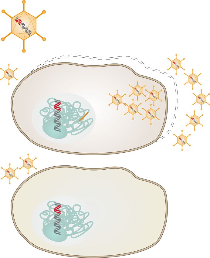
F.X. Heinz and K. Stiasny
2
vaccines that contain only parts of the S protein, in some instances
References
combined with components of other viral proteins. Therefore, the
97,141,142
19,62,63
landscape of vaccines becoming available for general use may
44,46
45,47
65,66
67,68
89,90
86,87
98,99
expand in the near future. Key features of the vaccines discussed
56
61
in this review are summarized in Table 1.
10×1010 adenovirus vector
5×10¹⁰ adenovirus vector
5×10¹⁰ adenovirus vector
5×10¹⁰ adenovirus vector
BIOSYNTHESIS AND KEY PROPERTIES OF THE SPIKE PROTEIN
4 µg proposed (2x)
3 µg proposed (2x)
6 µg proposed (2x)
100 µg RNA (2x)
Biosynthesis of S
5 µg S ( +50 µg
30 µg RNA (2x)
12 µg RNA (2x)
adjuvant) (2x)
particles (2x)
particles (2x)
particles (2x)
particles (1x)
In the course of cellular SARS-CoV-2 infection (Fig. 1a), the S
protein is synthesized from one of the viral subgenomic mRNAs
Eukaryotic production Dosage
and co-translationally transported into the lumen of the endo-
plasmic reticulum (ER) by the use of a signal sequence at its N-
terminus, comprising residues 1 to 13 of its total 1273 amino
acids14. The signal sequence is cleaved off by signal peptidase
attached to the inner ER membrane, generating the final
not applicable
not applicable
not applicable
N-terminus of the viral spike protein (14-QCVNL…). After
completion of translation, the protein remains attached to the
cell line
HEK293
HEK293
HEK293
PER.C6
ER membrane through a C-terminal membrane anchor, trimerizes
Vero
Vero
NIV2020-770 (D614G) Vero
Sf9
and moves to the ER-Golgi intermediate compartment (ERGIC)
where virus assembly occurs by budding into the ERGIC lumen
(Fig. 1a)15. During exocytosis, virus particles encounter the
Wuhan-Hu-1-like
Wuhan-Hu-1-like
protease furin in the trans-Golgi network (TGN), which cleaves
the S protein into its membrane-associated S2 subunit and the
Wuhan-Hu-1
Wuhan-Hu-1
Wuhan-Hu-1
Wuhan-Hu-1
Wuhan-Hu-1
Wuhan-Hu-1
Wuhan-Hu-1
Wuhan-Hu-1
HB02 strain
Virus strain
CN2 strain
(probably)
distal S1 subunit at a characteristic polybasic cleavage site16.
1234567890():,;
These subunits remain associated in the trimer through non-
covalent interactions, and the virus is probably secreted via
exocytic lysosomes with disrupted lysosomal functions17. Exten-
sive modifications by N- and O-glycosylation occur in the Stabilizing mutations
compartments encountered by S during its intracellular
yes (prolines, furin
yes (prolines, furin
transport18.
BBIBP-CorV, Sinopharm COVID-19 vaccine not applicable
not applicable
not applicable
cleavage site)
cleavage site)
Yes (prolines)
yes (prolines)
yes (prolines)
When S is synthesized as an isolated protein (Fig. 1b) (as in
mRNA and adenovirus vector vaccines as well as for production of
recombinant subunit vaccines), the pathway of biosynthesis is
very similar. However, the absence of interactions with other viral COVID-19 vaccine AstraZeneca, AZD1222, no
no
no
components for particle assembly may modulate glycosylation
mRNA-1273, COVID-19 Vaccine Moderna
Ad26.COV2.S, COVID-19 Vaccine Janssen
patterns and stability of the S trimers. Furthermore, S1 may
dissociate from recombinantly produced spikes after furin rAd26-S + rAd5-S, Gam-COVID-Vac,
cleavage in the TGN (shedding) and allow S2 to convert into its
ChAdOx1-S, Vaxzeria; Covishield
post-fusion conformation in the absence of mutations that
remove the cleavage site18. Manipulations of the authentic viral
signal sequence may cause inhomogeneities of the N-terminus
Ad5 nCoV, Convidecia
BNT162b2, Comirnaty
and impair native folding of S19 (see also section “Adenovirus-
Covaxin, BBV152
vector vaccines” and Fig. 5).
NVX-CoV2373
CoronaVac
Sputnik V
Structural properties of S
CVnCoV
Name
Each monomer of S is composed of several structural elements,
including the N-terminal domain (NTD) and receptor-binding
domain (RBD) in S1, which occlude the S2 moiety in the native S
Gamaleya Research Institute (Russia)
trimer (Fig. 2a–c)20,21. The RBD oscillates between an ‘up’ and
Institute of Biotechnology (China)
University of Oxford-AstraZeneca
Janssen-Johnson & Johnson (NL/
BioNTech-Pfizer (Germany, USA)
‘down’ position, and interaction with the cellular receptor (ACE2) is
CanSino Biological Inc., Beijing
Biological Products Co (China)
Sinopharm,Beijing Institute of
only possible with the transiently exposed RBD in the up
Vaccines discussed in this review.
position20,21. In its mature form, the S trimer is metastable and
Moderna-NIAID (USA)
Bharat Biotech (India)
ready to undergo triggered conformational changes that allow S2
CureVac (Germany)
to drive fusion of the viral and cellular membranes upon virus
Sinovac (China)
Novavax (USA)
entry22. The trigger comprises binding of RBD to ACE2 and a
Manufacturer
(UK, Sweden)
further proteolytic cleavage by cellular proteases (in addition to
the furin cleavage between S1 and S2) at the so-called S2’ site,
USA)
resulting in the removal of a small sequence element and the
exposure of the fusion peptide at the N-terminus of S2 (Fig. 2c)22–
24
. As a consequence of these changes, the S1 subunits dissociate
from the trimer, releasing S2 from its constraints in the pre-fusion
conformation to allow an irreversible conversion into a character-
Adenovector
Adenovector
Adenovector
Adenovector
whole virus
whole virus
whole virus
istic elongated post-fusion structure (Fig. 2d)24–26. The energy
Inactivated
Inactivated
Inactivated
Table 1.
Subunit
gained by the formation of this hairpin-like structure, in which the
mRNA
mRNA
mRNA
Type
fusion peptide is juxtaposed to the C-terminal membrane anchor,
is the driving force for viral membrane fusion during entry22.
npj Vaccines (2021) 104 Published in partnership with the Sealy Institute for Vaccine Sciences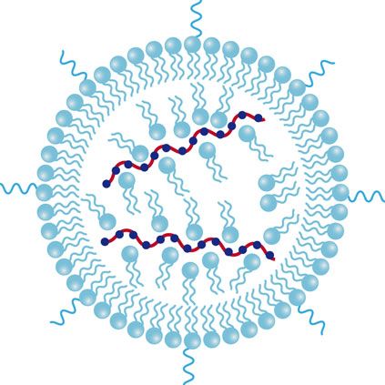
F.X. Heinz and K. Stiasny
                                                                                                                                                       3
                             a                                                 Lysosome

                                      ER
                                                                                                                    Virus
                                                           ERGIC
                                                                                                                   release
                                 E
                                 M
                                             S

                                                                                                                 TGN

                                                                                                           S2
                                                   S                                                        S1
                                                                                              Furin
                                                   M                RNP
                                                                          N                 cleavage
                             mRNAs
                                                   E
                                                   N      Assembly and Budding                             Exocytosis

                                                                                                      plasma membrane

                             b
                                       ER                ERGIC                            TGN

                                                                                                  Furin
                                                                                     S2         cleavage

                                                                                                 S1

                                                                                                  Shedding of S1

                                                                                                  S2*
                                                                              Exocytosis

Fig. 1 Biosynthesis and intracellular transport of S. a Infected cells: Subgenomic mRNAs for viral structural proteins are translated in
association with the ER (S, M, and E) or in the cytoplasm (N), and virus assembly takes place in the ERGIC. Virus particles are transported
through the TGN and released from the cells probably via lysosomes. During transport, S is cleaved into S1 and S2 by the cellular protease
furin in the TGN. Some spike molecules, not assembled into virions, are also transported to the plasma membrane despite the presence of an
ER retention signal15. b Transfected cells: Biosynthesis of S occurs in the absence of interactions with other viral proteins. Proteolytic cleavage
into S1 and S2 occurs in the TGN similar to that in infected cells, but some shedding of cleaved S1 and conversion of S2 into its post-fusion
structure (S2*) may occur in the absence of stabilizing mutations. ER—endoplasmic reticulum; ERGIC—endoplasmic reticulum Golgi
intermediate compartment; TGN—Trans Golgi Network; RNP—Ribonucleoprotein; Viral proteins: S—spike, M—membrane; E—envelope; N—
nucleoprotein.

   The potential of the S trimer to adopt different conformations             specific for the RBD27–34, but several strongly neutralizing
may pose a problem for its use in vaccines, because the native                antibodies also recognized the NTD27,34–37, and some were
structure—required to induce potently neutralizing antibodies—                dependent on the quaternary assembly of the trimer27,38.
may be disrupted during manufacturing of conventional vaccines                Neutralizing activity was also observed for antibodies against S2,
or when the protein is expressed in cells of the vaccinee after               but the potency was lower than of those against S127. Importantly,
genetic vaccination. Some manufacturers have therefore intro-                 the human neutralizing antibody response in SARS-CoV-2 infec-
duced stabilizing mutations that are intended to prevent                      tion appears to be dominated by RBD-specific antibodies, which—
inadvertent structural conversion of the labile S protein. These              on average—were shown to contribute 90% of the total
modifications (indicated in Table 1 and in the discussion of                   neutralizing activity of human post-infection sera39. It is therefore
individual vaccines below) include two proline mutations in S2                a major goal of all COVID-19 vaccines to present the spike and its
(K986P and V987P) at the junction between two alpha helices in                RBD in a most native conformation for inducing a high proportion
the pre-fusion form to avert their fusogenic conformational switch            of potently neutralizing antibodies after vaccination.
into a long alpha helix in the post-fusion form, and mutations that
abolish furin cleavage between S1 and S2 to maintain the pre-                 VACCINE-SPECIFIC DIFFERENCES OF S-ANTIGEN STRUCTURE
fusion trimer and to prevent shedding of S118 (Fig. 2c, d).                   AND PRESENTATION
                                                                              The different classes of currently available COVID-19 vaccines
Antigenic structure of S                                                      exhibit fundamental differences with respect to their modes of
A number of monoclonal antibodies were isolated from COVID-19                 action and the ways by which the spike antigen is presented to
patients and used for antigenic characterization of the S trimer,             the immune system. In the following sections, we will discuss
including 3D structure determinations of complexes between S (or              these basic differences, and provide information on variations and
parts thereof) and antibody Fab fragments. Collectively, these data           modifications that can affect the structural integrity of the spike in
showed that the most potently neutralizing antibodies were                    genetic and conventional vaccines.

Published in partnership with the Sealy Institute for Vaccine Sciences                                                       npj Vaccines (2021) 104
F.X. Heinz and K. Stiasny
4
a b c d
RBDs down RBD up RBD
NTD NTD
S1 NTD
2P
FP 2P
furin
cleavage
S2‘ site
cleavage
S2
site
FP
Fused viral and
Viral membrane cellular membrane
Fig. 2 Structures of the spike protein in pre- and post-fusion conformations. a Trimeric pre-fusion spike with all RBDs in ‘down’ position. b
Trimeric pre-fusion spike with one RBD in ‘up’ position. c Monomeric S protein of the pre-fusion spike with the RBD in red and NTD in gold, as
well as the following structural details: The two stabilizing prolines (2 P) are shown in pink, the FP in orange. The two protease cleavage sites
are indicated by arrows. d Trimeric post-fusion structure of S2, with the three dissociated S1 subunits, shaded in light colors. RBD—receptor
binding domain; NTD—N-terminal domain; FP—fusion peptide. The structures were generated with PyMol, using protein data bank (PDB) files
7KRR and 7KRS96 for the pre-fusion forms, 6XRA for the post-fusion form25. The domains were colored according to reference.20.
Genetic vaccines—general production48,49. In vitro transcription is followed by several steps
The uniting feature of current genetic COVID-19 vaccines is the of mRNA purification, including the removal of dsRNA, which
provision of mRNAs for the whole, membrane-anchored spike could lead to an excessive innate immune response and
protein (Figs. 1, 2) in tissues after intramuscular application. RNA concomitant reactogenicity48,50. Both mRNA vaccines have
vaccines contain fully functional mRNAs that can be translated modulated 5′ and 3′ untranslated sequences to optimize mRNA
directly into the S protein, whereas additional biosynthetic steps stability and translation efficiency44,45, and all uridines are
are required with adenovirus vector vaccines, including intra- replaced by N1-methylpseudouridine (m1Ψ) to further increase
nuclear transcription of the vector DNA into RNA and processing RNA stability and to reduce innate immune responses (Fig. 3a; see
to generate functional mRNAs. It is believed (but not system- section “Vaccine-specific differences of innate responses”)51,52.
atically studied and formally shown) that muscle cells, fibroblasts, Details of manufacturing processes may differ between the
endothelial cells, and/or immune cells such as dendritic cells companies, and subtle product-specific variations of RNA
contribute to the expression of S after intramuscular vaccina- sequences were recently confirmed by comparative analyses of
tion40–42. Production of potently neutralizing antibodies requires RNA extracted from original vials of the two vaccines (https://
the interaction of B cells with the native protein, most likely by github.com/NAalytics/Assemblies-of-putative-SARS-CoV2-spike-
recognition of the spike anchored in the plasma membrane of encoding-mRNA-sequences-for-vaccines-BNT-162b2-and-mRNA-
S-expressing cells (Fig.1b). In contrast, CD8 and CD4 T cells are 1273/blob/main/Assemblies%20of%20putative%20SARS-CoV2-
stimulated by complexes of peptides (derived from intracellular S spike-encoding%20mRNA%20sequences%20for%20vaccines%
after its proteolytic processing) with MHCI and MHCII, 20BNT-162b2%20and%20mRNA-1273.docx.pdf).
respectively43. For delivery, the RNA vaccines are formulated as complexes
with specific lipids in the form of lipid nanoparticles (LNP), which
mRNA vaccines not only provide protection from RNA degradation in tissues but
The two mRNA vaccines in current widespread application also facilitate cellular uptake and release into the cytoplasm for
(BioNTech-Pfizer and Moderna) (Table 1) are technologically very RNA translation (Fig. 3b)53,54. The components used for LNP
similar. They contain codon-optimized sequences for efficient formulation include phospholipids, cholesterol, special cationic
expression of the full-length S protein and use the authentic signal (ionizable) lipids and polyethylene glycol (PEGylated) lipids that
sequence for its biosynthesis44–47 (Fig. 1b). Both constructs include are mixed in a sophisticated and critical production step (parts of
the two stabilizing mutations in S2 (K986P and V987P) that were which are not documented in the published literature) to yield the
shown to prevent the conformational change of the pre-fusion final vaccine53. Specifically developed and improved ionizable
into the post-fusion structure of S (section “Introduction” and lipids are used in the Moderna and Biontech-Pfizer vaccines
Fig. 2c)20,21. (designated Lipid H, SM-102 and ALC-0315, respectively), which
The production process of vaccine mRNAs involves the cloning together with the molar ratios of the lipid components in LNPs
of the corresponding sequence into a plasmid DNA containing a play a critical role for RNA delivery54. The precise mechanisms of
DNA-dependent RNA-polymerase promoter. After amplification in how the RNA is taken up by different cells after vaccination and
bacterial cells, the plasmid DNA is linearized and impurities are escapes from LNPs and intracellular vesicles is incompletely
removed before in vitro transcription into RNA. The addition of a resolved53,55. Collectively, there are subtle differences between
5′ cap structure is a critical part of this production step that has the two vaccines, both with respect to the RNA and the LNP
been improved by new technology suitable for large-scale carriers, and a higher amount of RNA per dose is used in the
npj Vaccines (2021) 104 Published in partnership with the Sealy Institute for Vaccine SciencesF.X. Heinz and K. Stiasny
5
a b
5‘ UTR S gene 3‘ UTR
CAP Poly-A
N1-methylpseudouridine (m1Ψ)
modified modified
codon optimized
PEG
Fig. 3 Configuration of mRNA vaccines. a Schematic of the vaccine mRNA in BionTech-Pfizer and Moderna vaccines. UTR—untranslated
region. b Schematic of a lipidnanoparticle (LNP) used for delivery of mRNA vaccines. PEG—polyethyleneglycol.
Moderna vaccine (100 µg) than in the BioNTech-Pfizer vaccine chimpanzee adenovirus Y2564–66; Gamaleya Institute—human
(30 µg)46,47. adenovirus 26 for the first vaccination and human adenovirus 5
Another mRNA vaccine, manufactured by the company CureVac for the second67,68.
(current name CVnCoV; Table 1) is in an advanced stage of The unifying feature of all current adenovirus-vaccine vectors is
development56. The RNA in this vaccine is also codon-optimized the replacement of one of the early adenoviral genes (E1) for the
and contains modifications to improve its performance, but— full-length SARS-Cov-2 S gene in the adenoviral DNA (Fig. 4a) and
different from the BioNTech-Pfizer and Moderna mRNA vaccines the additional deletion of E319,61,62,64,65,69. The loss of the E1 gene
described above—it does not contain the m1Ψ nucleoside abolishes replication competence of the vector. Therefore, for
modifications57,58. Recently, data from a phase 3 clinical trial production of the engineered particles as a vaccine, immortalized
became available, showing a relatively low efficacy of 47% at helper cell lines are used that contain the E1 gene in their
preventing disease (https://www.curevac.com/en/2021/06/16/ chromosomal DNA and provide the missing function, allowing the
curevac-provides-update-on-phase-2b-3-trial-of-first-generation- biosynthesis of structural proteins, replication of modified
covid-19-vaccine-candidate-cvncov/), well below the efficacies genomic DNA, and finally assembly of replication-incompetent
reported for the BionTech-Pfizer and Moderna vaccines46,47 and virus particles in the cells (Fig. 4b)70. Production cell lines for the
below the requirement of at least 50% efficacy proposed by WHO Oxford-AstraZeneca, Gamaleya and CanSino vaccines are derived
(https://www.who.int/publications/m/item/considerations-for-the- from primary human embryonic kidney cells (HEK293), and for the
assessment-of-covid-19-vaccines-for-listing-by-who). The low per- Janssen vaccine from human embryonic retinal cells (PER.C6)
formance may be attributed in part to the high proportion of (Table 1). Quantitative recovery of adenoviral vector particles
variants that have caused infections in the study population. The involves lysis of the cells by detergents (Fig. 4B, right) and further
major problem, however, appears to reside in the relatively low downstream processes for the removal of cellular components
dose of 12 µg RNA that had to be chosen to avoid intolerably and free viral DNA71. Details of these processes, affecting the
strong side reactions in the absence of RNA modifications such as purity and quality of the final vaccines (containing at least 5 × 10¹⁰
the m1Ψ nucleoside modifications used in the two authorized particles per dose), are not accessible in the published literature
mRNA vaccines59. Results from a phase 1 clinical trial with the (see section “Contaminations from cell substrates”).
Curevac vaccine had indeed already shown relatively low titers of Similar to mRNA vaccines, adenovirus vector vaccines are
neutralizing antibodies induced by the dose used in the phase 3 intended to result in the production of native S proteins from a
clinical trial56,59. specific mRNA in cells of the vaccinee (Figs. 1b, 4c). The pathway
Head-to-head comparisons of current mRNA vaccines with to this mRNA however is substantially more complex than with
respect to possible differences in the efficiency of protein mRNA vaccines because it takes a detour of the adenoviral DNA
translation, stability or the stimulation of innate responses are through the nucleus (where it remains extrachromosomal) and
not available in the literature. Persistence of RNA and its requires a number of additional cellular processes, including RNA
expression after different routes of application (including intra- transcription and processing (Fig. 4c). Although in vitro model
muscular) appears to be short (at least in mice), with a maximum studies with one of the current adenovirus vector vaccines
of 10 days60. (ChAdOx1 nCoV-19; Table 1) have shown that S-coding transcripts
dominate the transcription patterns, rare aberrant splicing or
Adenovirus-vector vaccines polyadenylation site usage were observed72. Recent work by
Compared to mRNA vaccines, adenovirus-vector vaccines com- Kowarz et al.73 provides further evidence for alternative splice
prise several additional layers of complexity (including production events that might lead to the formation of C-terminally truncated
in mammalian cell cultures) that can lead to heterogeneities of and therefore soluble S protein. The authors speculate that such
immune reactions and adverse effects. Variations include (but are secreted forms may bind to ACE2-expressing endothelial cells and
not limited to) the type of adenovirus used as a vector, genetic could contribute to thrombotic events via antibody-mediated
modifications of the vector, the cell lines used for vaccine mechanism as observed after vaccination with adenovirus vector
production, procedures for purification, and the specific design COVID-19 vaccines74,75 (see also section “Reactions due to vaccine
of the gene for expressing S (Table 1). constituents other than the immunogen”).
Currently, four adenovirus-vector vaccines are in widespread In addition, background expression of remaining adenoviral
use. These are the products (in alphabetical order) of CanSino genes has been demonstrated in this as well as in other studies
Biological Inc./Beijing Institute of Biotechnology, Janssen-John- with human adenovirus-based vectors72,76. It is part of the
son&Johnson, Oxford-AstraZeneca and The Gamaleya Institute unknowns of current COVID-19 adenovirus vector vaccines, how
Moscow (Table 1). They use derivatives of different adenoviruses the patterns of background-vector DNA and protein expression
as vectors for reasons more specifically discussed in section look like after vaccination and whether immune reactions to such
“Distinguishing features of vaccines independent of immunogen”, proteins are induced.
as follows: CanSino—human adenovirus 561, Janssen-Johnson&- Although the constructs for all four adenovirus-vector vaccines
Johnson—human adenovirus 2619,62,63, Oxford-AstraZeneca— contain the full-length spike protein, there are some differences in
Published in partnership with the Sealy Institute for Vaccine Sciences npj Vaccines (2021) 104F.X. Heinz and K. Stiasny
6
a
ΔE1 ΔE3
Spike gene
b
E1 mRNA
and protein
Lysis
E1
Adenovirus
mRNAs
Nucleus and proteins
c
Intracellular
RNA transcription MHC
and processing pathways
S mRNA
Biosynthesis
alternative
and
splicing?
transport of S
?
Adenovirus
mRNAs
Nucleus and proteins
S2*
Shedded S1
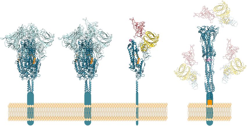
Fig. 4 Principle of adenovirus vector vaccines. a Schematic of replication-incompetent adenoviral vector particle and its DNA. E1 and E3:
Early adenovirus genes 1 and 3, respectively. b Formation of vaccine particles in production cell line complementing E1 from chromosomally
integrated E1 gene. Release of newly produced vector particles through cell lysis. c Expression of spike in cells of vaccinated individuals. More
or less shedding of S1 and conversion of S2 into its post-fusion structure (S2*) may occur in the absence of stabilizing mutations.
construct design with respect to mutations for stabilizing S as well Medicines Agency assessing thrombotic post-vaccination events,
as to the signal peptide at the N-terminus of S that require EMA/205598/2021) and may contain a P to A mutation at position
attention. Only the Janssen vaccine contains S-stabilizing muta- 22 to improve processing by signal peptidase80. The extended
tions (Table 1), comprising not only the two prolines in S2 but also N-terminal ‘leader sequence’ results in two consecutive signal
the S1/S2 furin cleavage site, which is mutated from 682-RRAS-685 peptides separated by an intervening stretch of tPA propeptide
to SRAG19,62. Both modifications are intended to avoid conversion (Fig. 5b). This complex artificial sequence element may lead to
of S into the post-fusion structure (Fig. 2d)19. some inhomogeneity in proteolytic processing and impairment of
Correct processing of the signal peptide by signal peptidase to correct formation of the S N-terminus during biosynthesis, as
generate the final N-terminus of S (Fig. 5a) may be especially recently shown in comparative model studies with similar
critical for obtaining natively folded S, because there is a cysteine constructs19.
immediately downstream of the cleavage site (amino acid 2) that Despite the absence of S2-stabilizing mutations, structural
has to form an S-S bond with the cysteine at position 136. studies of the S protein expressed in HeLa cells from the
Janssen-Johnson&Johnson and Gamaleya-Institute use the Oxford-AstraZeneca ChAdOx1 nCoV-19 vaccine provided evidence
authentic SARS-CoV-2 S protein signal sequence19,67, whereas for proper folding and presentation of the trimeric pre-fusion
CanSino replaced it with that of human tissue plasminogen conformation at the cellular plasma membrane78. However, the
activator (tPA) (Fig. 5a) (https://patents.google.com/patent/ authors discuss evidence of shedding of the cleaved S1 portion78,
CN111218459B/en). In the Oxford-AstraZeneca vaccine, an which has also been observed in model studies with unmodified S
extended form of the tPA signal sequence (containing the tPA proteins compared to mutationally stabilized proteins18 (Fig. 2c).
propeptide) was engineered in front of the authentic S protein The effect of dissociation of soluble S1 from the trimer complex on
signal sequence65,77,78 (Fig. 5b), based on a previous study with the quality of immune responses is incompletely understood, but
Middle East respiratory syndrome coronavirus (MERS-CoV79 some data suggest it may contribute to a higher proportion of
(Fig. 5b). Details of the engineered ‘leader sequence’ in the non-neutralizing relative to neutralizing antibodies19,81.
ChAdOX1-S vaccine are difficult to track, but likely comprise 32 to Animal experiments have shown that adenovirus-vector DNA
34 amino acids of tPA (according to a document of the European can remain detectable for months after inoculation in
npj Vaccines (2021) 104 Published in partnership with the Sealy Institute for Vaccine SciencesF.X. Heinz and K. Stiasny
7
a
SP of S BioNTech-Pfizer, Moderna,
Janssen-Johnson&Johnson, Gamaleya-Institute
VCQ
SP of tPA
CanSino
VCQ
SRP
Signal
peptidase C-ter of S N-ter SP of S
Cytosol
ER lumen
translocon SRP N-ter of S
receptor (QCV...)
b
SP of S SP of tPA
Oxford-AstraZeneca
VCQ tPA pro-peptide
?
?
SRP and
SRP-receptor C-ter of S N-ter SP of tPA
Cytosol
Interactions
ER lumen
translocon SRP ?
receptor N-ter of S ?
(QCV...)
?
Fig. 5 Signal sequence-mediated transport of S into the lumen of the ER. a Schematic of the process using the authentic viral signal
peptide only (as in the vaccines of BionTech-Pfizer, Moderna, Janssen-Johnson&Johnson and Gamaleya Institute). In the CanSino vaccine, the
signal peptide of S is replaced by that of human tPA (https://patents.google.com/patent/CN111218459B/en). b Schematic of the process using
an additional N-terminal leader sequence (signal peptide and propeptide of tPA), as used in the vaccine of Oxford-Astra Zeneca, based on
reference. 80). SP—signal peptide; SRP—signal recognition particle; tPA—tissue plasminogen activator; ER—endoplasmic reticulum; C-ter—C
terminus; N-ter—N terminus.
transcriptionally active form82 in contrast to rapidly degraded conformational integrity may vary depending on the conditions
RNA55,83. Persistence of antigen expression may be a distinctive used for vaccine preparation.
feature of adenovirus vector vaccines, and has been proposed to
contribute to induction of sustained immune responses and long- Inactivated vaccines
lasting immunity (reviewed in41). Published information about the production process is available
for two inactivated whole-virus vaccines manufactured by the
Chinese companies Sinopharm and Sinovac (Table 1). In both
Protein-based vaccines—general instances, the virus is grown in Vero cells and inactivated by beta-
In parallel to genetic vaccines, more conventional approaches of propiolactone (BPL), which is used as an inactivating agent for
vaccine development were pursued with similar intensity and led other viral vaccines, including rabies vaccines85. Literature data
to the authorization of inactivated whole-virus vaccines and a indicate that production of the Sinovac vaccine includes several
subunit vaccine that is in a stage of pre-authorization (Table 1). steps of virus purification, leading to a product that contains
Both of these technologies have already been applied successfully primarily the viral proteins and consists of essentially pure viral
to the production of vaccines against other viral diseases84. With particles86,87. The degree of purity of the Sinopharm vaccine
these vaccines—and in contrast to genetic vaccines—a prede- (referred to as BBIBP-CorV; Table 1) is less clear. Two pertinent
fined amount of the S immunogen/antigen is applied to the references do not indicate specific steps of purification after
vaccinee, but—as discussed in the following sections—its inactivation and removal of cell debris88,89, but one figure in a
Published in partnership with the Sealy Institute for Vaccine Sciences npj Vaccines (2021) 104F.X. Heinz and K. Stiasny
8
publication by Wang et al.90 suggests a step of chromatography, neutralization titers against some VOCs were observed with sera
albeit without providing details of this process and the purity of after immunization with mRNA and adenovirus vector vaccines106–
109
the vaccine. Both vaccines use aluminum hydroxide as an , pointing to the importance of the problem. On the positive
adjuvant. Another inactivated whole-virus vaccine using similar side, results of efficacy as well as field effectiveness studies in
technology is produced by the Indian company Bharat and has various countries using different vaccines indicated a high degree
received emergency use authorization in India even before of protection also against circulating VOCs, in particular against
completion of phase III clinical trials91 (Table 1). Details of the Alpha variant2,98,110,111. Prevention of infection with the Beta,
purification processes used for the manufacturing of this vaccine Gamma and Delta variants might be lower, although evidence
are not available in published literature. indicates substantial protection from severe disease after two
There are open questions concerning the structure of S in the vaccinations110,112,113. So far, no data are available that would
inactivated vaccines. Electron microscopical pictures of the viral allow a direct comparison of the various vaccines and their
spikes in the Sinovac vaccine have been interpreted differently, effectiveness against the different VOCs. Given the same antigenic
either as displaying the pre-fusion structure86 or the post-fusion difference of all vaccines relative to VOCs, the most important
structure25,92. Several lines of evidence suggest that BPL- parameter determining cross-protection may be the quantity of
inactivation in combination with purification processes can indeed neutralizing antibodies and relevant cellular immune reactivity at
lead to the formation of the post-fusion spike and the the time of infection. This quantitative aspect is important for
concomitant dissociation of S125,92. Preservation of the native future analyses of the impact of waning immunity on protection
trimeric pre-fusion conformation, in contrast, was observed in and decisions about optimal timings of booster immunizations.
structural studies with formalin-inactivated virus93, suggesting Efforts are also underway to replace existing vaccine strains and
that inactivation and/or purification procedures can influence the corresponding sequences for those of the most relevant circulat-
ratios of pre- and post-fusion conformations of S and thus the ing strains114.
qualities of killed whole-virus vaccines. Such factors may
contribute to variations in the efficacies reported in clinical trials
with current inactivated whole-virus vaccines94. There is indirect DISTINGUISHING FEATURES OF VACCINES INDEPENDENT OF
evidence that virus strains having emerged later in the pandemic IMMUNOGEN STRUCTURE
(e.g. containing the mutation D614G in S) may be more stable95,96 As outlined in the preceding sections, substantial differences
and therefore could serve as an improved substrate for the appear to exist among current vaccines that can affect the
production of inactivated vaccines. The vaccine produced by conformation of S and its presentation to the immune system.
Bharat is indeed based on a seed virus containing this mutation97 Independent of such antigenic effects, the fundamentally different
(Table 1). mechanisms of action and ways of production are likely to
introduce additional variation to the characteristics of immune
Subunit vaccines responses and possible adverse reactions. Adenovirus-vector and
So far, results of phase III clinical efficacy trials were reported and mRNA vaccines promote substantially different innate responses
published for a single subunit vaccine only98, which is manufac- that will certainly influence the nature of adaptive immune
tured by the company Novavax (NVX-CoV2373) (Table 1)98,99 and responses43. There is evidence that the Oxford-AstraZeneca
is still in a stage before regulatory approval100. It consists of the vaccine might induce higher levels of specific T cells, whereas
trimeric full-length spike that is produced as a recombinant mRNA vaccines might induce higher antibody titers115–117. The
protein in insect Sf9 (Spodoptera frugiperda) cells using a relevance of these differences for protection are not yet clear.
baculovirus expression system and contains mutations to stabilize Similarly, immune responses to protein-based vaccines are shaped
S2 (K986P and V987P) as well as to delete the furin cleavage site by the adjuvant used, for example by shifting CD4 T cells towards
(682-RRAS-685 changed to QQAQ)99 (Table 1 and Fig. 2). The either Th1 or Th2118,119. For meaningful conclusions, studies on
protein has its authentic membrane anchor and remains these topics will require head-to-head comparisons of vaccines,
associated with the membranes of the Sf9 production cells. and corresponding publications are expected to expand rapidly in
Therefore, isolation of the final product includes detergent the near future. Here, we briefly discuss existing data and describe
solubilization of the cells and several steps of purification101. The distinguishing features that can contribute to differences among
company formulates the S trimer as a nanoparticle in polysorbate vaccine responses independent of the structure and presentation
80 (PS80) detergent and uses a special proprietary saponin-based of the S immunogen.
adjuvant (Matrix-M™) that comprises 40 nm particles composed of
Quillaja saponins, cholesterol and phospholipids102,103. High- Contaminations from cell substrates
resolution structural analysis revealed that the purified protein is Contaminating cellular proteins can be present in all vaccines
stably locked in the preferred pre-fusion conformation, in part as involving production in cell culture. This pertains to many well-
free trimers and in part as multitrimer complexes103. The vaccine used licensed vaccines such as those against influenza, measles
thus presents the correctly folded immunogen in essentially pure and rabies120. The amount of impurities depends on the
form and in combination with a potent adjuvant. purification steps applied in the manufacturing process. Except
for mRNA vaccines, different eukaryotic cell cultures are used in
Effectiveness of vaccines against viral variants the production of current COVID-19 vaccines (see sections
A plethora of viral mutants arose and spread since the emergence “Biosynthesis and key properties of the spike protein” and
of SARS-CoV-2104,105. Some of these mutants are considered “Vaccine-specific differences of S-antigen structure and presenta-
‘Variants of Concern’ (VOCs) because of their highly efficient tion” and Table 1). Constituents in the Oxford-AstraZeneca vaccine
transmission, the concomitant replacement of previously circulat- were recently analyzed in the context of a search for potential
ing strains, and the presence of mutations in the spike protein that causes of venous sinus thrombosis as a rare post-vaccinal
can lead to immune escape (https://www.who.int/en/activities/ complication121. The study revealed that the vaccine contains
tracking-SARS-CoV-2-variants/). In principle, all current vaccines vast numbers and amounts of cellular proteins from the human
are affected similarly by VOCs, because they are all based on HEK293 production cell line, in addition to adenoviral proteins and
original wild-type strains from the early phase of the pandemic the S protein, which is apparently also synthesized already during
(see Table 1) and therefore their S protein sequences differ from the manufacturing process. The total amount of protein per dose
those of VOCs to the same degrees. Indeed, substantially reduced was found to be 35 to 40 µg, most of which can be assumed to be
npj Vaccines (2021) 104 Published in partnership with the Sealy Institute for Vaccine SciencesF.X. Heinz and K. Stiasny
9
cellular protein, because the protein of 5 × 10¹⁰ adenovirus Janssen-Johnson&Johnson vaccine19,62 as well as in the first shot
particles per dose would account for only about 8 µg (for of the Gamaleya-Institute vaccine67,68. These considerations of
calculation see122). Although corresponding data do not yet exist vector immunity also prompted the development of non-human
in the public domain for the other adenoviral vector vaccines, the adenovirus vectors such as ChAdOx1 derived from chimpanzee
problem of cellular impurities may be similar, because they all adenovirus Y2564, now used in the Oxford-AstraZeneca vaccine77.
depend on the lysis of production cells for releasing the In this case, seropositivity is negligible in Europe (zero in the UK,64)
engineered vector particles (section “Adenovirus-vector vaccines”). and low in Africa (9% in Gambian adults,64,131).
Details of purification procedures during manufacturing of the Irrespective of pre-existing immunity, all adenovirus vector
current adenovirus vector vaccines may differ but are not vaccines are prone to induce immune responses against the vector
published. particles129. Each dose contains 5 × 10¹⁰ or 10 × 1010 adenoviral
Cell cultures are also used for production of the inactivated particles (Table 1), which corresponds to 8 or 16 µg of adenoviral
whole-virus vaccines (Vero cells) of Sinopharm88, Sinovac86 and protein (for calculation see ref. 122). It is unclear, at present, which
Bharat97 as well as for the Novavax subunit vaccine (insect Sf9 influences anti-vector responses will have on necessary COVID-19
cells)99,101 (sections “Inactivated vaccines” and “Subunit vaccines” booster vaccinations in the future. Possible remedies are prime-boost
and Table 1). According to published literature, manufacturing of regimens as already used for vaccination with the Gamaleya-Institute
the Sinovac and Novavax vaccines involves extensive purification vaccine (Ad26 followed by Ad5) or combinations with other classes
procedures86,101, suggesting that the antigenic contents of these of vaccines such as mRNA vaccines. Corresponding studies are in
products consists primarily of the proteins of the virus particle or progress (Com-Cov study: Oxdorf-AstraZeneca and BionTech-Pfizer,
the isolated spike trimer, respectively. Details of purification launched in February132).
procedures of the Bharat vaccine and the degree of cellular
contaminants in the Sinopharm vaccine (which is less purified Vaccine-specific differences of innate responses
than that of Sinovac according to ref. 88) could not be found in the
literature. Specific features of adaptive immune responses are strongly
influenced and shaped by innate responses that are triggered by
pathogen-associated molecular patterns (PAMPs) and their sen-
Reactions due to vaccine constituents other than the sing by pattern recognition receptors (PRRs) (reviewed in ref. 133).
immunogen Current COVID-19 vaccines are very different with respect to their
Vaccination of millions or even billions of people within a short time compositions and modes of action, and therefore vaccine-induced
window allows identification of rare adverse reactions that would innate responses will vary considerably. Adenoviral vectors
otherwise be difficult to be linked causally to vaccination. Currently, a contain PAMPs that can be sensed by TLRs at the plasma
slightly but significantly increased risk of thrombotic events membrane (TLR2 and TLR4) and the endosomally located TLR9
(including cerebral venous sinus thrombosis) was reported after (reviewed in ref. 41). In addition, the viral DNA itself can be sensed
vaccination with Oxford-AstraZeneca and Janssen adenovirus vector after endosomal rupture by cytosolic DNA sensors such as cGAS
vaccines and has raised considerable concern74,75. Collectively, this and the inflammasome, resulting in downstream signaling
kind of adverse event is designated ‘vaccine-induced immune cascades for producing antiviral factors such as type I
thrombotic thrombocytopenia’ (VITT). Sophisticated analyses of the interferons41.
ChAdOx1 nCoV-19 vaccine to elucidate underlying pathogenic Innate responses to RNA that enters cells from the outside (such
mechanisms suggest that constituents such as viral DNA and/or as in RNA virus infections or mRNA vaccination) differ from those
cellular proteins can favor the formation of antibodies against stimulated by adenoviruses, because RNA is sensed by other PRRs,
platelet factor 4 (PF4), thus promoting VITT74,121. Information on including TLR3, TLR7 and TLR8, all located in endosomes134,135.
cellular impurities are so far restricted to ChAdOx1 and comparative Sensors in the cytoplasm, such as retinoic-inducible gene I (RIG-I)
analyses of all adenovector vaccines are not yet available. and melanoma differentiation-associated antigen 5 (MDA-5)
After vaccination with mRNA vaccines, rare events of anaphylactic recognize preferentially dsRNA, also leading to stimulation of
shock above the average incidence in the population have been type I IFN secretion134,135. Excessive innate responses can not only
reported, largely in individuals with a history of allergy123,124. Most of result in strong reactogenicity of vaccination but also restrict
the allergens are proteins, which are not contained in these antigen translation from the vaccine RNA, thus impairing adaptive
chemically defined vaccines (section “mRNA vaccines”). One of the immune responses. In the BionTech-Pfizer and Moderna vaccines
constituents discussed as being causally linked to anaphylaxis is this problem was taken into account by modifications of the RNA
polyethylene glycol (PEG), which is used in the formulation of LNPs sequence and the inclusion of m1Ψ (section “mRNA vaccines”),
that protect the RNA and facilitate its transfer into cells (section which is not contained in CureVac’s mRNA vaccine56. In addition
“mRNA vaccines”). It has been speculated that pre-existing PEG to direct triggers of innate immunity by RNA, other constituents of
antibodies might be involved in these allergic events124. Correspond- LNPs can contribute to vaccine-induced inflammatory reactions
ing scientific investigations into the mechanisms of vaccine-induced and provide adjuvant activity for adaptive immune responses.
anaphylactic reactions are ongoing125. Such effects have been specifically shown for the ionizable lipid
component in LNPs124. Head-to-head comparisons of mRNA
Vector immunity vaccines will be informative to identify and evaluate differences
Effects of pre-existing and vaccination-induced immunity against of innate and adaptive responses as well as reactogenicity
the vector are a special feature of adenovirus vector vaccines. High between representatives of this class of COVID-19 vaccines.
rates of seropositivity against adenovirus 5 (the pioneer of Due to their capacity to stimulate innate responses, the genetic
adenovirus vector development) have been reported in the vaccines are referred to as being ‘self-adjuvanted’55,136. Protein-
population126,127, and a number of studies have shown that pre- based vaccines such as inactivated whole-virus vaccines or
existing vector immunity can impair the response to the vaccine subunit vaccines are usually not sufficiently immunogenic on
antigen128–130. Adenovirus 5 is used in the CanSino vaccine and their own and require the addition of adjuvants. Alum is the most
the second dose of Gamaleya vaccines (section “Adenovirus- frequently used adjuvant in human vaccines and is used in the
vector vaccines” and Table 1). For reducing potential negative Sinopharm and Sinovac vaccines137,138. This adjuvant results in
effects of pre-existing immunity, alternative adenoviruses were polarization towards a Th2 response, which has been regarded as
developed as vectors, one of them adenovirus 26, which has lower unfavorable in the case of coronavirus and other viral infections
rates of seropositivity in the population127 and is now used in the and vaccinations118,139,140. Therefore, other adjuvants or
Published in partnership with the Sealy Institute for Vaccine Sciences npj Vaccines (2021) 104F.X. Heinz and K. Stiasny
10
combinations thereof with Alum have been developed for use in 5. Khoury, D. S. et al. Neutralizing antibody levels are highly predictive of immune
COVID-19 vaccines138. The inactivated whole virus vaccine protection from symptomatic SARS-CoV-2 infection. Nat. Med. 27, 1205–1211
produced by Bharat (Covaxin, Table 1) is adjuvanted with an (2021).
imidazoquinoline class molecule (IMDG, a TLR 7/8 agonist) 6. McMahan, K. et al. Correlates of protection against SARS-CoV-2 in rhesus
macaques. Nature 590, 630–634 (2021).
adsorbed on aluminum hydroxide gel (Algel-IMDG) that shifts
7. Corbett, K.S. et al. Immune correlates of protection by mRNA-1273 vaccine
the response towards Th197,141,142. Another BPL-inactivated against SARS-CoV-2 in nonhuman primates. Science. Preprint at https://www.
whole-virus vaccine in development (by the European company biorxiv.org/content/10.1101/2021.04.20.440647v2 (2021).
Valneva) makes use of Alum in combination with CpG to induce 8. Earle, K. A. et al. Evidence for antibody as a protective correlate for COVID-19
preferentially a desired Th1 response138, and a similar effect has vaccines. Vaccine 39, 4423–4428 (2021).
been attributed to the Matrix-MTM adjuvant used in the Novavax 9. Kustin, T. et al. Evidence for increased breakthrough rates of SARS-CoV-2 var-
subunit vaccine99,101,102. iants of concern in BNT162b2-mRNA-vaccinated individuals. Nat. Med. https://
doi.org/10.1038/s41591-021-01413-7 (2021).
10. Barnes, C. O. et al. Structures of human antibodies bound to SARS-CoV-2 spike
CONCLUSIONS reveal common epitopes and recurrent features of antibodies. Cell 182,
828–842.e816 (2020).
The severe consequences of the COVID-19 pandemic have created 11. Cai, Y. et al. Structural basis for enhanced infectivity and immune evasion of
a pressing need for vaccines that not only prevent serious disease SARS-CoV-2 variants. Science 373, 642–648 (2021).
but preferentially also transmission. Several of the 291 candidates 12. Jagannathan, P. & Wang, T. T. Immunity after SARS-CoV-2 infections. Nat.
listed in the COVID-19 vaccine pipeline by WHO (184 pre-clinical Immunol. 22, 539–540 (2021).
and 107 in clinical development) (https://www.who.int/ 13. Winkler, E. S. et al. Human neutralizing antibodies against SARS-CoV-2 require
publications/m/item/draft-landscape-of-covid-19-candidate- intact Fc effector functions for optimal therapeutic protection. Cell 184,
vaccines, accessed on July 9, 2021), have already reached the 1804–1820.e1816 (2021).
market and are used for mass immunization. They all proved to 14. Zhao, P. et al. Virus-receptor interactions of glycosylated SARS-CoV-2 spike and
human ACE2 receptor. Cell Host Microbe 28, 586–601.e586 (2020).
exceed initial hopes and maximal expectations of 50 % protec-
15. Duan, L. et al. The SARS-CoV-2 spike glycoprotein biosynthesis, structure,
tion143,144, displaying efficacies in preventing clinical disease of function, and antigenicity: implications for the design of spike-based vaccine
more than 90% in certain instances. Although all current vaccines immunogens. Front. Immunol. 11, 576622 (2020).
for which phase 3 efficacy data are available rely on the whole 16. Coutard, B. et al. The spike glycoprotein of the new coronavirus 2019-nCoV
viral spike protein as an antigen, its presentation to the immune contains a furin-like cleavage site absent in CoV of the same clade. Antiviral Res
system is strikingly different not only between genetic vaccines 176, 104742–104742 (2020).
and protein-based vaccines, but also between vaccines within 17. Ghosh, S. et al. β-Coronaviruses use lysosomes for egress instead of the bio-
these categories. In addition, approaches to cope with the synthetic secretory pathway. Cell 183, 1520–1535.e1514 (2020).
problem of the lability of the viral S protein cause variation across 18. Brun, J. et al. Assessing antigen structural integrity through glycosylation ana-
lysis of the SARS-CoV-2 viral spike. ACS Central Sci. 7, 586–593 (2021).
all current vaccines. These also differ with respect to their degree
19. Bos, R. et al. Ad26 vector-based COVID-19 vaccine encoding a prefusion-
of purity (presence of extraneous proteins from the production stabilized SARS-CoV-2 Spike immunogen induces potent humoral and cellular
process) and other vaccine constituents that can affect immune immune responses. npj Vaccines 5, 91 (2020).
responses and cause adverse events. We have reviewed the most 20. Wrapp, D. et al. Cryo-EM structure of the 2019-nCoV spike in the prefusion
apparent and significant differences among the vaccines as far as conformation. Science 367, 1260 (2020).
they can be recognized from published literature, which 21. Walls, A. C. et al. Structure, function, and antigenicity of the SARS-CoV-2 spike
unfortunately is still incomplete. Hopefully, more details will glycoprotein. Cell 181, 281–292.e286 (2020).
become available in the near future. Comparative analyses of 22. Rey, F. Structure-function relations of the SARS-CoV-2 spike protein and impact
antibody and T cell responses and their fine specificities will allow of mutations in the variants of concern. C. R. Biol. 344, 77–110 (2021).
23. Tang, T., Bidon, M., Jaimes, J. A., Whittaker, G. R. & Daniel, S. Coronavirus
indirect but important conclusions to be drawn. Studies are
membrane fusion mechanism offers a potential target for antiviral develop-
emerging that address antibody formation to the different ment. Antiviral Res. 178, 104792–104792 (2020).
domains of S and analyze the ratio of neutralizing and non- 24. Xu, C. et al. Conformational dynamics of SARS-CoV-2 trimeric spike glycoprotein
neutralizing antibodies as an important parameter of vaccine in complex with receptor ACE2 revealed by cryo-EM. Sci. Adv. 7, eabe5575
performance145,146. These data can serve as an indirect measure (2021).
for the structural integrity of S in the vaccines and the quality of B 25. Cai, Y. et al. Distinct conformational states of SARS-CoV-2 spike protein. Science
cell immune responses. Head-to-head comparisons of vaccinated 369, 1586 (2020).
cohorts will be especially insightful, considering the profound 26. Fan, X., Cao, D., Kong, L. & Zhang, X. Cryo-EM analysis of the post-fusion
differences of antigen presentation and principles of action of structure of the SARS-CoV spike glycoprotein. Nat. Commun. 11, 3618 (2020).
27. Andreano, E. et al. Extremely potent human monoclonal antibodies from COVID-
current COVID-19 vaccines.
19 convalescent patients. Cell 184, 1821–1835.e1816 (2021).
28. Brouwer, P. J. M. et al. Potent neutralizing antibodies from COVID-19 patients
Received: 1 June 2021; Accepted: 30 July 2021; define multiple targets of vulnerability. Science 369, 643 (2020).
29. Pinto, D. et al. Cross-neutralization of SARS-CoV-2 by a human monoclonal
SARS-CoV antibody. Nature 583, 290–295 (2020).
30. Robbiani, D. F. et al. Convergent antibody responses to SARS-CoV-2 in con-
valescent individuals. Nature 584, 437–442 (2020).
REFERENCES 31. Rogers, T. F. et al. Isolation of potent SARS-CoV-2 neutralizing antibodies and
protection from disease in a small animal model. Science 369, 956 (2020).
1. Hasan, T., Beardsley, J., Marais, B. J., Nguyen, T. A. & Fox, G. J. The imple-
32. Zost, S. J. et al. Potently neutralizing and protective human antibodies against
mentation of mass-vaccination against SARS-CoV-2: a systematic review of
SARS-CoV-2. Nature 584, 443–449 (2020).
existing strategies and guidelines. Vaccines (Basel) 9, 326 (2021).
33. Zost, S. J. et al. Rapid isolation and profiling of a diverse panel of human
2. Haas, E. J. et al. Impact and effectiveness of mRNA BNT162b2 vaccine against
monoclonal antibodies targeting the SARS-CoV-2 spike protein. Nat. Med. 26,
SARS-CoV-2 infections and COVID-19 cases, hospitalisations, and deaths fol-
1422–1427 (2020).
lowing a nationwide vaccination campaign in Israel: an observational study
34. Graham, C. et al. Neutralization potency of monoclonal antibodies recognizing
using national surveillance data. Lancet 397, 1819–1819 (2021).
dominant and subdominant epitopes on SARS-CoV-2 Spike is impacted by the
3. Rossman, H. et al. COVID-19 dynamics after a national immunization program in
B.1.1.7 variant. Immunity 54, 1276–1289.e1276 (2021).
Israel. Nat. Med. 27, 1055–1061 (2021).
35. Suryadevara, N. et al. Neutralizing and protective human monoclonal antibodies
4. Vasileiou, E. et al. Interim findings from first-dose mass COVID-19 vaccination
recognizing the N-terminal domain of the SARS-CoV-2 spike protein. Cell 184,
roll-out and COVID-19 hospital admissions in Scotland: a national prospective
2316–2331.e2315 (2021).
cohort study. Lancet 397, 1646–1657 (2021).
npj Vaccines (2021) 104 Published in partnership with the Sealy Institute for Vaccine SciencesYou can also read