Dr Catherine Westbrook Dr John Talbot - MRI Education
←
→
Page content transcription
If your browser does not render page correctly, please read the page content below
Dr Catherine Westbrook Dr John Talbot
MRI in Practice – The Course

   Welcome to MRI in Practice – The Course

   The book, MRI in Practice, was first published in 1993 and quickly
   became the one of the world’s leading MRI resources. Now in its 5th
   edition, it is considered an essential text for tens of thousands of MRI
   practitioners around the world. It is used to support learning of MRI
   theory by practitioners who wish to gain qualifications in MRI,
   including the MR Registry Review Exam in the USA and on post
   graduate MRI programmes in countries such as the UK and Australia.

   MRI in Practice course is based on this book, it was first delivered in
   1992 in Oxford in the UK. It has been continuously presented since
   then by the authors of MRI in Practice, Dr Catherine Westbrook and
   Dr John Talbot. Thousands of delegates, from over 20 countries,
   across 5 continents have attended the MRI in Practice course and it is
   now considered one of the world’s leading MRI programmes. MRI
   professionals from across the clinical and research spectrum have
   attended the MRI in Practice course including radiographers,
   radiotherapists, radiologists, physicists,, veterinarians and engineers.

   The popularity of the course is mainly due to state-of-the-art graphics
   that are purposed to present complex MRI theory in a user-friendly
   way. Catherine and John combine their extensive experience in MRI
   and education to uniquely enable delegates to apply MRI theory to
   their practice. The course also has a well-deserved reputation for its
   informal approach to learning that allows delegates to take advantage
   of networking opportunities that are fostered by this type of course.

   The MRI in Practice course is now also available as an online learning
   experience. The following information relates to the content of both
   live and online courses. The timing shown applies to the on-line course.

MRI in Practice The Course World Brochure – all text subject to copyright © 1992-2021 Talbot Westbrook. All graphics subject to copyright © 2000-2021 Talbot and must not be reproduced without permission.   2
Three-Day Programme

   The course is offered in different formats to cater for participant preferences. The programme shown below is the three-day course. This is intended for
   participants who wish to attend the entire course in one sitting and contains all you need to know in just three days. It is sometimes presented over a
   weekend for participants who find it difficult to get week-day leave. The timings shown relate to the online course.

MRI in Practice The Course World Brochure – all text subject to copyright © 1992-2021 Talbot Westbrook. All graphics subject to copyright © 2000-2021 Talbot and must not be reproduced without permission.   3
Four-Day Programme

   The programme shown below is the four-day flexible course. This version allows participants to choose any combination of days. It is, however, strongly
   recommended that all participants should attend day 01 (irrespective of previous experience). This is because all of the other presentations constantly
   make reference to the concepts covered on day 01. The timings shown relate to the online course.

MRI in Practice The Course World Brochure – all text subject to copyright © 1992-2021 Talbot Westbrook. All graphics subject to copyright © 2000-2021 Talbot and must not be reproduced without permission.   4

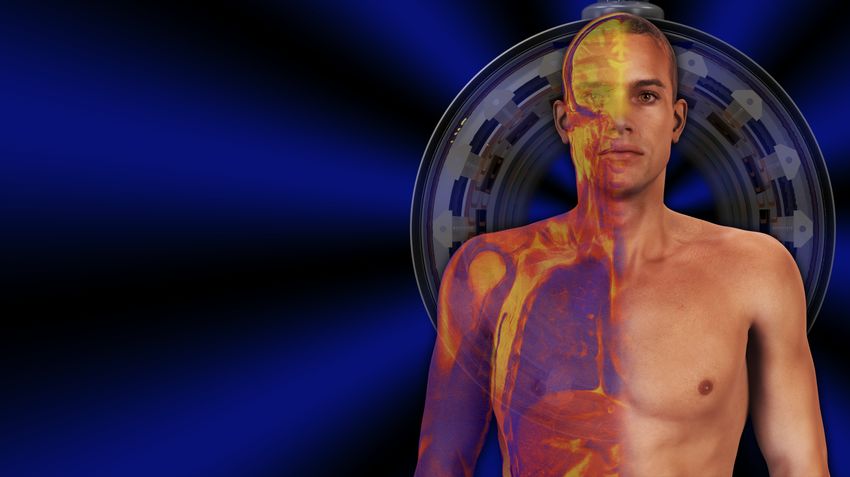
MRI in Practice – now available online

   MRI is still a rapidly evolving modality and poses many challenges for the MRI
   practitioner. An in-depth understanding of theory and how it is applied in clinical
   practice are necessary to exploit the full potential of the MR system. The MRI in
   Practice course has always had a reputation for its use of the latest multimedia and
   presentation technology. We were one of the first MRI courses to use digital data
   projectors and HD animated graphics to present complex MRI concepts. Delegates
   value our unique presentation style that combines sophisticated graphics and clear
   analogies to enable them to easily relate MRI physics to their practice. Protocol
   parameter manipulation, artefact recognition, appropriate selection of pulse
   sequences and image interpretation are all easier after attending the MRI in Practice
   course!

   MR professionals are advancing their knowledge of MRI using a variety of resources
   including online content. With this in mind, Catherine and John have developed an
   online version of the MRI in Practice course. The course is still presented by
   Catherine and John but via all-new video versions of the lectures that can be
   accessed on your PC, Mac or laptop.

   However, MRI in Practice Online is not like other online learning resources where
   delegates simply view presentations over the internet. Catherine and John have over
   20 year’s experience of distance learning delivery of MRI and have used this expertise
   to add value to your learning experience. The programme is highly interactive and
   includes live sessions with the presenters. We use a mobile application to incorporate
   quizzes, revision and feedback sessions on every day of the course. We are confident
   that the online version of the MRI in Practice course is even more valuable than the
   face to face offering and, of course, far more convenient for delegates with no
   requirement for travel or accommodation costs.

   We recognise that everyone has different levels of experience in MRI and different
   learning needs. For this reason, the course programme is designed to begin with first
   principles and then each day, progress onto to more advanced concepts. Whether you
   are new to MRI or a seasoned pro, we can guarantee that you will learn a great deal by
   attending this course.

   Take a look at the lecture topics on the following pages.

MRI in Practice The Course World Brochure – all text subject to copyright © 1992-2021 Talbot Westbrook. All graphics subject to copyright © 2000-2021 Talbot and must not be reproduced without permission.   5
The Fundamentals of MRI

    01: BASIC PRINCIPLES                                                                                               02: IMAGE CONTRAST
    Dr Catherine Westbrook                                                                                             Dr Catherine Westbrook
    (duration 00:57)                                                                                                   (duration 01:08)

    Aim: to investigate the fundamental principles of MRI:                                                             Aim: to explore and understand the processes of image contrast
                                                                                                                       generation:
         •    Nuclear structure
         •    MR active nuclei                                                                                              •    Contrast definition
         •    Alignment                                                                                                     •    MRI contrast parameters
         •    Precession and resonance                                                                                      •    TR
         •    Signal generation                                                                                             •    Fat and Water characteristics
         •    Relaxation processes                                                                                          •    Relaxation
                                                                                                                            •    T1 Recovery
                                                                                                                            •    T1 Recovery time
                                                                                                                            •    T1 Contrast and TR
                                                                                                                            •    T2 Decay
                                                                                                                            •    T2 Decay time
                                                                                                                            •    T2 contrast and TE
                                                                                                                            •    Weighting
                                                                                                                            •    T1 Weighting
                                                                                                                            •    T2 Weighting
                                                                                                                            •    Proton density weighting
                                                                                                                            •    Diffusion Weighting
                                                                                                                            •    Stejskal-Tanner gradient scheme
                                                                                                                            •    Diffusion-weighted images

MRI in Practice The Course World Brochure – all text subject to copyright © 1992-2021 Talbot Westbrook. All graphics subject to copyright © 2000-2021 Talbot and must not be reproduced without permission.   6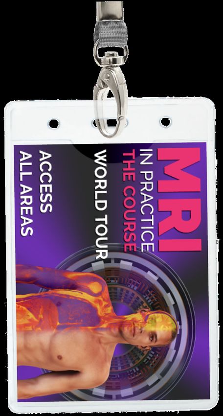
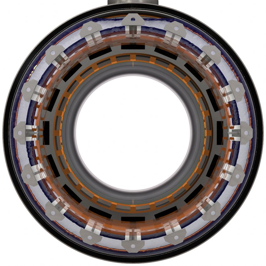
The Fundamentals of MRI

    03: INSTRUMENTATION
    Dr John Talbot
    (duration 01:40)

    Aim: to explore the components of an MRI scanner and relate them to
    the functions they provide during patient scanning.

         •    Cryostat
         •    Cold head
         •    Cryogen vent
         •    Cryogen chamber
         •    Helium
         •    Superconductivity
         •    Solenoids
         •    Ramping the magnet
         •    Flux density
         •    Magnet shielding
         •    Shim system
         •    Gradient system
         •    RF system
         •    RF transmitter
         •    RF receiver coils

MRI in Practice The Course World Brochure – all text subject to copyright © 1992-2021 Talbot Westbrook. All graphics subject to copyright © 2000-2021 Talbot and must not be reproduced without permission.   7
Image Quality

    04: IMAGE ARTEFACTS (EXTRINSIC)                                                                                    05: PROTOCOL OPTIMISATION
    Dr John Talbot                                                                                                     Dr Catherine Westbrook
    (duration 01:23)                                                                                                   (duration 01:03)

    Aim: to identify, and provide remedies for MRI image artefacts that                                                Aim: to explore how protocol parameters are manipulated to optimise
    arise due to factors external to the patient.                                                                      image quality:

         •    Aliasing                                                                                                      •    What is meant by a protocol.
         •    Array Processor Error                                                                                         •    Balancing image quality and scan time
         •    Cross-Excitation                                                                                              •    Signal to noise ratio.
         •    Damaged Coils                                                                                                 •    Field Strength.
         •    Extraneous RF                                                                                                 •    Coil choice.
         •    Field Inhomogeneity                                                                                           •    Coil position.
         •    Magnetic Susceptibility                                                                                       •    Time to Repeat.
         •    Moiré Fringing                                                                                                •    Time to Echo.
         •    Noise Spike                                                                                                   •    Flip angle
         •    Nyquist Ghost                                                                                                 •    Number of signal averages.
         •    Standing Wave                                                                                                 •    Receive bandwidth.
         •    Truncation                                                                                                    •    Contrast to noise ratio.
         •    Zipper                                                                                                        •    Spatial resolution
                                                                                                                            •    Voxel volume
                                                                                                                            •    Scan time.
                                                                                                                            •    Phase matrix.
                                                                                                                            •    Trade-offs

MRI in Practice The Course World Brochure – all text subject to copyright © 1992-2021 Talbot Westbrook. All graphics subject to copyright © 2000-2021 Talbot and must not be reproduced without permission.   8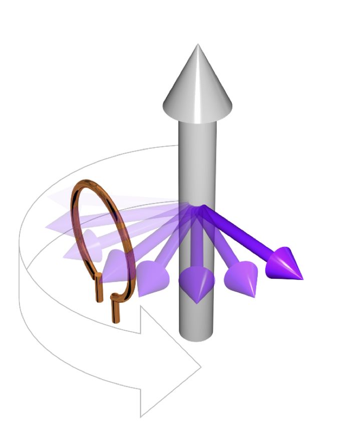
Image Quality
06: IMAGE ARTEFACTS (INTRINSIC)
Dr John Talbot
(duration 01:06)
Aim: to identify, and provide remedies for MRI image artefacts that
arise due to factors relating to the anatomy and physiology of the
patient.
• Chemical Shift
• Magic Angle
• Out of Phase Signal Loss
• Phase Mismapping
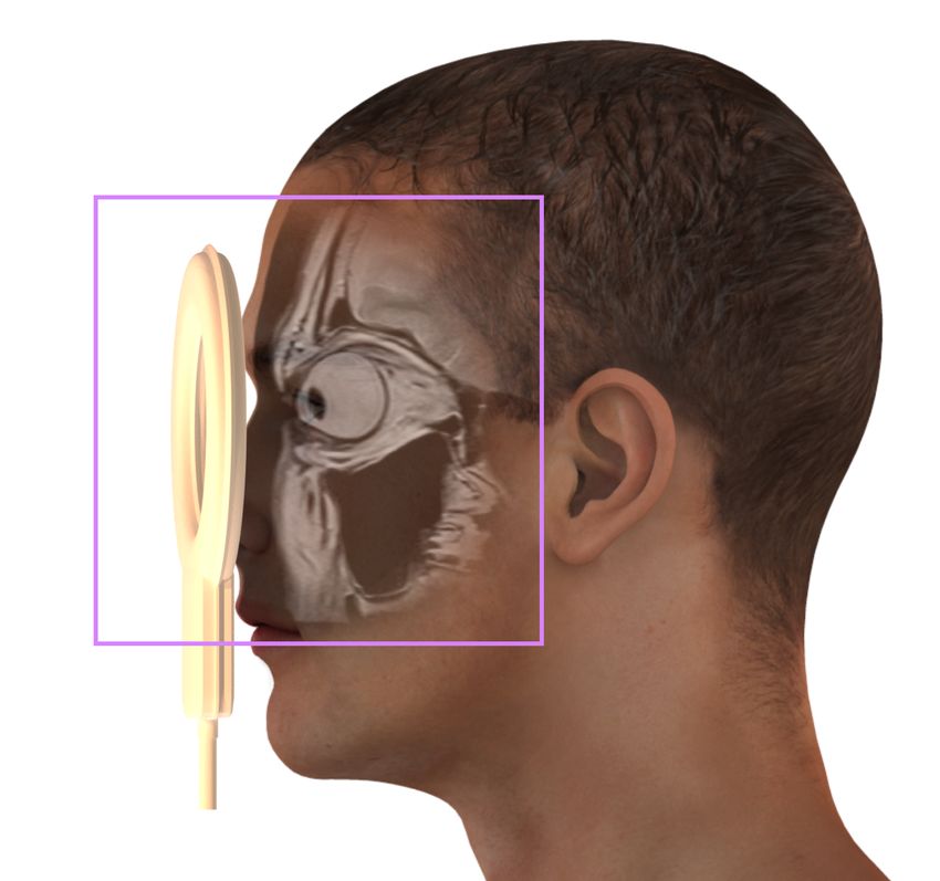
MRI in Practice The Course World Brochure – all text subject to copyright © 1992-2021 Talbot Westbrook. All graphics subject to copyright © 2000-2021 Talbot and must not be reproduced without permission. 9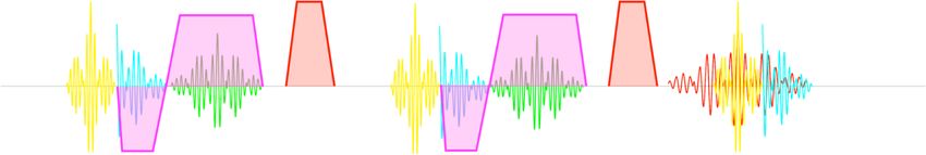
Spatial Encoding

    04: SPATIAL ENCODING                                                                                               05: k-SPACE 01 (INTRODUCTION)
    Dr John Talbot                                                                                                     Dr Catherine Westbrook
    (duration 01:34)                                                                                                   (duration 01:07)

    Aim: to facilitate understanding relating to the use of gradient                                                   Aim: to provide a useful introduction to k-space including:
    magnetic fields for the purpose of spatial encoding:

         •    Historic background                                                                                           •    Revision of gradient mechanisms in pulse sequences
         •    Gradients in spatial encoding                                                                                 •    k-space functions and characteristics
         •    Gradient functions                                                                                            •    The Chest of Drawers Analogy
         •    Slice selection                                                                                               •    Cartesian filling of k-space in a basic pulse sequence
         •    Slice location                                                                                                •    How k-space is used to optimise image quality
         •    Slice thickness
         •    Slice gap
         •    Field of view
         •    Phase and frequency
         •    Sampling waveforms
         •    Temporal frequencies
         •    Spatial frequencies
         •    A novel vector model (Plewes)
         •    Acquiring an image - phase and frequency encoding
         •    Relating the data to the image
         •    Fourier transformation

MRI in Practice The Course World Brochure – all text subject to copyright © 1992-2021 Talbot Westbrook. All graphics subject to copyright © 2000-2021 Talbot and must not be reproduced without permission.   10
Spatial Encoding
05: k-SPACE 02 (DATA ACQUISITION AND IMAGE PRODUCTION) 05: k-SPACE 03 (NON-CARTESIAN FILLING METHODS)
Dr Catherine Westbrook Dr Catherine Westbrook
(duration 01:40) (duration 01:20)
Aim: to explore, in depth, the principles that underpin data acquisition Aim: to investigate non-Cartesian k-space filling techniques including:
in MRI:
• Revision of spatial encoding • Partial Fourier
• Sampling and analogue to digital conversion (ADC) • Fast or Turbo spin echo pulse sequences
• The Wheel Analogy • Respiratory Ordered Phase Encoding
• The Runner on the Track Analogy • Centric k-space filling
• User-friendly Fourier Analysis! • Single and multi-shot
• How data points in k-space create MR images • Radial k-space filling
• The receive bandwidth - its importance in protocol optimisation • How k -space determines image geometry
• Advantages and disadvantages of each technique in clinical use • Parallel and compressed imaging
MRI in Practice The Course World Brochure – all text subject to copyright © 1992-2021 Talbot Westbrook. All graphics subject to copyright © 2000-2021 Talbot and must not be reproduced without permission. 11Pulse Sequences
05: SPIN-ECHO PULSE SEQUENCES 05: FAST SPIN ECHO & INVERSION RECOVERY PULSE SEQUENCES
Dr John Talbot Dr John Talbot
(duration 01:00) (duration 01:03)
Aim: to examine the purpose and mechanism of spin-echo pulse Aim: to examine the purpose and mechanism of fast (turbo) spin echo
sequences. and inversion recovery pulse sequences:
• Free induction decay • Fast (Turbo)Spin-Echo, mechanism
• The mechanism of the 180 degree rephasing pulse • Advantages and trade-offs
• Spin-spin dephasing • Blurring
• T1 vs. T2 • J-coupling
• Types of spin-echo sequence • RF deployment
• Conventional spin echo • Driven Equilibrium Fourier Transform
• Single echo acquisition • Inversion Recovery
• Multiple echo acquisition • "T1" Weighting at suboptimal field strength
• Clinical applications • Short Tau Inversion Recovery
• Fluid Attenuated Inversion Recovery
• Clinical applications
MRI in Practice The Course World Brochure – all text subject to copyright © 1992-2021 Talbot Westbrook. All graphics subject to copyright © 2000-2021 Talbot and must not be reproduced without permission. 12Pulse Sequences
05: GRADIENT ECHO PULSE SEQUENCES
Dr Catherine Westbrook
(duration 02:03)
Aim: to explore, in depth, the principles that underpin gradient echo
pulse sequences
• Flip Angles
• Gradient Mechanism
• Dephasing
• Rephasing
• Weighting Mechanisms
• Extrinsic Parameters
• The Steady State
• Echo generation
• Ernst Angle
• Gradient-Echo Sequence Types
• Gradient Echo Acronyms
• Rewound GE
• Spoiled GE
• Reverse GE
• Balanced GE
• Turbo GE
• Single Shot GE
• Echo-Planar Imaging
• Hybrid Sequences
MRI in Practice The Course World Brochure – all text subject to copyright © 1992-2021 Talbot Westbrook. All graphics subject to copyright © 2000-2021 Talbot and must not be reproduced without permission. 13Online Course Requirements
To fully benefit from this course all of the following requirements must be met;
• Three (or four) uninterrupted days of study for the duration of the daily programme (for example 8:00 am to 6:00 pm). Treat the attendance
requirements as though you were participating in a face-to-face course. Children, partners and friends are, regrettably, not invited. Access must
be from a domestic location (your home address), access from educational institutions, hospitals and commercial premises is prohibited.
• A fast broadband connection - the online lectures are streamed and contain many high resolution graphics and animations. The lectures are set
to automatically display at the highest quality that your internet connection speed will allow. The quality may be adversely affected if other
family members are on-line at the same time, especially if they are accessing streaming content such as Netflix.
• Headphones, or suitable audio, and a microphone on your device. There are regular meetings with the presenters and other delegates during
the course and you will be encouraged to actively participate.
• A webcam on your device. For CPD, copyright protection and to monitor access, it is necessary to have your web cam switched on during all
lectures and interactive sessions with the presenters. Lecture access will only be provided if you are on camera at all times.
• A second device such as a smartphone or tablet is strongly recommended. We run quizzes, revision sessions and Q and A sessions via a web app.
This can be accessed from any device including a PC or laptop but having the app on a second device makes these sessions a little easier to
manage. Older PCs and laptops may struggle to display both the lectures and the Zoom meeting simultaneously, in which case a tablet is an ideal
way to access the Zoom meeting whilst watching the lectures on your PC. It must have a camera (see above).
• A copy of the 5th edition of MRI In Practice - as the 5th edition of the book is the course notes. No other supporting material is provided. The
book is often referred to in the online lectures and it is therefore necessary to have a copy at hand throughout the course. The book can be
obtained here.
As part of the online course terms and conditions you agree not to film, photograph or otherwise capture the screen content during the lectures or
permit access to any other individuals such as colleagues. The course fee allows access by the single applicant only. Our software uses screengrab
detection and digital watermarking that personally identifies the participant and embeds the geolocation data directly onto the video stream. Any
participants who are found to have used screen-grabbing or any other form of prohibited copying of the lecture materials will be banned from course.
Legal action will be taken against any individual who copies and shares content from the course either via digital networks, on-line or via any other
media. Such participants will also be reported to their professional body and employer. We take intellectual property theft very seriously because our
books are shared as illegal downloads and piracy costs us our livelihood.
MRI in Practice The Course World Brochure – all text subject to copyright © 1992-2021 Talbot Westbrook. All graphics subject to copyright © 2000-2021 Talbot and must not be reproduced without permission. 14Delegate Feedback MRI in Practice The Course World Brochure – all text subject to copyright © 1992-2021 Talbot Westbrook. All graphics subject to copyright © 2000-2021 Talbot and must not be reproduced without permission. 15
International Excellence-Ratings and Comments Collected Anonymously via Mentimeter

                                                                                                      Word-cloud – participants asked to describe the course in three words

       Ratings rounded up or down to the nearest whole figure.

MRI in Practice The Course World Brochure – all text subject to copyright © 1992-2021 Talbot Westbrook. All graphics subject to copyright © 2000-2021 Talbot and must not be reproduced without permission.   16
Registration Information
AFRICA (Kenya) www.mrieducation.com/africa
AMERICA www.mrieducation.com/usa
AUSTRALIA www.mrieducation.com/australia
EMIRATES www.mrieducation.com/emirates
NETHERLANDS www.mrieducation.com/netherlands
NORWAY www.mrieducation.com/norway
PORTUGAL www.mrieducation.com/portugal
QATAR www.mrieducation.com/qatar
UK & IRELAND www.mrieducation.com/uk
MRI in Practice The Course World Brochure – all text subject to copyright © 1992-2021 Talbot Westbrook. All graphics subject to copyright © 2000-2021 Talbot and must not be reproduced without permission. 17Why Choose the MRI in Practice Course?
If you are trying to decide between MRI in Practice and another course, you will want to make a sound, evidence-based decision. please print out this quick summary
to see how we compare to other courses on the following important points:
The Course - MRI in Practice has been running continuously for over 25 years (formerly known as the Oxford MRI Course) and has been taught in 22 countries across
5 continents. We have presented in the UK since 1992, mainland Europe since 2005 and Australia since 2004. We engage and educate hundreds of delegates per
year, and their feedback sheets consistently rate our lectures as being excellent. Unlike many other MRI courses, MRI in Practice is completely independent of any
private “learning” company or institution. This is most important, because we can guarantee that our delivery is unbiased and we are free to say what we want to say.
The Cost - The online course is actually very expensive to create, run and maintain, however, we are able to offer it at a substantial discount. As a delegate, the biggest
cost saving will be the fact that there is no necessity for travel or accommodation. As a result it is likely that you will save approximately £400-£500 on the overall
cost of attending a live course.
The Content - MRI in Practice - is based on the World’s best-selling MRI book*. MRI in Practice is consistently at the top of its league on the Amazon.com bookstore
and overwhelmingly attracts five-star reviews from the public, which for us, are the ones that count. MRI in Practice is famed as the reference text for practitioners
taking the US registry exam - so you can rest assured that our course content is tried, trusted and relevant. You don’t have to take our word for it though - this course
has been accredited/endorsed by The UK College of Radiographers (CPD NOW), The British Institute of Radiology, Trinity College Dublin, The Australian Institute of
Radiology, The University of Sharjah (Emirates), The Romanian Radiology Society/College of Physicians, the American Society of Radiologic Technologists and many
other respected institutions.
The Presentation - MRI in Practice has evolved over the years, always taking advantage of the very latest technology. We were using data projection before anyone
else (as long ago as 1997), and our computer graphics have also developed in line with broadcast production values. The online course is a perfect showcase for this.
Delegates repeatedly tell us that our 3D graphics help to clarify difficult concepts, by bringing them to life in a way that a conventional book or PowerPoint lecture
can never achieve. Our feedback score (collected anonymously at the end of each course) is, at the time of writing, 98% excellence rating across all international
courses.
The Presenters - Some courses rely on the good-will of amateur external speakers, so the quality can vary from course-to-course. MRI in Practice is consistently
presented by authors Dr Catherine Westbrook and Dr John Talbot. We firmly believe that radiographers should be taught by radiographers; it seems obvious, we
speak the same language, and can apply the basic principles to the real world of scanning and patient care. It goes without saying that the presenters should know
their subject, Cathy and John are both clinical MRI specialists, but are also both educated in MRI to Masters level. It is (understandably) essential that course
presenters should be qualified in teaching and learning. As academics, Cathy and John have formal post-graduate qualifications in teaching and learning, both have
supervised students to Masters or Doctoral level and both hold Doctorates in education. We do not believe that any other course of this kind has a more highly-
qualified faculty for presenting MRI education.
If you are considering attending a course run by anybody else, we recommend that you check that the entire faculty are qualified to the level described above,
otherwise it is highly unlikely that they are qualified to be selling educational services, or creating and presenting educational materials. There are MRI courses
currently offered in the UK and on-line by providers who literally have no qualifications in MRI or in education.
MRI in Practice The Course World Brochure – all text subject to copyright © 1992-2021 Talbot Westbrook. All graphics subject to copyright © 2000-2021 Talbot and must not be reproduced without permission. 18The names MRI in Practice, and MRI in Practice / The Course are copyright (1992-Date) Blackwell Science (Wiley Books) & Catherine Westbrook. All graphics in this brochure and in the course materials are copyright (1992-Date) Blackwell Science (Wiley Books) and John Talbot. The course content is also protected by copyright, and as such, audio and video recording of the course content is strictly prohibited (including, but not limited to the use of mobile phones, voice recorders and digital cameras. Course pricing (live courses) may vary according to venue charges, location, AV charges, catering etc. and is set by the individual organisers. MRI in Practice (The Book) may be offered inclusive of the course fee in certain venues. Please check with the course organiser at the time of application. VERY IMPORTANT - In the unlikely event of a course cancellation the organiser is not responsible for reimbursing any costs incurred by delegates other than their registration fee. The course materials are updated constantly, to keep pace with changes within the field of MRI and to take advantage of the latest presentation technology and as such the course content may vary over time. The example programmes provided are indicative of a typical online course delivery. Live course timing may vary may vary slightly from country to country depending on the requirements of the local organiser, sponsors, climate and cultural norms. MRI in Practice is often commissioned by commercial companies and vendors for staff and customer training, some of our international course organisers also rely on sponsorship from private medical companies, however, MRI in Practice - The Course is completely independent of any private company, healthcare provider or equipment/consumables manufacturer. * Statements relating to the popularity of the book MRI in Practice are based on sales figures from Amazon.com. MRI in Practice is an eco-conscious course, we always seek to offset our carbon footprint and would encourage you not to print this brochure, but to share it electronically with anyone you feel might be interested MRI in Practice The Course World Brochure – all text subject to copyright © 1992-2021 Talbot Westbrook. All graphics subject to copyright © 2000-2021 Talbot and must not be reproduced without permission. 19
You can also read