Drosophila motor axons recognize and follow a Sidestep-labeled substrate pathway to reach their target fields
←
→
Page content transcription
If your browser does not render page correctly, please read the page content below
Downloaded from genesdev.cshlp.org on August 30, 2015 - Published by Cold Spring Harbor Laboratory Press

Drosophila motor axons recognize and
follow a Sidestep-labeled substrate
pathway to reach their target fields
Matthias Siebert, Daniel Banovic, Bernd Goellner, and Hermann Aberle1
Westfälische Wilhelms-Universität Münster, Institut für Neurobiologie, 48149 Münster, Germany

During development of the Drosophila nervous system, migrating motor axons contact and interact with different
cell types before reaching their peripheral muscle fields. The axonal attractant Sidestep (Side) is expressed in most
of these intermediate targets. Here, we show that motor axons recognize and follow Side-expressing cell surfaces
from the ventral nerve cord to their target region. Contact of motor axons with Side-expressing cells induces the
down-regulation of Side. In the absence of Side, the interaction with intermediate targets is lost. Misexpression of
Side in side mutants strongly attracts motor axons to ectopic sites. We provide evidence that, on motor axons,
Beaten path Ia (Beat) functions as a receptor or part of a receptor complex for Side. In beat mutants, motor axons
no longer recognize Side-expressing cell surfaces. Furthermore, Beat interacts with Side both genetically and
biochemically. These results suggest that the tracing of Side-labeled cell surfaces by Beat-expressing growth cones
is a major principle of motor axon guidance in Drosophila.
[Keywords: Drosophila; motor axon guidance; Sidestep; Beaten path; substrate pathway; in vivo imaging;
time-lapse movie]
Supplemental material is available at http://www.genesdev.org.
Received January 5, 2009; revised version accepted March 18, 2009.

Wiring of the nervous system is a precisely controlled                         The labeled pathways hypothesis postulates that a small
process that includes axon outgrowth, axon pathfinding,                     number of early differentiating neurons pioneer a stereo-
target recognition, and synapse formation. Outgrowing                       typic array of differentially labeled axonal pathways (Good-
axons of the same subtype often join and migrate collec-                    man et al. 1983; Raper et al. 1983b). These primary
tively toward their targets, a process called selective                     pathways are selectively recognized by subsequently de-
fasciculation. Upon arrival in the target region, a subset                  veloping growth cones (Raper et al. 1983a). While follower
of growth cones must inevitably defasciculate from the                      axons migrate along a homogenous substrate toward their
main pathway to select a specific synaptic target. Ad-                      target region, pioneer axons have to interact with many
vancing growth cones are believed to express appropriate                    different substrates, actively search for guidance cues, and
receptors that detect and evaluate relevant guidance                        interpret relevant guidance information. Pathfinding deci-
molecules presented by surrounding cells and tissues.                       sions for pioneer axons are greatly simplified if directional
Despite the discovery of several conserved regulators of                    cues would be present in the substrate. However, the
axonal pathfinding, the spatiotemporal sequence of mo-                      experimental evidence for the existence of such substrate
lecular events that steer growth cones is still unclear                     pathways is scarce. Ghysen and Janson (Ghysen 1978;
(Dickson 2002; Araujo and Tear 2003; Huber et al. 2003).                    Ghysen and Janson 1980) noted that projections of sensory
How exactly do growth cones recognize and interpret                         axons in homeotic mutants of Drosophila involved the
guidance cues to make pathway decisions? Over the                           specific recognition of ‘‘pre-existing trails.’’ Similarly,
years, several models have been proposed to describe                        axons emanating from transplanted eyes or Mauthner
general features of axon navigation across species, in-                     neurons in Xenopus have been observed to project along
cluding the chemoaffinity hypothesis (Sperry 1963), the                     defined tracks called ‘‘substrate pathways’’ (Katz and Lasek
guidepost cell hypothesis (Bate 1976), the blueprint hy-                    1979, 1981). Furthermore, Silver and Rutishauser (1984)
pothesis (Singer et al. 1979), and the labeled pathways                     reported that chick optic axons are guided along a pre-
hypothesis (Goodman et al. 1983).                                           formed adhesive pathway. Despite the appealing simplic-
                                                                            ity of substrate pathways, the molecular tags that provide
1
 Corresponding author.                                                      directional information have not been identified.
E-MAIL aberleh@uni-muenster.de; FAX 49-251-8324686.
Article published online ahead of print. Article and publication date are      One possible molecule that could serve as a directional
online at http://www.genesdev.org/cgi/doi/10.1101/gad.520509.               cue for Drosophila motor axons is the attractant Sidestep

1052       GENES & DEVELOPMENT 23:1052–1062 Ó 2009 by Cold Spring Harbor Laboratory Press ISSN 0890-9369/09; www.genesdev.org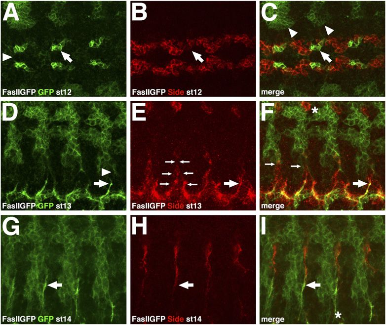
Downloaded from genesdev.cshlp.org on August 30, 2015 - Published by Cold Spring Harbor Laboratory Press

                                                                                                   Motor axons follow a Sidestep-labeled pathway

(Side), a transmembrane protein of the immunoglobulin                      are recognized by a specialized subset of axons expressing
superfamily that is dynamically expressed during em-                       the appropriate receptors.
bryogenesis but prominently enriched in muscles
when motor axons arrive in their target areas (Sink
                                                                           Results
et al. 2001). In side mutant embryos, motor axons fail to
defasciculate at key choice points and hence bypass                        Neuromuscular connectivity in Drosophila is established
their muscle targets, suggesting that Side functions as                    by efferent motor axons that project in a stereotypical
a target-derived attractant (Sink et al. 2001; de Jong et al.              pattern from the ventral nerve cord to peripheral body
2005). Interestingly, mutations in beaten path Ia (beat)                   wall muscles (Sink and Whitington 1991; van Vactor et al.
lead to similar axon guidance phenotypes, and like Side,                   1993). To visualize this dynamic process in living em-
Beat has been shown to regulate axon defasciculation at                    bryos, we examined exon trap lines in the fasciclin II
choice points (Fambrough and Goodman 1996; Holmes                          (fasII) locus that specifically label motor axons with green
and Heilig 1999; Sink et al. 2001). Based on primary                       fluorescent protein (GFP) (Rasse et al. 2005; Buszczak
structure predictions, Beat is a secreted protein of the                   et al. 2007; Stork et al. 2008). We decided to use the
immunoglobulin superfamily that has been shown to                          homozygous viable line FasIIGFPMue397 that contains an
function as an anti-adhesive factor on motor axons                         insertion in the extracellular domain of FasII (Rasse et al.
(Fambrough and Goodman 1996; Pipes et al. 2001).                           2005; Stork et al. 2008). FasIIGFPMue397 stains all tissues
Beat consists of two Ig domains and a Cysteine-rich                        known to express endogenous FasII, as recognized by the
C-terminal domain that shares similarities with cystine                    monoclonal anti-FasII antibody 1D4 (Grenningloh et al.
knots (Bazan and Goodman 1997; Mushegian 1997).                            1991). However, GFP was also detectable in the hemo-
   Here, we show that side encodes a candidate direc-                      lymph of FasIIGFPMue397 embryos (Supplemental Fig. S1).
tional cue that steers Drosophila motor axons from the                     Since FasIIGFPMue397 is expressed in motor neurons prior
place of their birth to their peripheral targets. The                      to axogenesis, it is an excellent marker for early de-
spatiotemporal expression pattern of Side thus delineates                  velopmental stages.
a linear pathway for motor axons. High levels of Side are
consistently found ahead of Beat-expressing growth
                                                                           Motor axons follow a Sidestep-labeled pathway
cones. Contact with motor axons induces the down-
regulation of Side and eliminates the pathway, preventing                  The axon guidance molecule Sidestep (Side) attracts
other outgrowing nerves from choosing the same route.                      motor axons and is expressed in a dynamic pattern in
Furthermore, we provide biochemical and genetic evi-                       neurons and muscles during embryogenesis (Sink et al.
dence that Beat functions as a membrane-associated                         2001), prompting us to examine its spatiotemporal re-
receptor or part of a receptor complex for Side. Taken                     lationship to motor axons. At embryonic stage 12, Side
together, these findings support the concept of pre-                       was expressed in a belt-like pattern along the ventral
existing pathways labeled with molecular markers that                      midline (Fig. 1A–C). The pioneering motor neuron of the

                                                                                      Figure 1. Motor axons follow Sidestep-labeled cell
                                                                                      surfaces. (A–I) FasIIGFPMue397 embryos stained with
                                                                                      anti-GFP and anti-Side antibodies to visualize the
                                                                                      spatiotemporal relationship of motor axons and Side-
                                                                                      expressing cells. (A–C) Ventral view of a stage 12 em-
                                                                                      bryo. (A) FasIIGFPMue397 is expressed in a cluster of four
                                                                                      to five cells in each neuromere, including the aCC and
                                                                                      pCC neurons. Arrowhead marks the ventral midline.
                                                                                      (B) Sidestep is expressed in a belt-like pattern flanking
                                                                                      the ventral midline. (C) FasIIGFPMue397-positive cells
                                                                                      are located next to Side-expressing cells (cf. arrows in
                                                                                      A–C). Arrowheads mark the tracheal precursors. Ante-
                                                                                      rior is left. (D–F) Ventrolateral view of a stage 13
                                                                                      embryo. (D) Pioneering axons of the ISN (arrow) project
                                                                                      toward the exit junction and developing ganglionic
                                                                                      branch of the trachea (arrowhead). (E) Sidestep is
                                                                                      expressed in a triangular area (small arrows) in the
                                                                                      CNS that points toward the trachea. (F) Motor axons
                                                                                      follow Sidestep-positive cell surfaces toward the tra-
                                                                                      chea (arrows in D–F). Small arrows in F mark the close
                                                                                      proximity of the tip of the triangle to the trachea.
                                                                                      Asterisk indicates the earliest expression of Side in
                                                                                      sensory neurons of the dorsal cluster. (G–I) Lateral view
                                                                                      of a stage 14 embryo. (G) ISN motor axons migrate
                                                                                      dorsally between the trachea and the developing mus-
cle field. (H) Sensory axons grow ventrally along similar routes. (I) Sensory axons and motor axons fasciculate in the ventrolateral region of
the embryo (arrows in G–I). The asterisk in I shows that Side is not detectable any more in the triangular area of the CNS (cf. F).

                                                                                                           GENES & DEVELOPMENT             1053
Downloaded from genesdev.cshlp.org on August 30, 2015 - Published by Cold Spring Harbor Laboratory Press
Siebert et al.
intersegmental nerve (ISN), the aCC neuron, developed the fasciculation of motor and sensory axons in
next to Side-expressing cells (Fig. 1A–C, arrows). At stage FasIIGFPMue397 embryos stained with anti-Futsch anti-
13, the Side expression domain had changed into a tri- bodies that recognize a microtubule-associated protein
angular pattern, with the tips pointing away from the (Hummel et al. 2000). In wild-type embryos, ISN motor
midline (Fig. 1E). Motor axons of the ISN projected along axons remained tightly fasciculated with sensory axons
the anterior edge of each triangle (Fig. 1D–F, arrows). until they reached the dbd neuron (Fig. 2A). Only 2% of
Remarkably, axons of the segmental nerve (SN) grew ISN nerves showed detachments >4 mm (Table 1). In
along the posterior edge. The triangular cell cluster contrast, in side mutants, 49% of ISN motor nerves were
guided motor axons directly toward the exit junction detached from sensory nerves of the anterior fascicle
and the incoming ganglionic branch of the trachea (Fig. (Fig. 2B; Table 1). This phenotype was even stronger when
1F, small arrows). At the trachea, motor axons fascicu- Side was overexpressed in muscles using the muscle-
lated with Side-positive, afferent sensory axons exactly at specific driver Mef2-Gal4 (Fig. 2C). In these embryos,
the lateral bidendritic neuron (lbd) (Fig. 1G–I, arrows). 84% of motor nerves were detached (Table 1). These
When the ISN reached the end of the sensory tracks at results show that the mutual recognition of motor and
the dorsal bidendritic neuron (dbd), expression of Side sensory axons is disturbed when levels of Side are altered.
in sensory neurons was no longer detectable but now Side thus functions in the attraction of motor axons to
could be observed in muscle fibers (Sink et al. 2001). sensory axons. The mutual recognition provides a station-
Side-expressing muscles likely attract motor axons, caus- ary substrate for the migration of either nerve, and
ing them to leave the sensory tracks. In general, cells and eventually results in the formation of coherent nerve
tissues located ahead of the growth cones express Side. bundles.
Upon contact with motor axons, the expression of Side is
down-regulated in the substrate, as monitored with anti- Growth cones in side mutants actively search
Side antibodies (Fig. 1I, asterisk) and in situ hybridiza- for pathway information
tions (Sink et al. 2001). The spatiotemporal expression
If motor axons use sensory tracks as growth substrates
pattern of Side is therefore consistent with a cell surface
the absence of Side might affect the rate of growth cone
marker that prefigures and potentially directs the path of
progression. We recorded time-lapse movies of the mi-
motor axons.
gration of the ISN in wild-type embryos expressing
FasIIGFPMue397 and compared them with side mutants
Sensory tracks cannot be recognized in side mutants
(see the Materials and Methods). In wild-type embryos,
If motor axons are attracted to and migrate along the ISN emerged as a thin bundle of axons in lateral body
Sidestep-expressing sensory axons, then this interaction wall regions at early stage 14 (Fig. 2D). The growth cone
should be disrupted in side mutants. We examined advanced continuously until it reached the dorsal trunk,
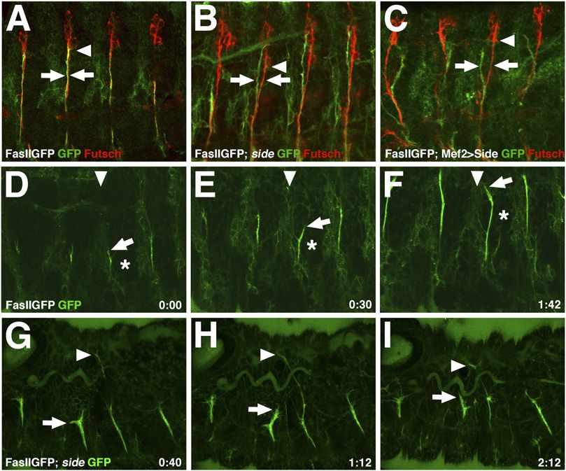
Figure 2. The ISN detaches from sensory axons
and shows migratory delays in side mutants. (A–C)
Motor axons are stained with anti-GFP and sensory
axons with anti-Futsch antibodies. (A) In stage 15
wild-type embryos, the ISN is tightly associated
with sensory axons of the anterior fascicle (arrows)
until it reaches the dbd neuron (arrowhead). The
dbd neuron stains only weakly with anti-Futsch
antibodies at this stage, as do motor axons. (B) The
ISN is partially detached from sensory axons
(arrows) prior to the dbd (arrowhead) in sideC137/
sideI1563 mutant embryos. (C) Wild-type embryos
overexpressing Side in muscles using Mef2-Gal4
show severe detachments of motor and sensory
axons (arrows). For quantification of the detach-
ment phenotypes see Table 1. (D–F) Stills from
Supplemental Movie 2-1 showing the migration of
the ISN in the lateral body wall of a FasIIGFPMue397
embryo at stage 14. The growth cone of the ISN
(arrows) travels between the transverse connective
(downward arrowheads) and the myogenic field
(asterisks). The growth cone marked with arrows
makes a small detour but corrects its path. Time is
indicated in hours and minutes (h:min). (G –I) Stills
from Supplemental Movie 2-2 showing the advance
of the ISN in a sideC137/sideI1563 mutant embryo at
stage 16. While one of the ISN nerves has already crossed the dorsal trunk (arrowheads), others are lacking behind (arrows). Delayed
growth cones are larger and sprout more filopodia.
1054 GENES & DEVELOPMENT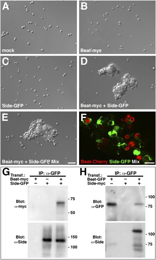
Downloaded from genesdev.cshlp.org on August 30, 2015 - Published by Cold Spring Harbor Laboratory Press

                                                                                            Motor axons follow a Sidestep-labeled pathway

Table 1. Frequencies of detachments of ISN motor axons and sensory axons of the anterior fascicle between the lbd and dbd
neurons in FasIIGFPMue397 (control), FasIIGFPMue397; sideC137/sideI1563 (loss-of-function), and FasIIGFPMue397; UAS-side/Mef2-Gal4
(gain-of-function) embryos
                                                                                   FasIIGFPMue397;                 FasIIGFPMue397;
                                                              Mue397
Detachments of the ISN/anterior fascicle             FasIIGFP                     sideC137/sideI1563             Mef2-Ga14/UAS-side
Percent detachments                                        2%                           49%                                84%
n hemisegments                                            62                            67                                 45
Only detachments >4 mm in stage 17 embryos have been evaluated.

with an average growth rate of 31.9 6 13.4 mm/h (n = 16).              using Serpent-Gal4, most, if not all, growth cones were
The entire fascicle employed only a single, relatively                 attached to hemocytes (Fig. 3D). Filopodial extensions of
small growth cone for steering, which increased in                     both cell membranes were tightly interdigitated. Time-
brightness and thickness over time (see Supplemental                   lapse movies in a FasIIGFPMue397 background showed
Movie 2-1). It is interesting to note that during its                  strong mutual adhesion between hemocytes and growth
migration along the transverse branch of the trachea,                  cones (Fig. 3E–H; Supplemental Movie 3-1). In a third
the ISN was not attracted into the nearby myogenic field               experiment, we expressed Side in muscles of wild-type
(Fig. 2D–F, asterisks). The growth cones occasionally                  embryos under control of Mef2-Gal4. This manipulation
diverged from their normal paths, which caused migra-                  results in the up-regulation of Side when muscles are not
tory delays but no permanent guidance defects, suggest-                yet developed but motor axons are just passing by. Under
ing that growth cones have the inherent ability to correct             this condition, the ISN nerves were strongly attracted to
minor misprojections (Fig. 2D–F, arrows). In side mutant               the developing muscle field and split aberrantly into
embryos, we observed severe delays in the dorsal migra-                several directions, suggesting directed growth toward
tion of the ISN (see Supplemental Movie 2-2). Growth                   individual muscle precursors (Fig. 3I,J). The split growth
cones eventually managed to cross the dorsal trunk (Fig.               cones prevented dorsal migration, causing strongly re-
2G–I, arrowheads), but a subset lagged behind (Fig. 2G–I,              duced growth rates (see Supplemental Movie 3-2). In
arrows). The growth rates were strongly reduced com-                   summary, the ability to redirect the path of motor axons
pared with wild-type nerves (15.2 6 4.6 mm/h; n = 12).                 shows that Side is an instructive cue for motor axons
Stalled growth cones appeared thicker and sprouted more                independent of the tissue in which it is expressed.
and longer filopodia. A small percentage of growth cones
exhibited highly disoriented directionality, extending in              Beaten path Ia (Beat) functions in the recognition
abnormal directions over the course of axonal growth                   of Side-labeled cell surfaces
(Fig. 2G–I, cf. arrows). Taken together, the reduced growth            Since mutations in beaten path Ia (beat) and side have
rates and the disoriented pattern of growth cone exten-                been reported to cause similar motor axon guidance
sions indicate that motor axons in side mutants increase               defects, we wondered whether Beat and Side cooperate
their search behavior, likely due to the absence of                    in pathfinding decisions (Fambrough and Goodman 1996;
attractive cues in the substrate.                                      Sink et al. 2001). To test this possibility, we examined
                                                                       the locations of neuromuscular junctions (NMJs) in
Side instructs the growth direction of motor axons
                                                                       beat and side mutant third instar larvae that reflect the
If Side marks a path for motor axons it should be possible             final outcome of embryonic projection errors at a high
to redirect the growth direction of motor axons by                     spatial resolution (de Jong et al. 2005). Guidance errors
creating an ectopic pathway. Misexpression of Side on                  lead to the irreversible formation of NMJs at aberrant
trachea or a subset of muscles in wild-type embryos                    positions that can be visualized with the post-synaptic
results in extended contact and exploration by motor                   marker CD8-GFP-Sh (Meyer and Aberle 2006). Compared
nerves (Sink et al. 2001). To circumvent the presence of               with wild-type larvae (Fig. 4A), NMJs were frequently
endogenous Side we expressed Side in the trachea of side               absent on dorsal muscles 1/9 in both beat and side
mutants using breathless-Gal4. Under these conditions,                 mutant larvae (Fig. 4B,C; Table 2). In addition, beat and
most, if not all, ISN growth cones were tightly attached to            side mutants showed similar innervation defects on a
tracheal branches (Fig. 3B, arrows). Even more strikingly,             variety of muscles, indicating that they function in a re-
the ISN followed the tracheal substrate and crossed and                lated process.
recrossed the transverse connective. In comparison, in                    We next tested for possible activating or antagonizing
side mutant control embryos, the ISN grew out into the                 effects of Beat with Side. We expressed Beat and Side
periphery but none of the growth cones was particularly                individually or together in muscles of wild-type animals
associated with tracheal branches (arrows in Fig. 3A). In              using Mef2-Gal4. Since expression of Side on muscle
a second experiment, we expressed Side in highly motile                precursors prematurely attracts motor axons (Fig. 3I,J)
hemocytes. Normally, these circulating cells function in               we assumed that the simultaneous overexpression of Beat
the immune system of Drosophila and do not establish                   might increase or suppress this effect. While increased
firm contacts with growth cones neither in wild-type nor               levels of Beat had only minor effects on the innervation
in side mutant embryos (Fig. 3C). Strikingly, when Side                pattern of dorsal muscles (Fig. 4D), ectopic expression of
was ectopically expressed in hemocytes of side mutants                 Side resulted in an almost complete lack of dorsal NMJs

                                                                                                       GENES & DEVELOPMENT          1055
Downloaded from genesdev.cshlp.org on August 30, 2015 - Published by Cold Spring Harbor Laboratory Press

Siebert et al.

due to premature attraction into ventral and lateral                 dence for a genetic interaction, we created double mutants.
muscle fields (Fig. 4E; Table 2). Simultaneous expression            In beat;side double mutants, the pattern of mislocalized
of Beat and Side in muscles completely abolished the Side            NMJs was qualitatively similar and quantitatively not
gain-of-function phenotype (Fig. 4F; Table 2). Thus, Beat            increased, when compared with single mutants of beat or
is able to suppress the function of exogenous Side when              side (Fig. 4G; Table 2). Since Beat is expressed in motor
coexpressed in the same tissue. To collect further evi-              axons (Fambrough and Goodman 1996), it might function
                                                                     in the detection of Side on substrates. If so, muscles
                                                                     overexpressing Side should not attract motor axons in
                                                                     a beat mutant background, as they normally would in
                                                                     a wild-type background (Figs. 3I,J, 4E). Indeed, overex-
                                                                     pression of Side in muscles of beat mutants did not
                                                                     increase the dorsal innervation defects of beat mutants
                                                                     (Fig. 4H; Table 2). Motor axons lacking Beat seem there-
                                                                     fore to be unable to respond to Side presented on muscle
                                                                     progenitors, and hence unable to recognize Side-labeled
                                                                     substrates. Genetic evidence thus suggests that beat and
                                                                     side cooperate in axonal pathway decisions.

                                                                     Beat regulates the expression levels of Side
                                                                     In wild-type embryos, Side is down-regulated shortly after
                                                                     contact with motor axons (e.g., Fig. 1I, asterisk). Since the
                                                                     genetic evidence presented above suggests that Beat is
                                                                     required to detect Side, the distribution of Side might be
                                                                     altered in beat mutants. We analyzed the expression level
                                                                     and subcellular distribution of Side in wild-type and beat
                                                                     mutant embryos employing anti-Side antibodies (Sink
                                                                     et al. 2001). Peripheral nerves normally lack detectable
                                                                     levels of Side at stage 16/17, since Side is efficiently
                                                                     cleared from sensory axons once motor axons have
                                                                     entered their target regions (Fig. 5A). In beat mutants,
                                                                     however, Side was not eliminated, and instead was
                                                                     expressed on sensory axons until late embryonic stages
Figure 3. Sidestep is sufficient to direct the path of motor         (Fig. 5B). To test whether Beat plays a role in the
axons. (A,B) Side attracts motor axons to tracheal branches.
                                                                     regulation of Side levels we ectopically expressed Beat
Axons are stained with anti-FasII antibodies, the tracheal lumen
is stained with a chitin-binding probe coupled to Rhodamine. (A)
                                                                     either in all post-mitotic neurons or in all muscles in
The ISN growth cones are not in direct contact with tracheal         a beat mutant background. Nerve-derived Beat fully
branches in a side mutant embryo at stage 16 (arrows) (genotype:     rescued the constitutive expression phenotype of Side
sideC137/sideI1563). (B) Ectopic expression of Side in trachea of    and induced the elimination of Side from sensory axons
side mutants strongly attracts motor axons to tracheal branches,     (Fig. 5C). Only low traces of Side expression remained
the only source of Side in these embryos (arrows) (genotype: btl-    (Fig. 5C, arrow). In contrast, muscle-derived Beat failed to
Gal4/+; UAS-side, sideC137/sideI1563). (C,D) Side attracts motor     down-regulate Side on sensory axons (Fig. 5D). In addi-
axons to hemocytes. (C) Stage 16 side mutant embryo express-         tion, pan-neuronal (Elav-Gal4) and motor neuronal (FasII-
ing transmembrane CD8GFP in hemocytes (red). ISN growth              Gal4), but not muscular Beat (Mef2-Gal4) expression,
cones misproject but are not in direct contact with hemocytes
                                                                     rescued the axon guidance phenotype, leading to a neuro-
(arrows) (genotype: UAS-mCD8GFP, sideC137/sideI1563, Serpent-
Gal4). (D) FasIIGFPMue397 embryo mutant for side and express-
                                                                     muscular innervation pattern almost indistinguishable
ing exogenous Side in hemocytes. In the absence of endogenous        from wild-type larvae (Table 2). These results suggest that
Side, almost every growth cone of the ISN is in contact with a       Beat is required cell-autonomously in motor neurons.
hemocyte (arrows) (genotype: FasIIGFPMue397; UAS-side,                  Based on the analysis of its primary sequence and its
sideC137/sideI1563, Serpent-Gal4). (E–H) Still images of Supple-     staining pattern ‘‘around axons and growth cones’’ Beat
mental Movie 3-1 (time in minutes and seconds) showing strong        has been suggested to be a secreted protein (Fambrough
adhesive interactions between an ISN growth cone (arrow) and         and Goodman 1996). However, no costaining with an
a Side-expressing hemocyte (arrowhead) in a side mutant em-          axonal membrane marker has been provided (Fambrough
bryo at stage 16 (genotype: FasIIGFPMue397; UAS-side, sideC137/      and Goodman 1996). Surprisingly, protein sequence
sideI1563, Serpent-Gal4). (I,J) Stills of Supplemental Movie 3-2
                                                                     analysis using several topology prediction algorithms
(time in hours and minutes) showing ISN growth cones in a
stage 15 FasIIGFPMue397 embryo overexpressing Side in muscles
                                                                     (HMMTOP, PredictProtein, TMpred, Phobius, and
(genotype: FasIIGFPMue397; Mef2-Gal4/UAS-side). The growth           TopPred) indicates a transmembrane region in Beat (data
cone of the ISN splits into several directions, preventing its       not shown). We therefore wanted to examine whether
dorsal migration and deflecting its route along presumptive          Beat is a secreted or membrane-associated protein. In
muscle fibers (arrows).                                              transiently transfected S2 cells, myc-tagged Beat was not

1056        GENES & DEVELOPMENT
Downloaded from genesdev.cshlp.org on August 30, 2015 - Published by Cold Spring Harbor Laboratory Press

                                                                                          Motor axons follow a Sidestep-labeled pathway

                                                                           Figure 4. Genetic evidence that Beat and Side function
                                                                           in a common pathway. (A–H) Confocal images of muscle
                                                                           pairs 1/9 and 2/10 in third instar larvae expressing the
                                                                           post-synaptic marker CD8-GFP-Sh. The percentages of
                                                                           noninnervated muscles in the respective genotypes are
                                                                           quantified in Table 2. (A) In wild-type larvae, dorsal-most
                                                                           muscles are innervated by centrally localized NMJs
                                                                           (arrows). (B,C) sideC137/sideI1563 (B) and beat3/beatC163
                                                                           (C) mutant larvae show similar innervation defects on
                                                                           a variety of muscles and frequently lack NMJs on
                                                                           muscles 1/9. (D) Ectopic expression of Beat in muscles
                                                                           of wild-type larvae using Mef2-Gal4 does not affect the
                                                                           innervation of dorsal muscles. (E) Ectopic expression of
                                                                           Side in muscles results in complete lack of NMJs on
                                                                           dorsal-most muscles. (F) Coexpression of Beat and Side
                                                                           in muscles suppresses the innervation defects caused by
                                                                           overexpression of only Side, resulting in a wild-type
                                                                           innervation pattern on dorsal muscles. (G) Phenotypic
                                                                           strength is not increased in beat3/beatC163; sideC137/
                                                                           sideC137 double mutants when compared with single
                                                                           mutants of beat or side. (H) Overexpression of Side in
                                                                           muscles of beat3/beatC163 mutant animals does not
                                                                           increase phenotypic strength of beat mutants, leading
                                                                           to the absence of NMJs only on muscles 1/9.

secreted into the supernatant (Supplemental Fig. S2).               complex. To test if Beat interacts with Side, we tran-
Similar to Side, most of the Beat protein was detected              siently transfected S2 cells with tagged constructs of
in the organelle fraction but a significant portion was             either protein and subjected these cells to aggregation
associated with the fraction containing integral mem-               assays. S2 cells expressing either Beat-myc or Side-GFP
brane proteins (Supplemental Fig. S2). These results                did not form cell–cell aggregates, indicating that neither
combined with the strict cell-autonomous function sug-              Beat nor Side interact homophilically (Fig. 6B,C). In
gest that Beat functions on cell surfaces.                          contrast, cells cotransfected with Beat-myc and Side-
                                                                    GFP formed large aggregates (Fig. 6D). Large cell clusters
                                                                    also formed when we mixed Beat-myc-expressing cells
Beat interacts with Side
                                                                    with Side-GFP-expressing cells (Fig. 6E). We obtained
Neuronal Beat might recognize Side-expressing cell sur-             similar results with Beat–Cherry constructs as well (Fig.
faces by directly binding to Side or a Side-containing              6F). The cell aggregates consisted almost exclusively of

Table 2. Percentage of dorsal and ventral muscles lacking NMJs in third instar larvae of the indicated genotypes expressing CD8-
GFP-Sh
Lack of NMJs on dorsal muscles                    Muscle 1               Muscle 9                Muscle 2                  Muscle 10
Wild type (CD8-GFP-Sh)                                0                      0                        0                          0
beat3/beatC163                                       31                     34                        8                         13
sideC137sideI1563                                    19                     24                       11                          9
beat3/beatC163; sideCl37/sideC137                    24                     42                       16                         20
24B-Gal4/UAS-side                                    88                     88                       39                         39
Mef2-Gal4/UAS-side                                   93                     93                       60                         57
UAS-dsRed; Mef2-Gal4/UAS-side                        88                     86                       60                         58
Mef2-Gal4/UAS-beat, UAS-side                         24                     30                       14                         14
Mef2-Gal4/UAS-beat                                   16                     21                        7                          5
beat3/beatC163; 24B-Gal4/UAS-side                    32                     31                       10                          5
beat3/beatC163; Mef2-Gal4/UAS-side                   25                     29                       11                          9
beat3/beatC163; Elav-Gal4/UAS-beat                    4                      2                        0                          0
beat3/beatC163; Mef2-Gal4/UAS-beat                   26                     18                        8                          8
FasII-Gal4; beat3/beatC163; UAS-beat                  5                      4                        0                          1
Lack of NMJs on ventral muscles                Muscle 12                Muscle 13                 Muscle 6                   Muscle 7
Wild type (CD8-GFP-Sh)                              0                        0                         0                         2
beat3/beatC163                                     25                       34                        38                        55
sideC137/sideI1563                                 58                       75                        77                        85
beat3/beatC163; sideC137/sideC137                  15                       48                        72                        84
n = 100 hemisegments for each genotype.

                                                                                                  GENES & DEVELOPMENT             1057
Downloaded from genesdev.cshlp.org on August 30, 2015 - Published by Cold Spring Harbor Laboratory Press
Siebert et al.
Beat-expressing motor axons recognize and follow Side-
labeled surfaces. In order to better visualize the complex
spatial and temporal dynamics of axon guidance pro-
cesses, we established a FasIIGFPMue397-based imaging
assay that allowed us to analyze the activity of growth
cones in living wild-type and mutant embryos. These
Figure 5. Expression of Beat in neurons but not in muscles
regulates the expression level of Side. (A) At stage 16, Side is no
longer detected on peripheral nerves of wild-type embryos but
remains weakly expressed in muscles and the neuropil. (B) In
beat3 mutant embryos, Side remains highly expressed on
peripheral nerves and the ventral neuropil until the end of
embryogenesis. (C) Expression of wild-type Beat in a beat3
mutant embryo using Elav-Gal4 rescues the constitutive ex-
pression phenotype and induces the down-regulation of Side on
peripheral nerves. A few Side-positive particles, however, re-
main (arrow). (D) Expression of wild-type Beat in muscles of
beat3 mutant embryos using Mef2-Gal4 fails to down-regulate
Side on peripheral nerves, indicating that Beat does act cell-
autonomously.
Beat- and Side-positive cells, and contained only marginal
amounts of nontransfected cells (Downloaded from genesdev.cshlp.org on August 30, 2015 - Published by Cold Spring Harbor Laboratory Press
Motor axons follow a Sidestep-labeled pathway
time-lapse observations revealed that in wild-type em- tion in wrong directions, whereas lack of Side leads to
bryos, the ISN migrates continuously through the lateral reduced attraction along the predestined path.
body wall until it reaches a choice point near the dorsal
trunk. As expected for migration along a substrate path-
Beat interacts with Side
way, the ISN employs a relatively small growth cone that
extends only few filopodia for steering. The observed Since mutations in both beat and side cause similar axon
growth rates are in good agreement with the growth rates guidance phenotypes in embryos (Fambrough and Good-
of the RP2 axon in filleted, semiviable embryo prepara- man 1996; Sink et al. 2001) and innervation defects in
tions (Murray et al. 1998). larvae (this study), we assumed that the products of both
genes might interact functionally. Several pieces of ge-
netic evidence suggest that beat and side function in
Side labels the path for motor axons
a common pathway. First, the double mutant phenotype
The absence of the attractant Side strongly interferes is similar to the respective single mutant phenotypes.
with axonal growth and results in delayed arrival of the Second, overexpression of Side in muscles of wild-type
ISN in its dorsal target regions, frequently failing to embryos leads to premature attraction of the ISN into
innervate the dorsal-most muscles. The lack of Side- ventral and lateral muscle fields. This gain-of-function
mediated attraction likely prevents the progression at a phenotype is completely suppressed by coexpression of
normal rate, causing the ISN to develop a complex Beat. Third, overexpression of Side in muscles of beat
growth cone that appears to actively search for guidance mutants renders motor axons unresponsive to ectopic
information. Since Side is a transmembrane protein, it is Side. Fourth, endogenous Side is not down-regulated in
predicted to function as a contact attractant. The spatio- beat mutants. In addition, Beat and Side interact in vitro.
temporal expression pattern should therefore provide S2 cells expressing Beat and Side form large cell clusters
pathway information. The growth cone of the ISN follows in aggregation assays when individually transfected cell
Side-positive cell surfaces from its first emergence in the populations are mixed. Moreover, Beat coimmunopreci-
ventral nerve cord to its dorsal target region. Conse- pitates with Side, supporting the idea that Beat interacts
quently, the tight association of motor axons and their with Side or a Side-containing complex. The formation of
substrates—e.g., sensory axons—is partially lost in side cell aggregates further argues that Beat–Side interactions
mutants. Since Side is expressed in all sensory clusters lead to the formation of heterophilic adhesion complexes.
(Sink et al. 2001), it likely also prefigures the SNa and SNc The spatiotemporal expression of Side appears to be
pathways. Based on the positions of the segmental sen- strictly regulated. The levels of Side expression are high-
sory clusters in the body wall, motor axons following est in front of motor axonal growth cones. Side disappears
Sidestep-labeled sensory axons are guided into the prox- from cell surfaces once these cells have been contacted by
imity of their target regions. At stage 15, motor axons motor axons, indicating that motor nerves neutralize
reach the end of the sensory tracks and begin to defas- attractive surfaces and thereby disguise the path they
ciculate into the muscle fields that up-regulate Side at this are following. Motor nerve bundles that exit the CNS at
developmental time point. In this respect, it is interesting a later time point are thus prevented from choosing the
to note that motor pathways terminate on the cell bodies same route. In beat mutants, this regulatory mechanism
of sensory neurons when deprived of their target muscles appears to be nonfunctional. Side is constitutively
(Landgraf et al. 1999). The opposed migration of efferent expressed in peripheral nerves. Expression of exogenous
motor axons and afferent sensory axons therefore provides Beat in post-mitotic neurons but not in muscles rescued
a robust mechanism for the establishment of the basic the regulatory defects, suggesting that Beat induces
neuromuscular connectivity pattern in Drosophila. In the the down-regulation of Side cell-autonomously. If Beat
brain of vertebrates, a similar mechanism controls the would be a secreted protein, one would expect that ex-
wiring of the thalamus and the cortex (Lopez-Bendito and pression in muscles down-regulates Side on sensory axons.
Molnar 2003). Corticothalamic and thalamocortical fibers The secreted metalloprotease tolloid-related (Meyer and
meet at a common intermediate target and continue to Aberle 2006; Serpe and O’Connor 2006) as well as the
grow along each other in opposite directions. secreted TGF-b ligand Dawdle (Parker et al. 2006) have
During its journey through the lateral body wall, the been shown to rescue axonal guidance defects tissue
ISN completely ignores the nearby ventral and lateral independently. Biochemical data from transiently trans-
muscle fields. At this stage, muscle fibers are not yet fected S2 cells further support a cell-autonomous func-
differentiated, and hence do not express endogenous Side. tion for Beat. Beat was not secreted into the medium, and
If these muscle precursors, however, are forced to express a fraction of it was associated with membranes. However,
Side prematurely under control of Mef2-Gal4, the ISN since Beat is not normally expressed in S2 cells the lack of
diverts from its normal path and grows straight into the a coreceptor or chaperone might prevent its secretion.
muscle field. The premature attraction drastically slows Candidate coreceptors are the remaining members of the
down the migration toward dorsal targets, leading to beat multigene family (Pipes et al. 2001). Several family
a permanent lack of NMJs on dorsal muscles. Thus, both members are expressed in the ventral nerve cord, and
gain and loss of Side cause migratory delays that result in might function together with Beat in the regulation of its
the lack of NMJs on dorsal muscles but for different subcellular localization and/or function (Pipes et al. 2001).
reasons. Ectopic expression of Side leads to excess attrac- Further experiments will be necessary to determine the
GENES & DEVELOPMENT 1059Downloaded from genesdev.cshlp.org on August 30, 2015 - Published by Cold Spring Harbor Laboratory Press
Siebert et al.
composition of Beat-containing complexes, and how they existing, Side-labeled substrate pathway that determines
transduce guidance signals into the growth cone. their growth direction (Fig. 7A). In the absence of the
labeled pathway, in side mutants, axonal migration is
Beat–Side complexes determine the growth direction delayed or growth cones head into aberrant directions.
of motor axons In either case they will miss their targets (Fig. 7B). In
beat mutants, the pathway is constitutively labeled but
If Side is an instructive signal for Beat-expressing motor
cannot be recognized, leading to similar phenotypes
axons it should be possible to redirect their paths in a side
(Fig. 7B). Since Beat and Side are conserved in insects,
mutant background; i.e., in the absence of endogenous
similar guidance principles might occur in all organisms,
Side but in the presence of exogenous Side on a defined
in which the peripheral nervous system develops from
tissue. Regardless of whether Side is expressed in trachea,
sensory organ precursors.
muscles, or hemocytes in a side mutant background,
motor axons head toward the ectopic source of Side. The
growth cones find and recognize Side-expressing cell Materials and methods
surfaces and adopt their route accordingly. In the most
extreme case, motor axons strongly interacted with Genetics and fly stocks
hemocytes, which are highly motile cells. Side therefore The exon trap line Mue397 was identified in the Muenster 2004
potently controls the path of motor axons. Although Side- exon trap screen performed by the DFG Consortium Cell
mediated attraction is likely not the only mechanism to Polarity (SPP1111) (Rasse et al. 2005; Stork et al. 2008). The
reach the muscle targets, these results, together with the sideC137 and sideI1563 alleles were isolated in an EMS mutagen-
high penetrance of sidestep mutant phenotypes, suggest esis screen for recessive mutations affecting the structure of
NMJs (Aberle et al. 2002). beat3, beatC163, and UAS-dsRED were
that it is one of the major mechanisms. In this respect,
obtained from the Bloomington stock center. UAS-side29A and
it is important to note that Side is not required for UAS-beat5 were kindly provided by C.S. Goodman. The follow-
motor axon outgrowth per se, but rather for the specifi- ing Gal4-lines were used: 24B-Gal4, Mef2-Gal4 (gifts of C.S.
cation of the growth direction. Based on the experimental Goodman), breathless (btlGal4; gift of M.A. Krasnow), Serpent-
evidence, we propose a model for the navigation of motor Gal4 (gift of R. Reuter), FasII-Gal4 (Mz507; B. Altenhein, pers.
axons from the ventral nerve cord to their target area comm.). As wild-type control strains, y w1 or w1; CD8-GFP-Sh
in Drosophila (Fig. 7). Beat-expressing motor axon fas- (Zito et al. 1999) were used.
cicles recognize, extend on, and subsequently mask a pre-
Molecular biology
Full-length beat Ia and side cDNAs (kindly provided by C.S.
Goodman) were amplified by PCR and cloned into Gateway
Entry Vectors (pENTR D-TOPO, Invitrogen). The inserts were
sequenced and subcloned into pTWH, pTWM, and pTWG
Destination Vectors (Drosophila Genomics Resource Center;
donated by Terence Murphy) by LR in vitro recombination,
containing UAST promotors and C-terminal 3xHA, 6xMyc,
and eGFP tags. The pTWC vector encoding a C-terminal Cherry
tag was a generous gift of F. Rodrigues and C. Klämbt. The
pUAST vector encoding Slit-myc was kindly provided by J.
Hillebrand and C. Klämbt, and the Actin5C-Gal4 vector was
provided by A. Wodarz. All steps were performed according to
the manual of the manufacturer (Escherichia coli Expression
System with Gateway Technology, Invitrogen). Sequence analy-
sis and topology prediction (HMMTOP, PredictProtein, TMpred,
and TopPred) were performed at the ExPASy and EBI Proteomics
servers.
Immunohistochemistry
For immunohistochemical stainings, embryos were dechorio-
Figure 7. Model: Beat-expressing motor axons follow a Side- nated, fixed with 3.7% formaldehyde and devitellinized. Em-
labeled substrate pathway. (A) In wild-type embryos, Beat- bryos were washed with PTx (PBS containing 0.1% Triton X-100)
expressing motor axons (green) recognize and follow Side- and blocked in PTx/5% normal goat serum. Primary antibodies
labeled cell surfaces (red). Contact with motor axons induces were added overnight at 4°C. Stainings were developed with
the down-regulation of Side (gray). Growth cones migrate until fluorescently labeled secondary antibodies. The dilutions of the
the end of the Side expression domain. Developmentally con- primary antibodies were as follows: mouse anti-Fasciclin II
trolled up-regulation of Side in another tissue induces growth (1D4) 1:20, mouse anti-Sidestep (9B8) 1:20, and mouse anti-Repo
cone turning. (B, left) In side mutants, growth cones fail to turn, (8D12) 1:40 (all gifts of C.S. Goodman), mouse anti-myc (9E10)
as substrates are not labeled. Other possible phenotypes such as 1:10 and mouse anti-Futsch (22C10) 1:100 (both from Develop-
delays or detours are not depicted. (Right) In beat mutants, the mental Studies Hybridoma Bank), mouse anti-GFP 1:400
Side-labeled pathway is constitutively marked but cannot be (Roche), and rabbit anti-GFP 1:1000 (Torrey Pines Biolabs).
recognized, thereby preventing growth cone turning. Cy3- and Alexa488-conjugated secondary antibodies were
1060 GENES & DEVELOPMENTDownloaded from genesdev.cshlp.org on August 30, 2015 - Published by Cold Spring Harbor Laboratory Press
Motor axons follow a Sidestep-labeled pathway
diluted 1:400 (Jackson Immunoresearch; Molecular Probes). The placed onto a greased glass ring (0.4 cm high, 1 cm diameter).
Rhodamine-conjugated chitin-binding probe (New England The glass ring was fixed before on a microscope slide with
Biolabs) was diluted 1:400 and consists of the maltose-binding vacuum grease and contained a drop of liquid agarose to support
protein fused to a C-terminal region of chitinase A1. Stained the agar block after solidifying. Time-lapse imaging was per-
embryos were imaged as whole mounts using a LSM510 confocal formed at an upright confocal laser scanning microscope
laser scanning microscope (Zeiss). Projections and single images LSM510 Meta (Zeiss) using a 403 objective. Stacks of four to
were adjusted for brightness, contrast, and color matching using five Z-planes were acquired in 45-sec intervals for up to 5 h.
linear functions of Adobe Photoshop. Stacks were processed and exported as Quicktime movies
using LSM software. Growth cone migration was quantified by
measuring the advance of its center in time.
Cell aggregation assays and biochemical methods
S2 cells were grown in complete Schneider medium supple-
Acknowledgments
mented with 10% fetal calf serum and 50 U/mL Penicillin/
Streptomycin. Cells (3 3 106) were seeded into six-well plates We are indebted to Christian Klämbt for providing laboratory
and transiently transfected using calcium phosphate precipita- space and support. We thank Stephan Sigrist for recommending
tion (5 mg per vector). The medium was replaced after 16 h. For the line Mue397 for live imaging, and Benjamin Altenhein for the
cell aggregation assays, cells were separated by pipetting and FasII-Gal4 (Mz507) line. Further fly stocks and reagents have
seeded into uncoated 6-cm plastic dishes 3 d post-transfection. been generously provided by Corey Goodman, Mark Krasnow,
Cells were agitated on a rotary shaker (100 rpm) for 2 h. Rolf Reuter, Floriano Rodrigues, Jens Hillebrand, Sven Bogdan,
Aggregates were transferred onto microscope slides and imaged Andreas Wodarz, and the Bloomington Stock Center. We are
using a Zeiss AxioPlan-2 microscope adjusted for DIC. For the grateful to Christian Klämbt, Sven Bogdan, Thomas Hummel,
detection of secreted proteins, transfected S2 cells (3-d-old and Joanne Yew for critical comments on the manuscript. This
cultures) were incubated for 24 h in serum-free medium. Cells project was funded by the Deutsche Forschungsgemeinschaft
were pelleted, washed and lysed in 23 SDS sample buffer. The (AB116/3-1, AB116/4-1).
medium was mixed with an equal volume of 50% (w/v) TCA
(trichloroacetic acid) (Sigma) and incubated for 30 min on ice.
References
Precipitated proteins were collected (14,000 rpm, 30 min, 4°C)
and washed with ice-cold isopropanol. The pellet was solubilized Aberle, H., Haghighi, A.P., Fetter, R.D., McCabe, B.D., Magalhaes,
in 23 SDS sample buffer and titrated with 1 M Tris (pH 7.5). For T.R., and Goodman, C.S. 2002. wishful thinking encodes
subcellular fractionation, S2 cells were lysed by sonification and a BMP type II receptor that regulates synaptic growth in
subjected to differential centrifugation (5000 rpm, 24,000 rpm, Drosophila. Neuron 33: 545–558.
and 75,000 rpm for 30 min at 4°C in an ultracentrifuge [Optima Araujo, S.J. and Tear, G. 2003. Axon guidance mechanisms and
Max-E, Beckman Coulter]). The pellet was incubated with 0.1 M molecules: Lessons from invertebrates. Nat. Rev. Neurosci.
Na2CO3 (pH 11.5) to release peripheral membrane proteins. 4: 910–922.
Bate, C.M. 1976. Pioneer neurones in an insect embryo. Nature
260: 54–56.
Immunoprecipitation and immunoblotting
Bazan, J.F. and Goodman, C.S. 1997. Modular structure of the
S2 cells from aggregation assays were collected by centrifugation Drosophila Beat protein. Curr. Biol. 7: R338–R339. doi:
and lysed in lysis buffer (50 mM Tris at pH 7.5, 150 mM NaCl, 10.1016/S0960-9822(06)00168-0.
1% NP40, protease inhibitor cocktail [Roche]). Insoluble debris Buszczak, M., Paterno, S., Lighthouse, D., Bachman, J., Planck,
was pelleted by centrifugation (15 min, 14,000 rpm, 4°C). The J., Owen, S., Skora, A.D., Nystul, T.G., Ohlstein, B., Allen,
supernatants were incubated with 1 mg of rabbit anti-GFP anti- A., et al. 2007. The carnegie protein trap library: A versatile
bodies (Invitrogen) for 1 h at room temperature on a rotary tool for Drosophila developmental studies. Genetics 175:
shaker. Protein complexes were collected by addition of equili- 1505–1531.
brated protein A sepharose beads and washed with washing de Jong, S., Cavallo, J.A., Rios, C.D., Dworak, H.A., and Sink, H.
buffer (50 mM Tris at pH 7.5, 150 mM NaCl, 1% NP40). Bound 2005. Target recognition and synaptogenesis by motor axons:
proteins were eluted by boiling in 23 SDS sample buffer. Responses to the sidestep protein. Int. J. Dev. Neurosci. 23:
Samples were separated on 7.5% SDS-PAGE gels (Bio-Rad) and 397–410.
transferred onto PVDF membranes (GE Healthcare) by semidry Dickson, B.J. 2002. Molecular mechanisms of axon guidance.
blotting (Biozym). Blots were incubated with anti-Side antibodies Science 298: 1959–1964.
(9B8) (1:10) or anti-myc antibodies (9E10) (1:100; Developmental Fambrough, D. and Goodman, C.S. 1996. The Drosophila beaten
Studies Hybridoma Bank) and developed with HRP-conjugated path gene encodes a novel secreted protein that regulates
secondary antibodies (1:10000; Dianova) followed by enhanced defasciculation at motor axon choice points. Cell 87: 1049–
chemiluminescence detection (ECL, GE Healthcare). Molecular 1058.
weights were standardized with Precision Plus Protein Dual Ghysen, A. 1978. Sensory neurones recognise defined pathways
Color Standards (Bio-Rad). in Drosophila central nervous system. Nature 274: 864–872.
Ghysen, A. and Janson, R. 1980. Sensory pathways in Drosophila
central nervous system. In Development and neurobiology
Time-lapse imaging
of Drosophila (eds. O. Siddiqi et al.), pp. 247–265. Plenum
Living, GFP-expressing embryos were dechorionated, mounted Publishing Corp., New York.
in 70% glycerol/PBS, and immediately examined for still images. Goodman, C.S., Raper, J.A., Chang, S., and Ho, R. 1983.
For time-lapse movies, embryos were dechorionated, placed on Grasshopper growth cones: Divergent choices and labeled
a coverslip, and gently covered with a piece of fruit agar (0.8 3 pathways. Prog. Brain Res. 58: 283–304.
0.8 3 0.2 cm). Liquid agarose (0.4%, 42°C) was then pipetted Grenningloh, G., Rehm, E.J., and Goodman, C.S. 1991. Genetic
between the coverslip and the fruit agar block to embed the analysis of growth cone guidance in Drosophila: Fasciclin II
embryos. The assembly (coverslip up, agar block down) was functions as a neuronal recognition molecule. Cell 67: 45–57.
GENES & DEVELOPMENT 1061Downloaded from genesdev.cshlp.org on August 30, 2015 - Published by Cold Spring Harbor Laboratory Press
Siebert et al.
Holmes, A.L. and Heilig, J.S. 1999. Fasciclin II and Beaten Sink, H. and Whitington, P.M. 1991. Location and connectivity
path modulate intercellular adhesion in Drosophila of abdominal motoneurons in the embryo and larva of
larval visual organ development. Development 126: 261– Drosophila melanogaster. J. Neurobiol. 22: 298–311.
272. Sink, H., Rehm, E.J., Richstone, L., Bulls, Y.M., and Goodman,
Huber, A.B., Kolodkin, A.L., Ginty, D.D., and Cloutier, J.F. 2003. C.S. 2001. sidestep encodes a target-derived attractant essen-
Signaling at the growth cone: Ligand–receptor complexes and tial for motor axon guidance in Drosophila. Cell 105: 57–67.
the control of axon growth and guidance. Annu. Rev. Neuro- Sperry, R.W. 1963. Chemoaffinity in the orderly growth of nerve
sci. 26: 509–563. fiber patterns and connections. Proc. Natl. Acad. Sci. 50:
Hummel, T., Krukkert, K., Roos, J., Davis, G., and Klambt, C. 703–710.
2000. Drosophila Futsch/22C10 is a MAP1B-like protein Stork, T., Engelen, D., Krudewig, A., Silies, M., Bainton, R.J., and
required for dendritic and axonal development. Neuron 26: Klambt, C. 2008. Organization and function of the blood–
357–370. brain barrier in Drosophila. J. Neurosci. 28: 587–597.
Katz, M.J. and Lasek, R.J. 1979. Substrate pathways which guide van Vactor, D., Sink, H., Fambrough, D., Tsoo, R., and Goodman,
growing axons in Xenopus embryos. J. Comp. Neurol. 183: C.S. 1993. Genes that control neuromuscular specificity in
817–831. Drosophila. Cell 73: 1137–1153.
Katz, M.J. and Lasek, R.J. 1981. Substrate pathways demon- Zito, K., Parnas, D., Fetter, R.D., Isacoff, E.Y., and Goodman,
strated by transplanted Mauthner axons. J. Comp. Neurol. C.S. 1999. Watching a synapse grow: Noninvasive confocal
195: 627–641. imaging of synaptic growth in Drosophila. Neuron 22: 719–
Landgraf, M., Baylies, M., and Bate, M. 1999. Muscle founder 729.
cells regulate defasciculation and targeting of motor axons in
the Drosophila embryo. Curr. Biol. 9: 589–592.
Lopez-Bendito, G. and Molnar, Z. 2003. Thalamocortical devel-
opment: How are we going to get there? Nat. Rev. Neurosci.
4: 276–289.
Meyer, F. and Aberle, H. 2006. At the next stop sign turn right:
The metalloprotease Tolloid-related 1 controls defascicula-
tion of motor axons in Drosophila. Development 133: 4035–
4044.
Murray, M.J., Merritt, D.J., Brand, A.H., and Whitington, P.M.
1998. In vivo dynamics of axon pathfinding in the
Drosophilia CNS: A time-lapse study of an identified motor-
neuron. J. Neurobiol. 37: 607–621.
Mushegian, A.R. 1997. The Drosophila Beat protein is related to
adhesion proteins that contain immunoglobulin domains.
Curr. Biol. 7: R336–R338. doi: 10.1016/S0960-9822(06)00167-9.
Parker, L., Ellis, J.E., Nguyen, M.Q., and Arora, K. 2006. The
divergent TGF-b ligand Dawdle utilizes an activin pathway
to influence axon guidance in Drosophila. Development 133:
4981–4991.
Pipes, G.C., Lin, Q., Riley, S.E., and Goodman, C.S. 2001. The
Beat generation: A multigene family encoding IgSF proteins
related to the Beat axon guidance molecule in Drosophila.
Development 128: 4545–4552.
Raper, J.A., Bastiani, M., and Goodman, C.S. 1983a. Pathfinding
by neuronal growth cones in grasshopper embryos. II. Selec-
tive fasciculation onto specific axonal pathways. J. Neurosci.
3: 31–41.
Raper, J.A., Bastiani, M.J., and Goodman, C.S. 1983b. Guidance
of neuronal growth cones: Selective fasciculation in the
grasshopper embryo. Cold Spring Harb. Symp. Quant. Biol.
48: 587–598.
Rasse, T.M., Fouquet, W., Schmid, A., Kittel, R.J., Mertel, S.,
Sigrist, C.B., Schmidt, M., Guzman, A., Merino, C., Qin, G.,
et al. 2005. Glutamate receptor dynamics organizing synapse
formation in vivo. Nat. Neurosci. 8: 898–905.
Serpe, M. and O’Connor, M.B. 2006. The metalloprotease
tolloid-related and its TGF-b-like substrate Dawdle regulate
Drosophila motoneuron axon guidance. Development 133:
4969–4979.
Silver, J. and Rutishauser, U. 1984. Guidance of optic axons in
vivo by a preformed adhesive pathway on neuroepithelial
endfeet. Dev. Biol. 106: 485–499.
Singer, M., Nordlander, R.H., and Egar, M. 1979. Axonal
guidance during embryogenesis and regeneration in the
spinal cord of the newt: The blueprint hypothesis of neuronal
pathway patterning. J. Comp. Neurol. 185: 1–21.
1062 GENES & DEVELOPMENTDownloaded from genesdev.cshlp.org on August 30, 2015 - Published by Cold Spring Harbor Laboratory Press
Drosophila motor axons recognize and follow a Sidestep-labeled
substrate pathway to reach their target fields
Matthias Siebert, Daniel Banovic, Bernd Goellner, et al.
Genes Dev. 2009 23: 1052-1062 originally published online April 15, 2009
Access the most recent version at doi:10.1101/gad.520509
Supplemental http://genesdev.cshlp.org/content/suppl/2009/04/16/gad.520509.DC1.html
Material
References This article cites 35 articles, 11 of which can be accessed free at:
http://genesdev.cshlp.org/content/23/9/1052.full.html#ref-list-1
Articles cited in:
http://genesdev.cshlp.org/content/23/9/1052.full.html#related-urls
Related Content Choosing the road less traveled by: a ligandreceptor system that controls target
recognition by Drosophila motor axons
Kai Zinn
Genes Dev. May 1, 2009 23: 1042-1045
Email Alerting Receive free email alerts when new articles cite this article - sign up in the box at the top
Service right corner of the article or click here.
To subscribe to Genes & Development go to:
http://genesdev.cshlp.org/subscriptions
Copyright © 2009 by Cold Spring Harbor Laboratory PressYou can also read