Drosophila PS2 and PS3 Integrins Play Distinct Roles in Retinal Photoreceptors-Glia Interactions
←
→
Page content transcription
If your browser does not render page correctly, please read the page content below
RESEARCH ARTICLE

   Drosophila PS2 and PS3 Integrins Play
Distinct Roles in Retinal Photoreceptors–Glia
                  Interactions
               Lıgia Tavares,1,2 Emiliana Pereira,1,2 Andreia Correia,1,2 Marılia A. Santos,1,2
               Nuno Amaral,1,2 Torcato Martins,1,2 Jo~  ao B. Relvas,1,2 and Paulo S. Pereira1,2

Cellular migration and differentiation are important developmental processes that require dynamic cellular adhesion. Integrins
are heterodimeric transmembrane receptors that play key roles in adhesion plasticity. Here, we explore the developing visual sys-
tem of Drosophila to study the roles of integrin heterodimers in glia development. Our data show that aPS2 is essential for retinal
glia migration from the brain into the eye disc and that glial cells have a role in the maintenance of the fenestrated membrane
(Laminin-rich ECM layer) in the disc. Interestingly, the absence of glial cells in the eye disc did not affect the targeting of retinal
axons to the optic stalk. In contrast, aPS3 is not required for retinal glia migration, but together with Talin, it functions in glial
cells to allow photoreceptor axons to target the optic stalk. Thus, we present evidence that aPS2 and aPS3 integrin have differ-
ent and specific functions in the development of retinal glia.

                                                                                                                                     GLIA 2015;00:000–000
Key words: integrins, aPS2, aPS3, axon targeting, migration, glia, Drosophila

Introduction                                                                        that progresses in a posterior to anterior direction (Chanut
                                                                                    and Heberlein, 1997; Dominguez and Casares, 2005; Jarman
G     lial cells are regarded as one of the most versatile cell
      types, with functions in the nervous system that go
much beyond that of neuronal support (Ndubaku and de
                                                                                    et al., 1994; Treisman and Rubin, 1995). Photoreceptors pro-
                                                                                    ject their axons (R-axons), first basally and then posteriorly to
Bellard, 2008). Integrins are a large family of heterodimeric                       enter the optic stalk (OS), a structure that connects the eye
cell membrane receptors important for nervous system devel-                         disc to the optic lobes. Glial cells migrate outward from the
opment, as they regulate neural precursor cell migration, pro-                      brain along the basal surface of the eye disc, a process that is
liferation and survival, and glia myelination (Jacques et al.,                      tightly coordinated with the ongoing photoreceptor differen-
1998; Leone et al., 2005; Relvas et al., 2001). Integrins medi-                     tiation (Silies et al., 2010; Tayler and Garrity, 2003). At least
ate cell–cell and cell–extracellular matrix (ECM) interactions,                     three types of glial cells are present in the eye imaginal disc:
linking these cues to modulation of signalling pathways                             the outermost glial cells, perineurial glia (PG), migrates from
within the cell (Hynes, 2002). Drosophila melanogaster offers                       the brain into the eye disc; once it reaches the newly formed
an attractive system to decipher the precise role of integrins                      axons it differentiates into wrapping glia (WG), that extends
in glial cell development. One of its advantages is having a                        its membranes to wrap bundles of axons (Hummel et al.,
relatively small integrin family with two b and five a subu-                        2002; Rangarajan et al., 2001); the third type of glia in the
nits (Brown et al., 2000). During larval development, photo-                        eye disc, the subperineurial glia (SPG or carpet cells), is pres-
receptors (R-cells) differentiate in the eye disc (ED) behind                       ent between the perineurial and wrapping glia layers and
the morphogenetic furrow (MF), an epithelial indentation                            forms the blood brain barrier (Silies et al., 2007, 2010).

                                           View this article online at wileyonlinelibrary.com. DOI: 10.1002/glia.22806

        Published online Month 00, 2015 in Wiley Online Library (wileyonlinelibrary.com). Received Sep 23, 2014, Accepted for publication Jan 28, 2015.

                    Address correspondence to Paulo S. Pereira. E-mail: paulop@ibmc.up.pt and Jo~
                                                                                                ao B. Relvas. E-mail: jrelvas@ibmc.up.pt

 From the 1Instituto de Investigaç~
                                   ao e Inovaç~       ude, Universidade do Porto, Portugal; 2IBMC—Instituto de Biologia Molecular e Celular, Universidade do
                                               ao em Sa
                                                                        Porto, Portugal

                                                PSP is a recipient of a Portuguese “Investigator FCT” contract.

                                     Additional Supporting Information may be found in the online version of this article.

                                                                                                                             C 2015 Wiley Periodicals, Inc. 1
                                                                                                                             V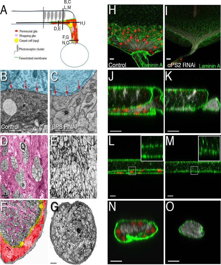
In the Drosophila peripheral nerves bPS (myspheroid—              Caspase-3 antibody at 1:200 (Cell Signaling). Appropriate Alexa
mys) integrin is broadly expressed in all glial cell types, while       Fluor-conjugated secondary antibodies were from Molecular Probes.
aPS2 integrin (inflated—if ) is expressed by the outermost              Images were obtained with the Leica SP5 confocal system and proc-
perineurial glia and aPS3 integrin (scab—scb) is expressed in           essed with Adobe Photoshop. Glial cell count was performed using
                                                                        Leica SP5 confocal stacks of 1 lM images covering the entire eye
the innermost wrapping glia (Xie and Auld, 2011). Using the
                                                                        imaginal disc. Cells were counted in Fiji using the maximum projec-
Drosophila eye imaginal disc we disclose different roles for
                                                                        tion image of each stack. Mean and standard deviation of the mean
different integrin alpha subunits in glia development. aPS2/
                                                                        were calculated for each case. Results from glial cell counting were
bPS integrin function in glial cells is essential for their migra-      analyzed by unpaired Student’s t test. Significant results were consid-
tion into the eye disc. The absence of glial cells in the eye           ered for P < 0.05.
disc caused by glial-specific knockdown of aPS2 or bPS did
not affect retinal axons entering the optic lobe. Surprisingly,         Transmission Electron Microscopy
we show that both aPS3 and Talin are nonautonomously                    Transmission electron microscopy (TEM) protocol was adapted from
required in glial cells for normal axonal targeting. Overall,           Pereanu et al. (2005). L3 larvae eye/antenna/brain tissues were fixed
our results suggest that glial expression of two different integ-       in 2% glutaraldehyde in PBS at 4 C for 2 h and post-fixated in 1%
rin heterodimers (PS2 and PS3) is important for glia migra-             osmium tetroxide in 0.15 M cacodylate buffer for 30 min (on ice).
tion and axonal targeting.                                              Specimens were washed several times in PBS and dehydrated by
                                                                        graded ethanol on ice, followed by immersion in propylene oxide for
                                                                        10 min. Samples were incubated overnight in a 1:1 mixture of Epon
Materials and Methods
                                                                        and propylene oxide and then 5 h in unpolymerized Epon. After
Fly Husbandry                                                           that, they were transferred to a mold, oriented and placed at 60 C
Most crosses were raised at 25 C under standard conditions. Excep-     for 48 h to permit Epon 812 polymerization. Blocks were then sec-
tions were the ones using the tub-Gal80ts (McGuire et al., 2003)        tioned (50–70 nm) and contrasted with uranyl acetate and lead
that were raised at 18 C for approximately 72 h and then moved to      citrate.
25 C. The following stocks (described in FlyBase, unless stated oth-          TEM images were obtained with a Jeol JEM-1400 with an
erwise) were used: If-GFP (DGRC #115467; Xie et al., 2014), repo-       Orius Sc1000 Digital Camera, acquired with GATAN software.
Gal4 (pan-glia; Xiong et al., 1994), moody-Gal4 (subperineurial glia;   Images were then exported to Photoshop CS2 for compilation and
Bainton et al., 2005), Mz97-Gal4, UAS Stinger (wrapping glia;           montage. Montage was done manually with panels of at least 6 3 5
Hummel et al., 2002), UAS-dicer-2 (Dietzl et al., 2007), UAS-lacZ,      images with an overlap of 15%.
UAS-CD8GFP (Lee and Luo, 1999), UAS-CD4tdTOM (Han et al.,
2011), UAS-aPS2 RNAi (JF02695, previously validated Liu et al.,
                                                                        Results
2013), UAS-bPS RNAi (VDRC KK100518; and GD15002, previ-
ously validated (Nishimura et al., 2014; Xie and Auld, 2011) and        Expression Patterns of a-Integrins in the Eye
TRIP JF02819), UAS-aPS3 RNAi (JF02696; and VDRC                         Imaginal Disc and Optic Stalk
KK106326, both previously validated by Vanderploeg et al., 2012),       Each integrin heterodimer is formed by one a and one b subu-
UAS-talin RNAi (HMS00799 and HMS00856), UAS-RpL7a RNAi                  nit (Hynes, 2002). Even though Drosophila melanogaster
(VDRC GD10934), UAS-eIF-4a RNAi (VDRC GD14111), UAS–                    encompasses five a and two b subunits, only the three hetero-
Ef1c RNAi (VDRC GD7427), UAS–aPS3EY10270, UAS–Egfrktop                  dimers with vertebrate orthologues (Takada et al., 2007) are
(Queenan et al., 1997), and repo4.3-Gal4 (Lee and Jones, 2005). For     commonly expressed: PS1 (aPS1/bPS), PS2 (aPS2/bPS), and
bPS, aPS3, and Talin the lines used were KK100518, JF02696, and         PS3 (aPS3/bPS). In the eye disc, aPS1 and aPS2 have been
HMS00799, respectively, except where otherwise stated. The effi-        proposed to have almost complementary expression patterns
ciency of aPS2, aPS3, and bPS knockdown was verified by immu-           (by in situ hybridization), with aPS1 being expressed in the
nofluorescence staining.                                                anterior region and aPS2 more posteriorly (Brower et al.,
                                                                        1984). Initially, we studied the pattern of aPS2 and aPS3 pro-
Immunohistochemistry                                                    tein expression in the eye disc and in the optic stalk (the
Eye-antennal imaginal discs were prepared for immunohistochemis-        regions analyzed are shown in Fig. 1A,B). For aPS2 expression
try using standard protocols. Primary antibodies used were: mouse
                                                                        we used a GFP trap inserted in the if gene (encoding aPS2-
anti-Repo antibody at 1:10 (8D12 anti-Repo, Developmental Stud-
                                                                        GFP) and an anti-aPS2 antibody. The expression patterns
ies Hybridoma Bank, DSHB; Alfonso and Jones, 2002), mouse anti-
bPS (CF.6G11, DSHB; Brower et al., 1984), mouse anti-aPS2 anti-
                                                                        obtained with both approaches were very similar (Fig. 1C–F
body at 1:5 (CF.2C7, DSHB; Brower et al., 1984), rabbit anti-aPS3       and Supp. Info. Fig. 1A). To visualize glial cell membranes, we
antibody at 1:300 (kind gift from Dr. Shigeo Hayashi; Wada et al.,      expressed a membrane-localized tdTomato (UAS-CD4tdTOM;
2007), goat anti-HRP antibody Cy5 conjugated at 1:100 (Jackson          Han et al., 2011) under the control of the glial-specific repo-
ImmunoResearch), rabbit anti-Laminin A antibody at 1:200 (kind          Gal4 driver (Campbell et al., 1994; Halter et al., 1995; Xiong
gift from Herwig O. Gutzeit; Gutzeit et al., 1991), and anti-cleaved    et al., 1994). aPS2 integrin was detected both in the cell bodies

2                                                                                                                          Volume 00, No. 00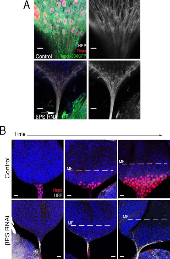
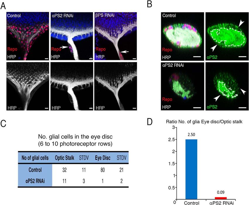
Tavares et al.: Integrin Dual Role in Drosophila Retinal Glia

FIGURE 1: aPS2 and aPS3 integrins are expressed in retinal glial cells. (A) Schematic representation of the Drosophila eye imaginal disc and optic
lobe indicating the different sections analyzed. (B) Electron microscopy analysis of a transversal cut from a control (repo>Dcr-2>lacZ) eye imaginal
disc. The R-cells on the apical side (blue) send their R-axons (gray) into the basal side where they contact glial cells. Wrapping glia (lilac) enwraps
each R1-R8 photoreceptor clusters laying apical to subperineurial glia (spg; yellow). Perineurial glial cells (red) form a layer basal to the spg. The
laminin-rich fenestrated membrane (FM; green) separates the R-axons soma from the glia region is magnified in the inset without false colouring.
Axons crossing the FM are highlighted (dashed box). (C, E, and F) aPS2-GFP;repo>CD4tdTOM was used to analyze aPS2 expression and glia mem-
branes. aPS2 is co-expressed with CD4tdTOM in perineurial glia (outermost glial cell layer). (D) aPS2-GFP stained with Repo and HRP. Square box
represents the region magnified in (D0 ) and (D00 ). aPS2 expression is visible underneath the axons and perineurial glia nuclei layer (in red). Dashed
line in (E) delimitates the axon region. (F) Confocal microscopy analysis of a region similar to the one in the dashed box in (B). Glial cell projections
into R-axon cell bodies were observed (dashed box). The inset shows close association between glial projections (CD4tdTOM) and axons (HRP).
CD4tdTOM and aPS2-GFP overlap in the wrapping glia/axonal layer. (G and H) repo>Dcr-2>lacZ (Control) eye imaginal discs stained with aPS3
antibody (green). Higher aPS3 expression is visible in the perineurial to subperineurial glia region (arrows) but it is also expressed in the perineurial
glia (arrowheads). (I and J) moody>CD8GFP>lacZ staining with aPS3 shows higher expression of aPS3 (J0 ) at subperineurial membranes (overlap
with CD8GFP, J00 ). HRP shows the photoreceptors in gray and DAPI stains DNA in blue. Scale bars correspond to 10 lm. [Color figure can be
viewed in the online issue, which is available at wileyonlinelibrary.com.]

Month 2015                                                                                                                                             3
and axons of photoreceptors and in the basal glial cell layers      cells (Supp. Info. Fig. 2B–E) further supporting a role for
(Fig. 1C–F). Our analysis showed that perineurial glia express      aPS2 in retinal glial cell migration.
aPS2 integrin both in the optic stalk (Fig. 1E) and upon enter-            To identify the b-subunit partner for aPS2 integrin in
ing the eye disc (Fig. 1C,D). aPS2 integrin immunolabeling          perineurial glia, we knocked down the expression of integrins
was found in the outermost membrane of perineurial glia             bm and bPS by glial-targeted RNAi. In agreement with the
where it contacts ECM (Fig. 1D–F). Both in the eye disc and         proposed lack of significant expression of bm outside the
in the optic stalk we observed high aPS2 levels in the region       midgut (Yee and Hynes, 1993), we failed to detect any retinal
where a high density of glial processes wraps R-axons bundles       glial defects when RNAi for bm was induced. In contrast,
(Fig. 1C–F). Interestingly, we detected glial cells projections     knocking down bPS phenocopied aPS2 interference, impair-
that were oriented apically into photoreceptors cell bodies         ing perineurial glia migration into the eye disc (Fig. 2A;
closely following R-axons (Fig. 1F). aPS2 expression is             Supp. Info. Fig. 2A). We observed that both aPS2 and bPS
observed in these projections (Fig. 1F) that belong to wrapping     are essential in Drosophila as their depletion in glial cells
glia, as they can be detected when glial cell membranes are         caused late L3 to pupa lethality (not shown). These results
labelled using the wrapping glia specific driver Mz97-Gal4          show that the heterodimer aPS2/bPS (PS2) is important for
(Supp. Info. Fig. 1B; Bainton et al., 2005; Poeck et al., 2001).    perineurial glial cell migration from the brain toward the eye
      To study aPS3 expression (Fig. 1G–J) we used a vali-          disc, suggesting a role in haptotactic and/or chemotactic cell
dated anti-aPS3 antibody (Wada et al., 2007; Xie and Auld,          migration.
2011; Xie et al., 2014; and Supp. Info. Fig. 1C–H). The                    Surprisingly, in both glia-specific knockdowns of either
strongest aPS3 expression is observed internally to the peri-       aPS2 or bPS no defects were observed in R-axons targeting
neurial glia layer, overlapping with subperineurial glial cell      to the OS in eye discs without any glia (Fig. 2A). Labelling
membranes both in the eye disc (Fig. 1I) and optic stalk (Fig.      of the glial cell membranes (with CD8GFP) in bPS RNAi
1G–J). This is visible by co-staining with the membrane             confirmed that both glial cell bodies and glia membrane pro-
marker CD8GFP expressed by the subperineurial-specific              jections were absent from the eye imaginal disc (Fig. 3A).
driver moody-Gal4 (Bainton et al., 2005; Fig. 1I,J). However,       Furthermore, when we depleted several essential proteins,
lower expression levels can also be observed in perineurial glia    such as Ef1g (translation elongation factor), RpL7a (ribo-
(Fig. 1H). The distinct expression patterns for aPS2 and            somal protein), and eIF-4a (translation initiation factor), we
aPS3 integrins suggest that they could play different roles in      also observed that axons target the optic stalk in eye discs
retinal glial cells.                                                where glia was absent (Supp. Info. Fig. 3B,C). These observa-
                                                                    tions indicate that glial cells are not essential for axonal tar-
                                                                    geting to the OS.
PS2 Integrin Is Essential for Glia Migration into the                      Because integrins are important for cell survival (Ben-
Eye Disc                                                            ninger et al., 2006; Colognato et al., 2002; Leone et al.,
To investigate the specific function of aPS2 in retinal glia we     2005) we wanted to exclude the possibility of glial cells hav-
knocked down aPS2 levels, by glia-specific expression of a          ing migrated into the eye disc and signalled the R-axons to
double-stranded RNA transgene (Liu et al., 2013) together           enter the OS before dying. For that, we analyzed earlier
with Dicer-2 (Dcr-2) to enhance RNA interference (Dietzl            developmental stages (L2 to L3) in aPS2 and bPS RNAi
et al., 2007; Fig. 2A–D). In the optic stalk, a strong reduc-       discs and we were also not able to observe any early glia
tion of aPS2 expression was observed in perineurial glial cells     migration at these stages (Fig. 3B and not shown). Addition-
peripheric to R-axons (Fig. 2B). Knockdown of aPS2 caused           ally, glial cell death was not detected in the eye discs or optic
a striking decrease in the number of glial cells in eye discs       stalks of wandering L3 larvae in these genotypes (Supp. Info.
(Fig. 2A). In the optic stalk the number of glial cells is also     Fig. 3A and not shown). Blocking cell death by expressing
reduced and very few glia migrate from the OS into the eye          baculovirus p35 together with bPS RNAi in glial cells also
disc (Fig. 2C). The observed decrease in the ratio of glial cells   did not rescue perineurial glia entry into the eye disc (data
between the eye disc and the optic stalk in the aPS2 RNAi           not shown).
suggested a role for aPS2 in glia migration (Fig. 2D). To
evaluate this role of aPS2 we expressed the activated Epider-       Eye/Optic Stalk Structure in the Absence of Glia
mal growth factor receptor (Egfrktop) in glia and, as expected,     Our data indicate that photoreceptors differentiate and pro-
we observed an increase in glial cell numbers in the eye disc       ject axons to the optic stalk even when glia are absent due to
(Supp. Info. Fig. 2B,C; Read et al., 2013; Reddy and Irvine,        aPS2/bPS integrin knockdown. Thus, this genotype allowed
2013; Witte et al., 2009). Importantly, co-expression of aPS2       for a detailed analysis of the role of glia in R-axon projec-
RNAi decreased the migration rate of Egfrktop expressing glial      tions, axonal fascicle organization, and ECM deposition and

4                                                                                                                  Volume 00, No. 00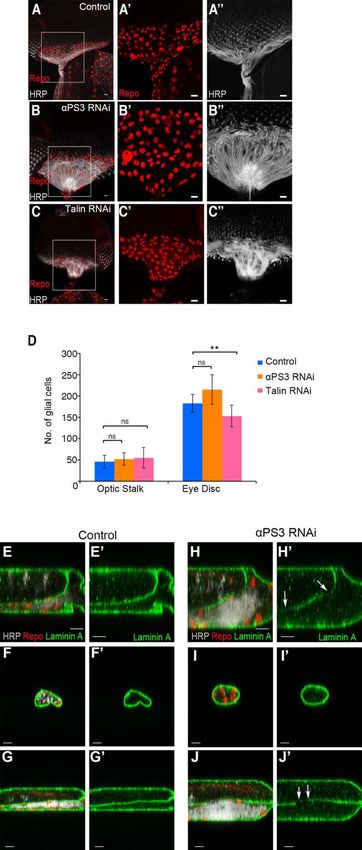
Tavares et al.: Integrin Dual Role in Drosophila Retinal Glia
FIGURE 2: aPS2 and bPS are required for glia migration into the eye imaginal disc but not for axonal pathfinding to the OS. (A) Exam-
ples of repo>Dcr-2>aPS2 RNAi and repo>Dcr-2>bPS RNAi eye imaginal discs where no glial cells were observed. Despite the absence
of glia, no difference in axon targeting to the OS was observed. repo>Dcr-2>lacZ represents the control. Arrows point to the limit of
glia migration. (B) Transverse view of the optic stalk of the control (repo>Dcr-2>lacZ) and aPS2 RNAi (repo>Dcr-2>aPS2 RNAi). aPS2
expression (green) in the perineurial glial, the outermost layer of the OS, was strongly reduced in the aPS2 RNAi demonstrating the
RNAi efficiency. Arrowheads point to the OS limits. aPS2 expression was detected by anti-aPS2 antibody (in green). Dashed line delimi-
tates the axon region. Repo shows glia in red, HRP shows the photoreceptors in gray and DAPI stains DNA in blue. Scale bars corre-
spond to 10 lm. (C) Table with the number of glial cells in the optic stalk and eye disc of control (repo>Dcr-2>lacZ) and aPS2 RNAi
(repo>Dcr-2>aPS2 RNAi). All the eye discs counted were age matched with 6–10 photoreceptor rows. N 5 10 (control OS), 12 (control
ED), 8 (aPS2 RNAi OS), and 19 (aPS2 RNAi ED). (D) Glia cell numbers (listed in panel C) were used to calculate the ratio between the
number of glial cells in the eye disc and optic stalk in the control (repo>Dcr-2>lacZ) and aPS2 RNAi (repo>Dcr-2>aPS2 RNAi) of age
matched discs (6–10 photoreceptor rows). [Color figure can be viewed in the online issue, which is available at wileyonlinelibrary.com.]
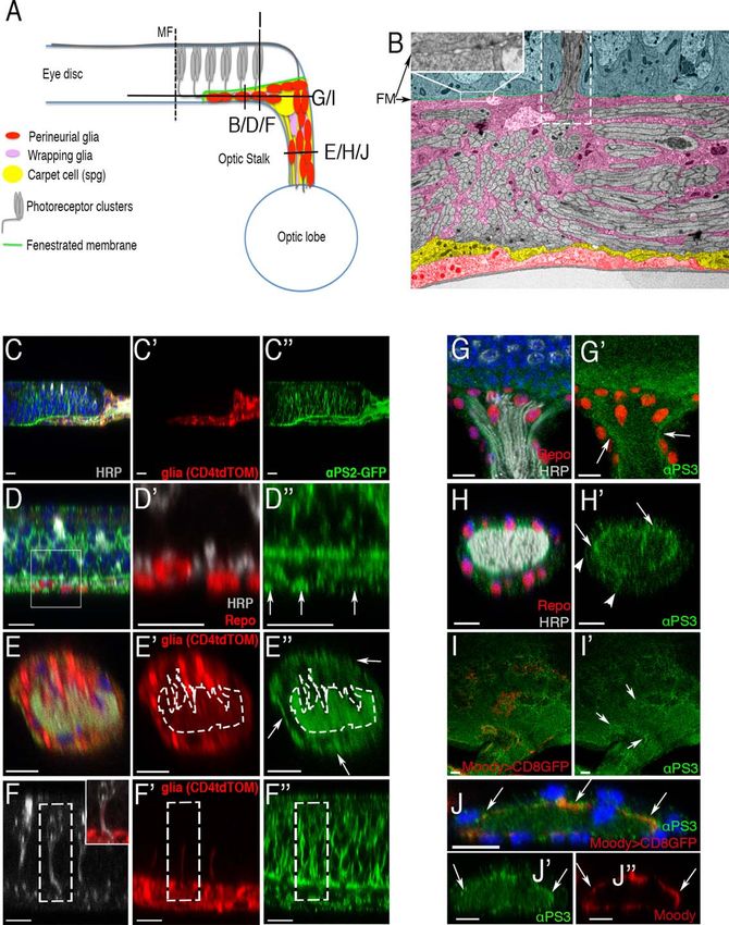
arrangement. Figure 4A shows a schematic representation of et al., 2007). In the bPS RNAi eye discs in which glial cells
the eye disc and indicates the approximate positions of the were absent, the photoreceptor axon bundles were not
tissue sections that were analyzed by transmission electron wrapped and exhibited a disorganized pattern, both in the
microscopy (TEM; Fig. 4B–G) and confocal analysis (Fig. disc proper (Fig. 4E) and in the OS (Fig. 4G). No fascicles
4H–O). could be distinguished, which supports a role for retinal glial
Our observation that wrapping glial cells project apically cells in axon fasciculation (Silies et al., 2007).
along R-axons toward the photoreceptors cell bodies (Fig. 1F) Basal to the photoreceptors soma layer lies the fenes-
suggested a possible role for glial cells in the axon guidance trated membrane (Figs. 1B and 4B,J,L; Supp. Info. Fig. 4A),
in the eye disc. However, in the absence of glial cells, R- formed by Laminin A (LanA; Fig. 4J,L) together with other
axons were still able to project basally in the eye disc and proteins (Cagan and Ready, 1989). LanA was also detected
redirect into the optic stalk, as observed in the control discs basal to the perineurial glia in the collagen-rich basement
(Supp. Info. Fig. 4A; Silies et al., 2007). In the basal axonal- membrane of the eye disc (Fig. 4H,J,L; Supp. Info. Fig. 4B).
glia layer and OS of control discs, wrapping glia ensheath the Upon glia-specific reduction of aPS2 and bPS expression the
8 R-axons from each photoreceptor cluster (Fig. 4D,F; Silies levels of LanA in the fenestrated membrane were strongly
Month 2015 5FIGURE 3: Axon targeting to the optic stalk is independent of glia. (A) repo 4.3>CD8GFP>lacZ (Control) and repo 4.3>CD8GFP>bPS RNAi are shown. Glia membranes (CD8GFP) were absent from the eye imaginal disc in the bPS RNAi. Arrow points to the edge of glia migration in the OS. (B) Late L2 (left panel), early L3 (middle panel) and middle L3 (right panel) are shown for repo>Dcr-2>lacZ (control) and repo>Dcr-2>bPS RNAi. Knocking down bPS blocks glia migration since early larval development. The morphogenetic furrow (MF) represents the edge of differentiation (dashed lines). Repo shows glia in red, HRP shows the photoreceptors in gray and DAPI stains DNA in blue. Scale bars correspond to 10 lm. [Color figure can be viewed in the online issue, which is available at wileyonlinelibrary.com.] reduced (Fig. 4K,M; and not shown). In fact, a detailed as previously shown in other contexts (Narasimha and Brown, TEM and confocal analysis showed that the fenestrated mem- 2004; Tanentzapf et al., 2007; Urbano et al., 2011). The brane itself was severely affected and almost residual (Fig. analysis of the OS of aPS2 and bPS knockdown discs that 4B,C,J–M; Supp. Info. Fig. 4A), whereas the basement mem- lack glial cells indicated that the wrapping processes that nor- brane was present (Supp. Info. Fig. 4B). This suggests that mally ensheath R-axons (Silies et al., 2007) were absent (Fig. glial cells are important for the dynamic reassembly and/or 4F,G,N,O). These data show that glial cells are important for accumulation of ECM components in the developing retina, the normal structure of the eye disc, namely for R-axon 6 Volume 00, No. 00
Tavares et al.: Integrin Dual Role in Drosophila Retinal Glia

FIGURE 4: Ultrastructure analysis of glia-specific PS2 depleted eye imaginal disc. (A) Representation of the views analyzed in panels (B)–(O).
(B–G) show TEM ultrastructure analysis of a bPS RNAi (repo>Dcr-2>bPS RNAi) and Control (repo>Dcr-2>lacZ) L3 eye imaginal disc. Red
arrows point toward the fenestrated membrane in the control and where the fenestrated membrane should be present in the bPS RNAi. Pho-
toreceptors soma is coloured in blue, wrapping glia in lilac, subperineurial glia in yellow and perineurial glia in red. (B,C,D,F,G) Scale—1 lm
and (E) Scale—2 lm. (H-O) Confocal analysis of repo>Dcr-2>lacZ (control, left panel) and repo>Dcr-2>aPS2 RNAi (right panel). White boxes
in insets show magnifications of the represented areas where a depletion/absence of the fenestrated membrane was detected in aPS2 RNAi.
Scale bars correspond to 10 lm. Laminin A staining is shown in green, Repo (glial cells) is shown in red, and HRP (photoreceptors) in gray.
[Color figure can be viewed in the online issue, which is available at wileyonlinelibrary.com.]

organization and wrapping, and for the formation/mainte-                  confirmed the results obtained with the membrane marker
nance of the fenestrated membrane. However, the absence of                CD8GFP (Fig. 3A).
glial cells did not compromise the formation of the neural
lamella, the outermost ECM (Fig. 4F,G,N,O). In spite of the               aPS3 and Talin Are Required in Retinal Glia for
absence of glial cells, no detachment of the axon layer from              Axon Targeting to the Optic Stalk
the neural lamella was observed (Fig. 4F,G,N,O). Addition-                We observed that unlike aPS2 RNAi, glia-specific RNAi downreg-
ally, TEM analysis of bPS RNAi eye imaginal discs did not                 ulation of aPS3, enhanced by Dicer-2 co-expression, did not have
detect any glial cell bodies or their membranes, which further            an impact on glial cell migration (Fig. 5A,B,D). Strikingly,

Month 2015                                                                                                                                   7
knockdown of aPS3 in glial cells caused a stalling of R-axons in the      induced a block in axonal targeting of photoreceptors to the optic
optic stalk, in a nonautonomous manner (Fig. 5A,B). It has previ-         stalk (Fig. 5A,C). Talin RNAi also caused a small decrease in glial
ously been reported that Talin (also known as rhea) is necessary to       cell migration (Fig. 5D), as previously described (Xie et al., 2014).
regulate integrin functions in Drosophila and mammals (Brown              The phenotype specificity for aPS3 and Talin RNAis was con-
et al., 2002; Helsten et al., 2008; Moser et al., 2009). Interestingly,   firmed using other independent RNAi lines, which also induced
in a similar manner to aPS3 RNAi, Talin knockdown in glial cells          axon blocking when expressed in glial cells (Supp. Info. Fig. 5A–C).
                                                                          We also observed axonal stalling when a weaker bPS RNAi line,
                                                                          which does not completely block glia migration, was used to drive
                                                                          glia-specific knockdown (not shown). Furthermore, aPS3 knock-
                                                                          down did not cause significant changes in the expression of both
                                                                          bPS and aPS2 (Supp. Info. Fig. 5D–G). As shown in Fig. 5A–C,
                                                                          R-axons in both aPS3 and Talin knockdown eye discs do not have
                                                                          a deficit in growth cone extension. In fact, stalled R-axons continue
                                                                          to grow forming a high-density ("plug-like") accumulation at the
                                                                          posterior region of the eye disc, exactly where axons should enter
                                                                          the optic stalk. To analyze if aPS3 depleted glial cells were efficient
                                                                          in the formation/rearrangement of ECM proteins to form the fenes-
                                                                          trated membrane we analyzed LanA expression (Fig. 5E–J). LanA
                                                                          levels were slightly decreased in the region where the R-axons accu-
                                                                          mulate but otherwise its expression pattern remains unchanged.
                                                                          Furthermore, we could not detect changes in the expression pattern
                                                                          of Wing blister (Wb), the second Laminin a protein in Drosophila
                                                                          (our own unpublished data).
                                                                                 Taken together, our results show that aPS3 and Talin
                                                                          depletion in glial cells induces axon stalling.

                                                                          Discussion
                                                                          The role of integrins in CNS and PNS glia has been vastly
                                                                          studied but despite its undoubtful importance, the functions
                                                                          and the in vivo mechanisms of action for the different integ-
                                                                          rin heterodimers are not well understood (Baron et al.,
                                                                          2005). The large number of a-subunits in mammals and its
                                                                          high redundancy makes their study even more difficult. The
                                                                          use of simpler model organisms helps to overcome those

                                                                          FIGURE 5: Glia-specific knockdown of aPS3 and Talin induce
                                                                          nonautonomous defects in axon targeting to the optic stalk. (A)-(C)
                                                                          Examples of repo>Dcr-2>aPS3 RNAi and repo>Dcr-2>Talin RNAi
                                                                          eye imaginal discs showing axonal stalling in the eye imaginal disc.
                                                                          repo>Dcr-2>lacZ was used as a control. White squares delimitate
                                                                          the magnified regions showed in (A0 —C0 ; Repo) and (A00 —C00 ; HRP).
                                                                          (D) Graph showing the number of glial cells in the optic stalk and eye
                                                                          disc of control (repo>Dcr-2>lacZ), aPS3 RNAi (repo>Dcr-2>aPS3
                                                                          RNAi) and Talin RNAi (repo>Dcr-2>Talin RNAi). N 5 7 (for control
                                                                          and aPS3 RNAi optic stalk), N 5 10 (for control, aPS3 RNAi eye disc
                                                                          and Talin RNAi), and N 5 11 (for Talin RNAi eye disc) of 16–20
                                                                          photoreceptor rows eye discs. Glial cell numbers were significantly
                                                                          different between the control and Talin RNAi eye discs (t test;
                                                                          P < 0.05). (E)–(J) Confocal microscopy images from repo>Dcr-2>lacZ
                                                                          (control, E–G) and repo>Dcr-2>aPS3 RNAi (H–J) showing ECM
                                                                          staining by Laminin A (green). Fenestrated membrane is slightly thin-
                                                                          ner in the region where axons stall in aPS3 RNAi eye discs (arrows).
                                                                          Glial cells were stained by Repo (red), and photoreceptors by HRP
                                                                          (gray). Scale bars correspond to 10 lm. [Color figure can be viewed
                                                                          in the online issue, which is available at wileyonlinelibrary.com.]

8                                                                                                                            Volume 00, No. 00
Tavares et al.: Integrin Dual Role in Drosophila Retinal Glia
issues and provides an in vivo system to address many of the later optic lobe axonal targeting (Tayler and Garrity, 2003). Our
unanswered questions. Recently, bPS was shown to be results contrast with other published genotypes where retinal glia
required in Drosophila retinal glia for migration and axonal was proposed to have axonal guidepost function to the optic
targeting (Xie et al., 2014). Our results confirm this key role stalk (Hummel et al., 2002; Rangarajan et al., 1999). Further-
for bPS, and show that bPS functions can be attributed to more, in the Drosophila central nervous system, some reports
distinct roles of aPS2/bPS and aPS3/bPS heterodimers in have presented evidence that glial cells control axonal fascicula-
photoreceptors–glia interactions. tion (Hidalgo, 2003; Sepp et al., 2001) and axon migration
aPS2, the Drosophila orthologue of a5, am and a8 (Takada (Klambt et al., 1991; Pielage and Klambt, 2001), whereas studies
et al., 2007), is required for glial cell migration (together with bPS) with gcm mutants or glial cell ablation have shown only minor
as described for its vertebrate counterpart amb1 in oligodendrocyte axonal defects despite the absence of glial cells (Hosoya et al.,
(Baron et al., 2005) and Schwann cell development (Milner et al., 1995; Jones et al., 1995; Vincent et al., 1996; Whitington et al.,
2001). We show that in the context of aPS2/bPS functions in reti- 2004). In other organisms, the role of glial cells as guideposts is
nal glial development, this heterodimer has a specific role in cell also not clear. For example, in zebrafish sox10/colorless mutant
migration from the optic stalk into the eye disc. In the peripheral embryos, in which glial cells fail to be specified, lateral line axons
nerves, knockdown of the heterodimer aPS2/bPS was previously still project along correct pathways (Gilmour et al., 2002). In
shown to cause only minor defects in perineurial glia wrapping, pos- mammals, neuronal precursors born in the subventricular zone
sibly due to functional redundancy with other aPS subunits (Xie of the neonatal and adult rodent brain can migrate without the
and Auld, 2011). Results presented here establish for the first time guidance of radial glia or axonal processes (Wichterle et al.,
an essential function for aPS2/bPS integrin in the developing visual 1997). Overall, these results reveal context-dependent glial func-
system making the eye imaginal disc an important system to further tions in axonal targeting that require further analysis.
understand the in vivo role of integrins in glia migration. In Dro- Both Drosophila (Xie and Auld, 2011; Xie et al., 2014) and
sophila hemocytes (equivalent to mammalian macrophages), aPS2 mammalian (Milner and Ffrench-Constant, 1994; Milner et al.,
integrin was shown to be necessary for migration into the tail of the 1997a, 1997b) glial cells can express more than one integrin het-
embryo, acting downstream of Rap1 and dizzy (a G-nucleotide erodimer at the same time pointing for different cellular func-
exchange factor for Rap1; Comber et al., 2013; Huelsmann et al., tions for different integrin heterodimers (Milner et al., 1999,
2006; Siekhaus et al., 2010). The integrin activators Talin (Dro- 1997b). This is in line with our observation that reduced levels
sophila rhea) and Kindlin 1 (Drosophila Fermitin 1) were also of aPS3 do not affect glia migration, as cell numbers in the eye
described as important for hemocyte migration (Moreira et al., disc remain similar, but affect R-axons by inhibiting their migra-
2013). However, other signalling regulators acting downstream of tion to the optic stalk leading to axonal misrouting. This pheno-
integrin activation, such as Focal adhesion kinase (FAK) and Vincu- type might be due to a direct effect on R-cells or R-axons or
lin, were shown not to have significant roles in hemocyte migration through alterations in the cell–ECM interactions that interferes
(Moreira et al., 2013) or retinal perineurial glia migration (Mura- with axonal migration. The latter mechanism has been suggested
kami et al., 2007). Recently, a dynamic role of aPS2 integrin in the for the role of bantam in epithelium rearrangement through the
remodelling of the ECM has been proposed to be important for the modulation of integrin–ECM interactions (Jiang et al., 2014).
correct migration of caudal visceral mesoderm cells (Urbano et al., No significant differences in Laminin protein levels were
2011). Furthermore, in the ventral nerve cord, aPS2 is required for observed in the aPS3 RNAi (Fig. 5E–J and data not shown for
the attachment of perineurial glial cells to the ECM and for the Wb). This might suggest that knockdown of aPS3 in glia indu-
maintenance of the structure of the neural lamella (Meyer et al., ces alterations in cell–cell interactions inhibiting axons to migrate
2014). Interestingly, we find that in retinal perineurial glia, aPS2 into the optic stalk. Axon pathfinding errors might be due to
integrin is also important for the dynamic control of laminin-rich changes on axonal acetylcholine nonvesicular release that
ECM deposition and/or rearrangement in the fenestrated mem- interferes with axon targeting and has been shown to be affected
brane but not in the neural lamella (Fig. 4). Thus, given the com- nonautonomously (Yang and Kunes, 2004). It has also been
plexity of integrin functions and their associated downstream reported that bantam expression in the optic lobe can mistarget
signalling events, genetic dissection of the roles of aPS2/bPS integ- axons (Li and Padgett, 2012). A nonautonomous role of glia in
rin in retinal glia will allow the identification of functionally relevant axon targeting is also known to exist in mammals but it is not
effectors. clearly understood (Beirowski, 2013). The function of aPS3 can
In this study we show in several ways (Figs. 2–4, and Supp. be envisaged in light of their mammalian counterparts a4 and
Info. Figs. 2 and 3) that in some genotypes (including aPS2 and a9 (Takada et al., 2007). Both this integrins are important for
bPS depletion in glia) the initial axonal targeting to the optic fibronectin binding but also for cell–cell contact through binding
stalk can occur in the absence of retinal glial from the eye disc to ADAM, Disintegrin and VEGF family proteins, between
proper, despite the importance of glia for axon development and others (Hoye et al., 2012). This, together with the enrichment of
Month 2015 9aPS3 in the subperineurial layer, might indicate that this integ- Comber K, Huelsmann S, Evans I, Sanchez-Sanchez BJ, Chalmers A, Reuter R,
Wood W, Martin-Bermudo MD. 2013. A dual role for the betaPS integrin
rin is important for cell–cell contacts that when lost could lead myospheroid in mediating Drosophila embryonic macrophage migration.
to axon misrouting. J Cell Sci 126:3475–3484.
Dietzl G, Chen D, Schnorrer F, Su KC, Barinova Y, Fellner M, Gasser B, Kinsey
K, Oppel S, Scheiblauer S and others. 2007. A genome-wide transgenic RNAi
Acknowledgment library for conditional gene inactivation in Drosophila. Nature 448:151–156.
Grant sponsor: FEDER funds through the Operational Com- Dominguez M, Casares F. 2005. Organ specification-growth control connection:
new in-sights from the Drosophila eye-antennal disc. Dev Dyn 232:673–684.
petitiveness Programme—COMPETE and National Funds
through FCT—Fundaç~ao para a Ci^encia e a Tecnologia; Gilmour DT, Maischein HM, Nusslein-Volhard C. 2002. Migration and function of
a glial subtype in the vertebrate peripheral nervous system. Neuron 34:577–588.
Grant number: FCOMP-01-0124-FEDER-029870 (EXPL/
Gutzeit HO, Eberhardt W, Gratwohl E. 1991. Laminin and basement
NEU-NMC/0380/2012), FCOMP-01-0124-FEDER-015675 membrane-associated microfilaments in wild-type and mutant Drosophila
(PTDC/SAU-BID/112250/2009), and FCOMP-01-0124- ovarian follicles. J Cell Sci 100:781–788.
FEDER-021333 (PTDC/SAU-NMC/119937/2010); Grant Halter DA, Urban J, Rickert C, Ner SS, Ito K, Travers AA, Technau GM. 1995.
sponsor: EMBO Intra European Fellowship; Grant number: The homeobox gene repo is required for the differentiation and maintenance
of glia function in the embryonic nervous system of Drosophila melanogaster.
ALTF 677-2012 (to LT); Grant sponsor: FCT Postdoc Fel- Development 121:317–332.
lowship; Grant number: SFRH/BPD/95336/2013 (to LT). Han C, Jan LY, Jan YN. 2011. Enhancer-driven membrane markers for analy-
The authors thank Christian Kl€ambt, Herwig O. Gutzeit, Dr. sis of nonautonomous mechanisms reveal neuron–glia interactions in Dro-
Shigeo Hayashi, the Bloomington Drosophila Stock Center, the sophila. Proc Natl Acad Sci USA 108:9673–9678.
Vienna Drosophila RNAi Center, the Drosophila Genetic Helsten TL, Bunch TA, Kato H, Yamanouchi J, Choi SH, Jannuzi AL, Feral CC,
Ginsberg MH, Brower DL, Shattil SJ. 2008. Differences in regulation of Dro-
Resource Center, and the Developmental Studies Hybridoma sophila and vertebrate integrin affinity by talin. Mol Biol Cell 19:3589–3598.
Bank for reagents; Paula Sampaio (ALMF, IBMC) and Rui Fer-
Hidalgo A. 2003. Neuron–glia interactions during axon guidance in Drosoph-
nandes (HEMS, IBMC) for technical assistance. ila. Biochem Soc Trans 31:50–55.
Hosoya T, Takizawa K, Nitta K, Hotta Y. 1995. glial cells missing: a binary
References switch between neuronal and glial determination in Drosophila. Cell 82:
1025–1036.
Alfonso TB, Jones BW. 2002. gcm2 promotes glial cell differentiation and is
required with glial cells missing for macrophage development in Drosophila. Hoye AM, Couchman JR, Wewer UM, Fukami K, Yoneda A. 2012. The new-
Dev Biol 248:369–383. comer in the integrin family: integrin alpha9 in biology and cancer. Adv Biol
Bainton RJ, Tsai LT, Schwabe T, DeSalvo M, Gaul U, Heberlein U. 2005. Regul 52:326–339.
moody encodes two GPCRs that regulate cocaine behaviors and blood-brain Huelsmann S, Hepper C, Marchese D, Knoll C, Reuter R. 2006. The PDZ-GEF
barrier permeability in Drosophila. Cell 123:145–156. dizzy regulates cell shape of migrating macrophages via Rap1 and integrins
Baron W, Colognato H, ffrench-Constant C. 2005. Integrin-growth factor in the Drosophila embryo. Development 133:2915–2924.
interactions as regulators of oligodendroglial development and function. Glia Hummel T, Attix S, Gunning D, Zipursky SL. 2002. Temporal control of glial
49:467–479. cell migration in the Drosophila eye requires gilgamesh, hedgehog, and eye
Beirowski B. 2013. Concepts for regulation of axon integrity by enwrapping specification genes. Neuron 33:193–203.
glia. Front Cell Neurosci 7:256.
Hynes RO. 2002. Integrins: bidirectional, allosteric signaling machines. Cell
Benninger Y, Colognato H, Thurnherr T, Franklin RJ, Leone DP, Atanasoski S, 110:673–687.
Nave KA, Ffrench-Constant C, Suter U, Relvas JB. 2006. Beta1-integrin signal-
Jacques TS, Relvas JB, Nishimura S, Pytela R, Edwards GM, Streuli CH,
ing mediates premyelinating oligodendrocyte survival but is not required for
ffrench-Constant C. 1998. Neural precursor cell chain migration and division
CNS myelination and remyelination. J Neurosci 26:7665–7673.
are regulated through different beta1 integrins. Development 125:3167–3177.
Brower DL, Wilcox M, Piovant M, Smith RJ, Reger LA. 1984. Related cell-
Jarman AP, Grell EH, Ackerman L, Jan LY, Jan YN. 1994. Atonal is the pro-
surface antigens expressed with positional specificity in Drosophila imaginal
neural gene for Drosophila photoreceptors. Nature 369:398–400.
discs. Proc Natl Acad Sci USA 81:7485–7489.
Brown NH, Gregory SL, Martin-Bermudo MD. 2000. Integrins as mediators of Jiang N, Soba P, Parker E, Kim CC, Parrish JZ. 2014. The microRNA bantam
morphogenesis in Drosophila. Dev Biol 223:1–16. regulates a developmental transition in epithelial cells that restricts sensory
dendrite growth. Development 141:2657–2668.
Brown NH, Gregory SL, Rickoll WL, Fessler LI, Prout M, White RA, Fristrom JW.
2002. Talin is essential for integrin function in Drosophila. Dev Cell 3:569–579. Jones BW, Fetter RD, Tear G, Goodman CS. 1995. glial cells missing: a
genetic switch that controls glial versus neuronal fate. Cell 82:1013–1023.
Cagan RL, Ready DF. 1989. Notch is required for successive cell decisions in
the developing Drosophila retina. Genes Dev 3:1099–1112. Klambt C, Jacobs JR, Goodman CS. 1991. The midline of the Drosophila
central nervous system: a model for the genetic analysis of cell fate, cell
Campbell G, Goring H, Lin T, Spana E, Andersson S, Doe CQ, Tomlinson A. migration, and growth cone guidance. Cell 64:801–815.
1994. RK2, a glial-specific homeodomain protein required for embryonic nerve
cord condensation and viability in Drosophila. Development 120:2957–2966. Lee BP, Jones BW. 2005. Transcriptional regulation of the Drosophila glial
gene repo. Mech Dev 122:849–862.
Chanut F, Heberlein U. 1997. Role of decapentaplegic in initiation and pro-
gression of the morphogenetic furrow in the developing Drosophila retina. Lee T, Luo L. 1999. Mosaic analysis with a repressible cell marker for studies
Development 124:559–567. of gene function in neuronal morphogenesis. Neuron 22:451–461.
Colognato H, Baron W, Avellana-Adalid V, Relvas JB, Baron-Van Evercooren A, Leone DP, Relvas JB, Campos LS, Hemmi S, Brakebusch C, Fassler R,
Georges-Labouesse E, ffrench-Constant C. 2002. CNS integrins switch growth Ffrench-Constant C, Suter U. 2005. Regulation of neural progenitor prolifera-
factor signalling to promote target-dependent survival. Nat Cell Biol 4:833–841. tion and survival by beta1 integrins. J Cell Sci 118:2589–2599.
10 Volume 00, No. 00Tavares et al.: Integrin Dual Role in Drosophila Retinal Glia
Li Y, Padgett RW. 2012. bantam is required for optic lobe development and cell proliferation and survival through TORC2-Akt signaling in glioblastoma.
glial cell proliferation. PLoS One 7:e32910. PLoS Genet 9:e1003253.
Liu ZC, Odell N, Geisbrecht ER. 2013. Drosophila importin-7 functions Reddy BV, Irvine KD. 2013. Regulation of Hippo signaling by EGFR-MAPK
upstream of the Elmo signaling module to mediate the formation and stabil- signaling through Ajuba family proteins. Dev Cell 24:459–471.
ity of muscle attachments. J Cell Sci 126:5210–5223.
Relvas JB, Setzu A, Baron W, Buttery PC, LaFlamme SE, Franklin RJ, ffrench-
McGuire SE, Le PT, Osborn AJ, Matsumoto K, Davis RL. 2003. Spatiotempo- Constant C. 2001. Expression of dominant-negative and chimeric subunits
ral rescue of memory dysfunction in Drosophila. Science 302:1765–1768. reveals an essential role for beta1 integrin during myelination. Curr Biol 11:
1039–1043.
Meyer S, Schmidt I, Klambt C. 2014. Glia ECM interactions are required to
shape the Drosophila nervous system. Mech Dev 133:105–116. Sepp KJ, Schulte J, Auld VJ. 2001. Peripheral glia direct axon guidance
Milner R, Ffrench-Constant C. 1994. A developmental analysis of oligoden- across the CNS/PNS transition zone. Dev Biol 238:47–63.
droglial integrins in primary cells: changes in alpha v-associated beta subunits Siekhaus D, Haesemeyer M, Moffitt O, Lehmann R. 2010. RhoL controls inva-
during differentiation. Development 120:3497–3506. sion and Rap1 localization during immune cell transmigration in Drosophila.
Milner R, Frost E, Nishimura S, Delcommenne M, Streuli C, Pytela R, Ffrench- Nat Cell Biol 12:605–610.
Constant C. 1997a. Expression of alpha vbeta3 and alpha vbeta8 integrins Silies M, Yuva Y, Engelen D, Aho A, Stork T, Klambt C. 2007. Glial cell migra-
during oligodendrocyte precursor differentiation in the presence and absence tion in the eye disc. J Neurosci 27:13130–13139.
of axons. Glia 21:350–360.
Silies M, Yuva-Aydemir Y, Franzdottir SR, Klambt C. 2010. The eye imaginal
Milner R, Huang X, Wu J, Nishimura S, Pytela R, Sheppard D, ffrench-
disc as a model to study the coordination of neuronal and glial development.
Constant C. 1999. Distinct roles for astrocyte alphavbeta5 and alphavbeta8
Fly (Austin) 4:71–79.
integrins in adhesion and migration. J Cell Sci 112:4271–4279.
Takada Y, Ye X, Simon S. 2007. The integrins. Genome Biol 8:215.
Milner R, Relvas JB, Fawcett J, ffrench-Constant C. 2001. Developmental reg-
ulation of alphav integrins produces functional changes in astrocyte behavior. Tanentzapf G, Devenport D, Godt D, Brown NH. 2007. Integrin-dependent
Mol Cell Neurosci 18:108–118. anchoring of a stem-cell niche. Nat Cell Biol 9:1413–1418.
Milner R, Wilby M, Nishimura S, Boylen K, Edwards G, Fawcett J, Streuli C, Tayler TD, Garrity PA. 2003. Axon targeting in the Drosophila visual system.
Pytela R, ffrench-Constant C. 1997b. Division of labor of Schwann cell integ- Curr Opin Neurobiol 13:90–95.
rins during migration on peripheral nerve extracellular matrix ligands. Dev
Biol 185:215–228. Treisman JE, Rubin GM. 1995. wingless inhibits morphogenetic furrow move-
ment in the Drosophila eye disc. Development 121:3519–3527.
Moreira CG, Jacinto A, Prag S. 2013. Drosophila integrin adhesion com-
plexes are essential for hemocyte migration in vivo. Biol Open 2:795–801. Urbano JM, Dominguez-Gimenez P, Estrada B, Martin-Bermudo MD. 2011.
PS integrins and laminins: key regulators of cell migration during Drosophila
Moser M, Legate KR, Zent R, Fassler R. 2009. The tail of integrins, talin, and embryogenesis. PLoS One 6:e23893.
kindlins. Science 324:895–899.
Vanderploeg J, Vazquez Paz LL, MacMullin A, Jacobs JR. 2012. Integrins are
Murakami S, Umetsu D, Maeyama Y, Sato M, Yoshida S, Tabata T. 2007. required for cardioblast polarisation in Drosophila. BMC Dev Biol 12:8.
Focal adhesion kinase controls morphogenesis of the Drosophila optic stalk.
Development 134:1539–1548. Vincent S, Vonesch JL, Giangrande A. 1996. Glide directs glial fate commitment
and cell fate switch between neurones and glia. Development 122:131–139.
Narasimha M, Brown NH. 2004. Novel functions for integrins in epithelial
morphogenesis. Curr Biol 14:381–385. Wada A, Kato K, Uwo MF, Yonemura S, Hayashi S. 2007. Specialized extraem-
bryonic cells connect embryonic and extraembryonic epidermis in response to
Ndubaku U, de Bellard ME. 2008. Glial cells: old cells with new twists. Acta
Dpp during dorsal closure in Drosophila. Dev Biol 301:340–349.
Histochem 110:182–195.
Whitington PM, Quilkey C, Sink H. 2004. Necessity and redundancy of
Nishimura M, Kumsta C, Kaushik G, Diop SB, Ding Y, Bisharat-Kernizan J,
guidepost cells in the embryonic Drosophila CNS. Int J Dev Neurosci 22:
Catan H, Cammarato A, Ross RS, Engler AJ and others. 2014. A dual role for
157–163.
integrin-linked kinase and beta1-integrin in modulating cardiac aging. Aging
Cell 13:431–440. Wichterle H, Garcia-Verdugo JM, Alvarez-Buylla A. 1997. Direct evidence for
homotypic, glia-independent neuronal migration. Neuron 18:779–791.
Pereanu W, Shy D, Hartenstein V. 2005. Morphogenesis and proliferation of
the larval brain glia in Drosophila. Dev Biol 283:191–203. Witte HT, Jeibmann A, Klambt C, Paulus W. 2009. Modeling glioma growth
Pielage J, Klambt C. 2001. Glial cells aid axonal target selection. Trends Neu- and invasion in Drosophila melanogaster. Neoplasia 11:882–888.
rosci 24:432–433. Xie X, Auld VJ. 2011. Integrins are necessary for the development and main-
Poeck B, Fischer S, Gunning D, Zipursky SL, Salecker I. 2001. Glial cells medi- tenance of the glial layers in the Drosophila peripheral nerve. Development
ate target layer selection of retinal axons in the developing visual system of 138:3813–3822.
Drosophila. Neuron 29:99–113. Xie X, Gilbert M, Petley-Ragan L, Auld VJ. 2014. Loss of focal adhesions in
Queenan AM, Ghabrial A, Schupbach T. 1997. Ectopic activation of torpedo/ glia disrupts both glial and photoreceptor axon migration in the Drosophila
Egfr, a Drosophila receptor tyrosine kinase, dorsalizes both the eggshell and visual system. Development 141:3072–3083.
the embryo. Development 124:3871–3880.
Xiong WC, Okano H, Patel NH, Blendy JA, Montell C. 1994. repo encodes a
Rangarajan R, Courvoisier H, Gaul U. 2001. Dpp and Hedgehog mediate glial-specific homeo domain protein required in the Drosophila nervous sys-
neuron–glia interactions in Drosophila eye development by promoting the tem. Genes Dev 8:981–994.
proliferation and motility of subretinal glia. Mech Dev 108:93–103.
Yang H, Kunes S. 2004. Nonvesicular release of acetylcholine is required for
Rangarajan R, Gong Q, Gaul U. 1999. Migration and function of glia in the axon targeting in the Drosophila visual system. Proc Natl Acad Sci USA 101:
developing Drosophila eye. Development 126:3285–3292. 15213–15218.
Read RD, Fenton TR, Gomez GG, Wykosky J, Vandenberg SR, Babic I, Yee GH, Hynes RO. 1993. A novel, tissue-specific integrin subunit, beta nu,
Iwanami A, Yang H, Cavenee WK, Mischel PS and others. 2013. A kinome- expressed in the midgut of Drosophila melanogaster. Development 118:845–
wide RNAi screen in Drosophila Glia reveals that the RIO kinases mediate 858.
Month 2015 11You can also read