Early Events in Japanese Encephalitis Virus Infection: Viral Entry - MDPI
←
→
Page content transcription
If your browser does not render page correctly, please read the page content below
pathogens
Review
Early Events in Japanese Encephalitis Virus Infection:
Viral Entry
Sang-Im Yun and Young-Min Lee *              ID

 Department of Animal, Dairy, and Veterinary Sciences, College of Agriculture and Applied Sciences,
 Utah State University, Logan, UT 84322, USA; sangim.yun@usu.edu
 * Correspondence: youngmin.lee@usu.edu; Tel.: +1-435-797-9667

 Received: 1 June 2018; Accepted: 6 August 2018; Published: 13 August 2018

 Abstract: Japanese encephalitis virus (JEV), a mosquito-borne zoonotic flavivirus, is an enveloped
 positive-strand RNA virus that can cause a spectrum of clinical manifestations, ranging from mild
 febrile illness to severe neuroinvasive disease. Today, several killed and live vaccines are available
 in different parts of the globe for use in humans to prevent JEV-induced diseases, yet no antivirals
 are available to treat JEV-associated diseases. Despite the progress made in vaccine research and
 development, JEV is still a major public health problem in southern, eastern, and southeastern Asia,
 as well as northern Oceania, with the potential to become an emerging global pathogen. In viral
 replication, the entry of JEV into the cell is the first step in a cascade of complex interactions between
 the virus and target cells that is required for the initiation, dissemination, and maintenance of infection.
 Because this step determines cell/tissue tropism and pathogenesis, it is a promising target for antiviral
 therapy. JEV entry is mediated by the viral glycoprotein E, which binds virions to the cell surface
 (attachment), delivers them to endosomes (endocytosis), and catalyzes the fusion between the viral
 and endosomal membranes (membrane fusion), followed by the release of the viral genome into the
 cytoplasm (uncoating). In this multistep process, a collection of host factors are involved. In this
 review, we summarize the current knowledge on the viral and cellular components involved in JEV
 entry into host cells, with an emphasis on the initial virus-host cell interactions on the cell surface.

 Keywords: Japanese encephalitis virus; flavivirus; viral replication; viral entry; attachment; binding;
 endocytosis; internalization; membrane fusion; virus-host interaction

1. Introduction: JEV Is a Mosquito-Borne Neurotropic Flavivirus
      Japanese encephalitis virus (JEV) is a member of the genus Flavivirus, family Flaviviridae [1,2].
Most flaviviruses replicate in both hematophagous arthropod vectors (i.e., mosquitoes and ticks)
and vertebrate animal hosts (e.g., mammals and birds) [3–7], but some infect only arthropods
(e.g., mosquitoes and sand flies) or almost exclusively vertebrates (e.g., bats and rodents) [8–12].
Based on the host range and choice of vector species, flaviviruses can be divided into four
groups [10–12]: mosquito-borne, tick-borne, arthropod-restricted, and vertebrate-restricted viruses,
of which the last group is commonly referred to as no known vector viruses. Many of the mosquito-
and tick-borne flaviviruses are the major emerging and re-emerging pathogens that present a global
challenge to human and animal medicine [13–16]. Of the mosquito-borne flaviviruses, JEV is the
prototype member of the Japanese encephalitis (JE) serogroup [17] that also includes the West Nile virus
(WNV), Murray Valley encephalitis virus (MVEV), St. Louis encephalitis virus (SLEV), and four other
lesser known flaviviruses, namely the Usutu virus, Koutango virus, Yaounde virus, and Cacipacore
virus [18,19]. Although antigenically distinct, JEV is genetically close to several medically important
mosquito-borne flaviviruses, such as the Zika virus (ZIKV), dengue virus (DENV), and yellow fever
virus (YFV) [19–21], as well as the tick-borne encephalitis virus (TBEV) [22].

Pathogens 2018, 7, 68; doi:10.3390/pathogens7030068                          www.mdpi.com/journal/pathogens
Pathogens 2018, 7, 68                                                                                2 of 38

     JEV is the etiological agent of JE, a serious neurological disease characterized by extensive
inflammation in the central nervous system [23,24]. JE is the most common form of viral encephalitis
occurring in the Asia-Pacific region, particularly in southern, eastern, and southeastern Asia, as well as
northern Oceania [25–29]. Initially seen in Japan, outbreaks of “summer encephalitis”, presumably
caused by JEV infection, were described as early as 1871, but it was not until 1924 that the first cases of
JE were diagnosed [30]. Since then, JEV has become prevalent in much of Asia, with a fatality rate of up
to ~30% [31] despite multiple JE vaccines having been made commercially available in this region [32]:
Its geographic boundaries have continued to expand southward into Papua New Guinea [33,34] and
Australia [35–40], eastward through the Pacific Islands [41], and westward into Pakistan [42] and China
(Tibet) [43,44]. Surprisingly, in Italy, JEV RNA was detected by RT-PCR assays in dead birds during
1997–2000 and field-collected mosquitoes in 2010 [45,46], raising concern regarding the long-distance
spread of the virus from the Asia-Pacific region and its potential autochthonous transmission in
Europe [47]. Likewise, the emergence of JEV in the Western Hemisphere is also conceivable [48].

2. JEV Is a Zoonotic Pathogen Capable of Infecting a Wide Range of Animal Species
      JEV is transmitted among multiple vertebrate hosts primarily through the bite of an infected
mosquito. In most Asian countries, Culex tritaeniorhynchus is known as the primary mosquito vector
for JEV transmission [49–54]; in Australia, on the other hand, Cx. annulirostris is identified as the
main vector involved in the introduction and spread of JEV [36–38,55]. Also, JEV has been isolated
or detected, albeit at various frequencies, in other wild-caught Culex mosquitoes (e.g., Cx. annulus,
Cx. bitaeniorhynchus, Cx. fuscocephala, Cx. gelidus, Cx. orientalis, Cx. pipiens, Cx. pseudovishnui, Cx.
quinquefasciatus, and Cx. vishnui), suggesting that they may play a role in local JEV transmission [45,50,
52,53,55–64]. Similarly, recent experimental studies on the vector competence of European mosquitoes
have shown that Cx. pipiens and three Aedes species (Ae. albopictus, Ae. detritus, and Ae. japonicus) are
susceptible to JEV infection in a laboratory setting [65–67]. Moreover, JEV infection has been detected
in field-collected or experimentally inoculated non-Culex mosquitoes, such as Ae. albopictus, Ae. vexans,
Armigeres subalbatus, and Mansonia uniformis, and three Anopheles species (An. minimus, An. sinensis,
and An. tessellatus), raising the question of whether they can act as potential vectors under certain
environmental conditions [50,68,69]. Furthermore, JEV can be passed directly from an infected female
Culex or non-Culex mosquito to her eggs, suggesting the transovarial transmission as a mechanism
by which the virus overwinters in the environment [70–73]. In addition, in terms of non-vector-borne
transmission, a recent report has indicated that JEV can be transmitted through the transfusion of
contaminated blood products [74].
      The natural cycle of JEV involves numerous vertebrate hosts. In Asia, domestic pigs and water
birds have been recognized as the two most important JEV-amplifying hosts, since they are generally
asymptomatic following infection, but develop high-titer viremias sufficient to transmit the virus to
engorging mosquitoes [75–82]. In sows, it is noteworthy that JEV infection during pregnancy often
causes abortions and stillbirths [83–86]. Bats, along with migratory birds, may play a role in the
overwintering and dispersal of JEV, as suggested by detection of the virus and its IgG antibody [87–92].
On the other hand, horses, like humans, are considered to be incidental hosts that sometimes develop
fatal encephalitis following JEV infection, but are not believed to be a significant source of the virus
for mosquitoes, although they may occasionally develop viremia that allows mosquito infection,
because of their small population size and long generation time [93–101]. In cows, JEV rarely causes
neurological disorders [102–106], and little or no viremia is typically detected [107]. Serological
surveys and experimental infection studies have suggested that JEV can subclinically infect other
vertebrate animals, such as dogs, goats, sheep, buffaloes, boars, raccoons, raccoon dogs, ducks,
and chickens [108–114], underlining the need to investigate their potential roles in JEV ecology [115].
Notably, ducklings and chicks under two weeks of age have been shown to develop considerably
high viremias following JEV infection, but the development of viremia is inversely correlated with
the age of the animals at infection [116]. Interestingly, JEV-infected pigs are demonstrated to shed the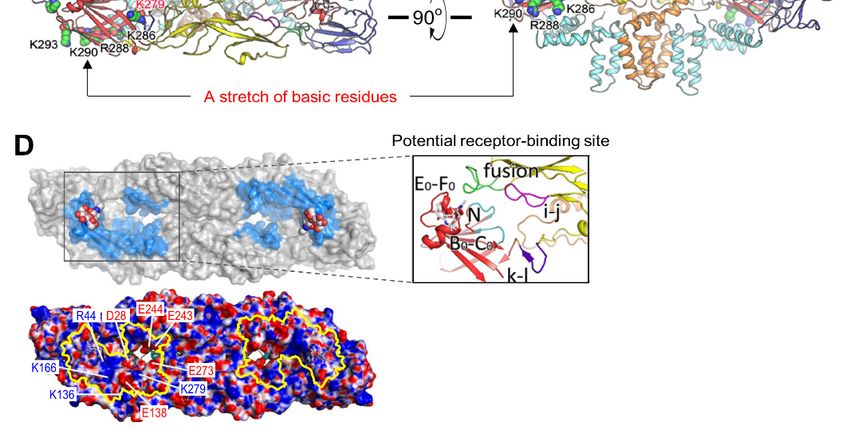
Pathogens 2018, 7, 68                                                                                 3 of 38

virus in oronasal secretions [117] and transmit it to co-housed naive pigs in the absence of mosquitoes,
suggesting a mode of viral transmission during mosquito-free seasons [118]. Further studies are
needed to understand the dynamic interactions between the virus, mosquito vectors, and vertebrate
hosts under certain geo-environmental and eco-agricultural conditions [119–121].

3. JEV Is a Small Enveloped Positive-Strand RNA Virus

3.1. Genome Structure and Gene Expression
      JEV is an enveloped RNA virus with a linear, single-stranded, and positive-sense RNA genome of
~11 kb in length (Figure 1A). The genomic RNA has a methylated cap structure at its 50 end, but lacks
a poly (A) tail at the 30 end [122–126]. It has one long open reading frame (ORF) encoded between
the two short, but highly structured, 50 and 30 non-coding regions (NCRs) that form a long-range
intramolecular RNA-RNA interaction to regulate viral translation and RNA replication [127–131].
In addition to the viral genomic RNA, a group of short non-coding subgenomic RNAs (~0.2–0.5 kb)
is also accumulated to high levels in a diverse range of mammalian and insect cells infected with
JEV and other flaviviruses as a result of incomplete degradation of the genomic RNA caused by the
stalling of the cellular 50 →30 exoribonuclease Xrn1 just upstream of a higher-order structure in the
30 NCR [132–136]. The generation of this subgenomic RNA may cause the suppression of Xrn1 and the
dysregulation of cellular mRNA stability [137], thereby disrupting the host’s innate immune responses
and contributing to viral replication and pathogenesis [138–140].
      The ORF in the JEV genomic RNA encodes a polyprotein precursor of ~3432 amino acids, which is
cleaved into at least 10 distinct products [141,142], i.e., three structural (capsid, C; premembrane,
prM; and envelope, E) and seven nonstructural (NS1, NS2A, NS2B, NS3, NS4A, NS4B, and NS5)
proteins (Figure 1B,C). In flaviviruses, the site-specific proteolysis of the polyprotein is catalyzed co-
and post-translationally by a set of four different proteases: (i) the host signal peptidase responsible for
cleaving at the C-prM, prM-E, E-NS1, and NS4A-NS4B junctions within the lumen of the endoplasmic
reticulum (ER) [143–148]; (ii) the two-component viral protease NS3 + NS2B [149] required for cleaving
at the NS2A-NS2B, NS2B-NS3, NS3-NS4A, and NS4B-NS5 junctions, as well as at internal sites within
the C and NS4A proteins on the cytoplasmic face of the ER membrane [143,144,150–155]; (iii) the host
furin or furin-like protease mediating the final cleavage of prM to M in the trans-Golgi network [156];
and (iv) an unknown host protease capable of cleaving at the NS1-NS2A junction [157–159]. In addition
to the aforementioned 10 proteins, an NS1 isoform (NS10 ) is also produced during infection with JEV
and other JE serogroup members as a result of −1 translational frameshifting occurring at codons
8–9 of NS2A [160–163].
Pathogens 2018, 7, 68 4 of 38
Pathogens 2018, 7, x FOR PEER REVIEW 4 of 36
Figure 1.
Figure 1. Genome
Genome organization
organization and and gene
gene expression
expression of of Japanese
Japanese encephalitis
encephalitis virus
virus (JEV).
(JEV). (A)
(A) Genome
Genome
organization. The genetically well-characterized JEV strain, CNU/LP2,
organization. The genetically well-characterized JEV strain, CNU/LP2, contains a single-stranded contains a single-stranded
positive-sense RNA
positive-sense RNA genome
genome of of 10,968
10,968 nucleotides
nucleotidesin inlength,
length,consisting
consistingofofa amethylated
methylatedcap capatat
thethe5′
end,
0 followed by a 95-nucleotide 5′ 0non-coding region (5′NCR),
0 a 10,299-nucleotide
5 end, followed by a 95-nucleotide 5 non-coding region (5 NCR), a 10,299-nucleotide open reading open reading
frame (ORF),
frame (ORF), andand aa 574-nucleotide
574-nucleotide 3′NCR
30 NCR[164,165].
[164,165].(B)(B)Gene
Geneexpression.
expression.The The single
single ORF
ORF encoded
encoded in
the viral genome produces two precursor polyproteins, a full-length 3432-amino
in the viral genome produces two precursor polyproteins, a full-length 3432-amino acid polyprotein acid polyprotein
(ppC-NS5)and
(ppC-NS5) and its
its C-terminally
C-terminally truncated
truncated1198-amino
1198-aminoacid acidpolyprotein
polyprotein(ppC-NS1
(ppC-NS1′), 0 ), the
thelatter
latter of
of which
which
is expressed
is expressed by by aa −
−11 ribosomal
ribosomal frameshift
frameshift (FS)
(FS) event
event that
that occurs
occurs between
between the the codons
codons 88 and and 99 of
of NS2A,
NS2A,
adding 52
adding 52 extra
extra amino
amino acids
acids to
to the
the C-terminus
C-terminus of of NS1
NS1 (designated
(designatedNS1 0 ). Each
NS1′). Each ofof the
the twotwo polyproteins
polyproteins
is cleaved
is cleaved by by host-
host- and
and virus-encoded
virus-encoded proteases
proteases toto yield
yield three
three structural
structural (green)
(green) and and atat least
least seven
seven
nonstructural (orange)
nonstructural (orange) proteins.proteins. The flavivirus-conserved cleavage sites and the length of cleavage
length of cleavage
productsare
products areindicated.
indicated. NS3NS3 consists
consists ofof an
an N-terminal
N-terminal serine
serine protease
protease (PRO)
(PRO) domain
domain and and aa C-terminal
C-terminal
RNA helicase
RNA helicase (HEL)
(HEL) domain,
domain, and
and NS5
NS5 consists
consists of
of an
an N-terminal
N-terminal methyltransferase
methyltransferase (MET) (MET) domain
domain
and aa C-terminal
and C-terminal RNA-dependent
RNA-dependent RNA RNA polymerase
polymerase (RDRP)
(RDRP) domain.
domain. Asterisks
Asterisks indicate
indicate fourfour N-linked
N-linked
glycosylationsites
glycosylation sites(NXT/S)
(NXT/S) found
foundin inthe
theprprportion
portionofofprMprM(Asn 15 ),
(Asn E (Asn154154),),and
15), E (Asn andNS1/NS1
NS1/NS1′ 0 (Asn
(Asn130130
and Asn 207
207). (C) Polyprotein membrane topology. The membrane orientation of the 10 major JEV
and Asn ). (C) Polyprotein membrane topology. The membrane orientation of JEV
proteins is
proteins is predicted
predicted on on the
the endoplasmic
endoplasmic reticulum
reticulum (ER)
(ER) membrane,
membrane, based based onon previous
previous work work with
with
other flaviviruses
other flaviviruses [142,166–169].
[142,166–169].
3.2. Viral
3.2. Viral Replication
Replication Cycle
Cycle
JEV is
JEV is aa flavivirus
flavivirus containing
containing an an inner
inner nucleocapsid
nucleocapsid [170],
[170], aa disordered
disordered structure
structure made
made ofof the
the
genomic RNA
genomic RNA and and helix-rich
helix-rich C C proteins
proteins [171–173].
[171–173]. TheThe nucleocapsid
nucleocapsid is is enclosed
enclosed by by aa lipid
lipid bilayer,
bilayer,
which is
which is in
in turn
turn encased
encased inin aa well-organized
well-organized outer outer protein
protein shell
shell composed
composed of of the
the membrane-anchored
membrane-anchored
prM/M and
prM/M and E E proteins
proteins [174–179].
[174–179]. Basically,
Basically, JEVJEV shares
shares aa common
common strategy
strategy forfor viral
viral replication
replication with
with
other flaviviruses
other flaviviruses (Figure
(Figure 2).2). Viral entry is
Viral entry is aa dynamic
dynamic process,
process, defined
defined byby aa series
series of
of interactions
interactions
between the virus and the host cell that starts with nonspecific binding of the viral glycoprotein EE to
between the virus and the host cell that starts with nonspecific binding of the viral glycoprotein to
one or more cellular attachment factors on the cell surface [180–185]. This attachment
one or more cellular attachment factors on the cell surface [180–185]. This attachment step serves to step serves to
concentratethe
concentrate the virions
virions at
at the
the cell
cell surface
surface to to facilitate
facilitate the
the specific
specific interaction
interaction of of the
the viral
viral E
E glycoprotein
glycoprotein
with a cellular entry factor(s) [186–188], directing the classical clathrin-dependent
with a cellular entry factor(s) [186–188], directing the classical clathrin-dependent endocytosis endocytosis [189–
200] or non-classical
[189–200] or non-classicalclathrin-independent
clathrin-independent endocytosis
endocytosis pathways
pathways [201–206],
[201–206], presumably
presumablyinin aa cell
cell
type-restricted fashion.
type-restricted fashion. OnceOnceinside
insidethetheendosome,
endosome, the the
viralviral
E glycoprotein
E glycoprotein undergoes low low
undergoes pH-
induced conformational
pH-induced conformational changes
changes [207–213],
[207–213], triggering
triggering thethefusion
fusionofofviral
viral and host endosomal
and host endosomal
membranes [214–221].
membranes [214–221]. Following
Following membrane
membrane fusion,
fusion, the
the genomic
genomic RNA RNA isis released
released into
into the
the cytoplasm,
cytoplasm,
where it is translated into two precursor polyproteins (with or without a ribosomal frameshifting at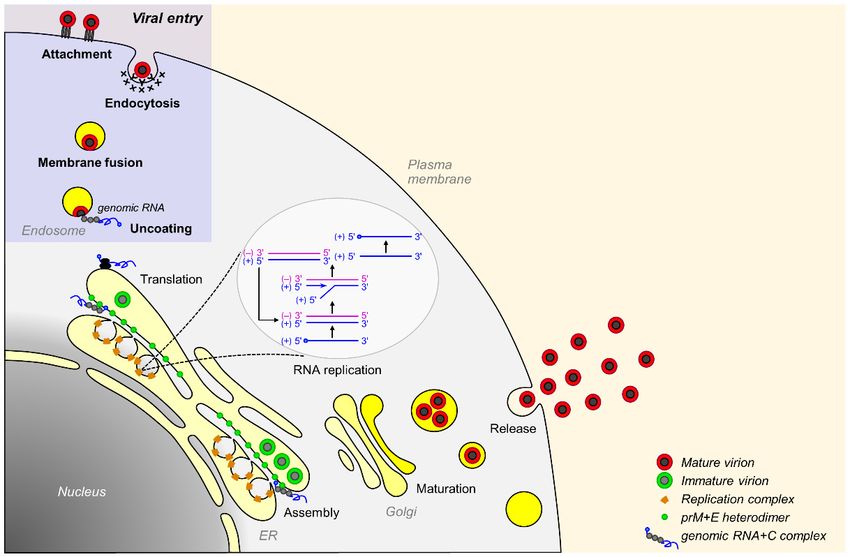
Pathogens 2018, 7, 68 5 of 38
Pathogens 2018, 7, x FOR PEER REVIEW 5 of 36
where it is translated into two precursor polyproteins (with or without a ribosomal frameshifting at
the beginning
beginning of
ofNS2A-coding
NS2A-codingregion)
region)that
thatare
arecleaved
cleavedtotoyield
yieldthree
threestructural (C,(C,
structural prM, and
prM, E) E)
and and at
and
least seven nonstructural (NS1 to NS5) proteins, along with NS1 0 [141,142].
at least seven nonstructural (NS1 to NS5) proteins, along with NS1′ [141,142].
Figure 2.
Figure 2. Replication
Replication cycle
cycle of
of JEV.
JEV. AA viral
viral particle
particle binds
binds initially
initially to
to aa target
target cell
cell through
through oneone oror more
more
attachment factors on the cell surface (Attachment). Subsequently, the virion interacts
attachment factors on the cell surface (Attachment). Subsequently, the virion interacts with an entry with an entry
factor(s), which
factor(s), whichtriggers
triggersreceptor-mediated
receptor-mediatedclathrin-dependent
clathrin-dependent oror clathrin-independent
clathrin-independent endocytosis
endocytosis of
of the bound virion (Endocytosis). Following internalization, the virion
the bound virion (Endocytosis). Following internalization, the virion travels through the endosomal travels through the
endosomal maturation
maturation pathway until pathway until acidification
endosomal endosomal acidification
triggers the triggers the low pH-induced
low pH-induced activation ofactivation
the viral
ofglycoprotein,
E the viral E glycoprotein,
enabling the enabling
fusion the fusionviral
between between
and viral and endosomal
endosomal membranes membranes
(Membrane (Membrane
fusion).
fusion). Upon release of the viral genomic RNA into the cytoplasm (Uncoating),
Upon release of the viral genomic RNA into the cytoplasm (Uncoating), it is translated into two it is translated into
two overlapping polyproteins as the result of a −1 ribosomal frameshift event
overlapping polyproteins as the result of a −1 ribosomal frameshift event in association with the in association with the
(Translation). The polyproteins are co- and post-translationally
ER (Translation). post-translationally cleaved to generate the mature
proteins that
viral proteins that are
are essential
essential for
for RNA
RNA replication
replication and
and virion
virion assembly.
assembly. The genomic RNA is
replicated in the replication complex within a structurally rearranged ER-derived membrane vesicles
replicated
(RNA replication).
replication). Viral assembly proceeds with the concomitant interaction of viral RNA with the
three structural
three structuralproteins
proteins(C,(C,prM,
prM,andandE),E),promoting
promotingthe thebudding
budding ofof immature
immature particles
particles into
into thethe lumen
lumen of
of the
the ER (Assembly).
ER (Assembly). The immature
The immature virions,virions, containing
containing heterodimers
heterodimers of prM and of E,
prMare and
then E, are then
transported
transported
through through thenetwork,
the trans-Golgi trans-Golgi network,
where prM iswhere
cleaved prM is cleaved
to form matureto form mature
virions virions
containing containing
homodimers
homodimers
of of M and EFinally,
M and E (Maturation). (Maturation). Finally,and
both partially both partially mature
completely and completely
virions aremature
releasedvirions are
into the
released intomilieu
extracellular the extracellular
(Release). milieu (Release).
After translation,
After translation, all
all seven
seven nonstructural
nonstructural proteins,
proteins, together
together with
with the
the poorly
poorly understood
understood host
host
factors [222], are involved directly or indirectly in the genomic RNA replication that
factors [222], are involved directly or indirectly in the genomic RNA replication that occurs in the occurs in the
virus-inducedER-derived
virus-induced ER-derived membraneous
membraneous organelle
organelle [223–227]
[223–227] housinghousing the replication
the replication complexes complexes
[123,228].
[123,228]. Viral RNA replication is catalyzed by NS3 and NS5 [229], the two
Viral RNA replication is catalyzed by NS3 and NS5 [229], the two largest and most conserved largest and most
conserved nonstructural
nonstructural proteins thatproteins that their
coordinate coordinate their
multiple multipleactivities
enzymatic enzymatic in activities in negative-
negative-strand RNA
strand RNA
synthesis, synthesis, RNA
positive-strand positive-strand
synthesis, RNARNA synthesis,
capping, RNA
and cap capping, [123,149,230].
methylation and cap methylation
During or
[123,149,230].
shortly During
after RNA or shortlya after
replication, RNAofreplication,
complex a complex ofgenomic
the newly synthesized the newly synthesized
RNA genomic
and C proteins is
RNA and C proteins is enveloped by two viral glycoproteins (prM and E [231]) on the
enveloped by two viral glycoproteins (prM and E [231]) on the ER membrane to produce the immature ER membrane
to produce
virion (~60 the
nmimmature
diameter)virion
covered(~60 nm60
with diameter)
protrudingcovered with
spikes, 60 protruding
each composed of spikes,
threeeach composed
parallel prM:E
of three parallel prM:E heterodimers [232–235]. The immature virions are believed to pass through
the constitutive secretory pathway to the extracellular space. During this exocytosis, viral maturation
occurs in the trans-Golgi network through the furin-mediated cleavage of the prM protein to M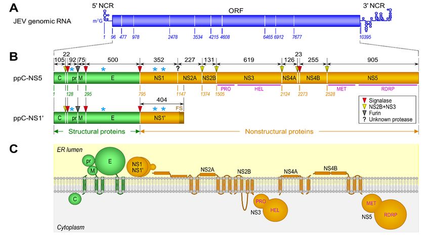
Pathogens 2018, 7, 68                                                                                6 of 38

heterodimers [232–235]. The immature virions are believed to pass through the constitutive secretory
pathway to the extracellular space. During this exocytosis, viral maturation occurs in the trans-Golgi
network through the furin-mediated cleavage of the prM protein to M [156,236–238], accompanied
by a significant structural rearrangement of the M and E proteins, to generate the mature virion
(~50 nm diameter), which is covered by 30 flat densely packed rafts, each composed of three parallel
E:M:M:E heterotetramers [232,239,240]. In addition to the M-containing completely mature virions,
prM-containing partially mature, but still infectious, virions are also shown to be produced [241–244],
although viral infectivity is likely compromised [245]. Overall, viral replication takes place entirely in
the cytoplasm; however, two viral proteins, C [246–250] and NS5 [251–257], are not only detected in
the cytoplasm, but are also found in the nucleus [258]. The precise role of their nuclear localization in
viral replication and pathogenesis requires further investigation.

4. Viral Entry Is the First Step in the Infection Process
     Viral entry is the first step in an orchestrated process of virus-host interactions that is not only
required for the initiation, dissemination, and maintenance of productive infection [259,260], but also
represents a critical determinant of cell/tissue tropism and pathogenesis [261]. JEV entry is thus a
promising target for antiviral therapy and offers multiple points for intervention [262]: attachment,
endocytosis, membrane fusion, and uncoating (see a recent review article for a detailed description
of small-molecule inhibitors targeting flavivirus entry [263]). Identifying the viral and host factors
involved in JEV entry is a prerequisite to elucidating the molecular mechanisms of viral entry and
developing novel therapeutic and preventive antivirals. In recent years, tremendous progress has been
made in understanding the viral components required for the various steps of JEV entry, but little is
known about the cellular components involved in this important process.

4.1. Virus Structure
      Using cryo-electron microscopy (EM) and image reconstruction techniques, Wang and coworkers
have determined the 4.3-Å three-dimensional structure of JEV [176]. On the surface of the mature JEV,
180 copies of each of the M and E proteins are organized into 30 flat, densely packed rafts. Each of
these rafts is composed of three parallel E:M:M:E heterotetramers, with the E proteins forming the
smooth outer protein shell and the M proteins being buried underneath it (Figure 3A), as seen in the
cryo-EM structures initially of DENV [174,179] and WNV [175], and lately ZIKV [177,178]. The JEV
E monomer, like that of other flavivirus E proteins, consists of three topologically distinct segments
(Figure 3B,C): (i) a banana-shaped ectodomain, which mediates receptor binding and membrane
fusion; (ii) a “stem” region, which includes three perimembrane helices lying nearly horizontal
on the viral membrane underneath the ectodomain; and (iii) an “anchor” region, which contains
two antiparallel membrane-embedded helices [240,264–266]. Notably, the E ectodomain adopts a
three-domain architecture, with domain I (E-DI) lying at the interface between domains II (E-DII) and
III (E-DIII) (Figure 3C): (a) E-DI has the glycan loop carrying an N-linked carbohydrate chain attached
to Asn154 and a string of six closely dispersed basic residues (Lys279 to Lys297 ) mapped in the last strand
I0 of E-DI and the linker between E-DI and DIII; (b) E-DII contains the fusion loop at its tip and several
potentially functionally important loops (e.g., h-i, i-j, and k-l loops) on its side; and (c) E-DIII has the
Arg-Gly-Asp (RGD) motif and is implicated in receptor binding and antibody neutralization [267–284].
In contrast, the JEV M monomer contains a flexible N-terminal loop, followed by an amphipathic
helix lying on the membrane and two antiparallel helices embedded in the membrane (Figure 3C).
The N-terminal loop of M participates in electrostatic and hydrophobic interactions with E-DI and
E-DII, and the amphipathic helix of M is involved in hydrogen-bond interactions with E-DII (centered
at Gln264 near helix αB) and the N-terminus of a neighboring M. The E-DI, E-DIII, and the helical stem
region of E are held together by charge interactions.
Pathogens 2018, 7, 68 7 of 38
Pathogens 2018, 7, x FOR PEER REVIEW 7 of 36
Figure 3. The cryo-electron microscopy (EM) structure structure of of JEV at 4.3 Å resolution. (A) The surface (left)
and cross-section
and cross-section(right)(right)ofofthe
thecryo-EM
cryo-EM mapmap of of mature
mature JEVJEV (strain
(strain P3).P3). An icosahedral
An icosahedral asymmetric
asymmetric unit
unit
is is indicated
indicated by a whiteby atriangle,
white with
triangle, with
the 2-, 3-, andthe 5-fold
2-, 3-,symmetry
and 5-fold axessymmetry
labeled. E:Maxes labeled. E:M
heterodimers of
the same colorofare
heterodimers theequivalent
same color byare
icosahedral
equivalent symmetry. (B) Side
by icosahedral view of the
symmetry. (B)E:M heterodimer
Side view of thefitted
E:M
into the electron
heterodimer density
fitted into map. Indicated
the electron are themap.
density ectodomain,
Indicatedstem,are and anchor regions
the ectodomain, of the
stem, E protein,
and anchor
along with the M protein buried underneath it. (C) Ribbon diagram of
regions of the E protein, along with the M protein buried underneath it. (C) Ribbon diagram of thethe E:M:M:E heterotetramer.
Color-coded are the structurally
E:M:M:E heterotetramer. distinct domains/regions
Color-coded are the structurally of Edistinct
and M proteins: E-DI (domain
domains/regions of EI),and
E-DII,
M
E-DIII,
proteins: E-H1
E-DI(helix
(domain1), E-H2, E-H3,
I), E-DII, E-TM1
E-DIII, (transmembrane
E-H1 (helix 1), E-H2, 1),E-H3,
E-TM2, M-H,(transmembrane
E-TM1 M-TM1, and M-TM2. 1), E-
Also
TM2,highlighted
M-H, M-TM1, are the functionally
and M-TM2. Also important structural
highlighted arecomponents in the Eimportant
the functionally ectodomain: glycan
structural
loop
components in the E ectodomain: glycan loop and a stretch of basic residues in E-DI; fusion loop,helx
and a stretch of basic residues in E-DI; fusion loop, h-i loop, i-j loop, k-l loop, and αB h-i
in E-DII;
loop, and k-l
i-j loop, Arg-Gly-Asp
loop, and αB (RGD)
helx inmotif inand
E-DII; E-DIII. In all cases,
Arg-Gly-Asp onemotif
(RGD) molecule is used
in E-DIII. forcases,
In all labeling.
one
The high-resolution
molecule is used forimages
labeling.of The
this high-resolution
figure were kindly provided
images of thisbyfigure
Dr. Xiangxi Wang provided
were kindly [176]. by Dr.
Xiangxi Wang [176].
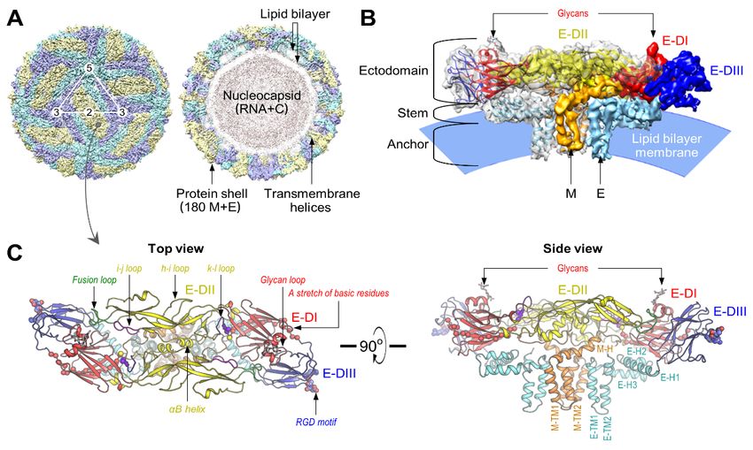
4.2. Attachment
4.2. Attachment
4.2.1. Viral Components
4.2.1.Despite
Viral Components
recent advances in our understanding of the near-atomic resolution cryo-EM structure of
JEV [176], therecent
Despite mechanisms
advances by which
in our the virion bindsof
understanding tothe
its cellular receptors
near-atomic are notcryo-EM
resolution fully understood.
structure
In
of JEV [285–287]
[176], the and other mosquito-borne
mechanisms by which the flaviviruses
virion binds[181–185,243,288–294],
to its cellular receptors theare
presence of
not fully
an N-linked glycan in E-DI (Figure 4A) and an RGD motif in E-DIII
understood. In JEV [285–287] and other mosquito-borne flaviviruses [181–185,243,288–294], the (Figure 4B) on the viral
membrane
presence ofsuggests a mechanism
an N-linked glycan inofE-DIrelatively nonspecific
(Figure 4A) and an interactions
RGD motif with
in the carbohydrate-binding
E-DIII (Figure 4B) on the
lectins and RGD-binding
viral membrane suggests aintegrins
mechanism on the cell surface,
of relatively respectively;
nonspecific in agreement
interactions with thewith this notion,
carbohydrate-
blocking/alteration
binding lectins and RGD-binding integrins on the cell surface, respectively; in agreementviral
of either the N-glycosylation or RGD motif generally negatively affects withentry
this
to varying
notion, degrees, but fails
blocking/alteration of to abolish
either the the process [182,243,285–295].
N-glycosylation or RGD motif Also, a string
generally of six closely
negatively affects
dispersed
viral entrybasic residues
to varying (Lys279but
degrees, to Lys 297
fails to) located
abolish in thethe last strand
process I0 of E-DI andAlso,
[182,243,285–295]. the linker
a stringbetween
of six
E-DI and
closely E-DIII (Figure
dispersed 4C), conserved
basic residues (Lys among
279 to Lys the
297 members
) located in of
thethe JEstrand
last and DEN I0 ofserogroups,
E-DI and the haslinker
been
proposed as a potential
between E-DI and E-DIIIbinding
(Figuresite forconserved
4C), glycosaminoglycans
among the (GAGs)
members [180,295].
of the JEHowever,
and DENthe cryo-EM
serogroups,
structure
has been of JEV indicates
proposed that, of thebinding
as a potential six basicsite
residues, four central residues
for glycosaminoglycans are buried,
(GAGs) suggesting
[180,295]. that
However,
conformational changesofare
the cryo-EM structure JEV required
indicates to make
that, ofthis
thepotential
six basicGAG-binding
residues, foursite accessible
central to GAGs
residues [176].
are buried,
Most intriguingly,
suggesting the cryo-EM structure
that conformational changes of JEV,
are combined
required to with a structure-based
make this potentialamino acid sequence
GAG-binding site
alignment of the E proteins from seven different flaviviruses (JEV, WNV, MVEV,
accessible to GAGs [176]. Most intriguingly, the cryo-EM structure of JEV, combined with a structure- SLEV, ZIKV, DENV,
and
basedYFV),
aminoreveals an unusual
acid sequence “hole” on
alignment theEviral
of the surface,
proteins fromwith
sevendistinct
differentelectrostatic
flaviviruses characteristics
(JEV, WNV,
MVEV, SLEV, ZIKV, DENV, and YFV), reveals an unusual “hole” on the viral surface, with distinct
electrostatic characteristics (Figure 4D) that could be a potential receptor-binding site for JEV andPathogens 2018, 7, 68 8 of 38
Pathogens 2018, 7, x FOR PEER REVIEW 8 of 36
(Figure 4D) that
other members ofcould be a potential
the JE serogroup receptor-binding
[176,296]. site
Thus, the viral for JEV and
components andother
theirmembers
interactingofcellular
the JE
serogroup
counterparts[176,296].
requiredThus, the viral components
for triggering and their interacting
flavivirus internalization cellular
after binding counterparts
on the cell surfacerequired
are still
for triggering flavivirus internalization after binding on the cell surface are still elusive.
elusive.
Figure 4.
Figure 4. Location
Location of of the
the viral
viral components
components involved
involved in in JEV
JEV entry
entry on on the
the protein
protein shell
shell ofof viral
viral particles.
particles.
(A) Location
(A) Location of of glycans.
glycans. The The carbohydrate
carbohydrate moieties
moieties attached
attached to Asn are
to Asn 154
154 are indicated
indicated on on the
the surface
surface ofof
the cryo-EM
the cryo-EM map map of of JEV
JEV atat aa lower
lower contour
contour level.
level. (B)
(B) Location
Location of of RGD
RGD motifs.
motifs. RGD RGD motifs
motifs areare shown
shown
on the
on the 2-,
2-, 3-,
3-, and
and 5-fold
5-fold symmetry
symmetry axes axes of
of the
the icosahedron.
icosahedron. (C) (C) Location
Location of of aa basic
basic residue-rich
residue-rich stretch
stretch
in the
the EE ectodomain.
ectodomain.AAcluster clusterofofsixsixbasic
basicresidues
residues 279 286 288 290
(Lys , Lys , Arg , Lys , Lys 293, and Lys
(Lys 279, Lys 286, Arg 288, Lys 290, Lys293 Lys297
297) in
the EEectodomain
ectodomain is shown
is shown on the ontopthe top
and sideand
viewsside views
of the of the
atomic model atomic model ofheterotetramer.
of an E:M:M:E an E:M:M:E
heterotetramer.
The The same
same color scheme as incolor scheme
Figure 3C isas in Figure
used, with the 3Cfour
is used,
resideswith the four
(black) being resides
buried(black)
inside being
the E
buried inside
ectodomain andthetheE two
ectodomain
resides (red)andbeing
the two resides
exposed (red) (D)
outside. being exposed
Location outside. receptor-binding
of putative (D) Location of
putative
sites. receptor-binding
A pair of the chargedsites. A pair ofputative
residue-rich the charged residue-rich putative
receptor-binding receptor-binding
sites is highlighted in blue, sites
withis
highlighted
the in blue, with
glycans attached to Asn 154
the glycans attached surface
on the external to Asn of an
154 on E:M:M:E
the external surface of an
heterotetramer (topE:M:M:E
panel).
heterotetramer
The (top panel).
charged residues The charged
are indicated on theresidues
externalare indicated surface
electrostatic on the external electrostatic
of the E:M:M:E surface of
heterotetramer,
the E:M:M:E
with the two heterotetramer, with the two
putative receptor-binding putative
sites outlinedreceptor-binding
in yellow (bottom sites panel).
outlinedAn in inset
yellow (bottom
shows the
panel). Aninteractions
molecular inset shows the molecular
formed interactions
in the putative formed insite,
receptor-binding theinvolving
putative the receptor-binding
fusion and i-j loops site,
involving
that interactthewith
fusiontheand i-j loops loop
N-terminal that interact
of the Awith
0 strandthe (labeled
N-terminal loopthe
as N), of Bthe
0 -CA strand
0 0and 0 loops as
E0 -F(labeled in
E-DI,
N), theand
B0-Cthe k-l loop
0 and E0-F0 in E-DII
loops in from
E-DI, the
andopposite monomer.
the k-l loop in E-DIIThe from images of this figure
the opposite monomer. wereThe graciously
images
provided by Dr.
of this figure wereXiangxi Wangprovided
graciously [176]. by Dr. Xiangxi Wang [176].
4.2.2. Cellular Components
JEV maintains a natural transmission cycle among birds, pigs, and other vertebrate hosts, with
mosquito vectors; in vitro, JEV can infect and replicate in a broad range of cell types originating from
many different vertebrate and invertebrate species [165], suggesting that there is probably more thanPathogens 2018, 7, 68 9 of 38
4.2.2. Cellular Components
JEV maintains a natural transmission cycle among birds, pigs, and other vertebrate hosts,
with mosquito vectors; in vitro, JEV can infect and replicate in a broad range of cell types originating
from many different vertebrate and invertebrate species [165], suggesting that there is probably more
than one host factor responsible for viral entry. The host factors documented to be involved in the
early steps of JEV entry to date are summarized in Table 1.
Table 1. Cellular components involved in the early steps of JEV entry.
Cellular Component Type/Property Host Cell Virus
Heparan sulfate Glycosaminoglycan Mammal JEV, WNV, MVEV, ZIKV, DENV, YFV, TBEV
DC-SIGN/L-SIGN C-type lectin Mammal JEV, WNV, DENV
MR C-type lectin Mammal JEV, DENV, TBEV
CLEC5A C-type lectin Mammal JEV, DENV
LSECtin C-type lectin Mammal JEV
mosGCTL-7, 1, and 3 C-type lectin Mosquito JEV, WNV, DENV
αv β3 Integrin Mammal JEV, WNV
HSP70/HSC70/GRP78 70-kDa heat shock protein Mammal, Mosquito JEV, DENV
HSP90 90-kDa heat shock protein Mammal JEV, DENV
37/67-kDa LR High-affinity laminin receptor Mammal JEV
CD4 Immunoglobulin superfamily Mammal JEV
CD14 Pattern recognition receptor Mammal JEV
Vimentin Type III intermediate filament Mammal JEV
LDLR Low-density lipoprotein receptor Mammal JEV
74-kDa protein Not characterized Mammal JEV
53-kDa protein Not characterized Mosquito JEV
Glycosaminoglycans (GAGs)
In JEV [297–300] as well as six other pathogenic flaviviruses [180,274,300–312], GAGs, a family of
linear, polydisperse, sulfated polysaccharides [313] (such as the heparan sulfates found in all animal
tissues), serve as one of the initial attachment factors for concentrating viral particles on the cell surface
prior to the interaction with other molecules. Negatively charged sulfate groups on the GAGs can
bind to a cluster of positively charged residues on the viral E glycoprotein [314]. In the case of JEV,
a role for GAGs in viral attachment has been demonstrated by (i) competition for viral binding to
hamster kidney-derived BHK-21 cells by highly sulfated GAGs, such as heparin and dextran sulfate;
(ii) pretreatment of BHK-21 cells with sodium chlorate, a potent sulfation inhibitor; (iii) a comparison of
the binding efficiency of the virus to the hamster ovary-derived wild-type CHO cell line and its mutants
with defects in GAG biosynthesis; and (iv) the identification of single net-positive-charge amino acid
changes (e.g., E49 K, E138 K, E306 K, D389 G/D389 N, and E390 G) in the E-DI or E-DIII region, with an
enhanced binding capacity for GAGs [297–300,315]. Interestingly, most if not all cell culture-adapted
JEVs, including a live-attenuated JE vaccine SA14 -14-2 strain derived from its virulent parental SA14
strain, exhibit an increased ability to bind heparin, a highly sulfated GAG [274,300,315]. Similar
results have also been observed for the live-attenuated YF vaccine 17D strain derived from its virulent
parental Asibi strain [301]. Thus, an increased binding affinity for highly sulfated GAGs seems to be
favorable for flavivirus growth in cell culture, and GAG-adapted flaviviruses tend to show attenuated
phenotypes in vivo.
C-Type Lectins
One set of attachment factors involved in flavivirus entry is the family of C-type lectins, which are
Ca2+ -dependent glycan-binding proteins that recognize carbohydrate moieties on the surface of invading
pathogens, act as the receptors for internalization, and deliver the pathogens to endosomes for antigen
presentation, thereby activating host defense systems [316,317]. Of particular interest are (i) the
dendritic cell-specific intercellular adhesion molecule (ICAM)-3-grabbing non-integrin (DC-SIGN,
also called CD209 and CLEC4L [318,319]), which is highly expressed on subsets of dendritic cells (DCs)
and macrophages; and (ii) the liver/lymph node-specific ICAM-3-grabbing non-integrin (L-SIGN,Pathogens 2018, 7, 68 10 of 38
also known as CD209L, CLEC4M, and DC-SIGNR for “DC-SIGN-related” [320–322]), which is mainly
expressed on endothelial cells in the liver and lymph nodes [323]. For JEV, siRNA knockdown and
antibody blocking experiments, combined with the characterization of a DC-SIGN mutant defective in
its internalization, have shown that DC-SIGN is important for viral binding to DCs that is mediated by
an N-linked mannose-rich glycan at Asn154 on the viral E protein, but it is dispensable for subsequent
internalization [287]. Similarly, for WNV [181,324] and DENV [182,184,325–329], both DC-SIGN and
L-SIGN have been shown to promote infection via an interaction with an N-glycan(s) at Asn154
(for WNV) and at both Asn67 and Asn153 (for DENV) on the viral E protein.
Based on the work with WNV and DENV, the use of DC-SIGN and L-SIGN as attachment factors
for flaviviruses varies, depending on the number and location of N-glycosylation sites on the viral E
protein [182,183,324,325], as well as on the type of N-glycans linked to these sites, which is determined
by the cells used for virus production [181,324–326]. Although glycosylation profiles may vary in
a given cell line, in general, the high-mannose N-glycans on mosquito cell-produced virions are
recognized well by both DC-SIGN and L-SIGN, whereas the complex N-glycans on mammalian
cell-produced virions are preferentially recognized by L-SIGN. There is a further added level of
complexity because “mosaic” partially mature flaviviruses contain a small, but detectable, amount of
the glycosylated uncleaved prM proteins [330,331], with one to three potential N-glycosylation sites
within the pr region [332] that may directly or indirectly contribute to the lectin-mediated attachment
of flaviviruses to the cell surface [324–326]. Notably, the physiologically relevant functional importance
of DC-SIGN in flavivirus replication and pathogenesis in humans is underlined by the association
of a single nucleotide polymorphism (SNP) found in the promoter region of the DC-SIGN gene
with a greater susceptibility to dengue hemorrhagic fever (SNP rs4804803 [333]) and severe forms of
tick-borne encephalitis (SNP rs2287886 [334]) in certain subpopulations.
There are three other C-type lectins that have been suggested to participate in the early steps of JEV
infection: mannose receptor (MR); C-type lectin domain family 5, member A (CLEC5A); and liver and
lymph node sinusoidal endothelial cell C-type lectin (LSECtin) [286,335–337]. (1) MR is expressed on
subsets of macrophages and DCs, as well as on nonvascular endothelium; it plays multiple important
roles in clearing endogenous molecules, promoting antigen presentation, and modulating cellular
activation and trafficking [338]. A study has shown that the extracellular region of MR binds broadly
to mosquito cell-produced DENV particles and pointedly to mammalian cell-expressed DENV E
ectodomains in a Ca2+ -dependent manner; in addition, the cell surface expression of human MR
in mouse embryo-derived 3T3 cells confers DENV binding, and anti-MR antibodies inhibit DENV
infection in human macrophages [335]. The same study has also reported that the extracellular region of
MR binds in enzyme-linked immunosorbent assays to formalin-inactivated JEV and TBEV, although the
nature of this binding has not been characterized [335]. (2) CLEC5A is exclusively expressed on myeloid
cells (e.g., macrophages and monocytes) and associates with a 12-kDa DNAX-activating protein
(DAP12), an adaptor molecule that transduces intracellular signaling involved in innate immunity;
thus, CLEC5A is also known as myeloid DAP12-associating lectin-1 (MDL-1) [339,340]. CLEC5A
has been shown to interact directly with JEV [336] and DENV [337], albeit in a Ca2+ -independent
manner, and is capable of inducing DAP12 phosphorylation in macrophages. Unlike DC-SIGN and
L-SIGN, the CLEC5A-JEV/DENV interaction does not promote viral infection, but rather stimulates
the release of proinflammatory cytokines (e.g., TNF-α and MCP-1), thereby potentially contributing to
the pathogenesis of virus-induced inflammatory diseases [336,337]. In the case of both JEV and DENV
infection, the inflammation-associated viral pathogenesis and lethality in mice can be ameliorated by
blocking the CLEC5A-JEV/DENV interaction with anti-CLEC5A antibodies [336,337]. (3) LSECtin
(also known as CLEC4G) is expressed on myeloid cells, as well as on sinusoidal endothelial cells of the
liver and lymph node; it mediates pathogen recognition, uptake, and internalization [317,341]. Using
human B lymphocyte-derived Daudi cells that are non-susceptible to JEV infection, researchers have
recently shown that the ectopic expression of LSECtin renders the cells susceptible to JEV infection.
This infection can be inhibited by N-acetylglucosamine β1–2 mannose (a target for LSECtin) but not byPathogens 2018, 7, 68 11 of 38
mannan (a target for DC-SIGN/L-SIGN) [286]; however, the underlying mechanism of LSECtin in JEV
entry has not been fully defined.
In addition to the mammalian C-type lectins described above, a family of mosquito
galactose-specific C-type lectins (mosGCTLs) has been reported to play a central role in the entry
steps of JEV [342], WNV [343], and DENV [344] in their major Culex/Aedes mosquito vectors.
The original work has shown that mosGCTL-1 (VectorBase accession no. AAEL000563) as a secreted
form of mannose-binding lectin (MBL) binds to WNV in a viral E protein-mediated Ca2+ -dependent
manner and brings the mosGCTL-1-WNV complex to its cell surface receptor, mosquito protein
tyrosine phosphatase-1 (mosPTP-1), thereby facilitating WNV entry both in vivo and in vitro [343].
The mosPTP-1 is a mosquito homolog of human CD45 that is expressed on all nucleated cells of
hemopoietic origin [345] and is critical for thymocyte development and activation [346,347] through its
interaction with human MBL [348]. Since CD45 is expressed on hematopoietic cells that are important
for flavivirus pathogenesis and host immunity [349], it will be interesting to determine whether
the human MBL-CD45 interaction can also mediate the entry of flaviviruses into human cells [350].
Moreover, mosquito blood-feeding experiments have demonstrated that WNV infection can be blocked
in vivo with anti-mosGCTL-1 antibodies [343], suggesting a promising new approach to interrupt the
life cycle of WNV in mosquito populations. Similarly, mosGCTL-7 (AAEL002524) and mosGCTL-3
(AAEL000535) have subsequently been shown to be able to mediate the mosquito cell entry of JEV and
DENV, respectively [342,344]. For DENV infection, a genetic association of the exon 1 polymorphisms
of the human MBL gene (MBL2) with dengue hemorrhagic fever has been suggested because variant
alleles and haplotypes related to low production levels of MBL are associated with the severity of
DENV-induced diseases [351]. Further investigation is required to elucidate the underlying mechanism
behind the variation in the usage of specific mosGCTLs for particular flaviviruses, along with distinct
mosPTPs, and the role of the mosGCTL-mosPTP pathway in flavivirus entry.
Integrins
Integrins are a family of cell surface receptors, each composed of two subunits (α and β),
that act as linkers between the extracellular matrix and the actin cytoskeleton, and play a critical
role in the activation and homing of hematopoietic cells [352]. Biochemical and molecular studies
have demonstrated that in monkey kidney-derived Vero or human cervical carcinoma HeLa cells,
the lineage-2 Sarafend strain of WNV binds to αv β3 integrin, and WNV infection is notably
decreased by pretreatment with anti-αv β3 antibodies, competition with recombinant αv or β3 protein,
or siRNA knockdown of the β3 subunit; however, somewhat unexpectedly, WNV infection is only
marginally affected by pretreatment with synthetic RGD peptides, an inhibitor of integrin-ligand
interactions [288,353]. It has also been noted that soluble αv β3 can block WNV infection of Vero cells in
a dose-dependent manner, and the expression of αv β3 increases the susceptibility to WNV infection of
hamster melanoma CS-1 cells lacking functional integrin [288]. In contrast, another study has shown
that the lineage-1 NY385-99 strain of WNV can infect and replicate in mouse embryonic fibroblasts
lacking functional αv β3 [354]. Therefore, the discrepancies in these studies suggest that the role of αv β3
in WNV entry is potentially strain-specific and/or cell type-dependent. Further investigation is needed
to define a potential role of αv β3 in WNV infection [355]. As previously seen for WNV, pretreatment
with anti-αv β3 antibodies has been shown to inhibit JEV entry into Vero cells [288]. A potential role
for αv β3 in JEV entry has also been proposed in hamster kidney-derived BHK-21 cells, based on
shRNA-based gene silencing and antibody/peptide-based blocking experiments using anti-αv β3
antibodies and synthetic RGD peptides, although their inhibitory effects varied significantly [285].
Other Host Factors
Heat shock proteins (HSPs) were long believed to be cytoplasmic proteins, but their protein- and/or
lipid-mediated association with intracellular and plasma membranes is now well documented [356].
To date, two families of HSPs have been proposed to participate in the early steps of JEV infection,Pathogens 2018, 7, 68 12 of 38
possibly in a cell type-dependent manner: (i) three members of the HSP70 family, namely the prototype
HSP70, heat shock cognate protein 70 (HSC70), and glucose-regulated protein 78 (GRP78, also referred
to as BiP for “binding immunoglobulin protein”); and (ii) the prototype HSP90 of the HSP90
family [196,357–362]. The details are as follows: (1) HSC70 derived from Ae. albopictus C6/36 cells
has been shown by co-immunoprecipitation experiments to bind to JEV [357]. In C6/36 cells, gene
knockdown experiments have identified HSC70 isoform D, which is involved in the clathrin-mediated
endocytosis of JEV [196]. (2) HSP70 derived from mouse neuronal Neuro-2a cells has been shown by
virus overlay protein binding assays to interact with JEV; the interaction between HSP70 and the JEV
E protein has been demonstrated by co-immunoprecipitation and immunoblotting [358]. Antibody
blocking experiments using anti-HSP70 antibodies have produced a significant reduction in JEV entry
into Neuro-2a cells [358]. (3) In human hepatoma Huh7 cells, the association of both HSP70 and JEV
E proteins with cholesterol-rich lipid rafts on the cell surface has been shown to be critical for JEV
infection [197,359]. In Huh7 cells, both antibody blocking and siRNA knockdown experiments have
revealed that HSP70, but not HSC70 or GRP78, is crucial for the host cell entry of vesicular stomatitis
virus-based pseudoviruses expressing JEV prM and E proteins [359]. (4) In Neuro-2a cells, however,
a combination of biochemical, genetic, and molecular experiments has shown that GRP78, capable of
interacting with JEV E-DIII, plays multiple roles in the entry and post-entry steps of JEV infection [360].
(5) HSP90 isoform HSP90β, but not HSP90α, is co-localized and co-immunoprecipitated with JEV
E proteins in hamster kidney-derived BHK-21 cells infected with JEV; HSP90β is also shown to be
secreted into the culture supernatant from JEV-infected BHK-21 cells, presumably in association
with released virus particles, promoting viral infectivity or the release of infectious particles [361].
Thus, all the data available to date suggest that several members of the HSP70 and HSP90 families
have pivotal, isoform-specific, and differential roles in JEV entry, depending to some extent on the cell
type, like those documented in DENV entry [363–370].
Several other host factors are thought to promote the infection of various cell types by JEV: (i) the
37/67-kDa high-affinity laminin receptor, CD4, and CD14 in mouse microglial BV-2 cells [362]; (ii) the
type III intermediate filament vimentin in mouse neuroblastoma N18 and human neuroblastoma
HTB-11 cells [371,372], as well as in porcine kidney PS cells [373]; (iii) the low-density lipoprotein
receptor in hamster kidney-derived BHK-21 cells [374]; (iv) a 74-kDa protein in monkey kidney-derived
Vero cells [375]; and (v) a 53-kDa protein in Ae. albopictus C6/36 cells [376]. However, the biological
function and physiological role of these molecules in JEV entry remain to be defined. In addition,
a handful of other cellular components have also been put forward as putative receptors for
one or more of other mosquito-borne flaviviruses, such as WNV, ZIKV, DENV, and YFV: (a) the
phosphatidylserine-recognizing TIM (TIM-1, -3, and -4) and TAM (TYRO3, AXL, and MER) family
members [185,377–388], (b) the phosphatidylserine- and phosphatidylethanolamine-binding protein
CD300a [389], (c) the tight junction component Claudin-1 [390,391], (d) the scavenger receptor
class B type I coupled with apolipoprotein A-I [392], (e) the 37/67-kDa high-affinity laminin
receptor [393,394], (f) CD14-associated molecules [395], (g) the carbohydrate β-N-acetylglucosamine
moiety of glycosphingolipids [396,397], (h) the natural killer cell-activating receptor, NKp44 [398],
and (i) the mosquito cell-derived prohibitin [399] (see two recent review articles for a detailed
description of these molecules [186,400]). Despite these research efforts discussed above, however,
the cell surface receptors and other host factors required for directing JEV, or any other flavivirus, into
the receptor-mediated endocytic pathway and low pH-dependent membrane fusion are still unknown.
4.3. Endocytosis
JEV is internalized from the plasma membrane of host cells to an endosomal compartment
via multiple endocytic routes in vitro [401], largely depending on the types of cell being infected:
(i) the classical clathrin-dependent pathway observed in the mosquito-derived C6/36 [196], hamster
kidney-derived BHK-21 [194], monkey kidney-derived Vero [198,201], porcine kidney-derived
PK15 [195], and mouse neural stem-like C17.2 [197] cells; and (ii) the non-classical clathrin-independentPathogens 2018, 7, 68 13 of 38
pathway (e.g., caveolin-dependent pathway) observed in the human neuroblastoma SK-N-SH [202],
mouse neuroblastoma Neuro-2a [201], and rat neuroblastoma B104 [203] cells. In almost all of these
cell lines (BHK-21, Vero, PK15, C17.2, SK-N-SH, Neuro-2a, and B104), the depletion of cholesterol from
the cell membrane with methyl-β-cyclodextrin reduces productive JEV infection [194,195,197,201–203],
suggesting an important role for cholesterol and possibly cholesterol-rich lipid rafts in both
clathrin-dependent and clathrin-independent endocytosis of JEV. Upon internalization, trafficking of
the endocytosed vesicles containing JEV particles to early and recycling endosomes is demonstrated
to be regulated by the two Rab GTPases, Rab5 and Rab11, for clathrin-dependent endocytosis in
BHK-21 cells [194]. In the case of clathrin-independent endocytosis in Neuro-2a cells, JEV-carrying
vesicles are transported to Rab5-positive early endosomes before the release of its genomic RNA into
the cytosol, and this vesicle trafficking is shown to be mediated by the actin-myosin II machinery
that is modulated by the major Rho GTPase RhoA [201]. Similarly, RhoA and Rac1 GTPase-mediated
actin rearrangements are documented to be critical for caveolin-dependent endocytosis of JEV in
SK-N-SH cells [202]. Considering the variations mentioned above, it is necessary to determine the
main endocytic pathway co-opted by JEV for its entry into human neurons in the brain, which are the
major target cells of JEV, and human monocytes and macrophages/DCs in the periphery, which are
likely to be of importance in mediating neuroinvasion.
Among other flaviviruses, WNV is reported thus far to adopt the clathrin-mediated endocytic
pathway for entry into C6/36 [190], Vero [191], and HeLa [402] cells. On the other hand, DENV is
shown to be able to enter the cytosol via an endosomal compartment, not only predominantly by
clathrin-dependent endocytosis (as described in C6/36 [189,192,403], Vero [205,206,404], BSC-1 [193],
NIH3T3 [328], HeLa [193,402], A549 [404], Huh7 [199], HepG2 [200,405], and ECV304 [406] cells and
human monocytes/immature DCs [328,407]) but also partially by clathrin-independent endocytosis
(as described in Vero [204–206,404] and HepG2 [200] cells). It has been noted in Vero cells, however,
that DENV can utilize both clathrin-dependent and clathrin-independent pathways for internalization,
depending on the viral serotype, and the virus can be transported to a different endosomal
compartment prior to membrane fusion, depending on the viral strain, even within the same
serotype [205,404]. Moreover, the main endocytic route employed by DENV in Vero cells is reported
to be altered from a clathrin-independent pathway for C6/36-grown virus to the clathrin-dependent
pathway for Vero-adapted virus [206]. In the case of both DENV and WNV, numerous studies
have indicated the functional importance of cholesterol [204,354,408], cytoskeleton and motor
proteins [189–192,199,328,409,410], and Rab GTPase-regulated vesicle trafficking [193,199,204,205,
328,402] in the process of their entry into various cell lines.
As is true for DENV, YFV is also able to enter HeLa cells via two distinct endocytic routes,
as demonstrated by the finding that the wild-type virulent Asibi strain primarily utilizes the
clathrin-dependent pathway, whereas its attenuated vaccine 17D strain exploits a pathway independent
of both clathrin and caveolin [411]. A mutagenetic analysis has indicated that the strain-specific use
of distinct endocytic pathways for YFV internalization is due to the 12 amino acid differences found
within the viral E protein between Asibi and 17D [411]. Interestingly, the 17D vaccine strain is shown
to enter HeLa and several other human cells more efficiently than does the parental Asibi strain,
resulting in a stronger induction of the cytokine-mediated antiviral response [411]. These data suggest
a potential link between viral entry and the host immune response. It will be interesting to examine
whether the strain-specific use of different endocytic pathways for YFV internalization is maintained
by an isogenic pair of JEV SA14 and SA14 -14-2 strains. In summary, JEV and other flaviviruses enter a
wide range of different host cells by viral E protein-directed endocytosis, but the precise endocytic
pathway used for viral internalization is determined by a combination of both the genetic composition
of the viral E protein and the availability of its interacting cellular components in a given cell type.
In cell biology, clathrin-mediated endocytosis is one of the best-studied processes, with a network
of various cellular proteins well characterized to date [412]. These previously known host factors are
generally required for those viruses that usurp the clathrin-mediated endocytic pathway [381,413–418].You can also read