Effect of 1-2 mmol/1 calcium, triamcinolone acetonide, and retinoids on low-calcium regulated keratinocyte differentiation
←
→
Page content transcription
If your browser does not render page correctly, please read the page content below
British Journal of Dermatology (1984) i i i , Supplement 27, 64-72.

        Effect of 1-2 mmol/1 calcium, triamcinolone
         acetonide, and retinoids on low-calcium
           regulated keratinocyte differentiation
         CYNTHIA L.MARCELO, R.C.GOLD AND JANET A.FAIRLEY
 University of Michigan Medical School, Department of Dermatology, Ann Arbor, Michigan 48109-0010, U.S.A.

                                               SUMMARY
Neonatal mouse keratinocytes cultured in low calcium (001 mmol/1) show rapid growth and
little stratification when compared with cells grown in normal i-2 mmol/1 calcium. The effect of
low calcium on the amount and synthesis of specific differentiation proteins was studied;
additionally, the effect of io~ ^ mol/1 triamcinolone acetonide, and 6 /ug/ml of retinoic acid and of
etretinate (Ro 10-9359) on low-calcium regulated keratinocyte hyperproliferation and differen-
tiation was determined. Low-calcium regulated keratinocytes contained less non-covalently
cross-linked and disulphide cross-linked keratins, less cell envelopes, much greater amounts of
SDS-soluble viable cell proteins, and slightly more keratohyaline granule-related proteins than
normal-calcium regulated keratinocytes. A 24 h switching time to 12 mmol/1 calcium medium
did not affect the amounts or synthesis of these proteins. Both retinoids and triamcinolone
acetonide inhibited by approximately 50% the proliferation of the low-calcium regulated
keratinocytes. Growth of low-calcium cells in these drugs for 9 days increased the amounts of
both keratins and cell envelope proteins in the cultures. We concluded that calcium-dependent
processes can regulate epidermal keratinocyte proliferation and differentiation. Our studies
suggest that these calcium-regulated events may occur via changes in calcium-dependent
proteins.

Epidermal keratinocytes grown in vitro form multi-layered, stratifying cultures that proliferate
and differentiate (Marcelo et al., 1978). The processes that control the formation of new basal
cells and the differentiation of the keratinocytes are regulated by a number of mediators. Among
these are cyclic AMP (Marcelo, 1979; Tong & Marcelo, 1983), retinoids (Marcelo & Madison,
1984) and glucocorticoids (Marcelo & Tomich, 1983).
  In vitro neonatal mouse keratinocyte proliferation and differentiation is also regulated by the
concentration of calcium ions in the growth medium. Hennings et al. (1980) and Hennings and
Holbrook (1983) have demonstrated that keratinocytes grown in low-calcium medium

 Correspondence: Cynthia L.Marcelo, Ph.D., Director of Dermatology Research Laboratories, University of
Michigan Medical School, Kresge I, R6558, Box 056, Ann Arbor, Michigan 48109-0010, U.S.A.
                                                   64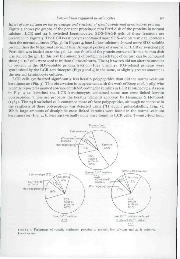
Low-calcium regulated keratinocytes                             65
(o-oi-oo6 mmol/1) proliferate rapidly, do not stratify and form no intracellular desmosomal
connections. Increasing the calcium levels to the normal 1-2 mmol/1 level causes the rapid
formation of desmosomes (1-2 h) and the eventual stratification, keratinization and cornifica-
tion of the cells (Hennings & Holbrook, 1983).
   We recently described a technique for quantitating the synthesis and processing of epidermal
differentiation proteins (Marcelo & Tong, 1983). This type of analysis detects cornified cell
formation, the presence of keratohyaline granule related proteins, and the amounts of
non-covalently and disulphide cross-linked keratins. Using this technique we investigated the
effect of a low-calcium to normal-calcium 24 h switch on the synthesis and processing of the
epidermal differentiation proteins, and the effect of two retinoids and the glucocorticoid,
triamcinolone acetonide, on the differentiation of low-calcium regulated keratinocytes.

                                           METHODS
  Growth of epidermal keratinocytes. The basal cells were prepared as previously described
(Marcelo et al., 197B). Low-calcium regulated (LCR) keratinocytes were grown as described by
Hennings et al. (1980). The cells were grown at 32°C and were fed on alternate days, or daily (the
LCR keratinocytes).

   Analysis of differentiation. The cultures were serially extracted with four buffers to yield six
fractions (Tong & Marcelo, 1983). These consisted of two keratohyaline granule (KG)-related
protein fractions, an SDS-soluble cell protein fraction, a non-covalently bound and a disulphide
cross-linked keratin fraction, and the residual cornified cell envelope fraction. These proteins
were analysed by Lowry assay and by SDS slab gel polyacrylamide electrophoresis (SDS-
PAGE) (Marcelo & Tong, 1983).

   Analysis of DNA and protein synthesis. Keratinocyte cultures were pulse labelled for 6 h with i
/iCi/ml of [^Hjthymidine and processed as previously described (Marcelo et al., 1983).
Seven-day-old normal and LCR keratinocytes were pulse labelled for 4 h with 10 /iCi/ml of
[•'HJleucine and cold-chased for 24 h in the appropriate medium (Tong & Marcelo, 1983).

   Addition of drugs. Six /Jg/ml of retinoic acid (RA), etretinate (RO, Ro 10-9359) or io~* mol/1
triamcinolone acetonide (TA) were added to 7-day-old LCR keratinocyte cultures. DMSO
(0-05%) was the vehicle control for the retinoids.

                                  RESULTS AND DISCUSSION
Growth properties of low-calcium regulated (LCR)     keratinocytes
Fig. I is a phase photomicrograph of a LCR keratinocyte (o-oi-o-O2 mmol/1 calcium). The cells
form a non-stratifying, non-cornifying monolayer. The keratinocytes show exceptionally wide
intracellular spaces. Hennings & Holbrook (1983) report that no intracellular desmosomal
complexes occur. Fig. 2 shows the proliferative rate as measured by [^HJthymidine incorpora-
tion per yug DNA of normal, LCR aind switched (low cialcium to 12 mmol/1 calcium)
keratinocytes. The LCR cells proliferate very rapidly, with a labelling index of 50% or more.
Switched keratinocytes ceased this rapid proliferative activity and reached normal keratinocyte
proliferative rates after 4-5 days.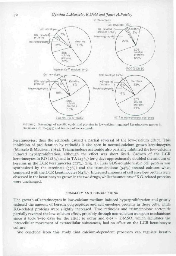
66                       Cynthia L.Marcelo, R.Gold and Janet A.Fairley

                                                               '^»Vl

                                    i^

     FIGURE I. Photomicrograph of epidermal keratinocytcs grown in o oi mmol/1 calcium medium, ( x 214,)

                             20,000 •

                                     0       1    2            4        7
                                                      Days after switch

     FIGURE 2- [^H]thymidine incorporation of low-calcium and normal-calcium regulated keratinocytes, and
     of low-calcium keratinocytes switched to normal-calcium growth medium.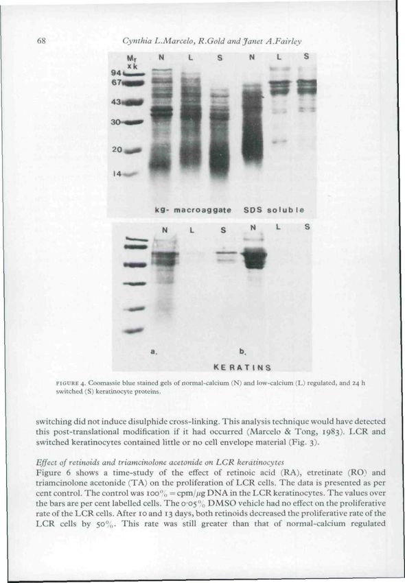
Low-calcium regulated keratinocytes                                67
Effect of low calcium on the percentage and synthesis of specific epidermal keratinocyte proteins
Figure 3 shows pie graphs of the per cent protein/60 mm Petri dish of the proteins in normal
calcium, LCR and 24 h switched keratinocytes. SDS-PAGE gels of these fractions are
presented in Figure 4. The LCR keratinocytes contained more SDS-soluble viable cell proteins
than the normal cultures (Fig. 3). In Figure 4, lane L (low calcium) showed more SDS-soluble
protein than the N (normal calcium) lane. An equal portion of a normal or LCR or switched (S)
Petri dish was loaded on to the gel, i.e. one-fourth of the protein extracted from a 60 mm dish
was run on the gel. In this way the amounts of protein in each type of culture can be compared
since 5 x 10'' cells were used to initiate all the cultures. The 24 h switch did not alter the amount
of protein in the SDS-soluble protein fraction (Figs 3 and 4). KG-rclated proteins were
synthesized by the LCR keratinocytes (Figs 3 and 4) in the same, or slightly greater amount as
the normal keratinocyte cultures.
   LCR cells synthesized significantly less keratin polypeptides than did the normal-calcium
keratinocytes (Fig. 3). This observation is in agreement with the work of Roop ^r a/. (1983) who
recently reported a marked absence of mRNA coding for keratins in LCR keratinocytes. As seen
in Fig. 4 (a. keratins) the LCR keratinocytes contained some non-cross-linked keratin
polypeptides. These are probably the keratin filaments reported by Hennings & Holbrook
(1983). The 24 h switched cells contained more of these polypeptides, although no increase in
the synthesis of these polypeptides was detected using [-'Hlleucine pulse-labelling (Fig. 5).
While large amounts of disulphide cross-linked keratins were found in the normal-calcium
keratinocytes (Fig. 4, b. keratins) virtually none were found in LCR cells. Twenty-four hour

                                                     Protein/petri

             Cell envelope (2%)

            KG-relQled]'
            proteins j.
         MocrooggregoteJ-

      FIGURE 3. Percentage of specific epidermal proteins in normal, low calcium and 24 h switched
      keratinocytes.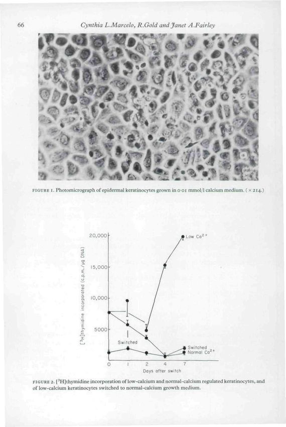
68                       Cynthia L.Marcelo, R.Gold and Janet A.Fairley
                           Mr        N        L        S         N        L       S

                                    kg- macroaggate             SDS s o l u b l e

                                      N        L        S         N       L

                                                      KE B A T I N S
     FIGURE 4. Coomassie blue suined gels of normal-calcium (N) and low-calcium (L) regulated, and 24 h
     switched (S) keratinocyte proteins.

switching did not induce disulphide cross-linking. This analysis technique would have detected
this post-translational modification if it had occurred (Marcclo & Tong, 1983). LCR and
switched keratinocytes contained little or no cell envelope material (Fig. 3).

Effect of retinoids and triamcinolone aceionide on LCR keratinocytes
Figure 6 shows a time-study of the effect of retinoic acid (RA), etretinate (RO) and
triamcinolonc acctonide (TA) on the proliferation of LCR cells. The data is presented as per
cent control. The control was 100",, ^ cpm//(g DNA in the LCR keratinocytes. The values over
the bars are per cent labelled cells. The oo5"o DMSO vehicle had no effect on the proliferative
rate of the LCR cells. After 10 and 13 days, both retinoids decreased the proliferative rate of the
LCR cells by 50"^,,. This rate was still greater than that of normal-calcium regulated
Low-calcium regulated keratinocytes
c.pm dish
Cell envelope (1%)
KG-reloted] V
prote ns I yS
t J
Mac
~^^=^ - - ^
SDS
soluble
proteins
61%
^ - - - - '
Normal Ca^* medium
,Kerolins Cell envelope Ke raiins 14-1%)
Cell envelope (0-5%) (0'90%)>>w
KG related] - /\67
^ ^ ^ A 5-5% ^ \ K6-relote
proteins \ \ protems \
MacroaqgregoteJ NV MacroaqgreqoieJ-V-* | ^ o / ^ \ \
127o
SDS SOS
( soluble soluble .
V protems
76 7o
Low Ca^* medium
/
)
Low Ca
proteins
73%
^ ^
medium switchec
/
/
to normol Ca^* medium
FIGURE 5. Perceniagc of [^Hjleucine incorporation into specific epidermal proceins by normal-calcium
and low-calcium regulated, and 24 h switched keratinocytes.
% Labelled
Ati cells
00 2l4
36
90 T *? I I9t2
i 6*3
1
i
1
80
70 -
1 5-8
2-6
tl-5 78
•Ifi •3 6 1%
60
50
1i 1 i 105
t\
10
D
M
R
A
R
0
T
A
D
M
R
A
R
0
T
A
D
i
R
T
S S M a A
S 0
30 0 0 0
20
10
10 13
growing in drug
FIGURE 6. l^Hlthymidine incorporation by low-calcium regulated keratinocytes growing in 6 /ig/ml of
reiinoicacid{RA)andofRo 10-9359 (etrciinate; RO) and 10 ' ** mol/1 triamcinoloneacetonide(TA). The
values over che bars are the per cent labelled cells lor each time point.70 Cynthia L.Marcelo, R.Gold and Janei A.Fairley
Protein/peirt
Cell envelope
KG-related|-
proteins y
MocroaggregofeJ-
Cell envelope
KG-reioied]-
proteins L
Mocfoaggregoiej-
FIGURE 7- Percentage of specific epidermal proteins in low-calcium regulated keratinocytes grown in
etretinate (Ro 10-9359) i>nd triamcinolone acetonide.
keratinocytes; thus the retinoids caused a partial reversal of the low-calcium effect. This
inhibition of proliferation by retinoids is also seen in normal-calcium grown keratinocytes
(Marcelo & Madison, 1984). Triamcinolone acetonide also partially inhibited the low-calcium
induced hyperproliferation, although the effect was short lived. Growth of the LCR
keratinocytes in RO (i8'\,) and in TA (23"',,) for 9 days approximately doubled the amount of
keratins in the LCR keratinocytes (12",,) (Fig. 7). Less SDS-soluble viable cell protein was
synthesized by the etretinate (55",,) and the triamcinolone (54",,) treated cultures when
compared with the LCR keratinocytes (64",,). Increased amounts of cell envelope protein were
observed in the keratinocytes grown in the two drugs, while the amounts of KG-related proteins
were unchanged.
SUMMARY AND CONCLUSIONS
The growth of keratinocytes in low-calcium medium induced hyperproliferation and greatly
reduced the amount of keratin polypeptides and cell envelope proteins in these cells, while
KG-related proteins were slightly increased. Two retinoids and triamcinolone acetonide
partially reversed the low-calcium effect, probably through non-calcium transport mechanisms
since it took 8~io days for the effect to occur and 005",, DMSO, which facilitates the
intracellular movement of extracellular substances, had no effect on the LCR keratinoeyte
culture.
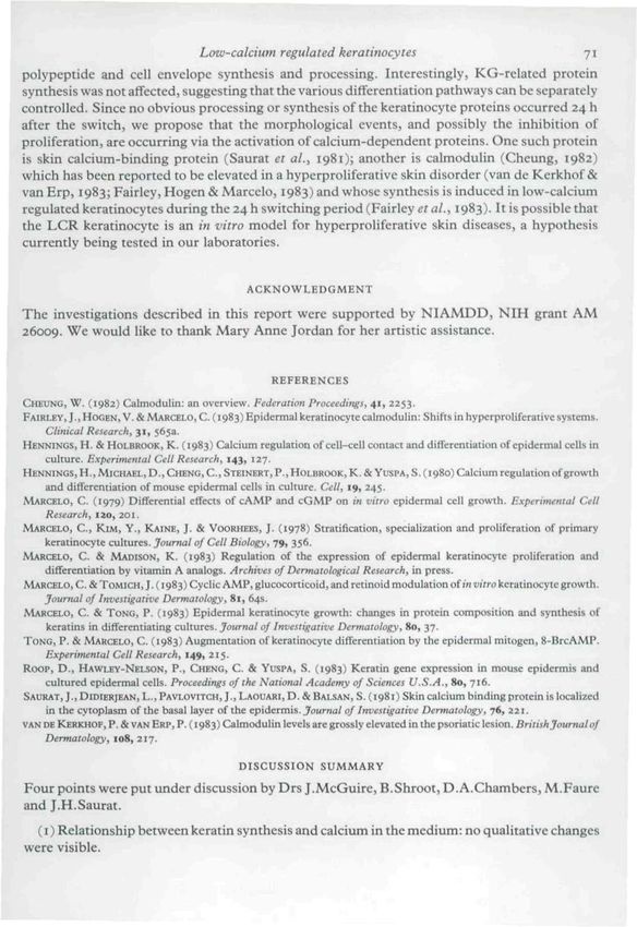
We conclude from this study that calcium-dependent processes can regulate keratinLow-calcium regulated keraiinocyies 71
polypcptide and cell envelope synthesis and processing. Interestingly, KG-rclated protein
synthesis was not affected, suggesting that the various differentiation pathways can be separately
controlled. Since no obvious processing or synthesis of the keratinocyte proteins occurred 24 h
after the switch, we propose that the morphological events, and possibly the inhibition of
proliferation, are occurring via the activation of calcium-dependent proteins. One such protein
is skin calcium-binding protein (Saurat ei al.> 1981); another is calmoduHn (Cheung, 1982)
which has been reported to be elevated in a hyperproliferative skin disorder (van de Kerkhof &
van Erp, 1983; Fairley, Hogen & Marcclo, 1983) and whose synthesis is induced in low-calcium
regulatedkeratinocytesduring the 24 h switching period {Fairley era/., 1983). It is possible that
the LCR keratinocyte is an in viiro model for hyperproliferative skin diseases, a hypothesis
currently being tested in our laboratories.
ACKNOWLEDGMENT
The investigations described in this report were supported by N I A M D D , NIH grant AM
26009. We would like to thank Mary Anne Jordan for her artistic assistance.
REFERENCES
CHEUNG, W . (1982) Calmodulin: an overview. Federation Proceedings, 41, 2253.
FAIRLEY, J ., HOGEN, V. & MARCELO, C . (1983) Epidermal kcrarinocyte calmodulin: Shifts in hyperproUferaiive sysiems.
Clinical Research, 31, 5653.
HENNINGS,H. &HOLBROOK, K . ( 1983) Calcium regulation of cell-cell contaa and ditferentiation of epidermal ceils in
culture. Experimental Celt Research, 143, 127.
HENNINGS,H,, MICHAEL, D., CHENG, C.,STEINERT, P.,HOLBROOK,K.&VuspA,S. {1980) Calcium regulation of growth
and differentiation of mouse epidermal cells in culture. Cell, 19, 245.
MARCELO, C . (1979) Differential cffecis of cAMP and cGMP on in vitro epidermal cell growth. Experimental Cell
Research, IZO, 2 0 1 .
MARCELO, C , K I M , Y . , KAINE, J . & VOORHEES, J . (1978) Stratification, specialization and proliferation of primary
keraiinocyte cultures. Journal of Cell Biology, 79, 356.
MARCELO, C . & MADISON, K . (1983) Regulation of the expression of epidermal keratinocyte proliferation and
differentiation by vitamin A analogs. Archives of Dermatological Research, in press.
MARCELO, C . & TOMICM, J, (1983) Cyclic AMP, glucocorticoid, and retinoid modulation of m t'lVrokeratinocyte growth.
Journal of Investigaiive Dermatology, 81, 64s.
MARCELO, C . & TONG, P. (1983) Epidermal keratinocyte growth: changes in protein composition and synthesis of
keratins in differentiating culiures. Jottrnal of Investigative Dermatology, 80, 37.
TONG, P , & MARCELO, C . (1983) Augmentation of keratinocyte differentiation by the epidermal mitogen, 8-BrcAMP.
Experimental Cell Research, 149, 215.
Roop, D,, HAWLEY-NELSON, P., CHENG, C . & YUSPA, S . (1983) Keratin gene expression in mouse epidermis and
cultured epidermal cells. Proceedings of the National Academy of Sciences U.S.A., 80, 716.
SAtntAT, J., DiDiERjEAN, L,, PAVi.oviTCH, J., LAOUARI, D, & BAL.SAN, S . (1981) Skin calcium binding protein is localized
in the cytoplasm of the basal layer of the epidermis. Journal of Investigative Dermatology, 76, 221,
VAN DE KERKHOF, P. & VAN ERF, P. (1983) Calmodulin levels are grossly elevated in the psoriatic lesion. British Journal of
Dermatology, 108, 217.
DISCUSSION SUMMARY
Four points were put under discussion hy Drs J.McGuire, B.Shroot, D.A.Chamhers, M.Faure
and J.H.Saurat.
(i) Relationship hetween keratin synthesis and calcium in the medium: no qualitative changes
were visible.72 Cynthia L.Marcelo, R.Gold and Jfa?iet A.Fairley (2) Effect of retinoids: over a long time period theretinoid will decrease. Delipidized sera was not used. (3) Calmodulin: calmodulin blockers were cytotoxic. Taking Ca'' away or adding cAMP to the culture had the same effect: hyperproliferation. It is not possible to correlate cAMP with calmodulin. (4) Intracellular calcium: it was not known from where the Ca"^' was coming after activation of the cells. It is possible that fluxes are increased in both directions with the switch. Two steps in this calcium activation: a fast initial step changing the morphology and a hyperproliferation with difTerentiation of the cultures.
You can also read