Effectiveness of Manual Therapy, Customised Foot Orthoses and Combined Therapy in the Management of Plantar Fasciitis-a RCT - MDPI
←
→
Page content transcription
If your browser does not render page correctly, please read the page content below
Article
Effectiveness of Manual Therapy, Customised Foot
Orthoses and Combined Therapy in the Management
of Plantar Fasciitis—a RCT
Casper Grim 1,*, Ruth Kramer 2, Martin Engelhardt 1, Swen Malte John 3, Thilo Hotfiel 1,4
and Matthias Wilhelm Hoppe 1,5
 1 Department of Orthopaedic, Trauma, Hand and Neuro Surgery, Klinikum Osnabrueck GmbH,
   49076 Osnabrueck, Germany; martin.engelhardt@klinikum-os.de (M.E.),
   thilo.hotfiel@klinikum-os.de (T.H.), matthias.hoppe@klinikum-os.de (M.W.H.)
 2 Physiopraxis Kramer, Westerkappeln 49492 Germany; ruth@physiopraxis-kramer.de

 3 Department of Dermatology, Environmental Medicine and Health Theory,

   University of Osnabrueck, 49076 Osnabrueck, Germany; sjohn@uos.de
 4 Department of Orthopedic Surgery, Friedrich-Alexander-University Erlangen-Nuremberg,

   91054 Erlangen, Germany
 5 Department of Movement and Training Science, University of Wuppertal, 42119 Wuppertal, Germany

 * Correspondence: casper.grim@klinikum-os.de

 Received: 30 March 2019; Accepted: 26 May 2019; Published: 28 May 2019

 Abstract: Background: Plantar fasciitis (PF) is one of the most common causes of plantar heel pain.
 Objective: To evaluate the effectiveness of three different treatment approaches in the management
 of PF. Methods: Sixty-three patients (44 female, 19 men; 48.4 ± 9.8 years) were randomly assigned
 into a manual therapy (MT), customised foot orthosis (FO) and a combined therapy (combined)
 group. The primary outcomes of pain and function were evaluated using the American Orthopaedic
 Foot and Ankle Society-Ankle Hindfoot Scale (AOFAS-AHS) and the patient reported outcome
 measure (PROM) Foot Pain and Function Scale (FPFS). Data were evaluated at baseline (T0) and at
 follow-up sessions after 1 month, 2 months and 3 months (T1–T3). Results: All three treatments
 showed statistically significant (p < 0.01) improvements in both scales from T0 to T1. However, the
 MT group showed greater improvements than both other groups (p < 0.01). Conclusion: Manual
 therapy, customised foot orthoses and combined treatments of PF all reduced pain and function,
 with the greatest benefits shown by isolated manual therapy.

 Keywords: plantar fasciitis; heel pain; manual therapy; joint mobilization; customised orthoses;
 insoles; back pain

1. Introduction
     Plantar fasciitis (PF) is reported as the most common cause of plantar heel pain and is referred
to as plantar fasciosis or fasciopathy, because these terms more accurately describe the inflammatory
degenerative nature of the disease [1–5]. The prevalence rate ranges from 4% in general to 7% in older
populations, and from 8% in athletes to 25% in runners [6]. In non-athletes, women are more
frequently affected [1,7,8] and have a higher risk of persisting symptoms [9] than men.
     The aetiology is largely unknown [2] and risk factors remain unclear. Obesity, prolonged
standing, running, limited ankle dorsiflexion, shortened triceps surae, hindfoot malalignment and
increased age are all considered as potential risk factors [2,4,6,10,11]; however, their scientific
evidence is weak. The high occurrence rate and level of impairment require a better understanding
not only of the diagnosis, but also of evidence-based recommendations for the therapy. Concerning
the latter, the quality of the studies is heterogeneous and often several forms of therapy are carried

Sports 2019, 7, 128; doi:10.3390/sports7060128                              www.mdpi.com/journal/sports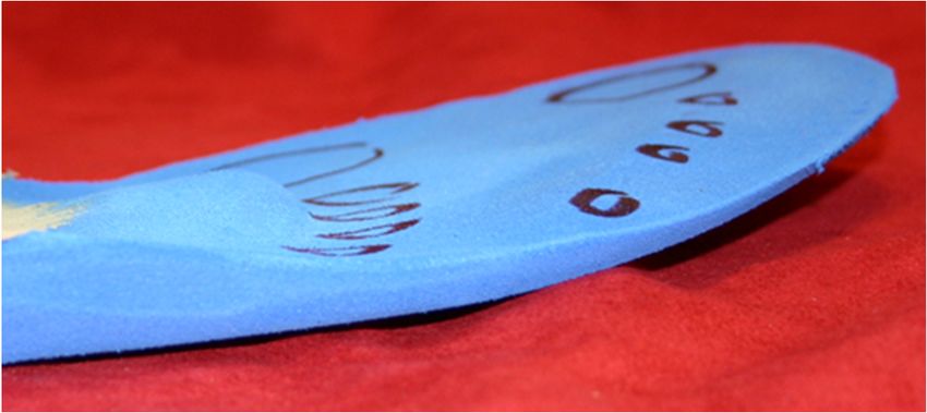
Sports 2019, 7, 128                                                                              2 of 12

out simultaneously [1,2,6]. This makes it difficult to determine the effectiveness of any individual
therapy or to rank therapies in order of their effectiveness [4]. Many patients report having persisting
or recurrent pain following treatment [12]. In addition to night splints, resistance training, corticoid
injections and extracorporeal shockwave therapy, manual therapy and foot orthoses are commonly
recommended interventions [13,14].
      Due to limited research, there are however no clear arguments for the use of foot orthoses. The
theoretical underpinning for their use includes improvement of the hindfoot alignment, the relief in
plantar pressure to the origin of the PF and modification in heel pitch, which may alter the mechanical
loading of the plantar fascia [15]. A lack of high quality evidence was found for the use of foot
orthoses [6,16]. Statistically significant differences were not found between customised or
prefabricated foot orthoses or soft and firm foot orthotic materials [2,6,13,17]. A longer duration of
foot orthoses use was associated with impairment in the plantar fascia and toe flexor muscle function
[12]. There are no two studies that used the same type of orthoses, which limits the comparisons
between studies and suggestions as to which orthoses features may be most effective. What is
considered most important is whether foot orthoses are beneficial to patients by effectively alleviating
their symptoms [6].
      There is only weak [1] or moderate evidence for short-term treatment [18,19] using manual
therapy interventions. However, compared to physical therapy, patients needed fewer sessions,
thereby reducing treatment costs [20]. Stretching of the calf muscles and improvement of the ankle
dorsiflexion is often recommended [13]; however, this additional mobilisation was no more effective
than stretching and ultrasound treatment alone [21]. In a study comparing customised foot orthoses
versus mobilisation of the foot and stretching, the mobilisation group had better results after two
weeks, but not after one or two months [17]. In only two of the reviewed studies [7,22], a single
treatment was applied in isolation in the experimental group. The other two well-rated studies
reporting significant improvements [1,23] used multiple interventions simultaneously. Thus, the
studies did not allow conclusions to be drawn with regards to the effectiveness of manual therapy
alone [4]. In an osteopathic study, the overall results were not significantly improved with three
treatments [24].
      After receiving conservative treatments, nearly 50% of patients still had symptoms when
interviewed after nine years [9,25]. Patients with plantar heel pain had a high prevalence of lumbar
back pain. Compared to the control group, more than twice as many patients with plantar heel pain
had lumbar back pain with the corresponding risk being five times higher. Treatment of local and
proximal restrictions, including those associated with back pain, may be justified for improving the
management of PF [25]. Hence, in the current study, the spine was also evaluated and treated. To
date, there has been no prospective, randomised, controlled trial in which manual therapy and
customised foot orthoses were investigated in relation to back pain in patients with PF.
      Thus, the aim of this study was to compare the effectiveness of manual therapy, customised foot
orthoses and combined therapy in the management of PF.

2. Materials and Methods
     The Ethics Committee of the University of Osnabrueck approved and accepted all procedures
involved in this study (4/7/1043.5). The patients were consecutively recruited over a 36-month period.
Patients were screened for eligibility by a foot and ankle surgeon. Inclusion criteria were: a clinical
diagnosis of PF with symptoms for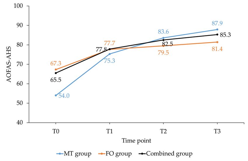
Sports 2019, 7, 128                                                                                     3 of 12

     All patients provided demographic information, medical history and previous treatments of PF.
They received a physical examination at baseline (T0) and at follow-up sessions after 1 month, 2
months and 3 months (T1–T3). The primary outcomes of pain and function were evaluated using the
American Orthopaedic Foot and Ankle Society-Ankle Hindfoot Scale (AOFAS-AHS) and the patient
reported outcome measure (PROM) Foot Pain and Function Scale (FPFS). An intention to treat
analysis was carried out using missing data from the last available value for the final evaluation
[26,27]. Figure 1 shows the patient recruitment and sample sizes, drop-outs and intention to treat of
the three groups.

      Figure 1. Flow-diagram of patient recruitment. Abbreviations: MT, manual therapy; FO, foot orthoses;
      ITT, Intention to treat; T0, baseline; T1–T3, follow-up sessions after 1–3 months.

2.2. Outcome Measures
      The AOFAS-AHS includes both subjective, patient reported items in pain and function (60%)
and objective, physician assessed items in function (40%). The AHS is scored from 0 to 100, where
higher values indicate a better outcome [28,29]. The AHS was preferred over the commonly used
Short Form 36 (SF-36), because the SF-36 has not been specifically studied in relation to foot and ankle
disorders [4]. Additionally, the time required for the evaluation of the nine items of the AHS is lower
than for the SF-36, which increases patient compliance in reporting data. Despite methodological
criticisms, the AHS is an established and frequently used rating system, making it possible to
compare the results with other studies [30,31]. The degrees of correlation and reliability provided an
acceptable validity for the subjective scores; however, the reliability of the objective component of the
AHS has yet to be reported [29,32]. There have been no reliable data published regarding the minimal
clinically important difference (MCID) related to the AOFAS score [29]. The MCID of the AHS in
hallux valgus surgery were indicated between 7.9 and 30.2, effect size derived 8.4 [33] or 8.9 out of
100 [34]. In the current study, the MCID was set at 10 out of 100. In the AHS, the subscale pain is one
single item. Pain is however subjective, with PROM providing the most valid measure of the
experience [35].
      In order to obtain more differentiated values for the typical pain of PF, the Foot Pain and
Function Scale (FPFS) was created with an 11-point numeric rating scale (NRS) from 0 to 10, where
higher values indicate better outcomes. The low gradation of the NRS for pain compensates for the
large variance in point values in the Ankle Hindfoot Scale [29]. The FPFS contains five questions on
pain (first steps, during rest, on pressure, while standing, weight bearing) and five questions about
function (limping, weakness, stiffness, restrictions in sports, at work). The highest possible total score
is 100. The FPFS uses 20 questions from the Visual Analogue Scale Foot and Ankle (VAS FA) [31], all
of which were validated against the SF-36 and the Hannover Questionnaire. VAS and NRS have a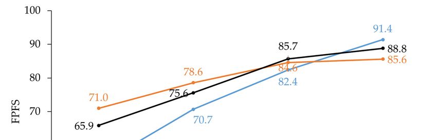
Sports 2019, 7, 128 4 of 12
well-documented reliability and validity in a variety of populations [1,4]. Due to the heterogeneity
between study results, no meaningful overall value for the MCID change can be determined. In the
subgroup pain, the NRS median for the MCID was 15% [36], those considered as clinically important
or “improved” ≥20%, clinically very important or ”much improved” ≥30% and ”very much
improved” ≥40%, respectively [37–39].
Blinding is barely achievable with the application of manual therapy in interventional studies,
making an even higher quality of the evaluation difficult [4]. Therefore, in the current study,
assignment of the patients to treatment groups was blinded for the therapist. The form of intervention
itself was recognisable to patients and therapists, though patients did not know if they were
participating in an intervention or control group.
2.3. Interventions
The patients were treated with manual therapy twice during the first week and subsequently
once per week for the remaining three-month period.
Patients in the manual therapy and combined group were evaluated with a standardised clinical
examination. The therapist used pre- and post-tests for each joint of the foot and intervertebral
segment of the spine. The order and type of treatment in therapy was standardised. The tests and
joint mobilisations were performed talocrural for dorsiflexion, subtalar for eversion and inversion,
and then tarsi transversal for pro- and supination. The sacroiliac joints and the symphysis pubica
were assessed and mobilised as well as the intervertebral joints in the supine position, partially in
lateral decubitus with rotation.
In the foot orthoses group, the orthopaedic technician used blueprints and foot scanners as
measuring instruments for the production of the orthoses. The orthoses were checked with
pedobarography and medilogic soles (T&T MediLogic Medizintechnik GmbH, Schoenefeld,
Germany). With the data obtained from the pressure distribution measurement, the orthoses
(Footpower, FSGmbH,Gummersbach, Germany) were milled with three layers (shore hardness A 50,
A 25 and A 35) and an additive support layer from ethylene vinyl acetate using computer-aided
design and computer-aided manufacturing techniques. To relieve pressure of the origin of the plantar
fascia, a canal (referring to the medial tuber calcanei) was milled and filled with soft material and a
cushion layer was applied to the heel. Subsequently, the footprints were produced, and the orthoses
were individually manufactured (Figure 2). The underlying idea of this type of foot orthoses is to
relieve the plantar fascia, reduce heel pressure and pain and obtain a positive, non-restrictive effect
on joint mobility without compromising the muscle activity of the foot itself. At the highest point of
the foot orthoses, a medial support for the sustentaculum tali was moulded. Through raising the toe
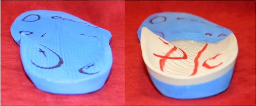
berries and using a retrocapital edged pelotte, pre-tensioning of the plantar fascia was expected.Sports 2019, 7, 128 5 of 12
Figure 2. Customised foot orthoses.
2.4. Statistical Analysis
The AOFAS-AHS and FPF Scale data were transferred into Microsoft Excel and were then
analysed with a statistical software package (IBM, SPSS version 23, Chicago, IL, USA). Descriptive
data were presented as relative frequency, mean and standard deviation. Differences in the
distributions were investigated by chi-square tests. Levene tests were applied to examine the variance
homogeneity between the three groups. Differences in the changes in scale values from T0 to T3
between the three groups were investigated using an analysis of variance (ANOVA) and Bonferroni
post hoc tests. Differences between T0 and T3 within each group were calculated using dependent
Student’s t-tests. Differences in the numbers of physiotherapy and evaluation of the foot orthoses
between the groups were investigated by independent Student’s t-tests. A p-value of ≤0.05 was
assumed to be statistically significant.
3. Results
Sixty-three patients met the eligibility criteria. The mean age and duration of PF symptoms were
48.4 ± 9.8 years and 4.4 ± 1.3 months, respectively. There were no statistically significant differences
for age, gender and body mass index between the three groups at T0 (p > 0.05). However, the patients
of the FO group had statistically significant shorter duration of symptoms, fewer had back pain with
shorter duration, lower FPFS values for work, and received fewer treatments and medications before
starting the study than both other groups (Table 1).
Table 1. Demographic data, symptoms, and therapies of the three groups at T0.
MT group (n FO group (n = Combined group (n p-
Variable
= 21) 21) = 21) value
Female sex, n (%) 16 (76.2) 14 (66.7) 14 (66.7) 0.741
BMI (kg/m2) 28.3 ± 6.2 30.4 ± 4.8 29.4 ± 4.2 0.432
Duration of PF symptoms
5.3 ± 0.8 2.9 ± 1.8 5.0 ± 1.2Sports 2019, 7, 128 6 of 12
A total of 58 (92%) patients appeared for the follow-up assessment after one month (T1). Five
patients from the FO group were counted as drop-outs (Figure 1). A total of 47 (75%) patients
completed the three-month follow-up (T0). Intention to treat was applied in the MT, FO and
combined group for one (5%), six (29%) and four (19%) patients, respectively. There were statistically
significant changes for the AOFAS-AHS (p < 0.01) and the FPFS (p < 0.01).
Between-group differences in the AOFAS-AHS and its subscales showed a greater improvement
from T0–T3 in the MT group (p < 0.01) than the FO and combined group (Figure 3, Table 2).
Figure 3. Changes in American Orthopaedic Foot and Ankle Score-Ankle Hindfoot Scale (AOFAS-
AHS) for the three groups from T0–T3. Abbreviations: MT, manual therapy; FO, foot orthoses; T0,
baseline; T1–T3, follow-up sessions after 1–3 months.
Likewise, between-group differences in the FPFS and in its subscales showed that the MT group
improved more from T0–T3 (p < 0.01) than the FO and combined group (Figure 4, Table 2).
Figure 4. Changes in Foot Pain and Function Scale (FPFS) for the three groups from T0–T3.
Abbreviations: MT, manual therapy; FO, foot orthoses; T0, baseline; T1–T3, follow-up sessions after
1-3 months.Sports 2019, 7, 128 7 of 12
Table 2. Comparison of the mean improvements in American Orthopaedic Foot and Ankle Score-
Ankle Hindfoot Scale (AOFAS-AHS), Foot Pain and Function Scale (FPFS) and their subscales for the
three groups from T0–T3.
MT group FO group Combined group
Variable
(n = 21) (n = 16) (n = 21)
AOFAS-AHS 33.9* 14.1 19.1
Pain subscale 48.8* 26.3 26.3
Function subscale 24.5* 6.1 6.1
FPFS 37.2* 14.6 22.9
Pain subscale 48.4* 28.4 28.4
Function subscale 32.8* 19.0 19.0
Abbreviations: MT, manual therapy; FO, foot orthoses; * Statistically significant higher (p < 0.01) than
in the other groups.
Besides the statistically significant differences, all three groups showed clinically meaningful
improvements over time. Differences in AHS from T0–T3 for the MT, FO and combined group were
35% (”much improved”), 15% (”minimally improved”) and 21% (”improved”), respectively. The
corresponding FPFS changes were 37% (”much improved”), 18% (”minimally improved”) and 24%
(”improved”), respectively. The FO and combined group did not reach the MCID in AHS subscale
function. In all groups, the improvement in subscale pain was higher than in subscale function.
One FPFS question was ”first step” pain after a period of rest. The improvement of the values in
the MT, FO and combined group were 50%, 32% and 43%, respectively. Effectiveness on pressure
pain and weight bearing were similar; in the MT group for 56% and 59%, in the FO group for 41%
and 41% and in the combined group for 45% and 42%. The values for pain during rest and while
standing were lower, just as in the subscale function for weakness and stiffness. Restrictions in sports
and work had an improvement of 51% and 30% in the MT group, 26% and 31% in the FO group and
28% and 20% in the combined group. The improvements in limping varied greatly between the
groups; 41% in the MT group, 36% in the FO group and 16% in the combined group.
The number of treatments did not differ between the MT and combined group (p > 0.05).
4. Discussion
The results of our study showed that all three interventions for PF achieved both a statistically
and clinically significant improvement over time. Furthermore, it suggests that manual therapy offers
greater clinical benefits, reducing pain and improving function compared to customised foot orthoses
and combined therapy. The application of manual techniques was standardised, and no additional
forms of therapy were used, making the therapy reproducible and comparable to other studies with
multiple concurrent therapies.
4.1. Comparison with Previous Studies
Cleland and colleagues [1] conducted a study in which 30 patients of a manual therapy group
underwent a five minute aggressive soft tissue mobilisation directed at the triceps surae and insertion
of the plantar fascia as well as a rear foot eversion mobilisation. It included an impairment-based
manual therapy at the hip, knee, ankle and foot on the clinical decision making of the treating
therapist. The other group with 30 patients was treated with electrophysical agents and exercises.
Three outcome measures were reported: Lower Extremity Functional Scale (LEFS), Foot and Ankle
Ability Measures (FAAM) and a numerical pain rating scale (NPRS). The overall group-by-time
interaction showed significantly better results for the manual therapy group. In comparison to the
Cleland study, the improvement reported in the pain subscale was greater at the three month follow-
up than the six month follow-up, and the AHS and FPFS total scores were higher than in LEFS of
Cleland et al. [1]. The underlying mechanism for improvements related to manual physical therapy
in the study of Cleland et al. [1] could not be determined as a first level of standardised intervention
was used in addition to a second level of intervention that utilised an impairments-based approach.Sports 2019, 7, 128 8 of 12
Hence, it could not be determined with any certainty which specific manual therapy and exercise
technique was most advantageous.
In our study, the focus was placed on treating local impairments of the joint structures of the
foot and the proximal impairments of the spine. No stretching of the soleus and gastrocnemius
muscle or plantar fascia took place, and no additional treatments of the knee and hip joints were
performed. McClinton and colleagues [25] also reported an association between PF and lumbar back
pain. The result of our study suggests that a therapy integrating the spine may help to alleviate the
often long-lasting symptoms of PF and ultimately achieve a better result.
The Burmeister study [24] used a similar intervention methodology. In addition to the spine, a
treatment of internal organs was conducted with 15 patients. Three osteopathic treatments were
conducted within a three-week period. Despite improvement on some items, no significant difference
could be detected between the verum and control group. It is possible that a higher number of
treatments could have led to a further improvement; however, this was not tested.
In our study, many patients actually required three months of therapy with a mean of 10.6
treatments to be symptom free. Cleland et al. [1] also questioned whether more than six therapies
would have resulted in a further improvement in function. The dosage also remained unclear in the
included studies of the clinical practise guidelines of Martin and colleagues [13]. The majority of the
studies selected by Mischke and colleagues in their review [4] evaluated the short-term effects of a
treatment. This might be less meaningful in the often long-lasting course of PF.
Custom made foot orthoses versus a combined treatment of manipulation and mobilisation of
the foot and stretching exercises with 10 patients per group were compared in a study by Dimou and
colleagues [40]. A review by Hawke and colleagues [17] reported that both groups in the Dimou study
had statistically significant reductions in pain on the NPRS. After only two weeks, there was a
statistically significant difference in foot pain favouring mobilisation of the foot versus stretching,
with no significant difference after one month and two months. The review indicated that the
customised foot orthoses did not reduce foot pain more than non-customised or sham foot orthoses,
including when combining them with stretching exercises or night splints. However, it is suggested
that using customised foot orthoses and night splints together may reduce foot pain. The foot
orthoses in our study complied with Hawke and colleague’s definition [17] of customised orthoses:
fabricated according to practitioner-prescribed specifications, the orthoses should be contoured,
removable in-shoe devices that are moulded or milled from an impression of the foot. In our study,
customised foot orthoses were found to be less effective compared to manual therapy or combined
treatment. The result also showed that wearing the foot orthoses over three months did not reduce
the number of manual therapies required in comparison to the MT group.
Overall, PF still remains a “black box”. Besides insoles and manual therapy, different treatment
options (e.g. resistance training, corticoid injections and extracorporeal shockwave therapy) seem to
be reasonable in the treatment of PF. It is, however, unclear what the underlying mechanisms are.
Additionally, it is important to highlight that individuals may respond differently to the various
treatment options, meaning that there is no general or overall recommendation for the treatment of
PF. In particular, the results of manual therapy for treating PF suggest that the pathophysiology
seems to be more complex and not fully understood. Our study shows that joint mobility and low
back pain play a role in treating PF. Joint dysfunction treated with manual therapy seems to lead to
a functional improvement and a relief of symptoms. If these dysfunctions lead directly to altered
mechanical loading of the plantar fascia or indirectly via the myofascial slings remains unclear. We
found in our study that only a one- to two-week manual therapy intervention altered the symptoms
of PF. It is unclear [41] if this easy-to-implement treatment can be used for preventive purposes, e.g.
in athletes, as it requires more research.
4.2. Strength and Limitations
In this study, manual therapy and customised foot orthoses treatments were carried out in
isolation, meaning that the methodology is reproducible, and any differences clearly assigned to the
treatment condition. Based on experience from other studies in which 3–6 weeks of therapy wereSports 2019, 7, 128 9 of 12
reported to be insufficient, the duration of treatment in our study was set at three months. Since the
disease is often long-lasting with severe discomfort, patients should receive treatment in the control
group, rather than to being exposed to placebo treatment over such a long period. The entire sample
population of our study was recruited from one clinical practice, which could be seen as a possible
limitation. As a limitation of our study, we did not perform a power analysis prior to the upcoming
recruitment. However, it must be considered that we were the first to investigate the effectiveness of
these three different treatment approaches in the management of PF within a randomised controlled
trial and that we nevertheless found statistically significant differences in our data.
The baseline outcome scores of the foot orthoses group were significantly higher at the
beginning of the study than those of the manual therapy and combined group. So, selection bias may
have occurred. High scores at baseline generally complicate an improvement over the course of the
study or even make it impossible. To enable an improvement, a limitation of the input values set on
the scales in the inclusion criteria would have been useful. An attrition bias occurred, because
participation in the follow-up assessment in the FO group with 10 patients, versus 20 in MT,
respectively 17 in MT and FO (combined group) was significantly lower. To counteract possible bias,
we performed an intention to treat analysis. The weekly treatment in the manual therapy group
represented a more intense support for the patients over three months. In contrast, the patients of the
foot orthoses group had one appointment with the orthopaedic technician for the footprints and a
second to receive the fabricated foot orthoses. After receiving their orthoses, patients may have seen
little reason for clinical follow-up appointments, resulting in a performance bias. This may have
reduced the success rate, because a higher number of FO patients no longer participated in the
evaluation compared to the manual therapy group.
The combination of manual therapy and customised foot orthoses should demonstrate whether
two simultaneously applied interventions would improve the outcome more than one treatment in
isolation. According to our study design, we intended to mobilise the local restrictions of the foot
before producing the foot orthoses. This was however not possible in all cases. The actual procedure
in the combination therapy could have contributed to the fact that the combined group hardly
achieved any improvements in the items function and alignment of the foot axis, whereas in the
manual therapy group, the results increased in both items. Some patients of the combined treatment
group reported foot pain, possibly caused by the fact that the orthoses no longer fitted optimally after
mobilisation of the restrictions of the feet. Therefore, differences were smaller in the manual therapy
group than in the foot orthoses group. Caution should be applied when interpreting these results, as
patients in the manual therapy group had significantly more complaints of accompanying back pain,
which could be another explanation as to why manual therapy was more successful in this group.
4.3. Clinical Implications
A meaningful overall minimal clinically important change (MCID) could not be reported for
NRS or AOFAS-AHS PHP (plantar heel pain) [36]. Additionally, it is a problem to calculate the mean
difference in pain score for the treatment group and to compare it to the MCID, because MCID is a
metric based on longitudinal differences in individuals and should be used in the same context [39].
Analyses of the relationships between changes in NPRS scores demonstrated a reduction of two
points, or 30%, to be clinically important and were measured using a standard seven-point patient
Global Impression of Change [37]. These results are similar to results that were found in lower back
pain patients compared after physical therapy using a 15-point Global Rating of Change scale [42].
The Global Rating of Change scale has been criticised because it is a transitional scale that requires
recall of prior health status [43]. The Global Rating of Change scale is not temporally stable, with a
finding in one week not associating to functional results the following week. The Global Rating of
Change scale is only correlated to functional measures up to three weeks [44], so it was not included
as an additional measure in the current study.
5. ConclusionsSports 2019, 7, 128 10 of 12
Manual therapy, customised foot orthoses and the combined treatments achieved statistically
and clinically significant improvements over time, with the greatest effect for the treatment of PF
being found in the manual therapy group. In addition, the results indicate that integrating spinal
treatment for patients experiencing back complaints together with PF could improve treatment
outcome.
Author Contributions: C.G., R.K., and M.E. designed the manuscript. C.G., R.K. and M.W.H. have made major
contributions in section “Results”. T.H., M.E. and S.M.J. have made major contributions in drafting and writing
the section “Discussion”. All authors read and approved the final manuscript.
Funding: This research received no external funding.
Acknowledgments: The authors would like to thank Joana Brochhagen for her linguistic revisions.
Conflicts of Interest: The authors declare no conflict of interest.
References
1. Cleland, J.A.; Abbott, J.H.; Kidd, M.O.; Stockwell, S.; Cheney, S.; Gerrard, D.F.; Flynn, T.W. Manual physical
therapy and exercise versus electrophysical agents and exercise in the management of plantar heel pain: A
multicenter randomized clinical trial. J. Orthop. Sports Phys. Ther. 2009, 39, 573–585.
2. Landorf, K.B. Plantar heel pain and plantar fasciitis. BMJ Clin. Evid. 2015, 2015, 11, 1–46.
3. McMillan, A.M.; Landorf, K.B.; Barrett, J.T.; Menz, H.B.; Bird, A.R. Diagnostic imaging for chronic plantar
heel pain: A systematic review and meta-analysis. J. Foot. Ankle Res. 2009, 32, 1–11.
4. Mischke, J.J.; Jayaseelan, D.J.; Sault, J.D.; Emerson Kavchak, A.J. The symptomatic and functional effects of
manual physical therapy on plantar heel pain: A systematic review. J. Man. Manip. Ther. 2017, 25, 3–10.
5. Pollack, Y.; Shashua, A.; Kalichman, L. Manual therapy for plantar heel pain. Foot 2018, 34, 11–16.
6. Whittaker, G.A.; Munteanu, S.E.; Menz, H.B.; Tan, J.M.; Rabusin, C.L.; Landorf, K.B. Foot orthoses for
plantar heel pain: A systematic review and meta-analysis. Br. J. Sports Med. 2018, 52, 322–328.
7. Ajimsha, M.S.; Binsu, D.; Chithra, S. Effectiveness of myofascial release in the management of plantar heel
pain: A randomized controlled trial. Foot 2014, 24, 66–71.
8. Renan-Ordine, R.; Alburquerque-Sendin, F.; de Souza, D.P.; Cleland, J.A.; Fernandez-de-Las-Penas, C.
Effectiveness of myofascial trigger point manual therapy combined with a self-stretching protocol for the
management of plantar heel pain: A randomized controlled trial. J. Orthop. Sports Phys. Ther. 2011, 41, 43–
50.
9. Hansen, L.; Krogh, T.P.; Ellingsen, T.; Bolvig, L.; Fredberg, U. Long-term prognosis of plantar fasciitis: A 5-
to 15-year follow-up study of 174 patients with ultrasound examination. Orthop. J. Sports Med. 2018, 6,
2325967118757983, doi:10.1177/2325967118757983.
10. Goff, J.D.; Crawford, R. Diagnosis and treatment of plantar fasciitis. Am. Fam. Physician. 2011, 84, 676–682.
11. van Leeuwen, K.D.; Rogers, J.; Winzenberg, T.; van Middelkoop, M. Higher body mass index is associated
with plantar fasciopathy/'plantar fasciitis': Systematic review and meta-analysis of various clinical and
imaging risk factors. Br. J. Sports Med. 2016, 50, 972–981.
12. McClinton, S.; Collazo, C.; Vincent, E.; Vardaxis, V. Impaired foot plantar flexor muscle performance in
individuals with plantar heel pain and association with foot orthosis use. J. Orthop. Sports Phys. Ther. 2016,
46, 681–688.
13. Martin, R.L.; Davenport, T.E.; Reischl, S.F.; McPoil, T.G.; Matheson, J.W.; Wukich, D.K.; McDonough, C.M.;
American Physical Therapy, A. Heel pain-plantar fasciitis: Revision 2014. J. Orthop. Sports Phys. Ther. 2014,
44, 1–33.
14. Huffer, D.; Hing, W.; Newton, R.; Clair, M. Strength training for plantar fasciitis and the intrinsic foot
musculature: A systematic review. Phys. Ther. Sport 2017, 24, 44–52.
15. Hotfiel, T.; Hotfiel, K.H.; Gelse, K.; Engelhardt, M.; Freiwald, J. Einlagenversorgung im leistungssport –
indikationen, wirkungsweise, sportspezifische versorgungsstrategien [The use of insoles in competitive
sports – Indications, effectiveness, sport specific treatment strategies]. Sports Orthop. Traumatol. 2016, 32,
250–257.
16. Whittaker, G.A.; Munteanu, S.E.; Menz, H.B.; Landorf, K.B. Should foot orthoses be used for plantar heel
pain? Br. J. Sports Med. 2018, 52, 1224–1225.Sports 2019, 7, 128 11 of 12
17. Hawke, F.; Burns, J.; Radford, J.A.; du Toit, V. Custom-made foot orthoses for the treatment of foot pain.
Cochrane. Database Syst. Rev. 2008, 16, 1–131.
18. Brantingham, J.W.; Bonnefin, D.; Perle, S.M.; Cassa, T.K.; Globe, G.; Pribicevic, M.; Hicks, M.; Korporaal, C.
Manipulative therapy for lower extremity conditions: Update of a literature review. J. Manipulative Physiol.
Ther. 2012, 35, 127–166.
19. Brantingham, J.W.; Globe, G.; Pollard, H.; Hicks, M.; Korporaal, C.; Hoskins, W. Manipulative therapy for
lower extremity conditions: Expansion of literature review. J. Manipulative Physiol. Ther. 2009, 32, 53–71.
20. Fraser, J.J.; Glaviano, N.R.; Hertel, J. Utilization of physical therapy intervention among patients with
plantar fasciitis in the united states. J. Orthop. Sports Phys. Ther. 2017, 47, 49–55.
21. Shashua, A.; Flechter, S.; Avidan, L.; Ofir, D.; Melayev, A.; Kalichman, L. The effect of additional ankle and
midfoot mobilizations on plantar fasciitis: A randomized controlled trial. J. Orthop. Sports Phys. Ther. 2015,
45, 265–272.
22. Wynne, M.M.; Burns, J.M.; Eland, D.C.; Conatser, R.R.; Howell, J.N. Effect of counterstrain on stretch
reflexes, hoffmann reflexes, and clinical outcomes in subjects with plantar fasciitis. J. Am. Osteopath. Assoc.
2006, 106, 547–556.
23. Saban, B.; Deutscher, D.; Ziv, T. Deep massage to posterior calf muscles in combination with neural
mobilization exercises as a treatment for heel pain: A pilot randomized clinical trial. Man. Ther. 2014, 19,
102–108.
24. Burmeister, S. Osteopathie und ihr effektivität bei fasciitis plantaris. Master’s Thesis, Donau Universität
Krems, Krems, Germany, 2012.
25. McClinton, S.; Weber, C.F.; Heiderscheit, B. Low back pain and disability in individuals with plantar heel
pain. Foot 2018, 34, 18–22.
26. Armijo-Olivo, S.; Warren, S.; Magee, D. Intention to treat analysis, compliance, drop-outs and how to deal
with missing data in clinical research: A review. Phys. Ther. Rev. 2009, 14, 36–45.
27. Gupta, S.K. Intention-to-treat concept: A review. Perspect. Clin. Res. 2011, 2, 109–112.
28. Kitaoka, H.B.; Alexander, I.J.; Adelaar, R.S.; Nunley, J.A.; Myerson, M.S.; Sanders, M. Clinical rating
systems for the ankle-hindfoot, midfoot, hallux, and lesser toes. Foot Ankle. Int. 1994, 15, 349–353.
29. Madeley, N.J.; Wing, K.J.; Topliss, C.; Penner, M.J.; Glazebrook, M.A.; Younger, A.S. Responsiveness and
validity of the sf-36, ankle osteoarthritis scale, aofas ankle hindfoot score, and foot function index in end
stage ankle arthritis. Foot Ankle. Int. 2012, 33, 57–63.
30. Kostuj, T.; Schaper, K.; Baums, M.H.; Lieske, S. Eine Validierung des aofas-ankle-hindfoot-scale für den
deutschen Sprachraum [German Validation of the AOFAS ankle hindfoot scale]. Foot & Ankle. 2014, 12,
100–106.
31. Richter, M.; Zech, S.; Geerling, J.; Frink, M.; Knobloch, K.; Krettek, C. A new foot and ankle outcome score:
Questionaire based, subjective, visual-analogue-scale, validated and computerized. Foot Ankle Surg. 2006,
12, 191–199.
32. Ibrahim, T.; Beiri, A.; Azzabi, M.; Best, A.J.; Taylor, G.J.; Menon, D.K. Reliability and validity of the
subjective component of the american orthopaedic foot and ankle society clinical rating scales. J. Foot Ankle
Surg. 2007, 46, 65–74.
33. Chan, H.Y.; Chen, J.Y.; Zainul-Abidin, S.; Ying, H.; Koo, K.; Rikhraj, I.S. Minimal clinically important
differences for american orthopaedic foot & ankle society score in hallux valgus surgery. Foot Ankle Int.
2017, 38, 551–557.
34. Dawson, J.; Doll, H.; Coffey, J.; Jenkinson, C.; Oxford; Birmingham, F.; Ankle Clinical Research, G.
Responsiveness and minimally important change for the manchester-oxford foot questionnaire (moxfq)
compared with aofas and sf-36 assessments following surgery for hallux valgus. Osteoarthr. Cartil. 2007, 15,
918–931.
35. Katz, J.; Melzack, R. Measurement of pain. Surg. Clin. North Am. 1999, 79, 231–252.
36. Olsen, M.F.; Bjerre, E.; Hansen, M.D.; Hilden, J.; Landler, N.E.; Tendal, B.; Hrobjartsson, A. Pain relief that
matters to patients: Systematic review of empirical studies assessing the minimum clinically important
difference in acute pain. BMC Med. 2017, 15, 1–18.
37. Farrar, J.T.; Young, J.P., Jr.; LaMoreaux, L.; Werth, J.L.; Poole, R.M. Clinical importance of changes in
chronic pain intensity measured on an 11-point numerical pain rating scale. Pain 2001, 94, 149–158.
38. Hawker, G.A.; Mian, S.; Kendzerska, T.; French, M. Measures of adult pain: Visual analog scale for pain
(vas pain), numeric rating scale for pain (nrs pain), mcgill pain questionnaire (mpq), short-form mcgill painSports 2019, 7, 128 12 of 12
questionnaire (sf-mpq), chronic pain grade scale (cpgs), short form-36 bodily pain scale (sf-36 bps), and
measure of intermittent and constant osteoarthritis pain (icoap). Arthrit. Care Res. 2011, 63 Suppl. 11, S240–
S252.
39. Katz, N.P.; Paillard, F.C.; Ekman, E. Determining the clinical importance of treatment benefits for
interventions for painful orthopedic conditions. J. Orthop. Surg. Res. 2015, 10, 1–11.
40. Dimou, E.; Brantingham, J.; Wood, T. A randomized, controlled trial (with blinded observer) of chiropractic
manipulation and achilles stretching vs orthotics for the treatment of plantar fasciitis. J. Am. Chiropr. Assoc.
2004, 41, 32–42.
41. Stecco, C.; Corradin, M.; Macchi, V.; Morra, A.; Porzionato, A.; Biz, C.; De Caro, R. Plantar fascia anatomy
and its relationship with achilles tendon and paratenon. J. Anat. 2013, 223, 665–676.
42. Childs, J.D.; Piva, S.R.; Fritz, J.M. Responsiveness of the numeric pain rating scale in patients with low back
pain. Spine 2005, 30, 1331–1334.
43. Michener, L.A.; Snyder, A.R.; Leggin, B.G. Responsiveness of the numeric pain rating scale in patients with
shoulder pain and the effect of surgical status. J. Sport Rehabil. 2011, 20, 115–128.
44. Garrison, C.; Cook, C. Clinimetrics corner: The global rating of change score (groc) poorly correlates with
functional measures and is not temporally stable. J. Man. Manip. Ther. 2012, 20, 178–181.
© 2019 by the authors. Licensee MDPI, Basel, Switzerland. This article is an open access
article distributed under the terms and conditions of the Creative Commons Attribution
(CC BY) license (http://creativecommons.org/licenses/by/4.0/).You can also read