Effectiveness of Ultrasound Therapy in Combination with Manual Therapy and Shoulder Exercises for Sub Acromial Impingement Syndrome
←
→
Page content transcription
If your browser does not render page correctly, please read the page content below
International Journal of Scientific and Research Publications, Volume 3, Issue 2, February 2013                               1
ISSN 2250-3153

     Effectiveness of Ultrasound Therapy in Combination
     with Manual Therapy and Shoulder Exercises for Sub
              Acromial Impingement Syndrome
                                                                  Praveena Thiruvasagar

                                              Department of Physiotherapy of Faculty of Allied Health Sciences
                                                            UNIVERSITY OF PERADENIYA
                                                                       SRI LANKA
                                                             Email: tpraveena29@yahoo.com

                                                                          I. INTRODUCTION

    01.1 Background
The shoulder joint has the greatest range of motion of any joint in the body. Because of this mobility the shoulder is more likely to be
injured or cause problems. They include sprains, strains, dislocations, separations, tendinitis, bursitis, torn rotator cuffs, frozen
shoulder, fractures and arthritis. Among these conditions sub acromial impingement syndrome is the most disabling condition of the
shoulder. (Hermoso, F.E, 2009).
Sub acromial impingement syndrome (SIS) is a painful impingement of the supraspinatus tendon and sub acromial bursa between the
head of the humerus and coracoacromial arch, which is a frequent cause of shoulder pain (Aktas I et al 2007). It is characterized by
severe pain in the anteroposterior and lateral shoulder extending to the deltoid and biceps area. It is caused by overuse or repetitive
micro trauma sustained in the overhead position (Williamson MP et al 1994). It is currently believed that stiffness and thickening of
the coracoacromial ligament (Hypvonen, P 2003), lesions to the long head of the biceps, sub acromial bursitis and partial or full
thickness tears of the rotator cuff (Calis HT et al 2011) and abnormal scapular kinematics(Hebert LG et al 2002) are the different
aetiologies for SIS

Figure 1 Sub acromial Impingement Syndrome
Physiotherapy is often the first choice of treatment for SIS. Between 10 to 30% of all shoulder patients seen in primary care are
referred to physiotherapy after initial presentation, Physiotherapy is widely used in the management of SIS which includes various
treatment methods such as shoulder exercises, manual therapy, and electrotherapy. Physiotherapy has been found to be effective in
reducing pain and disability in patients with shoulder impingement (Michener, L.A et al 2004).

                                                                                                                       www.ijsrp.org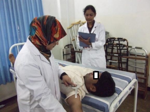
International Journal of Scientific and Research Publications, Volume 3, Issue 2, February 2013                                2
ISSN 2250-3153

Ultrasound is a commonly used electrotherapeutic modality for impingement as well as other forms of tendinitis and muscle injury.
Therapeutic ultrasound is a modality commonly used by physiotherapist. Ultrasound therapy works by driving alternating compression
and rarefaction of sound waves with a frequency of more than 20,000 cycles per seconds. Therapeutic ultrasound may have two types
of benefits, namely thermal effects and non-thermal effects. Thermal effects aid in pain relief whereas non-thermal effects enhance
cell-repair effects of the inflammatory response (H.D., et al 2004). Reduction in pain and induce tissue repair helps in regaining the
reduce range of motion due to SIS.

                                                      Figure 2 Therapeutic Ultrasound apparatus
When recovering from a shoulder injury physiotherapy exercises are an integral part in regaining the range of motion, muscle
elasticity, and strength. Therapeutic exercises can be defined as the use of active or assisted exercises aimed at improving the range of
motion, strength or dynamic neuromuscular control of joint motion, whereas manual therapy can be defined as the use of manually
and/or mechanically applied movement techniques to improve joint motion (Somty.R.2002). Both therapeutic exercises and manual
therapy are commonly used as part of physiotherapy programs aimed at improving shoulder kinematics. Therapeutic exercise focusing
on strengthening the rotator cuff and scapula stabilizing musculature has been shown to be effective in treating shoulder impingement
symptoms (Bang MD et al 2000)
Manual or manipulative therapy encompasses the treatment of health ailments of various etiologies through “hands-on”, physical
intervention. Various manual therapy techniques have proved to be effective for SIS. Stretching reduces capsular tightness (D’Hespeel
C.G., 2004) and a few studies have evaluated the effectiveness of incorporating glenohumeral joint mobilizations for SIS (Bang MD et
al 2000).

     Figure 3 Manual therapy and shoulder exercises

                                                                                                                        www.ijsrp.org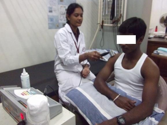
International Journal of Scientific and Research Publications, Volume 3, Issue 2, February 2013 3
ISSN 2250-3153
01.2 Problem Justification
The shoulder is the most movable joint in the body. However, it is an unstable joint because of the range of motion allowed. This
instability increases the likelihood of joint injury.
The most common cause of shoulder pain in the older population is soft tissue injuries. These include frozen shoulder, sub
acromial impingement, rotator cuff tendinopathy or rupture, arthritic conditions, degeneration and, or destruction of the joint and
referred pain from cervical radiculopathy. (Hermoso, F.E, 2009)
Shoulder instability and acromioclavicular joint disorders tend to affect younger people, particularly men who play certain sports
that involve repetitive shoulder movements, such as overarm bowling or throwing and contact sports, such as rugby.
Among the different diagnoses covered by the concept of shoulder pain, the most common is sub acromial impingement syndrome
which is increasingly more common in athletes whose sports involve repetitive overhead motions as well as in the older
population.
This condition is used in our study as majority of sub acromial impingement may be successfully managed with conservative
treatment. (Arcuni SE2000). Specific supervised exercises obtain improvements in the range of movement and muscular function
by restoring the shoulder's mobility and stability. Physiotherapeutic options include several electrotherapy techniques. Among
them Ultrasound (US) Therapy is more popular.
Although adequate literature provides evidence to prove the effectiveness of various therapeutic exercises and manual therapy
techniques (Lori A. et al 2004), the effectiveness of US therapy in the treatment of SIS is still under debate. According to some
studies ultrasound therapy added to conservative treatment of SIS do not provide an additional benefit to the patients, (Celik D et
al 2009, Valma, J et al 2001) and some other studies also reveal that the effectiveness of US in the treatment of SIS is limited.
(Santamato A et al 2009).
In spite of the limited evidence to prove the effectiveness of US in the treatment of SIS it is a widely used electrotherapeutic
modality to treat patients with soft tissue injuries including SIS. Based on this our study was designed to assess whether US
therapy helps in rehabilitation of SIS in the acute and sub-acute stages.
The patients were referred only from Kurunegala and Peradeniya teaching hospitals due to the limited accessibility. Occurrence of
SIS does not differ according to the age and sex of the patients and it was equally distributed in the age group from 18-55. The age
of selected patients was ranged from 18 to 55 according to the limited availability of the patients with pure impingement and the
equal distribution of this condition in this range.
01.3 Objectives
01.3.1 General objectives
To assess the effectiveness of ultrasound therapy when added to manual therapy and shoulder exercises in the
rehabilitation of patients with sub acromial impingement syndrome.
01.3.2 Specific objectives
To assess the effectiveness of the ultrasound with manual therapy and shoulder exercises.
To assess the effectiveness of manual therapy and shoulder exercises in treating sub acromial impingement syndrome
01.4 Hypothesis
01.4.1 Null hypothesis (H0)
Ultrasound therapy is not beneficial when combined with manual therapy and exercises in the physiotherapy
management of patients with SIS to reduce pain, increase range of motion and to reduce shoulder disability
www.ijsrp.org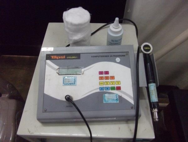
International Journal of Scientific and Research Publications, Volume 3, Issue 2, February 2013                                     4
ISSN 2250-3153

01.4.2 Alternative hypothesis (H1)
                      Ultrasound therapy is beneficial when combined with manual therapy and exercises in the physiotherapy
                      management of patients with SIS to reduce pain, increase range of motion and to reduce shoulder disability.

                                                                 II. REVIEW OF LITERATURE
What is subacromial impingement?
Brotzman (2003) reported that impingement is a chronic inflammatory process produced as the Rotator Cuff muscles (Supraspinatus,
Infraspinatus, Teres minor and Subscapularis) and the Subdeltoid bursa pinched against the coracoacromial ligament and the anterior
acromian when the arm is raised above 80 degrees. The supraspinatus, infraspinatus portion of the rotator cuff is the most common
area of the impingement. This syndrome is commonly seen in throwing sports, in racquet sports, and in swimmers, but can be present
in anyone who uses the arm repetitively in a position over 90 degrees of elevation. (Brotzman SB et al 2003)
02.2 Sub acromial impingement syndrome – most frequent reason of the painful shoulder syndrome.

Painful shoulder syndrome is a frequent cause of visit to a physician. The development of knowledge about the anatomy and
biomechanics of the shoulder allows, based on detailed examination, to precise localization of the shoulder dysfunction. Sub acromial
impingement syndrome is the most common cause of pain as well as of limited motion of the shoulder region. Misdiagnosis and
mistreatment can lead to serious damage of the structures placed in the sub acromial space including the rotator cuff, which along with
the deltoid are responsible for movements of the upper limb in the shoulder joint. If not taken seriously, the problem can cause
irreversible damages which will result in pain and limitations of upper limb movements. (Szyluk, K., et al 2008)

02.3Are ultrasound, laser and exercise superior to each other in the treatment of sub acromial impingement syndrome? A
randomized clinical trial
Calis HT et al, (2011) conducted a study to define and compare the efficacy of ultrasound, laser and exercise in the treatment of SIS.
This was a randomized controlled trial with-pre and post-treatment evaluations. This study was performed on 52 patients with SIS
who were out patients referred to the physical medicine and rehabilitation unit. The patients were put randomly into three groups.
According to this study result they concluded that ultrasound and the laser treatments were not superior to each other in the treatment
of SIS.
02.4Efficacy of standardized manual therapy and home exercise programme for chronic rotator cuff disease: randomized
placebo controlled trial
Bennell K, et al (2010) investigated the efficacy of a program of manual therapy and exercise treatment compared with placebo
treatment delivered by physiotherapists for the people with chronic rotator cuff disease. Participants were randomized and single
blinded, placebo controlled trial. A standardized programme of a manual therapy and home exercise did not confer additional
immediate benefits for pain and function compared with a realistic placebo treatment that controlled for therapists’ contact in middle
aged to older adults with chronic rotator cuff disease.

02.5 Adding ultrasound in the management of soft tissue disorders of the shoulder: a randomized placebo-controlled trial.

A study group (Kurtaiş et al 2004) from Ankara in Turkey conducted a randomized placebo controlled trial in2004, to assess the
effectiveness of ultrasound over a placebo intervention when added to other physical therapy interventions and exercise in the
management of shoulder disorders. This study included Forty patients who were diagnosed by ultrasonography or magnetic resonance
imaging to have a periarticular soft tissue disorder of the shoulder and were randomly assigned to either a group that received true US

                                                                                                                          www.ijsrp.org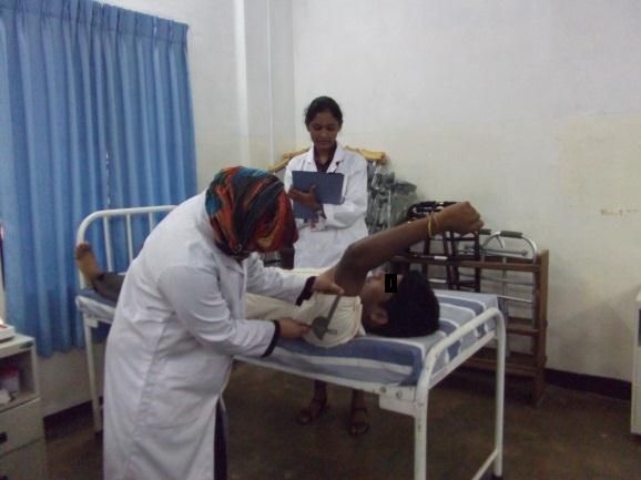
International Journal of Scientific and Research Publications, Volume 3, Issue 2, February 2013                                 5
ISSN 2250-3153

(n=20; mean time since onset of pain=8.7 months, SD=8.8, range=1-36) or a group that received sham US (n=20; mean time since
onset of pain=8.1 months, SD=10.8, range=1-42). Besides true or sham US (10 minutes), superficial heat (10 minutes), electrical
stimulation (15 minutes), and an exercise program (15-30 minutes) were administered to both groups 5 days each week for 3 weeks.
After the intervention subjects showed within-group improvements in reduction of pain, range of motion, Shoulder Disability
Questionnaire scores, and Health Assessment Questionnaire scores with the intervention, but the differences did not reach significance
when compared between the groups. Finally they concluded that the results suggested that true US, compared with sham US, bring no
further benefit when applied in addition to other physical therapy interventions in the management of soft tissue disorders of the
shoulder.

02.6 A systematic review of manipulative therapy for the treatment of shoulder pain.
Pribisevic M, (2010) conducted a systematic review to discuss the evidence for manipulative methods of management of shoulder pain
and chiropractic management techniques used within the literature. A literature search of MEDLINE,CINAHL,MANTIS, the
Cochrane Musculoskeletal Group trials register and the Cochrane Controlled Trials Register was conducted. Search terms included
chiropractic or manipulative therapy and shoulder pain, impingement, rotator cuff, shoulder instability, shoulder joint, treatment or
rehabilitation exercises. Publications were included if they contained shoulder pain or contained a specific clinical diagnosis of a
shoulder pain syndrome in the title; a detailed description of the treatment intervention which was typical of the profession; treatment
performed by a registered practitioner and outcome measures were included in the studies. Exclusion criteria included the diagnosis of
adhesive capsulitis or referred/pathological pain. The articles were reviewed and clinical trials ranked on the Physiotherapy Evidence
Database scale. From a total of 913 retrieved publications, 22 case reports, 4 case series and 4 randomized, controlled trials met the
inclusion and exclusion criteria for this review. The literature contains 2 articles of reasonably sound methodology. The evidence for
chiropractic management of shoulder pain is limited to low level evidence in the form of case reports and case series and 1 small
controlled trial. There is a need for more well-designed, trials investigating multi-modal chiropractic management for shoulder pain.
(Pribisevic M, 2010)

02.7 The value of intermittent ultrasound treatment in sub acromial impingement syndrome.
Celik D et al (2009) conducted this study with thirty-six patients (29 females, 7 males; mean age 51 years; range 40 to 69 years) with
type II SIS who were randomized to two groups to receive intermittent ultrasound (group 1, n=20) and placebo ultrasound (group 2,
n=16) for three weeks (15 sessions). All the patients received the same standard physical therapy and rehabilitation modalities besides
ultrasound treatment. Evaluations were made before and three and six weeks after treatment. Functional results were assessed by the
Constant score, pain was assessed by a visual analog scale, and range of motion was measured. Within-group comparisons showed
significant improvements in both groups three and six weeks after treatment According to the findings the study group concluded that
intermittent ultrasound added to conservative treatment of SIS do not provide an additional benefit to the patients (Celik D et al 2009).
02.8 A prospective double blind placebo-controlled randomized trial of ultrasound in the physiotherapy treatment of shoulder
pain.

A prospective double blind placebo controlled randomized trial to compare the effectiveness of manual therapy and ultrasound with
manual therapy and placebo ultrasound in the treatment of new episodes of unilateral shoulder pain referred for physiotherapy was
carried out by a study group from the University of Bermingham in UK in the year 2007. In a multicenter, double blind, placebo-
controlled randomized trial, participants were recruited with a clinical diagnosis of unilateral shoulder pain from nine primary care
physiotherapy departments in Birmingham, UK. Recruitment took place from January 1999 to September 2001. Participants were 18

                                                                                                                         www.ijsrp.org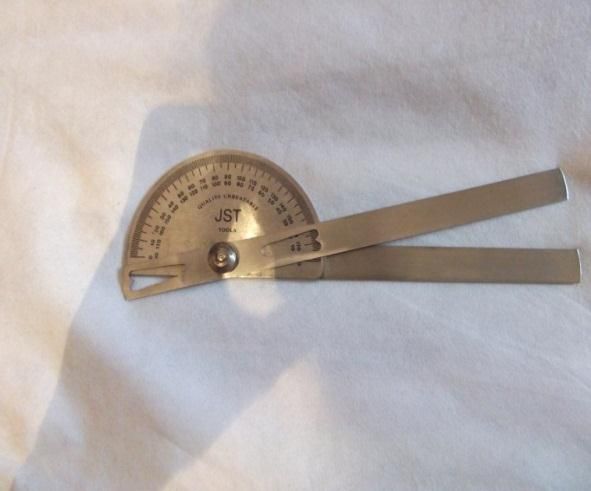
International Journal of Scientific and Research Publications, Volume 3, Issue 2, February 2013 6
ISSN 2250-3153
years old and above. Participants all received advice and home exercises and were randomized to additionally receive manual therapy
plus US or manual therapy plus placebo US. The primary outcome measure was the Shoulder Disability Questionnaire (SDQ-UK).
Outcomes were assessed at baseline, 2 weeks, 6 weeks and 6 months. Analysis was by intention to treat. A total of 221 participants
(mean age 56 years) were recruited. 113 participants were randomized to US and 108 to placebo US. There was 76% follow up at 6
weeks and 71% at 6 months. The mean (95% CI) reduction in SDQ scores at 6 weeks was 17 points (13-26) for US and 13 points (9-
17) for placebo US (P = 0.06). There were no statistically significant differences at the 5% level in mean changes between groups at
any of the time points. The results suggested that true US, compared with sham US, and brings no further benefit when applied in
addition to other physical therapy interventions in the management of soft tissue disorders of the shoulder.
III. METHODOLOGY
This chapter presents the research design, variables, study population and study area, selection criteria, measurements, materials and
apparatus and procedure.
03.1 Research design
This randomized control study was done to investigate the effectiveness of ultrasound therapy in patients with clinical signs and
symptoms of sub acromial impingement syndrome. Selected participants were divided into an intervention group and a control
group.
03.2 Variables
The following variables were used in the study. They can be categorized into two groups.
03.2.1 Independent variables
Manual therapy and shoulder exercises
Manual therapy, shoulder exercises and ultrasound therapy
03.2.2 Dependant variables
Shoulder pain
Shoulder ROM
Shoulder disability
03.3 Study population and study area
The study participants were referred by the orthopedic surgeons, physicians, and rheumatologists from the teaching hospital,
Peradeniya and from the teaching hospital, Kurunegalla. Referred participants were thoroughly assessed with a standard physical
examination by the investigators and the participants eligible with the selection criteria were confirmed for the study. This
research study was carried out at the Department of Physical Medicine (DPM), Teaching hospital, Kurunegalle and at the DPM,
Faculty of Allied Health Sciences, University of Peradeniya, Peradeniya.
03.4 Selection Criteria
The participants who were referred for the study were included and excluded from the study according to the following criteria
www.ijsrp.org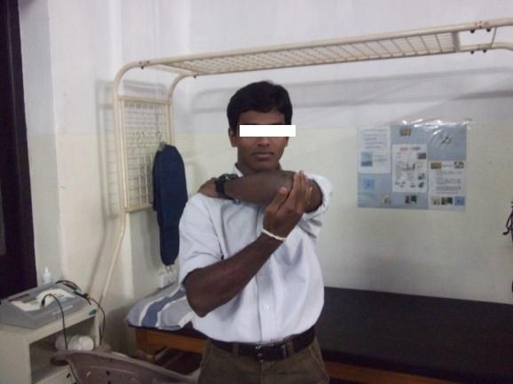
International Journal of Scientific and Research Publications, Volume 3, Issue 2, February 2013 7
ISSN 2250-3153
03.4.1 Inclusion criteria
Age between 18 and 55 year.
Main complaints in the glenohumeral joint region or the proximal arm.
Documented X-ray (XR)/ ultrasound (US) and/or evidence of sub acromial impingement through physical examination.
Presence of two of the following signs indicating SIS: Neer impingement test, Hawkins-Kennedy impingement test, painful arc with
active abduction or flexion.
Pain with two of the following resistance tests: external rotation, internal rotation, abduction, or flexion.
Shoulder disability: greater than or equal to 20/100 (0 = no disability).
Potentially available for the next two months.
Able to understand written and spoken English, Tamil, or Sinhala.
Pain at rest and/or with free movement and/or with movement against resistance with a score on the visual analogue scale of (1-
8)/10.
03.4.2 Exclusion criteria
Primary scapulothorasic dysfunction due to paresis.
Diagnosed instability or previous history of dislocation.
Adhesive capsulitis (frozen shoulder).
More than 1/3 restriction of elevation compared to the unaffected side.
Substantial shoulder weakness or loss of active shoulder function.
Shoulder surgery in the last 12 months on the involved side.
Involvement with sensory and muscular deficit.
Radiological findings of tumor lesions, avascular necrosis, glenoid development defects, acromial bone, severe degenerative signs
affecting inter-articular space and fractures
Ischemic cardiopathy in subacute phase
Cognitive deficit, psychiatric alterations or behavioral disorders that might compromise the patient's collaboration
Unsuitable for electrotherapy: pregnancy, epilepsy, pacemaker, osteosynthesis, undergoing treatment with Sintrom
www.ijsrp.org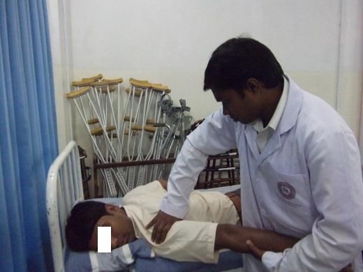
International Journal of Scientific and Research Publications, Volume 3, Issue 2, February 2013                                     8
ISSN 2250-3153

After a complete description of the study was provided, written informed consent was obtained from all subjects or their relatives. The
participants were instructed to abstain from the execution of painful activities of daily living involving the affected shoulder.

03.5 Sampling and sample size

All participants referred from the hospitals were screened for study eligibility. At the end of the evaluation, 30 patients who were
affected by SIS (Neer stage I , 19 right shoulders and 11 left shoulders), had sub-acute pain, fulfilled the selection criteria, agreed to
participate, and signed informed consent statements were enrolled in the study (16 women and 14 men; mean age = 40.22 years, SD
=9.0, range=18-60).

These participants were divided into 2 groups by convenient sampling. The participant who was referred first and eligible for the study
was assigned to the control group. And the second patient was referred to the intervention group. Thus participants were assigned to
groups alternatively. A group of 15 participants were assigned to the control group and 15 participants to the intervention group.

Among the selected participants only 26 participants completed the study and were included in the analysis. One participant was
excluded due to falling on the affected shoulder during the study period. One participant was unable to complete the study due to
family problems. Other 2 participants quit due to unknown reasons.

13 participants from the control group completed the study that received manual therapy and performed shoulder exercises (8 women
and 5 men; mean age = 37.62 years, SD =14.57, range=18-56). And 13 participants from the intervention group also completed the
study that received US therapy in addition to manual therapy and shoulder exercises (6 women and 7 men; mean age =39.92 years, SD
=16.6, range=18-60)

03.6 Interventions

The protocol for both the control and intervention groups involved the application of manual physical therapy and shoulder exercises
for a total of 15 treatment sessions of 40 minutes, over a period of 3 consecutive weeks (5 days per week). At the beginning of the
treatment all participants were given a brief explanation on anatomy and biomechanics of the shoulder complex and a short
description of the etiology and pathology of SIS.

The treatments in the first week aimed at reducing the pain intensity and to prevent further damage and consisted of manual therapy
techniques such as joint mobilization techniques and transverse friction massage and shoulder pendulum exercises.

                                                        Figure 0:4 Joint mobilization techniques

The second and third week aimed at restoring the functional level by increasing ROM, muscle strength and flexibility and consisted of
ROM exercises with rope and pulley, L bar exercises, self-capsular stretching exercises, joint mobilization techniques and

                                                                                                                          www.ijsrp.org
International Journal of Scientific and Research Publications, Volume 3, Issue 2, February 2013                            9
ISSN 2250-3153

strengthening exercises with weights, therapeutic bands, springs and push ups. The standard exercise protocol and manual therapy
were given in order to restore muscular deficits in strength, mobility, and coordination of the rotator cuff and the shoulder girdle
muscles to unload the subacromial space during active movements. And the participants were expected to return to their functional
level without recurrence at the end of the treatment.

                                         Figure 0:5 Abduction and flexion exercises with rope and pulley

                                                  Figure 0:6 Shoulder capsular stretching exercises

                                                   Figure 0:7 Strengthening exercises with weights

                                                                                                                    www.ijsrp.org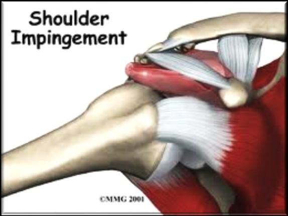
International Journal of Scientific and Research Publications, Volume 3, Issue 2, February 2013 10
ISSN 2250-3153
Figure 0:8 Wall pushups
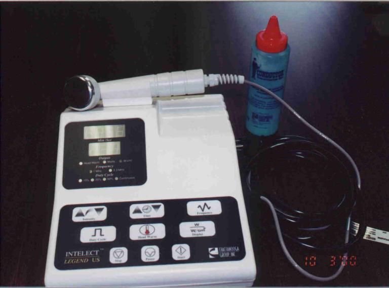
Participants in the intervention group received pulsed ultrasound for 5 minutes with a device that was operated at a frequency of 1
MHz, and an intensity of 1 W/cm2, The treating physical therapist, using the technique of slow circular movements, applied the
transducer head over the superior and anterior periarticular regions of the participant's glenohumeral joint and on the shoulder trigger
points. The treatment was continued from the first treatment day over the 15 day treatment period.
Figure 0:9 the participant being treated with Ultrasound therapy
The participants were treated by final year physiotherapy students with 3 years of clinical experience and supervised by the chief
physiotherapist from the department of physiotherapy, teaching hospital Kurunegala and the head of the department, Department of
Physiotherapy, University of Peradeniya
03.7 Data collection and Procedure
03.7.1 Methods of collection
Data were collected using a standard shoulder assessment form (Annexure 1). Participants were assessed by the group members at the
baseline (before the first treatment session), at the end of first and second weeks and at the end of physical therapy (after the last
treatment session).
03.7.2 Measurements
1. Shoulder pain intensity was measured using the visual analogue scale (scored on a 10 point visual analogue scale)
2. The shoulder disability was measured using the shoulder disability index.
www.ijsrp.orgInternational Journal of Scientific and Research Publications, Volume 3, Issue 2, February 2013 11
ISSN 2250-3153
3. Active painful joint ROM for shoulder flexion, extension, abduction, adduction and external and internal rotation were measured
using a goniometer.
4. And at the end of the treatment, patient satisfaction with treatment was assessed using a brief questionnaire. (Annexure 2)
03.7.3 Materials and apparatus
Materials and apparatus
Figure 10 Universal Goniometer
Figure 11 Ultrasound therapy apparatus
03.7.4 Procedure
1. All shoulder ROMs were measured using the universal goniometer. Shoulder flexion, abduction, and external rotation were
measured in the supine position whereas shoulder extension and external rotation were measured in the prone position.
The axis of goniometer was placed at 2.5cm inferior to the lateral aspect of the acromion process for shoulder flexion and
extension, at 1.3cm inferior and lateral to the coracoid process for abduction and at the olecranon process of the ulna for shoulder
internal and external rotation.
www.ijsrp.orgInternational Journal of Scientific and Research Publications, Volume 3, Issue 2, February 2013 12
ISSN 2250-3153
Figure 0:12 Goniometric measurement of shoulder ROM
2. Shoulder pain intensity was measured in various activities involving the shoulder. The participants were asked to mark the pain
intensity for each activity on separate visual analogue scales. The right end of the VAS was defined as "worst pain imaginable’’, the
left end as "no pain ". Each level was recorded in a table (Table). A score was then calculated out of 100 with higher scores
reflecting higher pain levels.
3. Shoulder disability level was measured in various activities involving shoulder. The right end of the VAS was defined as "so
difficult required help", the left end as "no difficulty".The level of difficulty in doing each activity is marked in the table. A score
was then calculated out of 100 with higher scores reflecting higher disability levels.
03.8 Ethical clearance
Ethical clearance was obtained by requesting from the ethical review committee of the faculty of Allied Health Sciences,
University of Peradeniya.
03.10 Data analysis
All analyses were performed with MINITAB statistical software (version14). Frequency distributions as well as means and standard
deviations were used for descriptive purposes. At the baseline, differences in age was analysed using the two-sample Wilcoxon rank
sum test. Differences between treatment groups in change scores at the baseline and at the end of each treatment sessions over a period
of 3 consecutive weeks were analysed with the two-sample Wilcoxon rank sum test. Repeated measurements obtained before and after
treatments within groups were also analysed with the two-sample Wilcoxon rank sum test. The Kruskal Wallis test was performed to
estimate differences between age groups for each studied outcome. The alpha level for significance was set at 0.05.
IV. RESULTS
Distribution of age groups among control and intervention groups
The age of the participants of both the control and intervention groups ranged from 18-55. The above age group was categorized into 4
groups.
www.ijsrp.orgInternational Journal of Scientific and Research Publications, Volume 3, Issue 2, February 2013 13
ISSN 2250-3153
Chart of age con
5
4
3
Count
2
1
0
18-29 30-40 41-50 51-60
age con
Figure 13 Histogram showing the distribution of age groups in the control group
The figure 12 shows the histogram of distribution of the age groups in the control group.
Chart of age int
5
4
3
Count
2
1
0
18-29 30-40 41-50 51-60
age int
Figure 14 Histogram showing the distribution of age groups in the intervention group
The figure 13 is a histogram that shows the distribution of different age groups in the intervention group.
The ‘Kruscal Wallis test’ was used to compare the distribution of age in the control and intervention groups. According to the
Wilcoxon signed rank test, there was no significant difference between distribution of age groups in the control group and the
intervention group.
04.2 Distribution of male and female participants in control group and the distribution of male and female participants in
intervention group
Both male and female participants were included in the study. The control groups had 5 male and 8 female participants. The
intervention group had 7 male and 6 female participants. The figure 14 is the pie chart that shows the distribution of gender in both
groups.
www.ijsrp.orgInternational Journal of Scientific and Research Publications, Volume 3, Issue 2, February 2013 14
ISSN 2250-3153
Pie Chart of sex control, sex intervention
sex control sex intervention Category
female
male
Figure 15 the pie chart showing the distribution of male and female participants in the control and the intervention group
04.3 Distribution of involved side (right/left) among selected participants in control group and the distribution of involved side
among selected participants in intervention group
Some participants involved in the study had shoulder impingement on the right side. And some of them had involved the left side. All
the participants were right handed.
Pie Chart of side con, side int
side con side int Category
left
right
Figure 16 the pie chart showing the distribution of affected sides in the control and intervention groups
The above pie charts show the distribution of participants involved side of shoulder in the control and intervention group.
04.4 Normality tests for baseline measurements of pain, disability and ROM in control and intervention groups
The normality test was used to find out whether the baseline measurements for pain, shoulder disability and shoulder ROM were
normally distributed. The results showed that none of the above baseline characteristics follow the normal distribution. Hence the
‘Wilcoxon signed rank test’ was used to compare for any difference between the control and intervention groups for baseline
measurements.The Wilcoxon signed rank test hypotheses were H0: median = hypothesized median versus H1: median ≠ hypothesized
median. The confidence level was set at 95.0.
www.ijsrp.orgInternational Journal of Scientific and Research Publications, Volume 3, Issue 2, February 2013 15
ISSN 2250-3153
04.5 The comparison for measurements between male and female participants at the baseline and the improvement
According to Wilcoxon Rank sum test;
Table 1 the comparison for measurements between male and female participants at the baseline and the improvement
Control Intervention
Measurement
Male Female Difference Male Female Difference
(M) (M) (M) (M)
Initial 44.4 57.625 NS 69.41 49.42 NS
Visual Analogue Scale Pain 49.25 29.32 NS 53.65 31.33 NS
reduction
Initial 43.34 54.33 NS 56.26 38.73 NS
Disability 47.08 33.73 NS 38.66 21.08 NS
Shoulder Disability
reduction
Index
Initial 120.6 167.00 NS 113.0 143.33 NS
Abduction
After 10.2 49.37 NS 24.28 51.16 NS
treatment
External Initial 40.0 44.0 NS 44.2 53.0 NS
rotation
Range of After 27.0 23.87 NS 15.0 16.0 NS
motion treatment
Initial 61.75 59.8 NS 61.83 61.42 NS
Internal
After 14.4 21.37 NS 22.85 13.16 NS
rotation
treatment
*= pInternational Journal of Scientific and Research Publications, Volume 3, Issue 2, February 2013 16
ISSN 2250-3153
04.6 Comparison between involved side of the shoulder for initial measurements and improvement in control and intervention
groups
According to the Wilcoxon rank sum test;
Table 2 Comparison between involved side of the shoulder for initial measurements and improvement in control and
intervention groups
Control Intervention
Right Left Difference Right Left Difference
(M) (M) (M) (M)
Initial 61.62 38 NS 63.56 47.6 NS
Visual Analogue Scale Pain 48.57 30.5 NS 43.78 38 NS
reduction
Initial 54.99 43.56 NS 52.33 34.45 NS
Disability 47.04 33.86 NS 32.87 25.46 NS
Shoulder Disability
reduction
Index
Initial 114.17 NS 112.5 136 NS
Abduction
After 53.4 NS 47.5 32.3 NS
treatment
External Initial 36 35 NS 44.44 53.67 NS
Range of
rotation
motion After 25.85 36.25 NS 18.33 29.7 NS
treatment
Initial 54.5 63.75 NS 52.86 57 NS
Internal
After 24.8 15 NS 21.43 9 NS
rotation
treatment
*= pInternational Journal of Scientific and Research Publications, Volume 3, Issue 2, February 2013 17
ISSN 2250-3153
04.7 Comparison between different age groups for initial measurements and improvement in the control and intervention
groups
According to the Kruskal Wallis test;
Table 3 Comparison between different age groups for initial measurements and improvement in the control group
18-29 30-39 40-50 51-60 Difference
(M) (M) (M) (M)
55. 42 62. 26 NS
Initial
Visual Analogue After treatment 44. 24 52. 14. NS
Scale
51. 53 47. 47. NS
Initial
Shoulder Disability After treatment 44. 40 40. 41. NS
Index
145 18 14 17 NS
Initial
Abduction After treatment 52. 0 53 NS
48 30 36. 70 NS
Shoulder External rotation Initial
Range of motion After treatment 66. 80 63 75 NS
63. 90 68. 50 NS
Internal rotation Initial
After treatment 16 0 10. 25 NS
*= pInternational Journal of Scientific and Research Publications, Volume 3, Issue 2, February 2013 18
ISSN 2250-3153
Initial
Abduction After treatment 21. 20 40 54 NS
49. 70 45 44 NS
Shoulder External rotation Initial
Range of motion After treatment 15. 20 15 19 NS
60. 90 60 62 NS
Internal rotation Initial
After treatment 27. 0 20 12 NS
*= pInternational Journal of Scientific and Research Publications, Volume 3, Issue 2, February 2013 19
ISSN 2250-3153
The above table shows the test results obtained by using Wilcoxon rank sum test for overall improvement after three weeks of
treatment in pain, disability, abduction, internal rotation and external rotation in both control and intervention groups. The difference
between initial measurements and measurements taken at the end of three weeks treatment period has been calculated using the above
test.
If the P value is less than 0.05 the null hypothesis is rejected at 5% of significant level that the two sample medians is not equal. The
measurements obtained before and after the treatment were taken as the two samples for this test. The above table shows that the P
values for improvements at the end of treatment is less than 0.05 and there is significant improvement at the end of three weeks of
treatment in both intervention and control groups.
Table 6 within group comparison at the baseline and after three weeks
Control group Intervention group
Initial After 3 Difference Initial After 3 Difference
(M) weeks (M) weeks
(M) (M)
Vas scale 52.53 10.95 ** 58.65 16.66 **
Disability 50.10 08.13 ** 46.82 16.23 **
Abduction 138.46 172.76 * 128.30 165 **
ROM
Ex. Rotation 42.46 67.53 ** 48.30 63.76 **
Int. rotation 65.53 79.69 ** 61.61 80.00 **
*= pInternational Journal of Scientific and Research Publications, Volume 3, Issue 2, February 2013 20
ISSN 2250-3153
Vas2 – Vas1- The difference between the initial measurement and at the end of the treatment at the 1 st week for pain.
Vas3 – Vas2- The difference between the measurement and at the end of the treatment at the 1 st week and the second week for pain.
Vas4 – Vas3- The difference between the measurement and at the end of the treatment at the 2nd week and the 3rd week for pain.
Vas4 – Vas1- The difference between the initial measurement and at the end of the treatment at the 3 st week for pain.
The table 2 shows the test results obtained by using Wilcoxon rank sum test for comparison of pain measured with VAS between
control and intervention groups for baseline measurements and improvements in first, second, third weeks and overall improvement at
the end of treatment.
Table 8 intergroup comparison for disability
Weekly N N for test Wilcoxon P Value Estimated
improvement statistic Median
Initial 13 13 54.0 0.576 2.470
Dis2 – Dis1 13 13 18.0 0.059 -7.000
DIs3 – dIs2 13 13 57.0 0.442 3.130
Dis4 – Dis3 13 13 45.0 1.000 -0.145
Dis4 – Dis1 13 13 64.0 0.208 10.40
Dis2 – Dis1- - The difference between the initial measurement and at the end of the treatment at the 1 st week for shoulder disability.
DIs3 – dIs2- The difference between the measurement and at the end of the treatment at the 1st week and the second week for shoulder
disability.
Dis4 – Dis3- The difference between the measurement and at the end of the treatment at the 2nd week and the 3rd week for shoulder
disability.
Dis4 – Dis1- The difference between the initial measurement and at the end of the treatment at the 3 st week for shoulder disability.
Table 3 shows the test results obtained by using Wilcoxon rank sum test for comparison of disability measured with shoulder
disability index between control and intervention groups for baseline measurements and improvements in first, second, third weeks
and overall improvement at the end of treatment
Table 9 Intergroup comparison for abduction ROM
Weekly N N for test Wilcoxon P Value Estimated
improvement statistic Median
Initial 13 11 43.0 0.398 10.00
Abd2 – Abd1 13 11 36.0 0.824 1.5
Abd3 – Abd2 13 10 26.0 0.919 -1.00
Abd4 – Abd3 13 10 40.0 0.221 5.00
Abd4 – Abd1 13 12 44.0 0.724 5.00
Abd2 – Abd1- The difference between the initial measurement and at the end of the treatment at the 1 st week for shoulder abduction
ROM.
Abd3 – Abd2 -The difference between the measurement and at the end of the treatment at the 1 st week and the second week for
shoulder abduction ROM.
www.ijsrp.orgInternational Journal of Scientific and Research Publications, Volume 3, Issue 2, February 2013 21
ISSN 2250-3153
Abd4 – Abd3 -The difference between the measurement and at the end of the treatment at the 2nd week and the 3rd week for shoulder
abduction ROM.
Abd4 – Abd1- the difference between the initial measurement and at the end of the treatment at the 3 st week for shoulder abduction
ROM.
The above table shows the test results obtained by using Wilcoxon rank sum test for comparison of abduction ROM measured with
the Universal Goniometer between control and intervention groups for baseline measurements and improvements in first, second, third
weeks and overall improvement at the end of treatment.
Table 10 Intergroup comparison for external rotation
Weekly N N for test Wilcoxon P Value Estimated
improvement statistic Median
Initial 13 12 27.0 0.367 -8.00
Ex.ro2 Ex.ro1 13 11 35.0 0.894 0.00
Ex.ro3 Ex.ro2 13 11 39.0 0.625 2.00
Ex.ro4 Ex.ro3 13 11 52.0 0.100 6.00
Ex.ro4 Ex.ro1 13 11 48.5 0.182 11.5
Ex.ro 2 – Ex.ro 1- The difference between the initial measurement and at the end of the treatment at the 1 st week for shoulder external
rotation ROM.
Ex.ro 3 – Ex.ro 2 -The difference between the measurement and at the end of the treatment at the 1 st week and the second week for
shoulder external rotation ROM.
Ex.ro 4 – Ex.ro 3 -The difference between the measurement and at the end of the treatment at the 2nd week and the 3rd week for
shoulder external rotation ROM.
Ex.ro 4 – Ex.ro 1- The difference between the initial measurement and at the end of the treatment at the 3 st week for shoulder external
rotation ROM.
The above table shows the test results obtained by using Wilcoxon rank sum test for comparison of external rotation ROM measured
with the Universal Goniometer between control and intervention groups for baseline measurements and improvements in first, second,
third weeks and overall improvement at the end of treatment.
Table 11 Intergroup comparison for internal rotation
Weekly N N for test Wilcoxon P Value Estimated
improvement statistic Median
Initial 13 10 33.0 0.610 9.00
In.ro2 –In.ro1 13 11 50.5 0.131 5.00
In.ro3 –In.ro2 13 11 50.5 0.131 5.00
In.ro4 –In.ro3 13 07 19.5 0.398 0.00
In.ro4 –In.ro1 13 8 15.5 0.779 0.00
In.ro 2 – In.ro 1- The difference between the initial measurement and at the end of the treatment at the 1st week for shoulder
internal rotation ROM.
In.ro 3 – In.ro 2 -The difference between the measurement and at the end of the treatment at the 1 st week and the second
week for shoulder internal rotation ROM.
www.ijsrp.orgInternational Journal of Scientific and Research Publications, Volume 3, Issue 2, February 2013 22
ISSN 2250-3153
In.ro 4 – In.ro 3 -The difference between the measurement and at the end of the treatment at the 2nd week and the 3rd week
for shoulder internal rotation ROM.
In.ro 4 – In.ro 1- The difference between the initial measurement and at the end of the treatment at the 3 st week for shoulder
internal rotation ROM.
The above table shows the test results obtained by using Wilcoxon rank sum test for comparison of internal rotation ROM
measured with the Universal Goniometer between control and intervention groups for baseline measurements and
improvements in first, second, third weeks and overall improvement at the end of treatment.
Table 12 Comparison between groups before treatments and after each week
Control group Intervention Differences
(M) group(M)
Initial 52.53 58.65 NS
1st week 35.69 40.00 NS
Vas scale nd
2 week 18.64 30.22 NS
3rd week 10.95 16.66 *
Initial 50.10 46.82 NS
1st week 31.43 34.77 NS
Disability 2nd week 16.40 24.93 NS
3rd week 08.13 16.23 NS
Initial 138.46 128.30 NS
1st week 156.76 143.08 NS
2nd week 167.23 157.07 NS
Abduction rd
3 week 172.76 165.00 NS
Initial 42.46 48.30 NS
1st week 51.23 56.38 NS
External nd
2 week 58.23 62.07 NS
ROM
rotation 3rd week 67.53 63.76 NS
Initial 65.53 61.61 NS
Internal st
1 week 71.92 77.23 NS
rotation nd
2 week 75.07 78.38 NS
3rd week 79.69 80.00 NS
*= pInternational Journal of Scientific and Research Publications, Volume 3, Issue 2, February 2013 23
ISSN 2250-3153
exercises alone and the groups which received ultrasound therapy in addition to them have shown no significant difference in the
improvement with the treatment regarding pain, disability and ROM.
Table 13 Overall improvements in percentage
Control group Improvement Interventio Improvement
(M) Percentage (%) n group Percentage (%)
(M)
I 52.53 58.65
1 35.69 40.00
VAS scale 2 18.64 79.15% 30.22 71.59%
3 10.95 16.66
I 50.10 46.82
1 31.43 34.77
Disability 2 16.40 83.77% 24.93 65.33%
3 08.13 16.23
I 138.46 128.30
1 156.76 143.08
2 167.23 24.77% 157.07 28.60%
Abduction 3 172.76 165.00
I 42.46 48.30
1 51.23 56.38
External 2 58.23 59.04% 62.07 32.00%
ROM
rotation 3 67.53 63.76
I 65.53 61.61
Internal 1 71.92 77.23
rotation 2 75.07 21.60% 78.38 29.84%
3 79.69 80.00
The above table shows the percentage for overall improvement in both control and intervention groups. The pain has been reduced by
79.15% in the control group whereas in the intervention group it has been reduced by 71.59%. The disability has been reduced by
83.77% in the control group whereas in the intervention group it has been reduced by 65.33%.
The Abduction has been improved by 24.77% in the control group whereas in the intervention group it has been improved by 28.60%.
Internal rotation it has been improved by 59.04% in the control group and in the intervention group it has been improved by 32.00%.
External rotation has been improved by 21.60% in the control group whereas in the intervention group it has been improved by
29.84%
www.ijsrp.orgInternational Journal of Scientific and Research Publications, Volume 3, Issue 2, February 2013 24
ISSN 2250-3153
V. DISCUSSION
The aim of this study was to identify whether ultrasound therapy has an additional effect when combined with manual therapy and
shoulder exercises in the treatment of patients with SIS. This is a randomized control trial which has not been done before in Sri
Lanka.
05.1 Major findings
The measurements in VAS, disability index and shoulder ROM shows significant improvement in both control and intervention
groups at the end of three weeks treatment period. In the control group VAS for pain measurement has reduced from 52.54±23.24 to
10.95 ± 9.52, the disability index has reduced from 50.11±19.11 to 8.13 ± 7.03. And the ROM for abduction external rotation and
internal rotation were increased from abduction 138.5 ±46.2 to 172.77 ±16.90, external rotation 45.38±19.73 to 67.54± 13.15, and
internal rotation 42.46 ±21.88 to 79.69 ± 12.33
In the intervention group VAS for pain measurement has reduced from 58.65 ±20.56 to 16.66± 13.02, the disability index has reduced
from 46.83±19.80 to 16.24 ± 13.26. And the ROM for abduction external rotation and internal rotation were increased from abduction
128.31 ±31.84 to 165.00 ±21.41, external rotation 48.31±17.72 to 80.00 ±14.72, and internal rotation 48.31±17.72 to 63.77± 13.40.
Between group comparisons for the improvement in patient condition regarding pain, disability and shoulder ROM for abduction,
internal rotation and external rotation after the three weeks treatment or at the end of first or second week have shown no statistically
significant difference between the groups.
The participants’ age ranged from 18-55. The distribution of age among the control and intervention groups has no significant
difference. And the comparison between difference for baseline measurements and overall improvement for pain, shoulder disability
and shoulder ROM has not shown significant difference according to the different age groups involved in this study either in the
control or intervention groups.
Most of the participants had been affected by their dominant shoulder. But some of them had been affected by their non-dominant
shoulder. But according to the results comparison between baseline measurements and overall improvement has no significant
difference in either group.
In this study both male and female participants were included. But comparison between female and male participants for baseline
measurements and overall improvement has no significant difference in control or intervention groups.
Thus the result shows that there is no gender, side or age associated with improvement.
05.2 Interpretation of results
It has been found by previous studies that there is an equal effectiveness of physiotherapist-led exercises compared with surgery
(Kromer, T.O., et al 2009). Other studies have concluded that manual therapy and exercise seem effective for shoulder impingement
(Michener, L.A., et al 2004) Results of the study conducted by Roy, J.S., et al. (2007) suggested that a 4-week program including
motor control and strengthening exercises reduces shoulder pain and improves function in persons with SIS.
In the present study both the control and intervention groups has significant improvement after three weeks of treatment. There is
significant improvement in pain reduction, disability and shoulder ROM from baseline measurements to the first, second and third
week after treatment in both the groups. The control group received manual therapy and shoulder exercises alone and these treatments
have led to the significant improvement in the control group. These findings support the previous study results and emphasize the
importance of manual therapy and shoulder exercises in the treatment of patients with SIS.
www.ijsrp.orgInternational Journal of Scientific and Research Publications, Volume 3, Issue 2, February 2013 25
ISSN 2250-3153
The effects of therapeutic ultrasound are still being disputed. To date, there is still very little evidence to explain how ultrasound
causes a therapeutic effect in injured tissue. Nevertheless practitioners worldwide continue to use this treatment modality relying on
personal experience rather than scientific evidence.
Therapeutic ultrasound may have two types of benefit: Thermal effects and non-thermal effects. Thermal effects aids in pain relief
whereas non thermal effects enhance the cell-repair effects of the inflammatory response. (H. D., et al. 2004).Reduction in pain and
induced tissue repair are believed to help in regaining the reduced ROM due to SIS.
According to the study conducted by Ceilik D et al (2009), intermittent ultrasound added to conservative treatment of SIS do not
provide an additional benefit to the patients with SIS. There was little evidence that active therapeutic ultrasound is more effective
than placebo ultrasound for treating people with pain or a range of musculoskeletal injuries or for promoting soft tissue healing
according to some systematic reviews.
The present study also shows that there is no significant difference between the control group which received manual therapy and
shoulder exercises and intervention group which received ultrasound therapy in addition. According to the present study results,
ultrasound therapy does not have an additional effect when combined with shoulder exercises and manual therapy.
05.3 Significance of the study
Although various studies have been done in UK, Turkey and some other country (Pribicevic, M, et al 2010) and the values of
individualized treatment according to the findings of patient’s physical assessments (Kromer, T.O, et al 2010) the studies have not
been done related to this in Sri Lanka.
The number of patients per physiotherapist is far more in Sri Lanka when compared with advanced countries. This has been a major
factor which leads to the limited availability of time that a therapist can spend for a patient. The patients also tend to believe that these
modalities are effective than exercises and pay less attention to exercises.
The present study aimed not only at proving that ultrasound therapy does not have an added effect when combined with shoulder
exercises and manual therapy, but also at emphasizing the importance of manual therapy and shoulder exercises in relieving
symptoms in patients with SIS without the use of any electrotherapeutic modality.
05.4 Strength of the study
The strength of this study is enhanced by its methods and design. Internationally accepted measuring tools and scales have been used
to collect data. The shoulder pain and disability index used in this study has shown to be valid and highly responsive in assessing
shoulder pain and function (MacDermid, J., et al 2006); it is therefore highly recommended for use in patients with SIS (Purdy, S., et
al 2005).
Participants were enrolled according to inclusion and exclusion criteria. All the group members have been involved in the
interventions, data collection and the measurements. Every group member treated a similar number of participants from both the
groups. Treatments were given according to standard treatment protocols and ultrasound therapy was given according to standard
parameters.
Special training regarding manual therapeutic techniques such as shoulder joint mobilization including anterior posterior and inferior
glides of 1st and 2nd grades, deep friction massage, shoulder capsular stretching and strengthening exercises was provided by the staff
of the Department of Physiotherapy, Faculty of Allied Health Sciences, to each member of the team to reduce potential errors in
treatments and taking measurements. Treatments were done under the supervision and instructions of qualified physiotherapists.
www.ijsrp.orgInternational Journal of Scientific and Research Publications, Volume 3, Issue 2, February 2013 26
ISSN 2250-3153
All the data has been analyzed through the MINITAB which is recognized as an accurate statistical software package. Data analysis
was done with the assistance from qualified and experienced statisticians and the Department of Statistics, of the Faculty of Science,
University of Peradeniya
05.5 Limitations of the study
This study has been done in a limited time period. Therefore duration of treatment has been only 3 weeks and has led to a lack of
follow up period.
Due to limited time and limited accessibility for the investigators to hospitals the sample size was limited to 30.
Among them only 26 participants completed the study and there were 4 drop outs because of distance to the faculty of Allied Health
Sciences.
Some patients referred from the Teaching Hospital, Peradeniya refused to give consent to participate in the study due to distance to the
faculty and hence, could not be included in the study.
05.6 Future directions
The present study has shown that ultrasound therapy has no additional effect when combined with other physiotherapy interventions
for SIS. The previous studies and systematic reviews also concluded that US therapy has no added effect.
Further studies are needed to compare the effectiveness of various other electrotherapy modalities when combined with manual
therapy and exercises in the treatment of musculoskeletal conditions to improve the knowledge of how and when to apply appropriate
modalities, manual therapy, therapeutic exercises and other physical therapy interventions for various conditions.
Studies to assess the effectiveness of individual assessment based treatment for various conditions should be encouraged.
Although the analytic studies regarding physiotherapy are being done, only few clinical trials have been done which are related to
physiotherapy, in Sri Lanka. Therefore more and more trials should be encouraged in the development of physiotherapy as a
profession and to improve the knowledge and the quality of physiotherapists in Sri Lanka
VI. CONCLUSION
The results of this research show that the participants who received manual therapy and shoulder exercises alone as well as the group
which received ultrasound therapy in addition to manual therapy and shoulder exercises had significant improvement at the end of
three weeks of treatment and at the end of each week.
The study finally concludes that ultrasound therapy has no additional benefit when combined with manual therapy and shoulder
exercises in the treatment of patients with SIS to reduce pain, disability and to improve ROM.
ACKNOWLEDGMENT
This research project would not have been succeeding without the support of many people who were supported us throughout the
whole project.
First we offer our most sincere gratitude to our supervisor, Dr (Mr.) Asela J.B. Ratnayeke who has supported us throughout our thesis
with his patience and knowledge whilst allowing us to work in our own way. This thesis had been completed due to his
encouragement and excellent guidance. He allowed us to engage with the orthopedic clinic in Teaching Hospital, Peradeniya where
we found some of the cases for our sample. We offer our special thanks to him for his immeasurable great contribution throughout our
carrier.
www.ijsrp.orgInternational Journal of Scientific and Research Publications, Volume 3, Issue 2, February 2013 27
ISSN 2250-3153
We would like to express our deepest gratitude to our co supervisor Mrs. Esther Liyanage for her guidance, encouragement, patience,
and for providing us with an excellent atmosphere for carrying out this research project. We would like to thank Mrs.
S.Wadugodapitiya who allowed us to use DPM of FAHS for our treatment procedures and who provided the best suggestions always.
We would like to thank Dr (Mr.) H.G. Suraweera for allowing us to receive patients from orthopedic clinic Teaching Hospital
Peradeniya.
We are obliged to the staff of ELTU of faculty of Allied Health Sciences for their invaluable support and guidance from the beginning
to the end, to complete this study successfully.
We offer our gratitude to Mrs. Padmini Sathkorala, Head of the Department, and the staff, Department of Physical Medicine, Teaching
Hospital Kurunegala. We had the opportunity to collect data in Kurunegala DPM as she accepted our request. Throughout the whole
period she and the Physiotherapy staff gave their maximum support for us.
Our special thanks should be given to Dr. (Mr.) R.D. Peris, Senior lecturer of Faculty of Dental Science and Demonstrations’ staff,
Faculty of Science who joined with us for the complex part of analyzing data in an accurate statistical method.
We wish to express our deep sense of gratitude to the each and every one who were in our sample. They spent their valuable time with
us without any problem. They were the people who played the key role of completing this research project.
Finally, with great respect, we would like to express our heartfelt thanks to our beloved parents for their blessings, encouragements,
caring and financial support. We express our deepest gratitude to our seniors, friends and classmates for their help and wishes for the
successful completion of this research
REFERENCES
[1] Aktas, I., Akgun, K., Cakmak, B., 2007. Therapeutic effect of pulsed electromagnetic field in conservative treatment of subacromial impingement syndrome. Clin
Rheumatol, 26(8), pp. 1234-9.
[2] Arcuni, S.E., 2000. Rotator cuff pathology and subacromial impingement. Nurse Pract, 25(5), pp. 58-61, 65-6.
[3] Bang, M.D., Deyle, G.D., 2000. Comparison of supervised exercise with and without manual physical therapy for patients with shoulder impingement syndrome.
J Orthop Sports PhysTher, 30, pp. 126–137.
[4] Bennell, K., Coburn, S., Green, S., Harris, A., Staples, M., Forbes, A., Buchbinder, R., 2010. Efficacy of standardised manual therapy and home exercise
programme for chronic rotator cuff disease. BMJ, 340, pp. 27-56.
[5] Brotzman, S.B., Wilk, K.E., 2003. Clinical orthopedic rehabilitation, 2nd ed., United Kingdom-Mosby.
[6] Calis, H.T., Berberoglu, N., Calis, M., 2011. Are ultrasound, laser and exercise superior to each other in the treatment of subacromial impingement syndrome? A
randomized clinical trial. Eur J Phis Retail Med.
[7] Celik, D., Atalar, A.C., Sahinkaya, S., Demirhan, M., 2009. The value of intermittent ultrasound treatment in subacromial impingement syndrome. Acta Orthop
Traumatol Turc. 43(3), pp. 243-7.
[8] D'Hespeel, C.G., 2004. Current concepts: Rehabilitation of patients with shoulder Impingement and tight posterior capsule. OrthopPract, 16, pp. 9–13.
[9] H.D., 2004. Influence of Therapeutic Ultrasound on Skeletal Muscle Regeneration Following Blunt Contusion. International Journal of Sports Medicine, 25, pp.
73-77.
[10] Hébert, L.J., Moffet, H., McFadyen, B.J., Dionne, C.E., 2002. Scapular behaviour in shoulder impingement syndrome. Arch Phys Med Rehabil, 83(1), pp. 60-9.
[11] Hermoso, F.E., Calvo, E., (2009), Shoulder pain in the elderly, Aging Health, 5, (5) , 711-718.
[12] Hyvönen, P., Melkko, J., Lehto, V.P., Jalovaara, P., 2003. Involvement of the subacromial bursa in impingement syndrome of the shoulder as judged by
expression of tenascin-C and histopathology. J Bone Joint Surg Br. 85(2), pp. 299-305.
[13] Kromer, T.O., de Bie, R.A., Bastiaenen, C.H.,(2010), Effectiveness of individualized physiotherapy on pain and functioning compared to a standard exercise
protocol in patients presenting with clinical signs of subacromial impingement syndrome. A randomized controlled trial. BMC Musculoskelet Disord.9; 11:114.
[14] Kromer, T.O., Tautenhahn, U.G., de Bie, R.A., Staal, J.B., Bastiaenen, C.H.,(2009) Effects of physiotherapy in patients with shoulder impingement syndrome: a
systematic review of the literature. J Rehabil Med. 41(11):870-80.
[15] Kurtaiş., Gürsel, Y., Ulus, Y., Bilgiç, A., Dinçer, G., van der Heijden, G.J., (2004) Adding Ultrasound in the management of soft tissue disorders of the
shoulder: a Randomized placebo-controlled trial. Phys Ther. 84(4):336-43
[16] Lori, A., Michener, Matthew, K., Walsworth, Evie, N., Burnet, 2004. Effectiveness of Rehabilitation for Patients with Subacromial Impingement Syndrome: A
Systematic Review. hand ther.17, pp. 152–164.
[17] MacDermid, J., Solomon, P., Prkachin, K.,(2006). The shoulder pain and disability index dmonstrates factor, construct and longitudinal validity. BMC
Musculoskeletal Disorders 7, 12.
[18] Michener, L.A., Walsworth, M.K., Burnet, E.N., (2004) Effectiveness of rehabilitation for Patients with subacromial impingement syndrome: a systematic review.
J Hand Ther.17(2),152-64.
[19] Pribicevic, M., Pollard, H., Bonello, R., De Luca, K., 2010. A systematic review of manipulative therapy for the treatment of shoulder pain, Manipulative
PhysiolTher, 33(9), pp. 679-89.
www.ijsrp.orgYou can also read