Effects of The Prolong Life With Nine Turn Method (Yan Nian Jiu Zhuan) Qigong On Brain Functional Changes in Patients With Chronic Fatigue ...
←
→
Page content transcription
If your browser does not render page correctly, please read the page content below
Effects of The Prolong Life With Nine Turn Method
(Yan Nian Jiu Zhuan) Qigong On Brain Functional
Changes in Patients With Chronic Fatigue Syndrome
in Terms of Fatigue and Quality of Life
Fangfang Xie
Shanghai University of Traditional Chinese Medicine
Ziji Cheng
Shanghai University of Traditional Chinese Medicine
Yuanjia Gu
Shanghai University of Traditional Chinese Medicine
Ziying Chen
Shanghai University of Traditional Chinese Medicine
Chaoqun Xie
Shanghai University of Traditional Chinese Medicine
Fei Yao ( doctoryaofei@126.com )
Shanghai University of Traditional Chinese Medicine
Yanli You
ChangHai Hospital, Naval Medical University
Research Article
Keywords: chronic fatigue syndrome, prolong life with nine turn method, fatigue, quality of life, ALFF, FC
Posted Date: October 20th, 2021
DOI: https://doi.org/10.21203/rs.3.rs-963598/v1
License: This work is licensed under a Creative Commons Attribution 4.0 International License.
Read Full License
Page 1/25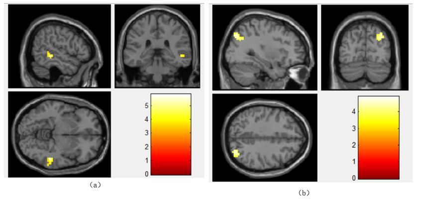
Abstract
Background: Chronic fatigue syndrome (CFS) is characterized by persistent fatigue, which often leads to
physical and psychological damage. The prolong life with nine turn method (PLWNT) Qigong is
considered one of the complementary treatments for improving symptoms in patients with CFS. However,
the neurophysiological relevance of these effects remains poorly understood. In this study, we used
functional magnetic resonance imaging (fMRI) to study the effects of PLWNT intervention on the neural
circuits in subjects with CFS.

Methods: Thirty four CFS patients were randomly divided into a PLWNT group (who received Qigong
exercises) and a control group (who received cognitive behavioral therapy, CBT). Both groups were taught
by a highly qualified professor at the Shanghai University of Traditional Chinese Medicine once a week
and were supervised online during the remaining 6 days at home, over 12 consecutive weeks. We
calculated the regional rs-fMRI index ALFF for all subjects. To study the changes of the brain network, we
used the brain regions with significant differences in ALFF as the regions of interest for whole-brain FC
analysis. The MFI-20 and SF-36 were used for clinical symptom assessment to explore the possible
correlation between the rs-fMRI indicators and clinical variations.

Results: The ALFF values of the right superior frontal gyrus (SFG), and left median cingulate gyrus (DCG)
were increased, whereas those of the left middle occipital gyrus (OG), right middle OG and left middle
temporal gyrus (MTG) were decreased in CFS patients. The FC values between the DCG and middle
temporal gyrus (MTG), and those between the left OG and the right OG were enhanced. In addition, the
SF-36 were positively with the left OG (r=0.542), SFG(r=0.517) and DCG(r=0.533), MFI-20 were negatively
with the left OG (r=-0.583), SFG(r=-0.542) and DCG(r=-0.578). These results were all corrected by FWE
(voxel level p < 0.001, cluster level p < 0.05).

Conclusion: In conclusion, PLWNT can relieve the fatigue symptoms of CFS patients and improve their
quality of life. CFS patients have abnormal regional spontaneous neuronal activity and abnormal
functional connections between regions after PLENT intervention. The study was approved by the Ethics
Committee of Yueyang Hospital of Integrated Traditional Chinese and Western Medicine (Ethics Approval
Number: 2018-043), and registered in the American Clinical Trial Registry (12/04/2018), Registration
Number is NCT03496961.

1. Introduction
Chronic fatigue syndrome (CFS), also called myalgic encephalomyelitis, is a complex multisystem
disease. It is commonly characterized by chronic fatigue lasting more than 6 months that is not alleviated
after resting, and accompanied by cognitive dysfunction, sleep problems, autonomic dysfunction, post-
exertional malaise, severely impaired activities of daily living, and/or pain in muscles or joints[1, 2]. CFS is
associated with poor health-related quality of life, even worse than cancer, multiple sclerosis, and
stroke[3]. In fact, approximately 25–29% of CFS patients are house- or bed-bound[4]; over half of the

                                                    Page 2/25
patients are unemployed[5], and only 19% work full time[6]. Although the mechanism underlying
neurological dysfunction in CFS is not yet clear, CFS may be considered a prototypical disorder of brain
connectivity[7, 8]. The prevailing neuroimaging studies have suggested that the brain responds differently
to a cognitive challenge in patients with CFS, with recruitment of wider regions to compensate for lower
or higher information capacity[9]. There is increasing neuroimaging evidence of functional and structural
abnormalities in the brain of CFS patients, suggesting that the central nervous system is involved in this
disorder and that at least some CFS patients may have an underlying neurological basis for their
illness[10]. Various drugs, such as nonsteroidal anti-inflammatory drugs (NSAIDS), antidepressants, and
COX-2 inhibitors, have been used to help relieve and manage the symptoms[11, 12]. However, the use of
antidepressants is controversial and has significant side effects. Complementary and alternative
medicine (CAM) is very popular among patients with diseases/illnesses of unknown etiology[13, 14].
Cognitive behavioral therapy (CBT) seems to be a promising CAM for CFS [15–17] however, persistent or
sustained significant outcomes have been shown in few CFS patients[18]. Many other CAM modalities,
such as traditional Japanese herbal medicine (Kampo)[19], acupuncture[20, 21], and Qigong[22, 23], have
demonstrated to be effective treatment and prevention methods in relieving fatigue, depression, and
insomnia.
Qigong (pronounced “chee gung”) is a therapeutic Chinese practice that has been used for thousands of
years to optimize and restore energy (Qi) to the body, mind, and spirit[24]; based on Taoist philosophy and
Chinese medicine theory[25], it promotes health and vitality through gentle exercises for the breath, body
and mind[26]. Prolong life with nine turn method (PLWNT) is a type of Qigong that includes eight kinds of
massage manipulations on the abdomen and a kind of upper body shaking. The exercise process
focuses on practicing muscles, bones and skin externally, and training the spirit, breath and energy
internally, so that the essence is sufficient, and the internal and external coordination is unique. It is a
unique method of prolonging life[27]. Other forms of Qigong have been found to be equally temporarily
effective[28, 29]. The findings of PLWNT Qigong interventions may open a new page for the study of the
effects of Qigong on humans, which can provide overall coordination to help the body achieve a state of
relative equilibrium of yin and yang, dredging meridians, and restoring physiological function. Some
studies have reported on the effects of massage techniques on fatigue and quality of life[30–32]. Other
studies have shown that abdominal massage manipulation therapy of PLWNT can relieve muscle tension
and regulate mood through electromyographic signals, neuromuscular synthesis, and nerve rhythm,
thereby significantly relieving sleep disorders, fatigue, and depression symptoms of CFS patients and
improving their quality of life[33]. Our recently published protocol for this project predicts that PLWNT
Qigong exercise can improve fatigue, sleep and depression in CFS patients, and improve the quality of
life[34]. However, the exact mechanism behind this phenomenon is still unknown.
Functional magnetic resonance imaging (fMRI) helps in noninvasive examination, localization, as well as
lateralization of brain functions such as language and memory[35]. It is a powerful imaging technique,
which has received substantial attention in brain disorder research[36–38]. Recently, there has been an
Page 3/25apparent shift in the focus of neuroscience research to studies dealing with the brain at 'resting state',
which involve measurement of ongoing spontaneous brain activity amplitude of low-frequency
fluctuations (ALFF) and mapping of interregional functional connectivity (FC)[39–41]. Related studies have
shown that the destruction of normal brain function may be the basis of the core symptoms of CFS,
including fatigue, pain, anxiety, and depression, all of which affect the quality of life[42, 43]. More than 80%
of CFS patients report symptoms of anxiety and depression, especially sleep problems[44]. Some
neuroimaging studies have shown that CFS patients have reduced gray matter volume and white matter
changes[45] in seed brain areas (regions of interest, ROI) related to CFS symptoms, such as memory
(parahippocampal gyrus, PHG), motor skills (globus pallidus), emotional processing (anterior cingulate
cortex), and higher-order neurocognitive functions, which have been widely used to assess the internal
state of brain function[46]. Compared with control group, CFS patients show more extensive activation of
brain regions, manifested by functional abnormalities in the prefrontal, parietal, and limbic areas[47].
However, few studies have investigated the effects of PLWNT intervention on CFS patients in terms of
fatigue and quality of life.
Here, we used fMRI to examine the effect of PLWNT intervention on fatigue and quality of life of CFS
patients. The neuronal activity and the distribution of high-level node connections play a vital role in the
transmission of information throughout the brain[48]. Based on functional correlation and physiological
basis, the entire neurological system of patients with CFS is usually affected[49, 50]. A number of studies
have shown that FC and ALFF indicators, which can detect central connections through voxel-based
graphical analysis methods, are closely related to physiological indicators such as regional cerebral
blood flow, oxygen, and glucose metabolism[51, 52]. Therefore, we used the fMRI fast Fourier transform
(FFT) algorithm to identify the changes in the central brain activity in the power spectrum observation
area and to evaluate the synergy and antagonism of the BOLD signal at the voxel level to explore the
effect of PLWNT on CFS. The purpose of this study was to investigate the neurological abnormalities in
CFS patients at resting state and the regulatory effect of PLWNT on the functional network of CFS
neuronal activity and functional connections.
2. Materials And Methods
2.1 Subjects
Thirty-four people were recruited for this study, 4 of them (2 in PLWNT group and 2 in control group) were
excluded because their head motion was more than 2.5 mm translation (FD standard) during the
scanning process. Thus, 30 subjects (15 in the PLWNT group and 15 in the control group) were finally
included. The recruitment of the subjects was conducted from December 2018 to September 2019 at the
Shanghai University of Traditional Chinese Medicine and Yueyang Hospital of Integrated Traditional
Chinese and Western Medicine. We included hospitalized patients with a preliminary diagnosis of CFS,
according to the latest Revise Guidelines for Treatment of Chronic Fatigue Syndrome in 2021[53]. The
inclusion criteria were as follows: (1) age between 20 and 60 years; no gender requirement; (2) severe
Page 4/25chronic fatigue that is unexplained after clinical evaluation and has a history of no less than 6 months;
fatigue was not caused by the work performed during the trial, and the fatigue was not alleviated after
rest; and (3) at least four of these eight symptoms (memory or concentration decline, failure to regain
energy after sleep, sore throat, headache, lymph node tenderness, muscle pain, multiple joint pain, and
myalgia after exertion for more than 24 hours). The exclusion criteria were as follows: severe
cardiovascular and cerebrovascular diseases, endocrine system diseases, motor system diseases,
autoimmune diseases, infectious diseases, use of medications that may affect the judgment of the
results. The detailed fundamental information of CFS subjects is available in our previously published
protocol[27].
2.2 Design
This study was designed as a randomized, parallel-controlled trial. The participants were randomly
assigned to the PLWNT group and the control group. Qigong or cognitive behavior education and learning
were conducted at Shanghai University of Traditional Chinese Medicine, each taught by a senior exercise
teacher and a psychology teacher. Exercise was practiced at home no less than six times a week. The
total duration of the study was 12 weeks. We distributed exercises/learning notebooks every week for
recording their exercises/learning until the end of the experiment. Detailed information is available in the
previously published protocol[27]. The study was conducted in accordance with the Declaration of
Helsinki and the International Code of Ethics for Biomedical Research Involving Human Subjects. It was
approved by the Ethics Committee of Yueyang Hospital of Integrated Traditional Chinese and Western
Medicine (Ethics Approval Number: 2018-043), and registered in the American Clinical Trial Registry
(12/04/2018), Registration Number is NCT03496961.
2.3 Intervention
PLWNT group
Qigong professors at Shanghai University of Traditional Chinese Medicine, who have been engaged in
Qigong education for at least 5 years, were in charge for the supervision of the exercise and corrected the
exercise posture during the entire intervention period for one hour every Sunday. Professional Qigong
teachers spent 10 minutes to perform stretching and relaxation exercises, as well as movement
introductions and demonstrations. In addition, they explained precautions during the process and
answered the participants’ questions. Subsequently, the teachers gave 30 minutes of action guidance and
correction to each of the participants. Finally, all of the participants practiced Qigong for 20 minutes
together. For the remaining 6 days of the week, all the participants in the WeChat cluster had to join
WeChat video supervision and practice for 30 minutes at 6 o'clock every day. If some of the participants
found it inconvenient, the private WeChat video surveillance exercise was conducted. Each participant
filled in the “Working Practice Record” after every exercise. The entire practice process lasted for 12
weeks. The content of PLWNT Qigong intervention was the same as in our previous research[27]. The nine
specific forms of manipulations are shown in Figure 1.
Page 5/25Step 1. Preparatory position.
First, relax the whole body, concentrate on your thoughts, evenly breathe, put your tongue against the
upper jaw, hold your dantian with your mind, and operate step by step
Step 2. PLWNT's first eight types of abdominal massage
1. Press the Danzhong acupoint (under the xiphoid process) with three fingers in both hands, make a
circle 21 times from the left and complete in 3 minutes.
2. With three fingers in both hands, rub down from Danzhong acupoint and move to the pubic
symphysis below the umbilicus, complete 21 times in 3 minutes.
3. With three fingers in both hands, rub up from the pubic symphysis to the two sides, and rub and walk
to Danzhong point until the hands were handed over, 21 times are completed in 3 minutes.
4. With two hands and three fingers, push down from Danzhong acupoint and push it straight to the
pubic symphysis 21 times, which is completed within 3 minutes.
5. Rub the abdomen with the right hand from the left 21 times and complete within 3 minutes.
6. Rub the abdomen with the left hand and from the right 21 times, within 3 minutes.
7. Place the left hand on the left side of the lower waist and kidney, with the thumb forward, and the
four fingers supporting the back, gently pinch it; With three fingers on the right hand, push straight
from the bottom of the left breast to the groin, completing 21 times in 3 minutes.
8. Place the right hand on the right side of the lower waist and kidney, with the thumb forward, and the
four fingers supporting the back, gently pinch it; With three fingers on the left hand, push straight
from under the right breast to the groin, completing 21 times in 3 minutes.
Step 3. Seated rocking method
Sit cross-legged, hold your hands slightly, and press your knees. The toes of both feet are also slightly
bent, and the upper body is turned clockwise 21 times before turning from the left and back from the
right. Then turn it counterclockwise 21 times.
Control group
CBT therapists with sufficient professional qualifications [e.g., diploma in CBT, or other professionally
accredited qualifications involving CBT as a major part of training (e.g., a clinical or counseling
psychologist degree)] were invited to conduct CBT by giving lectures or psychological consultations on
the prevention and treatment of CFS for one hour, once a week. For the remaining 6 days of the week, all
the participants in the WeChat cluster had to join WeChat video surveillance and listen to lectures for 30
minutes every day. If some of the participants found it inconvenient, the private WeChat video
surveillance Microsoft PowerPoint learning was conducted. Each participant was asked to fill in the
“Working Practice Record” after each video study to ensure that the other conditions were the same as
Page 6/25those of the PLWNT group. The entire practice process lasted for 12 weeks. The detailed information is
available in the previously published protocol[27].
2.4 Data acquisition
The fMRI data were obtained from all the participants, using the 3.0-T Trio Siemens System at Yueyang
Hospital of Integrated Traditional Chinese and Western Medicine, Shanghai University of Traditional
Chinese Medicine, Shanghai, China. The 32 head coils were used for scanning. In resting state BOLD
signal acquisition single excitation gradient echo-plane imaging (GE-EPI) sequence, 30 participants were
scanned as follows: repetition time (TR) = 1,900 ms; effective echo times (TE) = 2.93 ms; sagittal slices =
188; thickness/skip = 1.2/0.6 mm; field of view (FOV) = 256×256 mm2; matrix = 240×256 mm2; voxel size
= 1.0×1.0×1.0; phase encoding direction = A > > P; and flip angle (FA) = 90°. The subjects were asked to
close their eyes and rest for 10 minutes, and not to think about anything before the scan. They were
instructed not to move their head during data collection. We obtained 242 three-dimensional image
volume parameters as follows: TR = 2,000 ms; TE = 30 ms; section thickness = 1 mm; sagittal slices = 32;
FOV = 256×256 mm2; matrix = 64×64 mm2; and FA = 90°. Both groups of the subjects were tested before
and after the experiment.
The structural imaging data were processed using MATLAB 2015a (MathWorks, Natick, MA, USA) and
SPM12 (Wellcome Department of Cognitive Neurology, UK). MRICON was used to convert DICOM scan
format to NIFTI format and eliminate the 10 abnormal time points before the scan. Next, we eliminated
the difference in the scanning time of the fMRI signal compartment and the artifacts caused by slight
head movement. We excluded the subjects whose head motion was greater than 2.5 mm translation (FD
standard). Then, we match the functional image after head movement correction with the cut structure
image to achieve the mapping of the individual image to the standard brain template space, and then
unified it to the Montreal Neurological Institute (MNI) space, and re-sampled at 3 mm×3 mm×3 mm voxel
size. Subsequently, we co-registered the functional image with the T1 image, used 10 mm full-width half-
maximum (FWHM) to check the space for smoothing, de-trended the resampled image, and then
calculated the ALFF of each participant index. To study the changes of the brain network, we used the
brain areas with significant differences as the regions of interest for the whole-brain FC analysis.
2.5 Amplitude of low-frequency fluctuations (ALFF)
The ALFF uses the level of the BOLD signal and the FFT algorithm to convert the smooth signal of each
voxel from the time domain to the frequency domain, thereby obtaining changes in brain activity in the
power spectrum observation area. ALFF mainly calculates the value distributed in the range of 0.01–0.08
Hz after the square of the power spectrum, reflecting the strength of neuronal activity in each brain area.
2.6 Functional connectivity (FC)
FC is the degree of correlation of BOLD sequences in different brain regions in the time dimension. Each
voxel of the brain area contains a time series, which represents the level of the area-dependent signal
Page 7/25changes over time. We used brain regions with significant differences in ALFF as regions of interest to
perform whole-brain FC analysis to study the changes in the brain network. The changes in the time
series can determine that the brain regions that positively correlate with the BOLD signal are functionally
synergistic and negative. The related brain areas are antagonistic. The most common FC is voxel-wise
FC, that is, the functional connection based on seed points. The specific process is to select a seed point
and calculate the correlation between the seed point and the BOLD signal of all voxels in the whole brain.
2.7 Statistical analysis
The clinical data were analyzed using SPSS 21.0 software package (SPSS version 21.0, SPSS Inc.
Chicago, IL, USA). For measurement data, such as age and scale score, the average value ± standard
deviation (S) was used. The measurement data complying with the normal distribution and the test of
homogeneity of variance were tested by an independent sample t-test. For non-normally distributed
measurement data, pairwise comparisons between groups were based on the Mann–Whitney
nonparametric test for two independent samples. In addition, to evaluate the abnormal brain activity of
CFS patients, after data preprocessing, we used SPM12 to perform a two-sample t-test between the ALFF
average image groups, with p < 0.05 and the brain area corrected by FWE (family-wise error) considered
statistically significant at p < 0.05. Then, we took the ALFF different brain areas as the points of interest
and counted the differences in FC between the ROI and the whole brain. The specific process was to
make functional connections between the seed areas (ALFF different brain areas) and all voxels of the
whole brain. We used Fisher transform to obtain the correlation value of the normal distribution z scores
of the two sets of images with (r is the correlation coefficient), which obtained the FC
average image. A two-sample t-test was performed between the groups. Differences were statistically
significant at p < 0.05 and the brain area corrected by FWE. We used XJVIEW to present the results. The
Spearman correlation analysis was used to study the possible relationship between the ALFF brain nerve
activation area and the clinical features of the MFI-20 and SF-36 scale scores.
3. Results
3.1 Demographic and clinical characteristics
Thirty-four people were recruited for this study, 4 of them (2 in PLWNT group and 2 in control group) were
excluded because their head motion was more than 2.5 mm translation (FD standard) during the
scanning process. Finally, 15 in the PLWNT group and 15 in the control group were finished tested using
fMRI in a randomized controlled trial before and after the study. The clinical and demographic
characteristics and the intergroup differences are shown in Table 1. There were no significant differences
between PLWNT group and control groups in terms of age, weight, height, gender and education (p>0.05),
which confirmed the two groups were comparable.
Page 8/25Table 1
Demographic and clinical characteristics of the patients.
PLWNT(n=15) Control(n=15) P vaule
Age (year) 37.943±11.3447 37.343±9.864 0.935
Weight (kg) 59.804±10.893 61.943±12.061 0.557
Height (cm) 163.514±6.679 165.000±7.376 0.209
Gender(M:F) 7:8 6:9 0.420
Education(year) 11.823±3.25 11.232±2.86 0.641
MFI-20 10.556±2.896 10.932±2.872 0.513
SF-36 45.022±8.266 45.409±8.082 0.828
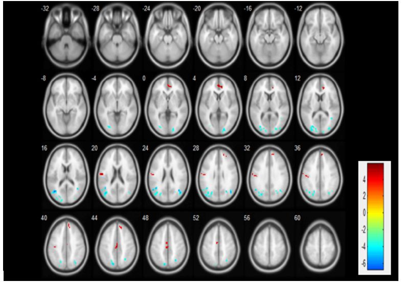
3.2 ALFF changes
We observed significant activation of ALFF neuronal activity in CFS patients (pCompared with the control group, the FC values between the DCG and MTG (Table 3, Figure 3), and those
between the left OG and the right OG were enhanced (Table 3, Figure 3).
Table 3 Compared with the control group, the FC values between the DCG and MTG.
ROI Regions X Y Z Voxel T- value P-value
ROI-1 DCG MTG 51 -39 -6 47 5.83 P<0.05
ROI-2 OG right middle occipital gyrus 30 -81 39 76 4.92 P<0.05
3.4 Correlation between the rs-fMRI parameters and the
clinical features
Since fatigue and quality of life were the focus of the study, the additional correlation analysis between
the scales and brain activation areas outcomes were performed. The Pearson correlation coefficient was
used for calculation to test the relationship between the scales of total score of the Multidimensional
Fatigue Inventory (MFI-20), Health Survey Short Form (SF-36) and the brain activation of OG, SFG, and
DCG. PLWNT interfered with neuronal activity and had a significant impact on the scores of SF-36 and
MFI-20. The clinical symptoms in SF-36 were positively with the left OG (r=0.542), SFG(r=0.517) and
DCG(r=0.533), and clinical symptoms in MFI-20 were negatively with the left OG (r=-0.583), SFG(r=-0.542)
and DCG(r=-0.578). P-values were all less than 0.05. The results of the correlation analysis are shown in
Figure 4 (a-f).
4. Discussion
This study used resting-state fMRI to analyze the effect of PLWNT intervention on the activation of brain
network neurons and FC changes in CFS patients. The change in resting state network (RSN) is related to
the lack of mental activity, which seriously affects the quality of life[54]. It is currently believed that RSN
has strong spontaneous activity, and it is also the most common neural network for evaluating quality of
life, involving cognitive control, attention, language processing, and working memory[55], including the OG,
angular gyrus, MTG, SFG and DCG[56]. RSN brain area is related to the maintenance of the brain’s
alertness to the outside world and introspection[57]. Previous studies have shown that CFS can lead to
impaired RSN function, which is manifested when performing externally targeted tasks such as cognitive
memory tasks[58, 59]. As shown in our study, the OG and MTG of the PLWNT group of patients belong to
RSN and have a lower ALFF value compared with the control group. This suggests that long-term fatigue,
insomnia, and poor quality of life in CFS patients can cause damage to the brain’s advanced cognitive
memory function. The injury of a certain center does not permanently remove the function managed by
the center, the function can be compensated by other areas to restore the function to a certain extent after
Page 10/25exercises[60]. This may be a neural compensation mechanism for the functionally damaged brain areas in
CFS patients after PLWNT treatment. The ALFF values of the SFG and DCG were all increased. This
shows that when CFS patients suffer from fatigue and sleep disturbance that affect their quality of life,
there are new strengthened brain areas that continue to complete specific neuronal activities and brain
functional activities.
Our study showed that 12 weeks of PLWNT intervention had a positive effect on ALFF and FC of
abnormal brain regions of OG, SFG, and DCG in patients with CFS. The FC values between the DCG and
MTG, and between the left OG and the right OG were all enhanced. In previous studies, these brain areas
have been linked to fatigue and quality of life[61–63]. We used correlation analysis to observe the
relationship between ALFF, FC, and the improvement of clinical symptoms. The results of our study
showed that among patients receiving Qigong, these brain activation areas positively correlated with the
clinical symptoms in SF-36 and negatively correlated those in MFI-20 in terms of fatigue, physical pain,
and lack of energy, thereby suggesting that PLWNT may objectively reflect the quality of life of CFS
patients through the DCG, SFG, and OG neuronal activity. Compared with other analyses, ALFF analysis
can suppress nonspecific signals more effectively, thereby significantly improving the neuron specificity
for detecting spontaneous activity in brain regions[64]. The FC analysis focuses on the similarity of
spontaneous brain activity within and between regions, with the ALFF activation area as the point of
interest[65]. Changes in the ALFF and FC values of DCG, SFG, and bilateral OG suggest that patients with
CFS have increased hemodynamic response to local neural activity or the brain's compensatory response
to fatigue. These findings provide support that PLWNT Qigong intervention may actively improve the
clinical symptoms of CFS patients. DCG, SFG, and OG can reflect the spontaneous neural activity of the
brain and the activation of CFS abnormal brain areas. The changes in DCG, SFG, and OG can help to
understand the changes in brain nerve function in CFS patients after exercises.
Higher-order level cognitive dysfunctions, such as those of memory and cognition, are well-known in
CFS [66], and recent studies have also documented the effects of basic sensory processing deficits on
quality of life[67, 68]. In CFS patients, there are also perceptual defects of the visual system[67–69]. In the
human brain, the OG is the main brain region of DMN for visual processing, which is involved in memory
acquisition. The ratio of occipital neurons to glial cells is the smallest, and the efficiency of removing
potassium ions is the lowest, which in turn affects the membrane potential and ultimately reduces
excitability, reduces exercise capacity, and includes symptoms such as fatigue, which reduces the quality
of life. Therefore, the OG is the most common site of CFS [70]. Bilateral OG cortex contains topographic
maps of size and orientation preference, in which neural responses to stimulus sizes and stimulus
orientations are modulated by intraregional lateral connections. We propose that these lateral
connections may underlie the selective influence of PLWNT Qigong on visual perception[71, 72]. Using
diffusion-tensor imaging, researchers have found that the white matter of the right suboccipital tract of
Gulf War syndrome veterans with visual neglect was damaged in connection with the occipital cortex,
which was manifested by severe fatigue, sleep, and decreased quality of life[73]. A resting-state ASL-fMRI
study also pointed out that the functional connection between the OG structure and the cerebral cortex of
Page 11/25CFS patients was damaged, which was related to the degree of fatigue and quality of life[46]. In our study,
the OG structure (middle occipital gyrus, supraoccipital gyrus) of CFS patients showed decreased
neuronal activity. These convergent results emphasize the possibility that memory decline, unrecoverable
fatigue, and the decreased quality of life in CFS patients may be related to OG dysfunction. Apart from
the increase in OG neural activity, this study revealed that long-term Qigong exercises actively increased
FC between the bilateral OG. These effects may be related to the ability of Qigong to change the brain's
functional networks related to the processing of external visual stimuli[74, 75]. To our knowledge, the
enhancement of FC in OG has not been reported to play a role in CFS quality of life, which may suggest
that the FC of the OG dysfunction may affect the quality of life of CFS patients and may also be involved
in the pathogenesis of CFS.
The defects of the somatic motor center seem to be related to the higher levels of fatigue in CFS, somatic
pain, energy disorders, and other aspects of the quality of life[76–78]. Researchers have early recognized
the importance of the motor function of the somatic motor center in explaining mental fatigue, but the
structure of the somatic motor center and OG is not sufficient to explain the model. Subsequently, brain
area networks including SFG, DCG, and MTG have also been shown to be related to fatigue[79, 80]. SFG
corresponds to the somatosensory center, which is mainly responsible for processing spatial information,
attention control, and somatosensory information. SFG is of great significance to the adjustment of the
quality of life such as fatigue, anxiety, and depression of CFS patients, as well as for the improvement of
functional activities, learning, and memory[74, 81, 82]. A recent study has found that the SFG area is widely
activated when CFS patients participate in activities. Although it is not clear whether these activations are
caused by positive or negative emotions, it shows that severely fatigued brains need to activate the right
frontal lobe and adjacent areas[74]. This may be caused by the accumulation of free radicals caused by
excessive neural activity of SFG in chronic fatigue, which induces brain oxidative stress. It is speculated
that the overactivity of the SFG area of the brain may be related to the pathophysiological changes of
CFS structure, neurotransmitter dynamics, and frontal mitochondrial function[83, 84]. Compared with the
control group, the SFG neuron activity in the PLWNT group was abnormally increased. Consistent with the
results of our study, research has shown that the white matter of the brain area related to cognition
promotes information transmission in the brain and makes the nervous system fast and effective[85]. Any
disorder of these neurological functions will affect the quality of life, including memory, attention, energy,
and executive function, as shown in CFS [85–87]. In addition, SFG is related to deficits in working memory,
impaired attention, poor motor coordination, and inability to concentrate vision. This area plays an
important role in connecting the frontal and temporal lobes[88, 89]. The above findings may reflect that
PLWNT intervention increased the activation of SFG neurons and the functional integration with MTG.
This change may improve higher-level processes such as fatigue and quality of life.
The fatigue symptoms of CFS and the decline in quality of life are closely related to the transition
network that connects cognitive and emotional feelings[55, 90]. There is numerous research evidence[91–93]
that DCG participates in a series of functions; not only it can process emotions, feelings, and attention,
but it can also participate in the regulation of sleep. The gray matter creatine phosphate of insomnia
Page 12/25patients is reduced, indicating that insomnia consumes more energy than normal sleep. The
enhancement of these activated abnormal brain regions and the enhancement of the functional
connection of MTG may be related to the high-energy compensation mechanism. Beyond that, pain is
also a common symptom of CFS and an important factor that affects the quality of life. The decline in
quality of life as in patients with CFS has been reported in many types of pain disorders, including
chronic back pain, physical pain disorders, and lack of energy[94–96]. These pain disorders can occur in
multiple locations, from the cerebral cortex to the spinal cord, and is considered to be the damage to the
central nervous system may cause the neurotransmitter involved in analgesia to be abnormal release[97].
At the same time, studies have further pointed out that physical pain in CFS patients may be caused by
hyperalgesia, that is, the brain area that regulates pain perception information is abnormal[98]. If the brain
has obstacles in receiving and processing pain information, then it is more sensitive to pain[99]. Previous
studies have also confirmed that this was mainly related to the core activation of the anterior cingulate
gyrus, SFG, occipitotemporal area and DCG in our study, which was typical features of pain
management[100]. More importantly, Zack et al.[9] found that SFG and MOG neuron activities in CFS
patients positively correlated with SF-36 by comparing CFS patients with healthy people, which is
consistent with our findings that the brain activation areas positively correlate with the clinical symptoms
in SF-36. Based on these observations, we can infer that PLWNT can reduce the fatigue symptoms and
improve the quality of life in CFS patients by activating related brain areas and regulating the patients’
sleep and physical pain.
Although our findings provide new and objective insights into the effects of PLWNT intervention on the
brain function of CFS patients (including fatigue symptoms and quality of life), there are still some
limitations that need to be further addressed. First of all, the selection criteria for CFS patients in this
study were only based on self-rating scales, and there was no equipment for objectively measuring
fatigue, energy, and pain; this may have resulted in irregular requirements for the inclusion of patients.
However, we limited the age of participants to 20–60 years to reduce the likelihood of chronic fatigue
symptoms and poor quality of life caused by diseases and age. Second, there were potential limitations
in the experimental design. Ideally, participants should be blinded, but this is difficult to achieve in non-
drug trials. However, we worked hard to ensure that laboratory technicians, data management personnel,
and statisticians did not participate in recruitment and data processing, which to a certain extent ensured
the authenticity of the data. Finally, our results indicating the brain regions with enhanced neuronal
activity and functional connectivity in patients with CFS after PLWNT intervention need to be verified in a
larger sample.
Conclusions
This study showed that PLWNT can relieve the fatigue symptoms of CFS patients and improve their
quality of life. We also found that CFS patients had abnormal regional spontaneous neuronal activity and
abnormal functional connections between regions after PLWNT intervention. There were also changes in
the activation of the brain regions, improving the quality of life related to fatigue symptoms and physical
Page 13/25pain, which were linearly related to the clinical symptoms in MFI-20 and SF-36. These findings provide a
new perspective for the role of traditional medical interventions such as Qigong in medicine, and may
provide guidance for the diagnosis and prevention of CFS.
Declarations
Ethics approval and consent to participate
Informed consent was obtained from the participants of this study. The study was approved by the Ethics
Committee of Yueyang Hospital of Integrated Traditional Chinese and Western Medicine Affiliated to
Shanghai University of Traditional Chinese Medicine (Ethics Approval Number: 2018-043), and registered
in the American Clinical Trial Registry (12/04/2018), Registration Number is NCT03496961.
Consent for publication
All authors agreed to publish this article.
Availability of data and materials
All data generated or analysed during this study are included in this published article.
Competing interests
The authors declare that they have no competing interests.
Funding
This work was supported by the National Natural Science Foundation of China under Grant 81774443
and 82105038, The funder is the corresponding author Professor Fei Yao and the first author Dr.
Fangfang Xie. The funding sources were not involved in study recruitment, data processing and
publication of papers.
Authors’ Contributorship
The project was conceived and designed by all the authors. F.F.X. performed the search. F.Y. and Y.L.Y.
contributed to data analysis and interpretation F.F.X., the manuscript draft that was revised by all co-
authors. All authors approved the final version of the manuscript and agree to be accountable for all
aspects of the work.
Acknowledgements
We are grateful to Guanwu Li for advice relating to the analysis and thanks are due to Ruiping Wang for
guidance. We also would like to thank Professor Min Fang from the University of shanghai university of
traditional Chinese medicine for proofreading the manuscript.
Page 14/25References
1. Sapra A, Bhandari P. Chronic Fatigue Syndrome. StatPearls. Treasure Island (FL): StatPearls
Publishing Copyright © 2020, StatPearls Publishing LLC.; 2020.
2. Lim EJ, Ahn YC, Jang ES, Lee SW, Lee SH, Son CG. Systematic review and meta-analysis of the
prevalence of chronic fatigue syndrome/myalgic encephalomyelitis (CFS/ME). Journal of
translational medicine. 2020;18(1):100.
3. Sotzny F, Blanco J, Capelli E, Castro-Marrero J, Steiner S, Murovska M, et al. Myalgic
Encephalomyelitis/Chronic Fatigue Syndrome - Evidence for an autoimmune disease. Autoimmunity
reviews. 2018;17(6):601–9.
4. Pendergrast T, Brown A, Sunnquist M, Jantke R, Newton JL, Strand EB, et al. Housebound versus
nonhousebound patients with myalgic encephalomyelitis and chronic fatigue syndrome. Chronic
illness. 2016;12(4):292–307.
5. Castro-Marrero J, Faro M, Zaragozá MC, Aliste L, de Sevilla TF, Alegre J. Unemployment and work
disability in individuals with chronic fatigue syndrome/myalgic encephalomyelitis: a community-
based cross-sectional study from Spain. BMC public health. 2019;19(1):840.
6. Crawley E. The epidemiology of chronic fatigue syndrome/myalgic encephalitis in children. Archives
of disease in childhood. 2014;99(2):171–4.
7. Washington SD, Rayhan RU, Garner R, Provenzano D, Zajur K, Addiego FM, et al. Exercise alters brain
activation in Gulf War Illness and Myalgic Encephalomyelitis/Chronic Fatigue Syndrome. Brain
communications. 2020;2(2):fcaa070.
8. VanElzakker MB, Brumfield SA, Lara Mejia PS. Neuroinflammation and Cytokines in Myalgic
Encephalomyelitis/Chronic Fatigue Syndrome (ME/CFS): A Critical Review of Research Methods.
Frontiers in neurology. 2018;9:1033.
9. Shan ZY, Finegan K, Bhuta S, Ireland T, Staines DR, Marshall-Gradisnik SM, et al. Brain function
characteristics of chronic fatigue syndrome: A task fMRI study. NeuroImage Clinical. 2018;19:279–
86.
10. Wostyn P, De Deyn PP. The putative glymphatic signature of chronic fatigue syndrome: A new view
on the disease pathogenesis and therapy. Medical hypotheses. 2018;118:142–5.
11. Kahl KG, Westhoff-Bleck M, Krüger THC. Effects of psychopharmacological treatment with
antipsychotic drugs on the vascular system. Vascular pharmacology. 2018;100:20–5.
12. Bolton MJ, Chapman BP, Van Marwijk H. Low-dose naltrexone as a treatment for chronic fatigue
syndrome. BMJ case reports. 2020;13(1).
13. Alraek T, Lee MS, Choi TY, Cao H, Liu J. Complementary and alternative medicine for patients with
chronic fatigue syndrome: a systematic review. BMC complementary and alternative medicine.
2011;11:87.
14. Geraghty KJ, Blease C. Cognitive behavioural therapy in the treatment of chronic fatigue syndrome: A
narrative review on efficacy and informed consent. Journal of health psychology. 2018;23(1):127–
Page 15/2538.
15. Abdollahi L, Mirghafourvand M, Babapour JK, Mohammadi M. Effectiveness of cognitive-behavioral
therapy (CBT) in improving the quality of life and psychological fatigue in women with polycystic
ovarian syndrome: a randomized controlled clinical trial. Journal of psychosomatic obstetrics and
gynaecology. 2019;40(4):283–93.
16. Anderson E, Parslow R, Hollingworth W, Mills N, Beasant L, Gaunt D, et al. Recruiting Adolescents
With Chronic Fatigue Syndrome/Myalgic Encephalomyelitis to Internet-Delivered Therapy: Internal
Pilot Within a Randomized Controlled Trial. Journal of medical Internet research. 2020;22(8):e17768.
17. Wilshire CE, Kindlon T, Courtney R, Matthees A, Tuller D, Geraghty K, et al. Rethinking the treatment of
chronic fatigue syndrome-a reanalysis and evaluation of findings from a recent major trial of graded
exercise and CBT. BMC psychology. 2018;6(1):6.
18. Jing L, Jin Y, Zhang X, Wang F, Song Y, Xing F. The effect of Baduanjin qigong combined with CBT on
physical fitness and psychological health of elderly housebound. Medicine. 2018;97(51):e13654.
19. Wang XQ, Takahashi T, Zhu SJ, Moriya J, Saegusa S, Yamakawa J, et al. Effect of Hochu-ekki-to (TJ-
41), a Japanese Herbal Medicine, on Daily Activity in a Murine Model of Chronic Fatigue Syndrome.
Evidence-based complementary and alternative medicine: eCAM. 2004;1(2):203–6.
20. Schweiger V, Secchettin E, Castellani C, Martini A, Mazzocchi E, Picelli A, et al. Comparison between
Acupuncture and Nutraceutical Treatment with Migratens(®) in Patients with Fibromyalgia
Syndrome: A Prospective Randomized Clinical Trial. Nutrients. 2020;12(3).
21. Sung WS, Kang HR, Jung CY, Park SS, Lee SH, Kim EJ. Efficacy of Korean red ginseng (Panax
ginseng) for middle-aged and moderate level of chronic fatigue patients: A randomized, double-blind,
placebo-controlled trial. Complementary therapies in medicine. 2020;48:102246.
22. Chan JS, Li A, Ng SM, Ho RT, Xu A, Yao TJ, et al. Adiponectin Potentially Contributes to the
Antidepressive Effects of Baduanjin Qigong Exercise in Women With Chronic Fatigue Syndrome-Like
Illness. Cell transplantation. 2017;26(3):493–501.
23. Jiao J, Russell IJ, Wang W, Wang J, Zhao YY, Jiang Q. Ba-Duan-Jin alleviates pain and fibromyalgia-
related symptoms in patients with fibromyalgia: results of a randomised controlled trial. Clinical and
experimental rheumatology. 2019;37(6):953–62.
24. McCaffrey R, Fowler NL. Qigong practice: a pathway to health and healing. Holistic nursing practice.
2003;17(2):110–6.
25. Xiong X, Wang P, Li X, Zhang Y. Qigong for hypertension: a systematic review. Medicine.
2015;94(1):e352.
26. van Dam K. Individual Stress Prevention through Qigong. International journal of environmental
research and public health. 2020;17(19).
27. Xie F, Guan C, Cheng Z, Yao F, You Y. Effects of the prolong life with nine turn method (Yan Nian Jiu
Zhuan) Qigong on patients with chronic fatigue syndrome: study protocol for a randomized
controlled trial. Annals of palliative medicine. 2020;9(5):3571–83.
Page 16/2528. Zou L, Pan Z, Yeung A, Talwar S, Wang C, Liu Y, et al. A Review Study on the Beneficial Effects of
Baduanjin. Journal of alternative and complementary medicine (New York, NY). 2018;24(4):324-35.
29. Chan JS, Ho RT, Wang CW, Yuen LP, Sham JS, Chan CL. Effects of qigong exercise on fatigue,
anxiety, and depressive symptoms of patients with chronic fatigue syndrome-like illness: a
randomized controlled trial. Evidence-based complementary and alternative medicine: eCAM.
2013;2013:485341.
30. Noh GO, Park KS. Effects of aroma self-foot reflexology on peripheral neuropathy, peripheral skin
temperature, anxiety, and depression in gynaecologic cancer patients undergoing chemotherapy: A
randomised controlled trial. European journal of oncology nursing: the official journal of European
Oncology Nursing Society. 2019;42:82–9.
31. Zengin L, Aylaz R. The effects of sleep hygiene education and reflexology on sleep quality and
fatigue in patients receiving chemotherapy. European journal of cancer care. 2019;28(3):e13020.
32. Bender PU, Luz CMD, Feldkircher JM, Nunes GS. Massage therapy slightly decreased pain intensity
after habitual running, but had no effect on fatigue, mood or physical performance: a randomised
trial. Journal of physiotherapy. 2019;65(2):75–80.
33. Langhorst J, Klose P, Dobos GJ, Bernardy K, Häuser W. Efficacy and safety of meditative movement
therapies in fibromyalgia syndrome: a systematic review and meta-analysis of randomized
controlled trials. Rheumatology international. 2013;33(1):193–207.
34. 34.. !!! INVALID CITATION !!! [24].
35. Smitha KA, Akhil Raja K, Arun KM, Rajesh PG, Thomas B, Kapilamoorthy TR, et al. Resting state fMRI:
A review on methods in resting state connectivity analysis and resting state networks. The
neuroradiology journal. 2017;30(4):305–17.
36. Agrawal U, Brown EN, Lewis LD. Model-based physiological noise removal in fast fMRI. NeuroImage.
2020;205:116231.
37. Crofts A, Kelly ME, Gibson CL. Imaging Functional Recovery Following Ischemic Stroke: Clinical and
Preclinical fMRI Studies. Journal of neuroimaging: official journal of the American Society of
Neuroimaging. 2020;30(1):5–14.
38. Huber L, Uludağ K, Möller HE. Non-BOLD contrast for laminar fMRI in humans: CBF, CBV, and
CMR(O2). NeuroImage. 2019;197:742–60.
39. Kang D, Qin Z, Wang W, Zheng Y, Hu H, Bao Y, et al. Brain functional changes in tibetan with
obstructive sleep apnea hypopnea syndrome: A resting state fMRI study. Medicine.
2020;99(7):e18957.
40. Lai W, Li X, Zhu H, Zhu X, Tan H, Feng P, et al. Plasma luteinizing hormone level affects the brain
activity of patients with polycystic ovary syndrome. Psychoneuroendocrinology. 2020;112:104535.
41. Zhang XH, Shi JY, Zhan C, Zhang L, Chen HJ. Intrinsic Brain Abnormalities in Patients with Hepatitis
C Virus Infection with Cognitive Impairment: A Preliminary Resting-State fMRI Study. BioMed
research international. 2020;2020:1693043.
Page 17/2542. Arnett SV, Alleva LM, Korossy-Horwood R, Clark IA. Chronic fatigue syndrome--a neuroimmunological
model. Medical hypotheses. 2011;77(1):77–83.
43. Nijs J, Meeus M, Van Oosterwijck J, Ickmans K, Moorkens G, Hans G, et al. In the mind or in the
brain? Scientific evidence for central sensitisation in chronic fatigue syndrome. European journal of
clinical investigation. 2012;42(2):203–12.
44. Knight S, Harvey A, Lubitz L, Rowe K, Reveley C, Veit F, et al. Paediatric chronic fatigue syndrome:
complex presentations and protracted time to diagnosis. Journal of paediatrics and child health.
2013;49(11):919–24.
45. Amihăesei IC, Cojocaru E. Main neuroendocrine features, diagnosis and therapeutic possibilities in
the chronic fatigue syndrome, an underdiagnosed entity. Revista medico-chirurgicala a Societatii de
Medici si Naturalisti din Iasi. 2014;118(3):688–91.
46. Boissoneault J, Letzen J, Lai S, O'Shea A, Craggs J, Robinson ME, et al. Abnormal resting state
functional connectivity in patients with chronic fatigue syndrome: an arterial spin-labeling fMRI
study. Magnetic resonance imaging. 2016;34(4):603–8.
47. Caseras X, Mataix-Cols D, Rimes KA, Giampietro V, Brammer M, Zelaya F, et al. The neural correlates
of fatigue: an exploratory imaginal fatigue provocation study in chronic fatigue syndrome.
Psychological medicine. 2008;38(7):941–51.
48. Achard S, Salvador R, Whitcher B, Suckling J, Bullmore E. A resilient, low-frequency, small-world
human brain functional network with highly connected association cortical hubs. The Journal of
neuroscience: the official journal of the Society for Neuroscience. 2006;26(1):63–72.
49. Barnden LR, Shan ZY, Staines DR, Marshall-Gradisnik S, Finegan K, Ireland T, et al. Intra brainstem
connectivity is impaired in chronic fatigue syndrome. NeuroImage Clinical. 2019;24:102045.
50. Provenzano D, Washington SD, Rao YJ, Loew M, Baraniuk J. Machine Learning Detects Pattern of
Differences in Functional Magnetic Resonance Imaging (fMRI) Data between Chronic Fatigue
Syndrome (CFS) and Gulf War Illness (GWI). Brain sciences. 2020;10(7).
51. Yang M, He H, Duan M, Chen X, Chang X, Lai Y, et al. The Effects of Music Intervention on Functional
Connectivity Strength of the Brain in Schizophrenia. Neural plasticity. 2018;2018:2821832.
52. Boissoneault J, Letzen J, Lai S, Robinson ME, Staud R. Static and dynamic functional connectivity in
patients with chronic fatigue syndrome: use of arterial spin labelling fMRI. Clinical physiology and
functional imaging. 2018;38(1):128–37.
53. UK Moves to Revise Guidelines for Treatment of Chronic Fatigue Syndrome. The American journal of
nursing. 2021;121(3):16.
54. Whitfield-Gabrieli S, Thermenos HW, Milanovic S, Tsuang MT, Faraone SV, McCarley RW, et al.
Hyperactivity and hyperconnectivity of the default network in schizophrenia and in first-degree
relatives of persons with schizophrenia. Proceedings of the National Academy of Sciences of the
United States of America. 2009;106(4):1279–84.
55. Lois G, Linke J, Wessa M. Altered functional connectivity between emotional and cognitive resting
state networks in euthymic bipolar I disorder patients. PloS one. 2014;9(10):e107829.
Page 18/2556. Greicius MD, Supekar K, Menon V, Dougherty RF. Resting-state functional connectivity reflects
structural connectivity in the default mode network. Cerebral cortex (New York, NY: 1991).
2009;19(1):72-8.
57. Smigielski L, Scheidegger M, Kometer M, Vollenweider FX. Psilocybin-assisted mindfulness training
modulates self-consciousness and brain default mode network connectivity with lasting effects.
NeuroImage. 2019;196:207–15.
58. Carhart-Harris RL, Roseman L, Bolstridge M, Demetriou L, Pannekoek JN, Wall MB, et al. Psilocybin
for treatment-resistant depression: fMRI-measured brain mechanisms. Scientific reports.
2017;7(1):13187.
59. Dutta A, McKie S, Downey D, Thomas E, Juhasz G, Arnone D, et al. Regional default mode network
connectivity in major depressive disorder: modulation by acute intravenous citalopram. Translational
psychiatry. 2019;9(1):116.
60. Fischer-Jbali LR, Montoro CI, Montoya P, Halder W, Duschek S. Central nervous activity during implicit
processing of emotional face expressions in fibromyalgia syndrome. Brain research.
2021;1758:147333.
61. Lim J, Wu WC, Wang J, Detre JA, Dinges DF, Rao H. Imaging brain fatigue from sustained mental
workload: an ASL perfusion study of the time-on-task effect. NeuroImage. 2010;49(4):3426–35.
62. Pellicano C, Gallo A, Li X, Ikonomidou VN, Evangelou IE, Ohayon JM, et al. Relationship of cortical
atrophy to fatigue in patients with multiple sclerosis. Archives of neurology. 2010;67(4):447–53.
63. Kohl AD, Wylie GR, Genova HM, Hillary FG, Deluca J. The neural correlates of cognitive fatigue in
traumatic brain injury using functional MRI. Brain injury. 2009;23(5):420–32.
64. Zhang Z, Bo Q, Li F, Zhao L, Wang Y, Liu R, et al. Increased ALFF and functional connectivity of the
right striatum in bipolar disorder patients. Progress in neuro-psychopharmacology & biological
psychiatry. 2020:110140.
65. Bonifazi P, Massobrio P. Reconstruction of Functional Connectivity from Multielectrode Recordings
and Calcium Imaging. Advances in neurobiology. 2019;22:207–31.
66. Kawatani J, Mizuno K, Shiraishi S, Takao M, Joudoi T, Fukuda S, et al. Cognitive dysfunction and
mental fatigue in childhood chronic fatigue syndrome--a 6-month follow-up study. Brain &
development. 2011;33(10):832–41.
67. Josev EK, Malpas CB, Seal ML, Scheinberg A, Lubitz L, Rowe K, et al. Resting-state functional
connectivity, cognition, and fatigue in response to cognitive exertion: a novel study in adolescents
with chronic fatigue syndrome. Brain imaging and behavior. 2020;14(5):1815–30.
68. Thapaliya K, Marshall-Gradisnik S, Staines D, Barnden L. Mapping of pathological change in chronic
fatigue syndrome using the ratio of T1- and T2-weighted MRI scans. NeuroImage Clinical.
2020;28:102366.
69. Eller-Smith OC, Nicol AL, Christianson JA. Potential Mechanisms Underlying Centralized Pain and
Emerging Therapeutic Interventions. Frontiers in cellular neuroscience. 2018;12:35.
Page 19/2570. Eyl RE, Xie K, Koch-Gallenkamp L, Brenner H, Arndt V. Quality of life and physical activity in long-term
(≥5 years post-diagnosis) colorectal cancer survivors - systematic review. Health and quality of life
outcomes. 2018;16(1):112.
71. Song C, Sandberg K, Andersen LM, Blicher JU, Rees G. Human Occipital and Parietal GABA
Selectively Influence Visual Perception of Orientation and Size. The Journal of neuroscience: the
official journal of the Society for Neuroscience. 2017;37(37):8929–37.
72. Wandell BA, Dumoulin SO, Brewer AA. Visual field maps in human cortex. Neuron. 2007;56(2):366–
83.
73. Rayhan RU, Stevens BW, Timbol CR, Adewuyi O, Walitt B, VanMeter JW, et al. Increased brain white
matter axial diffusivity associated with fatigue, pain and hyperalgesia in Gulf War illness. PloS one.
2013;8(3):e58493.
74. Mizuno K, Tanaka M, Tanabe HC, Joudoi T, Kawatani J, Shigihara Y, et al. Less efficient and costly
processes of frontal cortex in childhood chronic fatigue syndrome. NeuroImage Clinical.
2015;9:355–68.
75. Möller MC, Nordin LE, Bartfai A, Julin P, Li TQ. Fatigue and Cognitive Fatigability in Mild Traumatic
Brain Injury are Correlated with Altered Neural Activity during Vigilance Test Performance. Frontiers in
neurology. 2017;8:496.
76. Bonhof CS, van de Poll-Franse LV, Wasowicz DK, Beerepoot LV, Vreugdenhil G, Mols F. The course of
peripheral neuropathy and its association with health-related quality of life among colorectal cancer
patients. Journal of cancer survivorship: research and practice. 2021;15(2):190–200.
77. Braz NFT, Rocha NP, Vieira É LM, Barbosa IG, Gomez RS, Kakehasi AM, et al. Muscle strength and
psychiatric symptoms influence health-related quality of life in patients with myasthenia gravis.
Journal of clinical neuroscience: official journal of the Neurosurgical Society of Australasia.
2018;50:41–4.
78. Michiels S, van der Wal AC, Nieste E, Van de Heyning P, Braem M, Visscher C, et al. Conservative
therapy for the treatment of patients with somatic tinnitus attributed to temporomandibular
dysfunction: study protocol of a randomised controlled trial. Trials. 2018;19(1):554.
79. Shallice T, Stuss DT, Alexander MP, Picton TW, Derkzen D. The multiple dimensions of sustained
attention. Cortex; a journal devoted to the study of the nervous system and behavior.
2008;44(7):794–805.
80. Stålnacke BM. Postconcussion symptoms in patients with injury-related chronic pain. Rehabilitation
research and practice. 2012;2012:528265.
81. Cook DB, O'Connor PJ, Lange G, Steffener J. Functional neuroimaging correlates of mental fatigue
induced by cognition among chronic fatigue syndrome patients and controls. NeuroImage.
2007;36(1):108–22.
82. van der Schaaf ME, De Lange FP, Schmits IC, Geurts DEM, Roelofs K, van der Meer JWM, et al.
Prefrontal Structure Varies as a Function of Pain Symptoms in Chronic Fatigue Syndrome. Biological
psychiatry. 2017;81(4):358–65.
Page 20/25You can also read