Emodin-loaded polymer-lipid hybrid nanoparticles enhance the sensitivity of breast cancer to Doxorubicin by inhibiting epithelial mesenchymal ...
←
→
Page content transcription
If your browser does not render page correctly, please read the page content below
Emodin-loaded polymer-lipid hybrid nanoparticles
enhance the sensitivity of breast cancer to
Doxorubicin by inhibiting epithelial mesenchymal
transition
Tengteng Zou
Jinan University
Meng Lan
Jinan University
Lihong Li
Jinan University
Tiange Cai
Liaoning University
Yu Cai ( caiyu8@sohu.com )
Jinan University
Research
Keywords: Emodin, polymeric lipid nanoparticles, breast cancer chemoresistance, Doxorubicin, Epithelial
mesenchymal transition
Posted Date: May 13th, 2021
DOI: https://doi.org/10.21203/rs.3.rs-451946/v1
License: This work is licensed under a Creative Commons Attribution 4.0 International License.
Read Full License
Version of Record: A version of this preprint was published at Cancer Nanotechnology on August 11th,
2021. See the published version at https://doi.org/10.1186/s12645-021-00093-9.
Page 1/21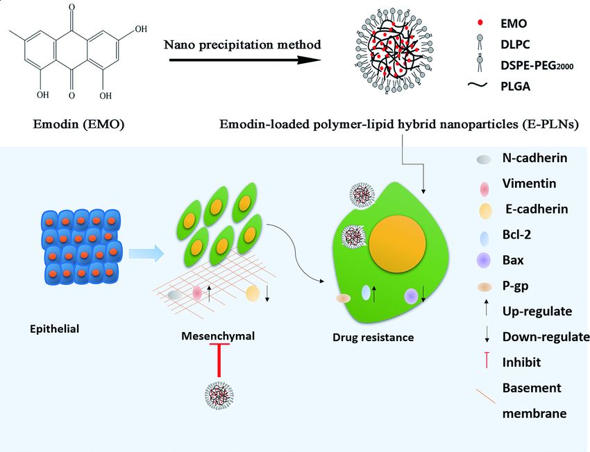
Abstract
Background
The role of epithelial mesenchymal transition (EMT) involved in breast cancer metastasis and
chemoresistance has been increasingly recognized. However, it’s necessary to search for more effective
strategies to inhibit EMT thereby increase the sensitivity of breast cancer cells to chemotherapy drugs.
Emodin has a potential in overcoming tumor drug resistance and restraining the development of EMT, but
the poor internalization into breast cancer cells limited the application.

Results
MCF-7/ADR cells have more EMT characteristics than MCF-7 cell. EMT in MCF-7/ADR cells promotes the
development of drug resistance via apoptosis resistance and facilitating the expression of P-gp. The anti-
cancer effect of DOX enhanced by the decreasing of drug resistance protein P-gp and apoptosis related
proteins after EMT inhibited in MCF-7/ADR cells. E-PLNs increase the cellular uptake of EMO and restore
DOX sensitivity in MCF-7/ADR cells by inhibiting EMT.

Conclusion
E-PLNs inhibit EMT to enhance the sensitivity of breast cancer to DOX. The combination of E-PLNs and
DOX can improve the efficacy of DOX in the treatment of breast cancer, which provides a new method to
prevent or delay clinical drug resistance.

Background
Despite significant advances in cancer diagnosis and treatment, breast cancer to be the number one
cancer in the world by 2020, more than 90% of the death causes of breast cancer patients are related to
chemotherapy resistance (Siegel et al. 2021, Sung et al. 2021). The role of epithelial mesenchymal
transition (EMT) involved in chemotherapy resistance has been increasingly recognized. Asiedu MK and
his colleagues found that EMT induced by TGF-β pathway in breast cancer cells decreased the sensitivity
to Doxorubicin (DOX) and paclitaxel (Asiedu et al. 2014). Reversal tumor drug resistance through EMT
inhibition is a new strategy and search for a safe and effective EMT inhibitor is necessary.

Emodin (EMO), the active component of Rheum rhabarbarum, is proved to have antitumor activity in
breast cancer (Sun et al. 2015). In addition, EMO has a potential in overcoming tumor drug resistance and
restraining the development of EMT (Liu et al. 2020, Fu et al. 2012, Lu et al. 2016, Gu et al. 2019). It can
effectively restore the effect of DOX, vincristine and other anticancer drugs by downregulating the
expression of P-gp in MCF-7/ADR cells. It was found that the combination of DOX and EMO effectively
inhibit the expression of snail, twist and slug, thereby inhibiting cell proliferation and promote cell

                                                   Page 2/21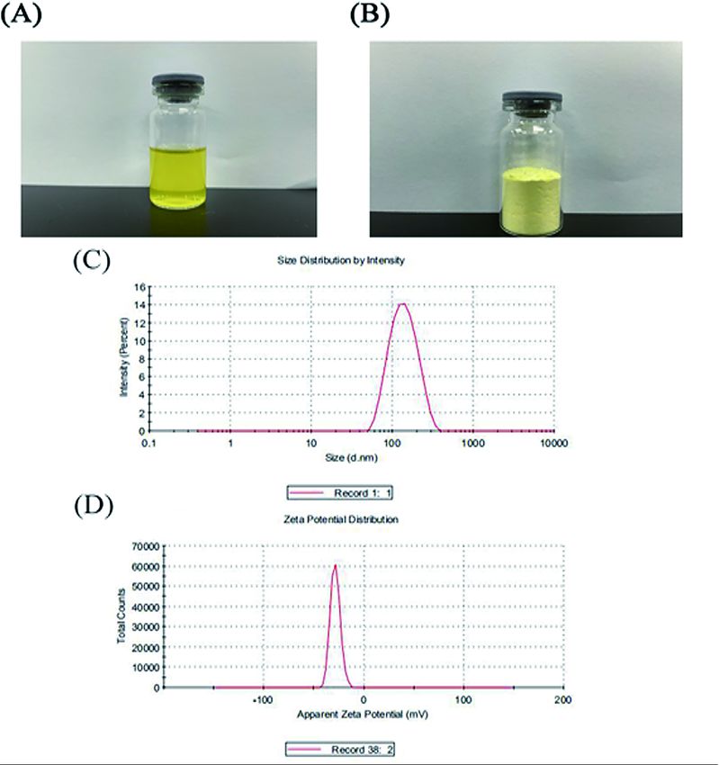
apoptosis. This combination results the suppression of cell migration and invasion. However, the poor
internalization into the breast cancer cells limited the application of EMO. Hence, improving EMO
internalization in breast cancer cells tend to be a reliable way to enhance the anti-cancer effect of DOX.
Furthermore, the drug reversal effect of EMO whether rely on the repression of EMT still needed to be
elucidated.

Nanoparticles loaded with anticancer drugs become an ideal formulation for reversing chemoresistance
(Li et al. 2016, Markman et al. 2013). The tumor targeting effect of nanoparticles is basely depended on
the enhanced permeability and retention (EPR) in tumor microenvironment (TME), thus drug
accumulation increased in TME (Kalyane et al. 2019). Polymeric lipid nanoparticles (PLNs), designed to
combine the advantages of both liposomes and polymeric nanoparticles, can meet the structural and
property differences of various anticancer drugs, as well as the combination therapy strategy, making
them a promising drug carrier (Jain et al. 2020, Mukerjee, Ranjan and Vishwanatha 2012).

Targeting EMT is becoming one of the new strategies for drug resistance reversal in various tumors. In
this study, the relationship of EMT and drug resistance were discussed. Here, we provided evidence for E-
PLNs to improve the therapeutic effect of DOX on breast cancer. Further, the evidence concerning drug
resistance reversal mechanism of E-PLNs in suppressing EMT is provided.

Results
EMT is associated with DOX resistance in breast cancer cells

We compared the effect of DOX on the viability of MCF-7 cells and MCF-7/ADR cells. As Fig. 1A, the
toxicity of DOX on MCF-7/ADR cells than MCF-7 cells were less, IC50 was 5.35 μM, while the IC50 of
MCF-7/ADR cells were 124.1 μM. MCF-7/ADR cells were resistant to DOX with a resistance index of 23.2.
EMT is characterized by loss of E-cadherin expression and upregulated Vimentin and N-cadherin (Fig. 1B
and C). DOX induced drug resistant cells have EMT characteristics, and the ability of invasion
significantly enhanced (Fig. 1D and E).

EMT inhibiting cell proliferation and invasion

TGF-beta signaling has been shown to play an important role in EMT. Galunisertib, a selective TGF-β
receptor type I (TGF-βRI) kinase inhibitor to block TGF-β signaling reverse EMT, is applicated in this part to
investigate the relationship of EMT, cell proliferation and invasion. It can be seen from Fig 2A that
different concentrations of Galunisertib can inhibit the proliferation of MCF-7/ADR cells, and the IC50
value calculated is 248.2 μM. When the concentration of Galunisertib was 30 μM, the cell viability was
more than 90%. Therefore, the concentration of 30 μM Galunisertib is selected to application for the
subsequent experiment to exclude the interference of Galunisertib cytotoxicity. Galunisertib reversed EMT
marker protein expression in the selected concentration (Fig. 2B and C). The therapeutic effect of DOX
was improved under the incubation of 10 μM DOX and 30 μM Galunisertib (Fig. 2D) MCF-7/ADR cells
have higher invasive ability than the DOX sensitive MCF-7 cells, but invasive ability of MCF-7/ADR cells
                                                  Page 3/21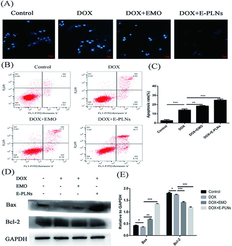
treated with DOX did not change. Compared with DOX (10 μM) group, Galunisertib (30 μM) group cells
was decreased and the invasion activity was inhibited. When Galunisertib and DOX were used together,
the inhibition effect was stronger (Fig. 2E and F). These results suggest that the inhibition of EMT may
weaken the drug resistance of MCF-7/ADR cells.

Blocking EMT restrain the expression of P-gp and induced apoptosis

Apoptosis is an important manifestation of anti-tumor effect of chemotherapy drugs. We can see from
Fig 3A and B that the apoptosis rate of DOX (10 μM) group is only 13.81%, Galunisertib (30 μM) group
(the apoptosis rate is 7.85%) has little effect on apoptosis. When DOX was combined with Galunisertib,
the number of apoptotic cells increased, and the apoptotic rate was 23.88%. It can be seen from Fig. 3C
and D that compared with DOX group, Bax/Bcl-2 ratio increased in DOX+Galunisertib (30 μM) group.
These results suggest that inhibition of EMT can enhance DOX induced apoptosis in MCF-7/ADR cells.

EMT was closely related to the process of drug resistance, one of which was upregulation of drug
resistance related transporters. As can be seen in Fig. 3E and F, the difference of expression level of P-gp
protein in Control group and DOX group was not significant. However, when MCF-7/ADR cells were
treated with Galunisertib, the expression levels of P-gp protein were down-regulated. More importantly, the
expression of P-gp in DOX+Galunisertib group was significantly suppressed than DOX group, resulting in
decreasing of DOX efflux. Therefore, blocking EMT down-regulate the expression level of P-gp, thereby
restraining the DOX efflux in MCF-7/ADR cells.

E-PLNs increase the cellular uptake and cytotoxity of EMO

E-PLNs prepared by nano precipitation method are light yellow liquid (Fig. 4A). In order to preserve the
samples for a long time, we keep E-PLNs samples through freeze-drying (Christ ALPHA 1-4 LD plus). The
particle size of E-PLNs was 122.7±1.03 nm (Fig. 4C), zeta potential was -28.5±1.55 mV (Fig. 4D) and PDI
was 0.118. The results show that the nanoparticles have uniform size and good stability.

As E-PLNs have no fluorescence, the C6-PLNs, characterized by green fluorescence, is formulated to
analyze the cellular uptake of PLNs carriers. After the coumarin-6-labeled nanoparticles were internalized,
the green fluorescence mainly distributed in the cytoplasm (Fig. 5A), indicating that PLNs carriers
promote the cellular uptake of the loaded drugs. Furthermore, flow cytometry quantitatively analyzes
cellular uptake, and the uptake intensity was expressed as the average fluorescence intensity of cells. The
average fluorescence intensity of cells was 500, indicating that the cells had a high uptake of
nanoparticles (Fig 5B and C). The cytotoxicity of EMO group (Fig.5D) and E-PLNs (Fig.5E) group on MCF-
7/ADR cells gradually increased with the increase of EMO concentration. The IC50 of EMO and E-PLNs
were 150.5 μM and 138.7 μM, respectively, which indicated that E-PLNs could enhance the antitumor
activity of EMO. In order to further explore whether EMO or E-PLNs can enhance the toxicity of DOX, a
suitable concentration was selected and combined with DOX to detect the effect on cell viability (Fig.5F).
We selected 30 μM EMO and E-PLNs for the subsequent combined administration experiment. It can be

                                                  Page 4/21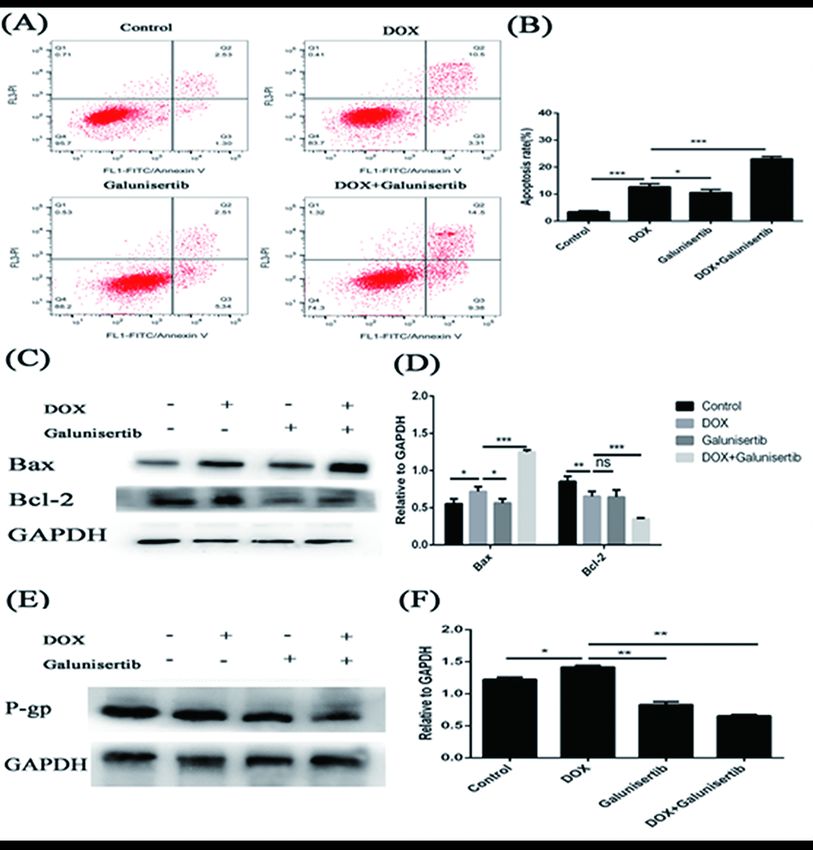
seen from Fig. that 30 μM E-PLNs can enhance the cytotoxicity of different concentrations of DOX.
Finally, 10 μM DOX was selected to explore how E-PLNs improve the therapeutic effect of DOX.

E-PLNs enhance the effect of DOX in apoptosis induction

MCF-7/ADR cells were treated with DOX, DOX+EMO, DOX+E-PLNs to investigate whether E-PLNs induced
cell apoptosis. The morphological changes of apoptotic cells are shown in Fig 6A. We observed that the
untreated cells were round or oval blue nuclei, while the cells in the treatment group were fragmented and
apoptotic cells were formed, and the number of apoptotic bodies was significantly reduced in the E-PLNs
group. It can be seen from Fig. 6B and C that compared with DOX (10 μM) group (15.52%), the apoptosis
rates of DOX+EMO (30 μM) and DOX+E-PLNs (30 μM) groups were 18.49% and 24.48%, respectively, and
the apoptosis rate was significantly increased. Western blot results also showed that the ratio of Bax/Bcl-
2 was increased and the expression of Fas was up-regulated in DOX+EMO (30 μM) and DOX+E-PLNs (30
μM) groups, which promoted cell apoptosis. At the same time, the expression level of Bcl-2 was down
regulated. DOX+E-PLNs group showed a more significant effect on the expression of apoptotic protein
(Fig. 6D and E). Flow cytometry combined with Western blot showed that E-PLNs may enhance the
apoptosis inducing effect of DOX on MCF-7/ADR cells through affecting apoptosis related pathways.

E-PLNs enhance DOX accumulation and restrain the expression of P-gp

The accumulation of DOX in MCF-7/ADR cells treated by different administration groups (Control, DOX,
DOX+EMO, DOX+E-PLNs) was analyzed quantitatively by flow cytometry (Fig. 7A). From Fig. 7B, it can be
seen that the peak map of DOX+EMO (30 μM) and DOX+E-PLNs (30 μM) groups shifts to the direction of
high fluorescence intensity compared with DOX (10 μM) group. The average fluorescence intensity of
DOX+E-PLNs is higher than that of DOX group,indicated that E-PLNs promote the uptake of DOX and
increase the DOX accumulation in MCF-7/ADR cells. In addition, the expression of P-gp significantly
decreased in DOX+EMO and DOX+E-PLNs groups compared with control and DOX groups (Fig 7C and D),
indicated that E-PLNs could effectively inhibit the expression of P-gp protein thus enhancing the effect of
DOX.

E-PLNs inhibit the expression of EMT marker protein and cell invasion

The expression of E-cadherin decreased, while the expression of N-cadherin and Vimentin increased in
MCF-7/ADR cells, which indicated the occurrence of EMT. It can be seen from Fig. 8A and B that there is
no significant difference in the protein levels of E-cadherin and Vimentin between the Control group and
the DOX group, but the protein level of N-cadherin increases significantly, which indicates that DOX
administration to MCF-7/ADR cells may further promote EMT. In DOX+E-PLNs (30 μM) group, the
expression of E-cadherin protein was increased, and the expression of vimentin and N-cadherin protein
was up-regulated. These results indicate that E-PLNs can effectively inhibit EMT, and the enhancement of
DOX sensitivity of MCF-7/ADR cells by E-PLNs is related to the inhibition of EMT.

                                                 Page 5/21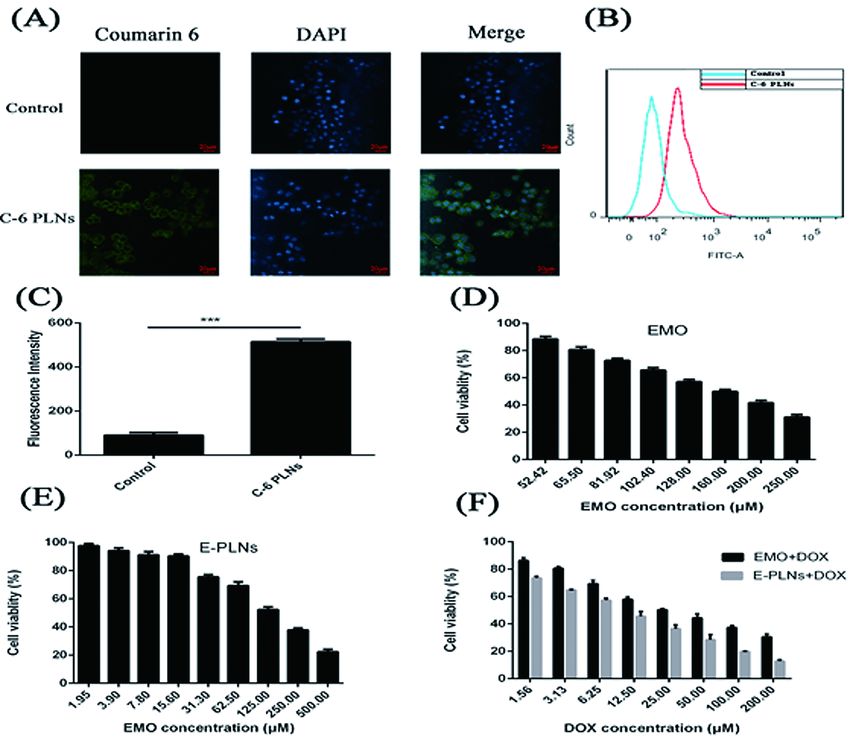
Cell invasion is an important manifestation after EMT induction. In Fig. 8C and D, there was no significant
difference in the number of cell invasion between the Control group and DOX group. Compared with DOX
group, the number of invasive cells in DOX+EMO (30 μM) group and DOX+E-PLNs (30 μM) group
decreased, and DOX+E-PLNs group was more significant. These results shown that the combination of E-
PLNs and DOX can inhibit the occurrence of EMT and the tendency to invasion in MCF-7/ADR cells.

Discussion
Chemotherapy is the main treatment for early stage of breast cancer, but the sensitivity of tumor cells to
chemotherapeutic drugs decreases with long-term chemotherapy. Increasing evidence shown that EMT is
involved in the development of drug resistance in breast cancer (Jiang et al. 2017). After EMT was
induced by snail eukaryotic expression vector, the expression of BCRP were significantly increased and
the drug resistance of MCF-7 cells was increased to 9.93 times (Du and Shim 2016). In this article, we
find that appears MCF-7/ADR cells have more significant characteristics of EMT than DOX sensitive MCF-
7 cells, manifested as the down-regulated of E-cadherin protein and the up-regulated of N-cadherin
protein and Vimentin. In fact, Li and his colleagues also confirmed that DOX could induce EMT and
apoptosis in MCF-7 cells (Li et al. 2020b). The invasion and metastasis are significantly enhanced in
tumor cells with EMT characteristics. Indeed, the invasion test showed that MCF-7/ADR cells had more
invasive cells than MCF-7 cells, which proved that MCF-7/ADR cells had stronger invasion ability.

Transforming growth factor-beta (TGF-β) signaling plays a key role in epithelial-mesenchymal transition
(EMT) of tumors (Xu, Lamouille and Derynck 2009). Galunisertib is a selective TGF-β receptor type I (TGF-
βRI) kinase inhibitor which blocking TGF-β signaling reverse EMT (Xu et al. , Rodón et al. 2015). The
direct toxicity of DOX to cells was enhanced by Galunisertib, which decreased cell viability and increase
cell invasion. Moreover, the apoptosis induction in MCF-7/ADR cells of DOX restored after EMT
suppression. In addition, previous studies have shown that both P-gp and ATP binding cascade
transporters are regulated by EMT. In this study, as the number of cells with EMT characteristics were
decreased, and the expression of P-gp protein was restrained. Therefore, it is worth to confirm that EMT
participates in the drug resistance of cancer cells by regulating the processes of apoptosis and drug
resistance proteins expression.

EMO has been found to inhibit to increase sensitivity to DOX and PTX in breast cancer (Li et al. 2020b, Gu
et al. 2020), and inhibits EMT in triple negative breast cancer (Song et al. 2018). It’s reported that solid
lipid nanoparticles of EMO can significantly block the cell cycle and induce apoptosis of MCF-7 cells
compared with free EMO (Wang et al. 2012), which indicates that lipid nanoparticles delivering EMO may
be a promising method for cancer treatment. In this research, we prepared PLNs encapsulated EMO to
enhance cellular uptake. As E-PLNs have no fluorescence, the C6-PLNs is formulated to analyze the
cellular uptake of PLNs carriers. The result shown that C6-PLNs can be absorbed by MCF-7/ADR cells,
and quantitative analysis of the average fluorescence intensity shown that PLNs structure improve the
uptake of C6. Furthermore, the cellular uptake and anti-tumor effect of DOX improved with the
combination of E-PLNs, indicated that EMO enhanced the therapeutic effect of DOX on tumor cells. The
                                                 Page 6/21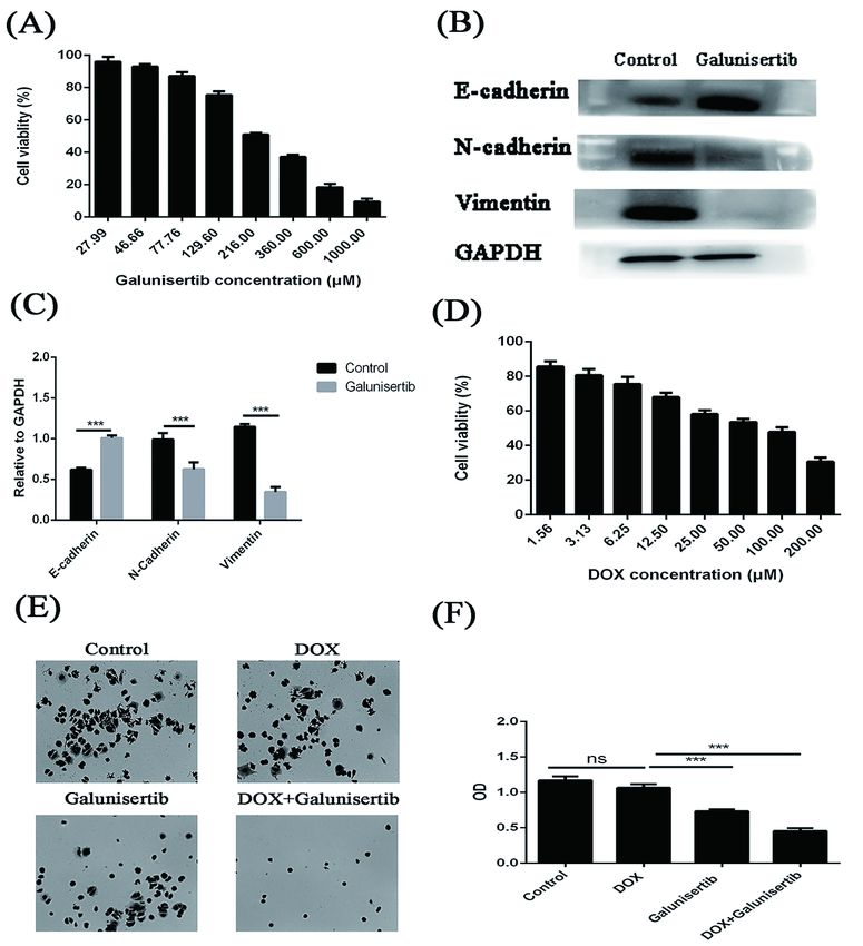
main mechanisms of the anti-cancer effect in DOX is apoptosis induction, and the apoptosis rate
increased to 24.48% combined with E-PLNs. The apoptotic pathway was activated by the increase of
Bax/Bcl-2 ratio. The expression of P-gp is restrained, resulting in the decrease of DOX efflux. The up-
regulated expression of E-cadherin, and down-regulated N-cadherin and Vimentin in MCF-7/ADR cells
incubated with E-PLNs indicate that E-PLNs inhibit EMT, therefore reverse DOX resistance in MCF-7/ADR
cells.

In this study, we find that EMT is involved in drug resistance, which is related to the regulation of
apoptosis and the expression of drug resistance transporters. EMT is one of the key factors to induce
DOX resistance in breast cancer. We also found that the expression of E-cadherin in DOX+E-PLNs group
was higher than that in DOX group, while the expression of Vimentin and N-cadherin was down regulated.
In addition, when E-PLNs were combined with DOX, the number of invasive cells decreased significantly.
These results suggested that E-PLNs enhancing the sensitivity of breast cancer to DOX depended on
inhibiting EMT.

Conclusion
Drug resistance in tumor is a phenomenon which the continuous administration of chemotherapeutic
drugs induces drug tolerance. EMT is one of the possible ways causing DOX resistance in breast cancer.
Our data suggested that the DOX resistance in breast cancer is associated with EMT. E-PLNs enhance the
sensitivity of DOX in breast cancer via the suppression of EMT. We confirmed that E-PLNs enhance the
effect of DOX, which is closely related to the regulation of EMT thereby inhibiting of apoptosis resistance
and P-gp expression. The combination of E-PLNs and DOX provides a new scheme for clinical treatment.
It’s potential that E-PLNs turn into an adjuvant therapy drugs of breast cancer in the feature. Further
investigations in animal models and mechanism are warranted.

Materials And Methods
Materials

Emodin (>98% purity) were provided by institute for drug control (Guangzhou, China). Mannitol was
purchased from Aladdin Biotech Co., Ltd. (Shanghai, China). Doxorubicin hydrochloride was obtained
from Selleck Chemicals (TX, USA). Galunisertib (LY2157299) were obtained from Beyotime Biotechnology
(Shanghai, China). P-glycoprotein primary antibodies and was purchased from Abcam (Cambridge, UK).
Annexin V-APC dye was provided by KeyGEN BioTECH (Jiangsu, China). Propidium iodide (PI) staining
buffer was obtained from BD Biosciences Pharmingen (Shanghai, China). E-cadherin, N-cadherin and
Vimentin primary antibodies were purchased from CST, Inc (Massachusetts, USA). Human breast MCF-7
cells and MCF-7/ADR human breast cancer DOX resistant cells were purchased from KeyGen BioTECH
(Jiangsu, China).

Preparation of E-PLNs lyophilized powder

                                                 Page 7/21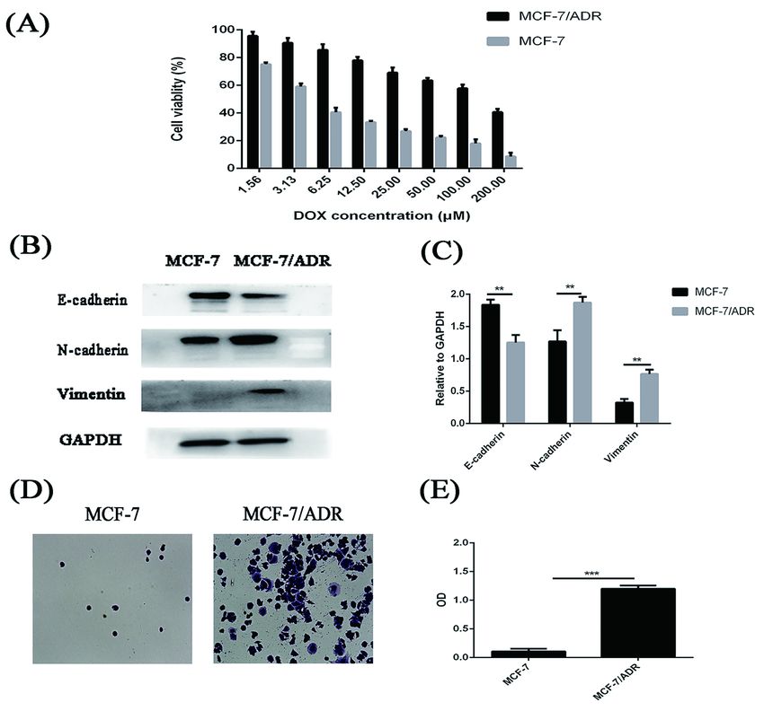
We prepared E-PLNs by nano precipitation method (Tahir et al. 2019). Briefly, EMO and PLGA were
dissolved in acetone. Another soybean phospholipid and DEPE-PEG 2000 were dissolved in 5% (W/V) F68
aqueous solution. The organic phase was slowly and evenly added to Pluronic F-68 aqueous solution at
75 ℃, and stirred (8000 rpm for 40 min) until the acetone completely evaporated. The supernatant was
collected by centrifugation (1000 rpm for 5 min). The particle size and Zeta potential of E-PLNs were
measured by Malvern Zetasizer Nano ZS90. The prepared E-PLNs were added with 10% (W/V) freeze-
drying protective agent (Mannitol). After being frozen overnight at - 80 ℃, E-PLNs were freeze-dried for 24
h and stored at 4 ℃.

Cell culture

MCF-7 cells were cultured in 10% FBS of RPMI-1640 medium under defined circumstances. In order to
maintain cell resistance to DOX, MCF-7/ADR cells were cultured in RPMI-1640 medium containing DOX
(250 ng/mL) every third passage.

Cellular uptake

Coumarin-6 (200 µg/mL) was encapsulated in PLNs instead of EMO to prepare coumarin 6-PLNs (C-6
PLNs). MCF/ADR were incubated with C-6 PLNs for 2 h, fixed by 4% paraformaldehyde, and treated with
Hochest staining solution for 5 min. The uptake of C-6 PLNs in MCF/ADR was observed under laser
confocal microscope and analyzed by Flow cytometry.

MTT assay

Cells were cultured in 96 well plate for 24 h. Then the fresh culture medium containing free DOX, free
EMO, E-PLNs, DOX+EMO, DOX+E-PLNs were added to the culture medium, and cultured for more than 48h
for cell test. After incubation with MTT (5 mg/mL, 20 μ L/well) for 4 h, the supernatant was removed. The
cells were exposed to DMSO solution (150 μL/well) and incubated for 15 min. Finally, the OD value was
detected (Microplate Reader, 490 nm) and cell viability was calculated.

Cell invasion assay

Cells were resuspended in RPMI-1640 medium without FBS, then seed in the upper chamber for 24 h, and
500 μL culture medium containing different drugs was added into the lower chamber. Cells were fixed
with 4% paraformaldehyde for 10 min and stained with 200 μL crystal violet staining solution for 15 min.
5 fields of vision were randomly selected under a 100 × microscope to observed the cells passing through
the filter membrane. After taking photos, 500 μL 33% acetic acid solution was added, shaking for 10 min,
the OD value was measured to indirectly reflect the number of invasive cells.

Apoptosis study

MCF-7/ADR cells (3×10⁵ cells/well, 6 well plate) were cultured, then treated with fresh medium containing
different drugs for 48 h. The cells were digested with trypsin without EDTA. After centrifugation again,

                                                 Page 8/21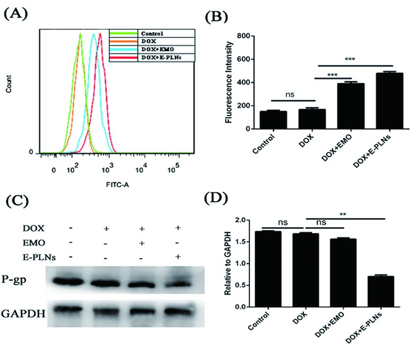
cells were suspend with 500 μL Binding Buffer. Then, Annexin-V/FITC and PI staining solution was added
with 5 μL each and mixed thoroughly. All experimental groups were sincubated in the dark for 15 min.
Finally, the apoptosis was detected on the flow cytometer.

Western blotting analysis

Treated with different treatments for 48 h, cells were lysed with lysate, and the protein was obtained by
centrifugation (10000 rpm for 5 min). BCA method was used to determine the protein concentration. After
the expressed proteins (30 μg each) were separated by SDS-PAGE (Bio RAD), and transferred to PVDF
membrane. The unbound protein sites were blocked with 5% skim milk for 2 h. The PVDF membrane was
incubated with solution containing with different primary antibodies at 4 ℃ overnight. After washed with
PBST for 1 h, The PVDF membrane incubated with solution containing horseradish peroxidase bound
secondary antibody for 2 h. The PVDF membrane was evenly coated with ECL developer. According to the
manual, the exposure was carried out with chemiluminescence imaging system and analyzed with Image
J software.

Statistical analysis

The experiment was repeated three times, and the average value ± SD value was calculated. GraphPad
Prism software analyzes and draws pictures, and the ONE-WAY ANOVE analysis is used for significance.
* p
Zou designed the experimental protocols, performed the experiments and analyzed the results. Lan, Li,
Tiange Cai, and Yu Cai contributed to writing the manuscript. All authors reviewed the final draft of the
manuscript. All authors read and approved the final manuscript.

Acknowledgements

Authors acknowledges the Natural Science Foundation of Guangdong for providing financial support to
carry out the work under the funding 2021A1515011241.

Abbreviations
EMT: epithelial mesenchymal transition; EMO: Emodin; E-PLNs: Emodin-loaded polymer-lipid hybrid
nanoparticles; DOX: Doxorubicin; TGF-β: Transforming growth factor beta; TME: Tumor
microenvironment; P-gp: P-glycoprotein; PDI: Polydispersity index; Bcl-2: B-cell lymphoma 2; Bax: Bcl-2-
associated X protein; GAPDH: Glyceraldehyde 3-phosphate dehydrogenase; PI: Propidium iodide; DAPI:
4′,6-diamidino-2-phenylindole; PLGA: Poly (lactic-co-glycolic acid).

References
Siegel RL, Miller KD, Fuchs HE, et al. Cancer Statistics, 2021. CA Cancer J Clin. 2021 Jan;71(1):7-33.

Sung H, Ferlay J, Siegel RL, et al. Global cancer statistics 2020: GLOBOCAN estimates of incidence and
mortality worldwide for 36 cancers in 185 countries. CA Cancer J Clin. 2021 Feb 4.

Asiedu, M. K., F. D. Beauchamp-Perez, J. N. Ingle, M. D. Behrens, D. C. Radisky & K. L. Knutson (2014) AXL
induces epithelial-to-mesenchymal transition and regulates the function of breast cancer stem cells.
Oncogene, 33, 1316-24.

Sun, Y., X. Wang, Q. Zhou, Y. Lu, H. Zhang, Q. Chen, M. Zhao & S. Su (2015) Inhibitory effect of emodin on
migration, invasion and metastasis of human breast cancer MDA-MB-231 cells in vitro and in vivo. Oncol
Rep, 33, 338-46.

Liu, Q., J. Hodge, J. Wang, Y. Wang, L. Wang, U. Singh, Y. Li, Y. Yao, D. Wang, W. Ai, P. Nagarkatti, H. Chen,
P. Xu, E. A. Murphy & D. Fan (2020) Emodin reduces Breast Cancer Lung Metastasis by suppressing
Macrophage-induced Breast Cancer Cell Epithelial-mesenchymal transition and Cancer Stem Cell
formation. Theranostics, 10, 8365-8381.

Lu, J., Y. Xu, X. Wei, Z. Zhao, J. Xue & P. Liu (2016) Emodin Inhibits the Epithelial to Mesenchymal
Transition of Epithelial Ovarian Cancer Cells via ILK/GSK-3β/Slug Signaling Pathway. Biomed Res Int,
2016, 6253280.

Gu, J., C. F. Cui, L. Yang, L. Wang & X. H. Jiang (2019) Emodin Inhibits Colon Cancer Cell Invasion and
Migration by Suppressing Epithelial-Mesenchymal Transition via the Wnt/β-Catenin Pathway. Oncol Res,

                                                   Page 10/21
27, 193-202.
Fu, J. M., J. Zhou, J. Shi, J. S. Xie, L. Huang, A. Y. Yip, W. T. Loo, L. W. Chow & E. L. Ng (2012) Emodin
affects ERCC1 expression in breast cancer cells. J Transl Med, 10 Suppl 1, S7.
Song, X., X. Zhou, Y. Qin, J. Yang, Y. Wang, Z. Sun, K. Yu, S. Zhang & S. Liu (2018) Emodin inhibits
epithelial‑mesenchymal transition and metastasis of triple negative breast cancer via antagonism of
CC‑chemokine ligand 5 secreted from adipocytes. Int J Mol Med, 42, 579-588.
Li, W., H. Zhang, Y. G. Assaraf, K. Zhao, X. Xu, J. Xie, D. H. Yang & Z. S. Chen (2016) Overcoming ABC
transporter-mediated multidrug resistance: Molecular mechanisms and novel therapeutic drug strategies.
Drug Resist Updat, 27, 14-29.
Markman, J. L., A. Rekechenetskiy, E. Holler & J. Y. Ljubimova (2013) Nanomedicine therapeutic
approaches to overcome cancer drug resistance. Adv Drug Deliv Rev, 65, 1866-79.
Kalyane, D., N. Raval, R. Maheshwari, V. Tambe, K. Kalia & R. K. Tekade (2019) Employment of enhanced
permeability and retention effect (EPR): Nanoparticle-based precision tools for targeting of therapeutic
and diagnostic agent in cancer. Mater Sci Eng C Mater Biol Appl, 98, 1252-1276.
Jain, V., H. Kumar, H. V. Anod, P. Chand, N. V. Gupta, S. Dey & S. S. Kesharwani (2020) A review of
nanotechnology-based approaches for breast cancer and triple-negative breast cancer. J Control Release,
326, 628-647.
Mukerjee, A., A. P. Ranjan & J. K. Vishwanatha (2012) Combinatorial nanoparticles for cancer diagnosis
and therapy. Curr Med Chem, 19, 3714-21.
Jiang, Z. S., Y. Z. Sun, S. M. Wang & J. S. Ruan (2017) Epithelial-mesenchymal transition: potential
regulator of ABC transporters in tumor progression. J Cancer, 8, 2319-2327.
Du, B. & J. S. Shim (2016) Targeting Epithelial-Mesenchymal Transition (EMT) to Overcome Drug
Resistance in Cancer. Molecules, 21.
Li, S., Y. Fan, A. Kumagai, E. Kawakita, M. Kitada, K. Kanasaki & D. Koya (2020b) Deficiency in Dipeptidyl
Peptidase-4 Promotes Chemoresistance through the CXCL12/CXCR4/mTOR/TGFβ Signaling Pathway in
Breast Cancer Cells. Int J Mol Sci, 21.
Xu, J., S. Lamouille & R. Derynck (2009) TGF-beta-induced epithelial to mesenchymal transition. Cell Res,
19, 156-72.
Rodón, J., M. Carducci, J. M. Sepulveda-Sánchez, A. Azaro, E. Calvo, J. Seoane, I. Braña, E. Sicart, I.
Gueorguieva, A. Cleverly, N. S. Pillay, D. Desaiah, S. T. Estrem, L. Paz-Ares, M. Holdhoff, J. Blakeley, M. M.
Lahn & J. Baselga (2015) Pharmacokinetic, pharmacodynamic and biomarker evaluation of transforming
Page 11/21growth factor-β receptor I kinase inhibitor, galunisertib, in phase 1 study in patients with advanced cancer.
Invest New Drugs, 33, 357-70.
Gu, M., W. Zheng, M. Zhang, X. Dong, Y. Zhao, S. Wang, H. Jiang & X. Zheng (2020) LncRNA
NONHSAT141924 promotes paclitaxel chemotherapy resistance through p-CREB/Bcl-2 apoptosis
signaling pathway in breast cancer. J Cancer, 11, 3645-3654.
Song, X., X. Zhou, Y. Qin, J. Yang, Y. Wang, Z. Sun, K. Yu, S. Zhang & S. Liu (2018) Emodin inhibits
epithelial‑mesenchymal transition and metastasis of triple negative breast cancer via antagonism of
CC‑chemokine ligand 5 secreted from adipocytes. Int J Mol Med, 42, 579-588.
Wang, S., T. Chen, R. Chen, Y. Hu, M. Chen & Y. Wang (2012) Emodin loaded solid lipid nanoparticles:
preparation, characterization and antitumor activity studies. Int J Pharm, 430, 238-46.
Tahir, N., A. Madni, A. Correia, M. Rehman, V. Balasubramanian, M. M. Khan & H. A. Santos (2019) Lipid-
polymer hybrid nanoparticles for controlled delivery of hydrophilic and lipophilic doxorubicin for breast
cancer therapy. Int J Nanomedicine, 14, 4961-4974.
Figures
Page 12/21Figure 1
EMT is associated with DOX resistance in breast cancer cells. (A) Cell viability in vitro on MCF-7 and MCF-
7/ADR cells after treatment with different DOX concentration (1.56 μM-200 μM) for 48 h. (B) and (C) The
expression of E-cadherin, N-cadherin and Vimentin in MCF-7 and MCF-7/ADR cells was analyzed by
Western blotting. (D) and (E) The invasion activity of MCF-7 and MCF-7/ADR cells were observed and
photographed with 200× microscope, and the OD value was detected at 570 nm to indirectly reflect the
number of cells. N=3, **p < 0.01, ***p < 0.001.
Page 13/21Figure 2
The effect of Galunisertib combined with DOX on cell proliferation and invasion. Cell viability of MCF-
7/ADR cells after treatment with different Galunisertib concentration (27.9-1000 μM). (B) and (C) The
effect of 30μM Galunisertib on the expression of EMT marker protein. N=3, **p < 0.01, ***p < 0.001
compared with Control group. (D) Cell viability on MCF-7/ADR cells after treatment with 30 μM
Galunisertib combined with DOX of different concentrations (1.56-200 μM). (E) and (F) The invasion
Page 14/21activity of MCF-7/ADR cells after incubation with DOX (10 μM), Galunisertib (30 μM), DOX (10 μM) +
Galunisertib (30 μM) over 48 h. N=3, **p < 0.01, ***p < 0.001 compared with DOX group.
Figure 3
Effect of Galunisertib combined with DOX on apoptosis and expression of drug resistance protein. (A)
and (B) The apoptotic rate of MCF-7/ADR cells treated with different groups (DOX, Galunisertib, DOX+
Galunisertib) was measured by flow cytometry. (C) and (D) The expression of Bax and Bcl-2 in MCF-
Page 15/217/ADR cells treated with DOX, Galunisertib, DOX+Galunisertib. Relative levels of the proteins were
quantified by densitometric analysis. (E) and (F) The expression of P-gp and MRP1 in MCF-7/ADR cells
treated with different groups (DOX, Galunisertib, DOX+Galunisertib) . N=3, **p < 0.01, ***p < 0.001
compared with DOX group.
Figure 4
Page 16/21The characterization of E-PLNs. (A) The morphology of E-PLNs. (B) Lyophilized powder of E-PLNs. (C)
and (D) The particle size and Zeta potential of E-PLNs was measured by the laser particle size analyzer.
Figure 5
Cellular uptake and cytotoxicity study of E-PLNs. (A) confocal imaging of C-6 PLNs in MCF-7/ADR cells
was observed. (B) and (C) Flow cytometry and fluorescence intensity of nanoparticles uptake by MCF-
7/ADR cells, N=3, **p < 0.01, ***p < 0.001 compared with Control group. (D) and (E) Cell viability in vitro
on MCF-7/ADR cells after treatment with free EMO and E-PLNs. (F) The cell viability of MCF-7/ADR cells
treated with 30 μM EMO or E-PLNs combined with different concentrations of DOX (1.56-200 μM).
Page 17/21Figure 6
E-PLNs enhanced DOX induced apoptosis by regulating apoptosis related proteins. (A) Morphological
changes of nuclei in MCF-7/ADR cells in the different treated groups using DAPI staining. (B) and (C)
Apoptosis assay of MCF-7 cells after incubation with different treated groups. (D) and (E) The expression
of Bax and Bcl-2 in MCF-7/ADR cells were determined by Western blot analysis. N=3, **p < 0.01, ***p <
0.001 compared with DOX group.
Page 18/21Figure 7
E-PLNs increase the accumulation of DOX by inhibiting the expression of drug resistance protein. (A) and
(B) The accumulation of in MCF-7/ADR cells treated with DOX, DOX+EMO, DOX+E-PLNs measured by
flow cytometry. (C) and (D) The expression of P-gp in MCF-7/ADR cells detected by Western blotting
analysis. N=3, **p < 0.01, ***p < 0.001 compared with DOX group.
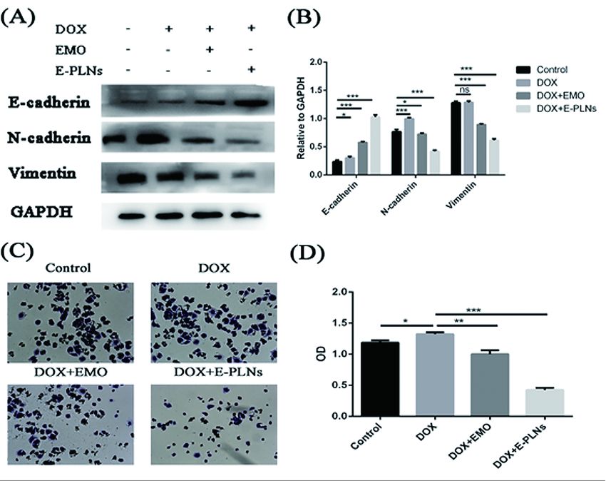
Page 19/21Figure 8
E-PLNs combined with DOX inhibited EMT marker protein expression and cell invasion. (A) and (B) After
MCF-7/ADR cells treated with DOX, DOX+EMO, DOX+E-PLNs for 48 h, the expression of E-cadherin, N-
cadherin and Vimentin were detected by Western blotting analysis. (C) and (D) The invasion activity of
MCF-7/ADR cells after incubation with DOX, DOX+EMO, DOX+E-PLNs for 24 h. N=3, **p < 0.01, ***p <
0.001 compared with DOX group.
Page 20/21Figure 9
E-PLNs enhance the sensitivity of breast cancer to Doxorubicin by inhibiting epithelial mesenchymal
transition.
Page 21/21You can also read