Endogenous fluorescence of hemosiderin in endometriosis to improve clinical detection
←
→
Page content transcription
If your browser does not render page correctly, please read the page content below
Cabe et al. Translational Medicine Communications                (2019) 4:9
https://doi.org/10.1186/s41231-019-0038-3
                                                                                                                             Translational Medicine
                                                                                                                                  Communications

 RESEARCH                                                                                                                                        Open Access

Endogenous fluorescence of hemosiderin
in endometriosis to improve clinical
detection
Andrew G. Cabe1,6* , Arnold D. Estrada3, Taylor Hoyt1, Xiao Yang1, Scott Jenney3, Philip T. Valente4, Bryan Cox5,
Jessica E. McLaughlin2, Randal D. Robinson2, Thomas E. Milner3 and Marc D. Feldman1

  Abstract
  Background: Endometriosis impacts 6–10% of all reproductive- age women. Studies have shown the more effectively
  endometriosis is removed, the better the patient outcomes for pain reduction and fertility (2, 3). Hemosiderin, glands,
  and stroma are the histologic markers of endometriosis; optical coherence tomography (OCT) can identify glands and
  hemosiderin has a known endogenous fluorescence than can be detected by two-photon microscopy (TPM). The
  hypothesis was that the identification of optical properties of endometriosis using OCT and TPM combined would
  improve a surgeon’s ability to diagnose and treat by improving endometriosis detection compared to current
  standards of visual diagnosis.
  Methods: Forty-one women with clinically suspected endometriosis undergoing laparoscopy were consented. Women
  were enrolled at two clinical sites: University of Texas Health Science Center, San Antonio and Methodist Healthcare
  System, San Antonio. The surgeon made a clinical diagnosis of suspected endometriosis as 1) yes present 2) maybe
  present, and 3) not present (controls) from the peritoneum without suspected disease. One-hundred-twenty biopsies
  were collected from 27 women with visually suspected endometriosis. All three patient biopsy classes were excised and
  underwent histologic examination as the gold-standard diagnosis for endometriosis. The samples were imaged ex-vivo
  for optical markers of endometriosis; OCT for endometrial glands and TPM for hemosiderin. Histologic markers were co-
  registered with optical properties. Biopsies were embedded in agar to maintain orientation during imaging and
  histological processing. TPM used the endogenous fluorescence of hemosiderin as a marker. OCT used glands as a
  marker. Sensitivity, specificity, and positive (PPV) and negative predictive values (NPV) were calculated.
  Results: The main-outcome-measure was the statistical comparison of clinical impression, imaging results, and histologic
  truth. Glands, stroma and hemosiderin were present in 49, 72 and 86% of endometriosis samples confirmed by histology.
  Clinical suspicion of endometriosis had 98% sensitivity, 53% specificity, 68% PPV, and 96% NPV. In 31 samples of
  endometriosis maybe being present, 39% were histologically confirmed. Eighty-eight samples were analyzed using OCT-
  TPM. OCT-TPM had 93% sensitivity, 100% specificity, 100% PPV, and 93% NPV.
  Conclusions: OCT-TPM is useful in identifying endometriosis’ presence or absence. Evaluation of suspected endometriosis
  by OCT-TPM improves surgeons’ abilities to diagnose and treat endometriosis.
  Keywords: Endometriosis, Optical coherence tomography, Auto-fluorescence

* Correspondence: cabe@uthscsa.edu
1
 Departments of Cardiology, University of Texas Health Science Center at San
Antonio, 7703 Floyd Curl Drive, San Antonio, TX 78229, USA
6
 Department of Medicine, University of Texas Health Science Center, 7703
Floyd Curl Drive, San Antonio, TX 78229, USA
Full list of author information is available at the end of the article

                                        © The Author(s). 2019 Open Access This article is distributed under the terms of the Creative Commons Attribution 4.0
                                        International License (http://creativecommons.org/licenses/by/4.0/), which permits unrestricted use, distribution, and
                                        reproduction in any medium, provided you give appropriate credit to the original author(s) and the source, provide a link to
                                        the Creative Commons license, and indicate if changes were made. The Creative Commons Public Domain Dedication waiver
                                        (http://creativecommons.org/publicdomain/zero/1.0/) applies to the data made available in this article, unless otherwise stated.
Cabe et al. Translational Medicine Communications (2019) 4:9 Page 2 of 11 Background standard, histologic diagnosis. The histologic diagnosis Endometriosis impacts 6–10% of all reproductive- age was made by a gynecologic pathologist (PTV) blinded to women [1]. The gold standard for diagnosis and treatment the results of the OCT-TPM imaging. of endometriosis is laparoscopic inspection and excision or ablation with histologic confirmation. Studies have shown the more effectively endometriosis is removed, the better Methods the patient outcomes for pain reduction and fertility [2, 3]. The tissue samples were collected at two clinical sites, However, of the tissue removed during laparoscopy, less University of Texas Health Science Center at San Anto- than 70% is confirmed as endometriosis on histologic diag- nio, and Methodist Healthcare System in San Antonio. nosis [4]. With a 30–40% recurrence rate for symptomatic Participants were enrolled between December 18, 2014 endometriosis after laparoscopic surgery, due to incomplete and June 6, 2017 in a prospective manner. Two experi- surgical excision [5], there are clearly short-comings in the enced gynecologic surgeons (RDR, BC), each with current surgical diagnosis and management of endometri- greater than 25 years’ experience diagnosing and treating osis. During laparoscopic surgery, the surgeon’s goal is iden- endometriosis performed all of the surgical procedures. tification of each endometriosis lesion and sequential excision of each implant. Complicating the time consuming process of identification and removal is the potential wide- Ethical approval spread distribution of endometriosis lesions throughout the IRB approval for this study was obtained at both clinical abdominal and pelvic cavities. The distribution of these le- sites. Background information was collected on patients’ sions presents a challenge to all surgeons whose diagnostic age and race, medical history, and list of medications. imaging tools are normally limited to an unaided eye and Subjects were approached for consent if they were laparoscope with some endometriosis not being immedi- undergoing surgery for suspected endometriosis for clin- ately discernible from surrounding tissue. The nature of the ical indications. Informed consent was obtained for all iceberg geometry of endometriosis lesions complicates the participants. Subjects were withdrawn if no suspected surgery since frequently only a fraction of the lesion is vis- endometriosis was identified at the time of surgery. The ible with the remaining endometriosis lesion located below mean participant age was 37 (range 22–46). the surface. Because the surgeon is faced with the divergent challenges of removing all endometrial lesions without dam- aging delicate underlying tissues and structures, prolonged Sample collection surgeries are common and only partially successful. Patient Peritoneal biopsy samples were collected at the time of outcomes suffer because no effective imaging tools are cur- laparoscopic surgery and the surgeons were asked to make rently available in the operating room to allow endometri- a clinical diagnosis of each sample removed as “yes”, “no”, osis lesions to be discerned from surrounding tissues [6]. or “maybe” for endometriosis. The location of origin of The current study presents novel solutions for the de- these samples was also recorded. Clinically defined endo- tection of endometriosis, using optical coherence tomog- metriosis was described as red, black, or white lesions. raphy and two photon microscopy (OCT-TPM) imaging. The samples designated in the “no” category were control Hemosiderin is known to have specific excitation/emis- samples of normal appearing peritoneal tissue biopsied by sion wavelengths [7, 8]. We hypothesized that we could the surgeon. These three categories allowed us to develop identify hemosiderin as a marker in endometriosis with optical signatures of endometriosis. The “maybe” category TPM imaging of the same excitation/emission pair. He- allowed us to determine if these optical properties could mosiderin is particularly relevant because is present in improve the sensitivity and specificity of the surgeon’s early-onset endometriosis [9]. Further, it is known that clinical diagnosis. The “maybe” samples represent sus- columnar epithelial cells, found in endometriosis glands, pected disease that some surgeons might remove and form fluid filled glandular structures and OCT is an op- others might not depending on location and appearance. tical technique capable of easily identifying fluid filled Once the samples were received, imaging was per- spaces in tissues [10]. Therefore, we hypothesized that formed to categorize optical properties. Samples were OCT could be a second optical tool in the detection of embedded in agar using a 4x4x2 mm mold and imaged endometriosis. We tested these hypotheses on ex vivo in the mold. The mold allowed for co-registration with tissue samples collected by gynecologic surgeons from histology. Specifically, pre-embedding in a mold women suspected of having endometriosis. Samples allowed the preservation of tissue orientation and lo- were classified by the surgeon as yes or maybe for the cation throughout imaging and histological processing. presence of endometriosis, with suspected negative con- Knowledge of tissue orientation allows exact 3D trols also excised, and these samples were then evaluated co-registration between the OCT-TPM images and with both optical techniques and compared to the gold stained histologic slides.
Cabe et al. Translational Medicine Communications   (2019) 4:9                                                   Page 3 of 11

Imaging procedures                                               features in the images were identified as matching the
Optical imaging was performed with three imaging sys-            histologic markers.
tems, Optical Coherence Tomography (OCT), Two-Pho-
ton Microscopy (TPM), and Wide-Field/Single-Photon               Statistical analyses
Fluorescence. Imaging was performed within 24 h of sam-          Sensitivity, specificity and negative and positive predict-
ple excision. OCT imaging was performed on a benchtop            ive values for the accuracy of both the imaging systems
OCT system built in our lab, using Alazar (Pointe-Claire,        and the surgeons’ impressions were calculated. Sensitiv-
QC, Canada) and Santec (Hackensack, NJ, USA) sources,            ity was calculated by taking the number of true positives
which operate between 1250 and 1376 nm. These systems            (histologically confirmed endometriosis) and dividing by
recorded b-scans for the entire sample volume (up to             the sum of the number of true positives and false nega-
6.25 × 6.25 × 2 mm). Lateral resolution was 6.1–12.21 μm/        tives (histologically confirmed endometriosis that was
pixel. Axial resolution was 3.75–8 μm/pixel. The targets         not detected by the surgeon or imaging systems). Speci-
for OCT imaging were glands. The presence of endomet-            ficity was calculated by taking the number of true nega-
riosis detected by OCT was a void in the image which cor-        tives (histology confirmed to lack endometriosis) and
responded to a fluid-filled gland in the histology. OCT          dividing by the sum of the number of true negatives and
penetration depth and wide-field also provided a full vol-       false positives (histology confirmed to lack endometriosis
ume view of the samples, ranging in size from 12 to 27           but identified as disease positive by the surgeon or im-
mm3, useful for later co-registration with histology. The        aging system). Positive predictive value was calculated by
second optical target for endometriosis was the intrinsic        taking the number of true positives and dividing by the
fluorescence of hemosiderin. Hemosiderin has an excita-          sum of true positives and false positives. Negative pre-
tion peak of 450 nm, and an emission peak near 680-690           dictive value was calculated by taking the number of true
nm. TPM imaging and Single Photon fluorescence im-               negatives and dividing by the sum of true negatives and
aging were performed on a Prairie View microscope sys-           false negatives.
tem (Middleton, WI, USA). TPM was performed using an
excitation wavelength of 900 nm (twice the one-photon            Results
excitation peak). Fluorescence images using emission             OCT was used to identify fluid filled glands and two-pho-
channels, 500 nm, 560 nm, 620 nm, and 690 nm, were re-           ton microscopy (TPM) to detect the endogenous fluores-
corded. Hemosiderin was identified by increased pixel in-        cence of tissue without exogenous labels, specifically
tensity in the 690 nm emission channel. TPM images were          hemosiderin. Results show that glands are visible via OCT
taken with a field of view of 735 μm. Z-series with 10 μm        imaging (Fig. 1a) and hemosiderin has endogenous fluor-
step-size were taken and stitched together to image the          escence that is present both in multi-photon (Fig. 1b) and
entire sample. Z-series (stacks of images) were taken in         single-photon fluorescence imaging (Fig. 1c).
depth until the image was no longer interpretable (ap-
proximately 200 μm). TPM had a resolution of 1.44 μm/            Clinical impression
pixel. Additionally, single-photon wide-field fluorescence       This study enrolled 41 women. Based on the clinical
images were recorded using an excitation source emitting         diagnosis of the physician, 27 women had endometriosis
at 420 + − 20 nm and an emission filter of 690 + − 20 nm         and 14 did not. These 14 women were determined to
to demonstrate hemosiderin detection with standard               have no endometriosis lesions by the surgeon and no bi-
fluorescence imaging as seen in increased pixel intensity.       opsies were obtained for ethical reasons. The results out-
This method is less expensive than TPM and therefore             lined below focus on the remaining 27 subjects. Of these
more translatable to a clinical device.                          27 subjects, 18 had advanced optical imaging performed
                                                                 with OCT and TPM.
Histologic processing
The slides underwent histologic processing. Slides were          Histology
taken every 40 μm for the first 400 μm and every 100 μm          For the set of 27 subjects the surgeon’s diagnosis of
subsequently. Paraffin slides were stained with Hematoxylin      120 biopsies was compared to the histologic gold
and Eosin. For the histologic evaluation, each sample was        standard. The surgeon diagnosed 65 of these as defin-
categorized for the presence of the three histologic markers     ite endometriosis, 31 as maybe for endometriosis, and
for endometriosis: glands (columnar endothelial cells),          24 as negative for endometriosis. Fifty-seven biopsies
stroma, and hemosiderin. The histologic diagnosis was used       were interpreted blindly by the gynecologic patholo-
as the gold standard against which the surgeons’ and im-         gist to have endometriosis; 44 of the definite biopsies,
aging systems’ findings were compared. Due to the exact-         12 of the maybe biopsies and one of the negative bi-
ness of the match from the use of agar, the optical              opsies. The accuracy of the surgeons’ clinical impres-
properties of specific markers were obtained. Unique             sion is summarized in Table 1.
Cabe et al. Translational Medicine Communications (2019) 4:9 Page 4 of 11
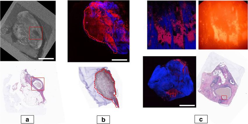
Fig. 1 Imaging Results. 1a Sample 29-1a, the top image is the OCT en face image, the bottom image is the histology, the identified gland is
boxed in red, scale bar is 1 mm 1b Sample 20–6, TPM image on top, histology on bottom, hemosiderin is circled in red and appears as brown in
the histology due to H&E stain, the TPM image is colored based on emission channel, the red represents 690 nm (hemosideran), blue is 500 nm
(collagen), scale bar is 1 mm 1c: Sample 34–1 The image on the top left is the single photon microscopy/ wide field fluorescence, very bright
spots match with TPM and histology as hemosiderin, the image on the top right is the corresponding TPM image location colored as stated in
1b these are scale to 470 um × 470 um, the bottom right is the full TPM image and bottom left is the corresponding histology, the red box
represents the area of the top images, scale bar is 1 mm
Hemosiderin was a prevalent marker seen in 49 of 57 samples where it was confirmed by histology and did
(86%) endometriosis samples proven positive by histology. not have a single false positive in the other 50 samples
Glands were found in pathology in 28 of the 57 (49%) of (Fig. 2). Glands were visible in OCT for 17 of the 21
endometriosis positive samples. The final marker, stroma, samples for which they were seen in histology. OCT for
was found in 41 of the 57 samples (72%), although not an glands lacks a PPV and specificity due to lack of a true
optical target for advanced imaging. false positive. This is due to OCT’s optical marker for
glands being able to be co-registered with its histologic
Advanced optical imaging counterpart, but it is difficult to examine the OCT image
It was possible to obtain full optical imaging sets for 18 of and read a false positive that corresponds to histology.
the 27 patients outlined above. This resulted in 113 optical
imaging sets of co-registered OCT, TPM and histology from Single photon results
86 biopsy samples. Results are shown in Tables 1, 2, and 3. To allow for clinical translation of hemosiderin as an op-
TPM confirmed the presence of hemosiderin with a tical target, 4 subjects were further examined by an add-
visible endogenous fluorescence signal in all 36 of those itional imaging technique, single-photon fluorescence.
Table 1 Statistical results of surgical impression/ advance optical imaging vs histologic gold standard
PPV NPV SENS. SPEC.
Surgical Impression (27 subjects) 68 96 98 53
OCT-TPM Combined (18 subjects with advanced imaging) 100 93 93 100
TPM for Hemosiderin (113 optical imaging sets) 100 100 100 100
OCT for Glands (113 optical imaging sets) 96 82
PPV is positive predictive value, NPV is negative predictive value, SENS is sensitivity, SPEC is specificity
Surgical Impression is based on the surgical diagnosis of all samples taken from 27 subjects against histological diagnosis. OCT-TPM combined is the optical
markers from both imaging type predication of endometriosis against histological diagnosis. OCT-TPM was only available in 18 of 27 patients with endometriosis.
One hundred thirteen optical imaging sets were available from these 18 patients for OCT-TPM. OCT for glands lacks a PPV and specificity due to lack of a true
false positive. TPM for hemosiderin and OCT for glands is specifically the identification of the marker in the imaging data against histologic confirmation of
the markerCabe et al. Translational Medicine Communications (2019) 4:9 Page 5 of 11
Table 2 Clinical Impression vs. Histology Gold Standard
Physician Subjects
clinical
7 8 9 10 11 12 13 15 16 20 22 23 24 25 27 28 29 30 32 34 36 38 39 42 45 46 47
impression
at time of
surgery
Yes No No No Yes Yes No No Yes Yes No No Yes Yes Yes Yes Yes Yes Yes Yes Yes Yes Yes Yes Yes Yes
No No Yes Yes Yes No No Yes Yes No Yes Yes Yes Yes No Yes Yes No Yes Yes
Yes Yes No Yes Yes No Yes No No Yes
Yes Yes No Yes No No
Yes Yes
Yes Yes
Maybe No Yes No Yes No Yes No No No No No No Yes Yes Yes Yes No Yes Yes Yes
No No Yes Yes No No No No No
No
No
No No No No Yes No No No No No No No No No No No No No No No No No No No No
Histological diagnosis
Blank = no sample obtained; each yes or no entry represents each individual sample of tissue biopsied; columns represent the histological diagnosis per the
reading of an expert Ob/Gyn pathologist; rows represent Ob/Gyn physician/surgeon clinical impression of the sample at the time of surgery
This evaluation examined 6 biopsies from these 4 pa- results from studies with larger cohorts, Mettler re-
tients identified by TPM to be hemosiderin positive, and ported a 53.8% positive rate in biopsies of suspected
2 controls shown by TPM to be hemosiderin negative. endometriosis [11] and Stratton a 61% rate [12]. Steg-
Single-photon was able to detect hemosiderin similar to mann reported in her 133 patient, 611 sample study, a
TPM, in the exact areas and identical pattern as TPM, true positive rate of 65.0% rate which is very similar to
and was confirmed by histology (Fig. 1c). our 67% rate but she found a much higher false negative
rate of 12% compared to our 4% [4].
Discussion There are several implications of our results regarding
Our results demonstrate the benefit of adding advanced surgeons removing too much tissue in 1/3 of cases and
optical imaging in the diagnosis of endometriosis. The missing lesions that should be removed. If any endometri-
clinician made a correct clinical diagnosis in 67% of osis is being missed and left in the patient, the chances for
histologic positive samples. Endometrial glands are help- improving patient outcomes of pain reduction and pos-
ful if present but were only found in histology in 28 of sibly increased fertility, and preventing the high persist-
the 57 endometriosis positive samples or 49% of the ence rates common in endometriosis, are diminished. The
time. Glands were visible in OCT for 19 of the 23 im- low specificity and PPV reveals how much non-endomet-
aging sets for which they were seen in histology. The sig- riosis tissue the surgeons are removing during the proced-
nificant finding was the frequency that hemosiderin was ure and how much unproductive surgical time is added to
present. Hemosiderin was found in 86% percent of all the procedure. The one false negative result reveals that
histologic samples, and correctly identified with TPM there is diseased tissue that is being left behind because
100% of the time; as well as, in single photon fluores- the physician could not determine that a negative biopsy
cence imaging, which will allow clinical translation of was actually endometriosis. This study also examined
hemosiderin identification to the operating room. “maybe” samples, where the surgical diagnosis was un-
Specifically, these data demonstrate the need for a bet- clear. These samples proved to be positive for endometri-
ter detection method than a surgeon’s clinical diagnosis. osis 39% of the time. Our combined TPM-OCT imaging
Of the 65 samples of endometriosis removed where the accurately classified every “maybe” case. This shows the
gynecologic surgeon was certain that endometriosis was additive value of an advanced optical imaging approach,
present, the surgeon was incorrect 21 times, confirming which would assist the surgeon in accurately identifying
prior reports of the relatively poor visual diagnosis of endometriosis where it is unclear whether endometriosis
endometriosis lesions during laparoscopic surgery for is present or not.
endometriosis. We had two experienced gynecologic sur- TPM-OCT detection overcomes many of the limita-
geons at two institutions and generated 120 total tissue tions of other methods developed to improve detection.
samples for evaluation. Our data is comparable with Studies have shown that specific endometriosis lesionsCabe et al. Translational Medicine Communications (2019) 4:9 Page 6 of 11
Table 3 Full Imaging Results
Subject Sample Subsec. CI Type Path. TPM-hemo OCT-glands
20 1 Yes Red H Yes
2 Yes Red H Yes
3 b Yes Red H,S Yes
4 b Yes Dark chocolate cyst H Yes
c Yes S No
5 a Yes Cyst H Yes
c Yes H,S Yes
d Yes H Yes
6 Yes Endometroma H Yes
22 1 a Yes Red+black – No
b Yes G,S No No
2 No Control – No
23 1 No Control – No
2 a Yes Red – No
3 a Yes red – No
b Yes – No
4 b Yes Black+white – No
24 1 a Yes Red – No
b Yes – No
2 Yes Black + white – No
3 No Control – No
25 1 a Yes Red S No
b Yes G,S No No
2 a Yes Black H,S Yes
b Yes H,S Yes
3 No Control – No
27 1 a Yes Red + black G,S No Yes
b Yes – No
2 Maybe White – No
3 No Control – No
4 a Yes Endometroma H Yes
b Yes H,S Yes
c Yes H Yes
5 Yes Black H Yes
6 Yes Red – No
28 1 Maybe White – No
2 a Yes Red H Yes
b Yes – No
c Yes H Yes
3 a Yes Red – No
b Yes – No
c Yes – No
4 No Control – No
29 1 a Yes Red G,H,S Yes YesCabe et al. Translational Medicine Communications (2019) 4:9 Page 7 of 11
Table 3 Full Imaging Results (Continued)
Subject Sample Subsec. CI Type Path. TPM-hemo OCT-glands
b Yes G,H,S Yes Yes
2 No Control – No
3 a Yes Red + black – No
b Yes G,H,S Yes Yes
4 Maybe Black – No
30 1 Yes Black H Yes
2 maybe White – No
3 a Yes Black H Yes
b Yes H Yes
4 Yes E, H Yes
5 a Yes H Yes
b Yes E,H Yes
7 No Control – No
32 1 Yes Red G,H,S Yes Yes
2 Yes Red G,H,S Yes Yes
3 Maybe Black E,H,S Yes
4 Maybe White G,H,S Yes Yes
5 No Control – No
34 1 Yes Black G,H,S Yes Yes
2 a Yes Red G,H,S Yes Yes
b Yes G,S No Yes
3 Maybe White H, S Yes
4 No Control – No
36 1 No Control – No
2 a Maybe Black H,S Yes
b Maybe – No
3 a Yes Red G,S No No
b Yes E,H,S Yes
4 Maybe Black + white H,S Yes
38 1 Yes Red G,S No Yes
2 Yes Red – No
3 Yes Black – No
4 Maybe White H Yes
5 No Control – No
6 Maybe White – No
39 1 Yes S No
2 Maybe – No
3 No Control – No
4 Maybe – No
5 a Yes H,S Yes
b Yes H Yes
c Yes – No
d Yes – No
42 1 No – NoCabe et al. Translational Medicine Communications (2019) 4:9 Page 8 of 11
Table 3 Full Imaging Results (Continued)
Subject Sample Subsec. CI Type Path. TPM-hemo OCT-glands
2 Maybe – No
3 Yes – No
4 a Maybe G, H, S Yes Yes
b Maybe – No
5 Yes G, S No Yes
6 Yes G, H, S Yes Yes
7 Yes G, S No Yes
45 1 Maybe – No
2 Maybe H, S Yes
3 Yes G, S No Yes
4 Yes – No
5 Yes – No
6 No – No
46 1 Yes H Yes
2 a Yes – No
b Yes – No
3 a Yes G, H, S Yes Yes
4 No – No
47 1 Maybe G, S No Yes
2 Yes G, H, S Yes No
3 Yes G, H, S Yes Yes
5 Yes – No
6 Maybe – No
7 Yes H, S Yes
8 No – No
Columns: Subject: Subject number, Sample: sample number from that subject, Subsec.: is the subsection if the sample had to be divided to be fully imaged due
to sample size, CI (Clinical Impression): yes is positive for endo, maybe is possible endometriosis normally at the surgeon’s discretion for removal, no is control
tissue, Type: the surgeon reading of type of endometriosis, Path: Pathology reading - G is gland, fluid filled sac, E is endothelial cells with no fluid filled sac, H is
hemosiderin, and S is stroma. TPM-hemo: yes is a present optical marker for hemosiderin, no is no optical marker as determined from the TPM image, OCT-glands:
yes if the glands is seen and co-registered between the OCT image and histology, no if no such co-registration was possible
are not easily identified laparoscopically or by CT, MRI widespread adoption. Thus, newer techniques are
or ultrasound, unless there are large masses [13, 14]. needed that are both diagnostic based on the true histo-
This limits the surgical relevance of these technologies. logic pathology of endometriosis, and marker free as will
Limited success in detecting endometriosis has been be shown in a more detailed discussion of these compet-
seen in comparing blue light adsorption of surrounding ing optical techniques below.
tissue with endometriosis, which does not absorb blue Demco and co-workers published [17] an endometri-
light. However blue light techniques do not provide a osis blue light reflectance/absorption imaging method.
mechanistic link with a histological gold standard, such The authors hypothesized that lesions not visible under
as stroma, glands, or hemosiderin and thus its value to white light were visible under blue light reflectance be-
clinicians has been questioned and not adopted [15]. cause porphyrin molecules in endometriosis absorb blue
Other imaging methods use exogenous materials such as light. This is not an auto fluorescence technique as it
the photo-sensitizer 5-ALA, Firefly™ fluorescence im- looks at absorption of blue light and not emission from
aging (ICG), and blue dye [15, 16]. All have had limited excitation. Our TPM detection of hemosiderin looks at
success in identifying markers associated with endomet- the emission signal from excited hemosiderin. Demco’s
riosis, including angiogenesis and enhanced vascularity, method has not been rigorously tested since there are
changes in the peritoneal tissue surrounding endometri- no sensitivity and specificity results published [17]. Fur-
osis, and continued epithelial bleeding. However, the in- thermore, the spectral analysis of Demco’s technique will
direct nature of these approaches has also limited not reveal endometriosis below the surface such asCabe et al. Translational Medicine Communications (2019) 4:9 Page 9 of 11 Fig. 2 Endogenous Fluorescence of Hemosiderin to Improve Clinical Detection of Endometriosis. Two-Photon Microscopy (TPM), Histology positive/negative for endometriosis based on pathologist’s determination of glands, stroma, and/or hemosiderin unrecognized intra-ovarian endometriosis, since it relies emission wavelength of the porphyrin in hemosiderin, on surface reflection. Our technique depends on en- which might explain why they did not see the enhanced dogenous fluorescence emission and will identify lesions fluorescence from hemosiderin as we did [7, 8]. Looking below the surface since we are detecting emitted pho- for a reduction in fluorescence is flawed in that this ap- tons which can pass through layers of tissue. proach detects a negative image or the absence of a signal. Buchweitz et al. developed an auto-fluorescence tech- Another approach utilizes exogenous fluorescence nique to detect differences between normal tissue and with application of δ-Aminolevulinic acid (5-ALA) endometriosis [18]. They hypothesized that well known which was originally developed for photodynamic ther- auto-fluorescence of NADH, and ATP has a different apy in cancer. It has been applied for the detection and fluorescence pattern in endometriosis tissue compared to treatment of endometriosis. 5-ALA follows the heme normal tissue. The reasoning behind their approach is that pathway and goes into proto-porphyrin IX. They thus endometriosis has a different metabolic activity than sur- are using a similar marker with a similar excitation/ rounding tissues, and different metabolic rates can be re- emission as our TPM detection of hemosiderin. How- vealed in auto-fluorescence imaging due to the different ever, 5-ALA has several drawbacks. First as an exogen- endogenous fluorescence’s of NAD+ and NADH, and ous material it requires a 24-h hospital stay due to its ATP and ADP. With Buchweitz’s method endometriosis nature as a photo-sensitizer [19]. 5-ALA is a precursor was identified by decreased fluorescence compared to sur- of protoporphyrin IX (PPIX) in the heme pathway. How- rounding tissue, looking for a reduction in auto- fluores- ever, PPIX is not in hemosiderin [20], thus 5-ALA tar- cence as the marker for endometriosis. The emission gets the increased degree of angiogenesis and vascular wavelengths of those molecules are shorter than the endothelial growth factor expression consistent with
Cabe et al. Translational Medicine Communications   (2019) 4:9                                                                  Page 10 of 11

endometriosis. Our techniques present better diagnostic          glands seen by OCT and hemosiderin seen by TPM im-
options because they are label free and target physical          prove a surgeon’s ability to correctly diagnose endomet-
components of endometriosis rather than metabolic                riosis. This is demonstrated in improved negative and
pathways expressed by endometriosis.                             positive predictive values as compared to the clinical im-
   Another competing technology is Firefly™ fluorescence         pression of the surgeon. The improved diagnosis by
imaging which uses indocyanine green (ICG) as an ex-             OCT-TPM may result in fewer false positive results and
ogenous marker which identifies blood vessels. It has            increased disease tissue removed, although this will have
been used in endometriosis detection as it can identify          to be tested in a prospective clinical trial.
neovascularization associated with endometriosis [21].
                                                                 Abbreviations
Beyond being an exogenous marker, it has several other           5-ALA: δ-Aminolevulinic acid; ICG: Indocyanine green; NPV: Negative
shortcomings in detection. Specifically, as not all endo-        predictive values; OCT: Optical coherence tomography; PPIX: Protoporphyrin
metriosis presents as ‘red’ vascularized endometriosis, its      IX; PPV: and positive; TPM: Two photon microscopy
ability to accurately detect black and white endometri-
                                                                 Acknowledgements
osis is poor and other methods that do not rely on               Not applicable.
angiogenesis are needed [22].
   A criticism of using TPM to identify hemosiderin is           Funding
                                                                 Clayton Foundation for Biomedical Research, Houston, Texas, USA.
the high cost of the two photon instrument to be used            The funders had no role in the initiation or design of the study, collection of
in the operating room. As a result, we evaluated whether         samples, analysis, interpretation of data, writing of the paper, or the
the low cost option of single photon fluorescence could          submission for publication.
be implemented to also identify hemosiderin in endo-
                                                                 Availability of data and materials
metriosis and found similar accuracy. Clinical translation       The datasets used and/or analysed during the current study not included in
of these results to a cost-effective device has been built,      this published article are available from the corresponding author on
programed, and its camera and electronic components              reasonable request.
have been tested. We are currently repeating our studies         Authors’ contributions
using this single photon device which can easily be              AGC planned the study, performed all experiments, collected the data,
inserted into a laparoscope for clinical translation at low      analyzed the results, and wrote the manuscript. ADE helped conduct
                                                                 imaging, devised image processing methods, and contributed to study
cost. The clinical application of a laparoscope using the        design. RDR, BC, and JEM preformed surgeries and provided all biopsies. TH
specific fluoroscopy detailed in this paper could provide        and XY processed imaging data and assisted in image co-registration. SJ
surgeons with the ability to more accurately detect and          contributed to designing image processes and data acquisition. PTV was the
                                                                 expert pathologist and analyzed all histology slides. TEM, RDR, and MDF con-
removed endometriosis.                                           tributed to study design. ADE, JEM, RDR, TH, TE M, and MDF all assisted in re-
   There are some limitations to our study. Glands and           vising the manuscript. All authors read and approved the final version.
hemosiderin are not present in every endometriosis le-
sion. Even a TPM imaging system would miss some le-              Ethics approval and consent to participate
                                                                 IRB approval was received and maintained at both sites. Informed consent
sions due to their lack of hemosiderin. However it does          was received from every patient.
present an improved option compared to white light, as
TPM has better statistical accuracy. Only 18 of 27 pa-           Consent for publication
                                                                 Not applicable.
tients identified by the surgeon as having endometriosis
had OCT and TPM imaging performed. The clinical                  Competing interests
study was started before we fully solved the difficult           The following authors have potential conflicts of interest and have
technical issue of co-registration of histology and the re-      completed International Committee of Medical Journal Editors (ICMJE) forms
                                                                 per their guidelines.
gion of tissue optically imaged. Embedding the tissue in         AGC reports personal fees from Clayton Foundation for Research, during the
a cube of agar allowed us to maintain tissue orientation         conduct of the study; In addition, AGC has a patent International Patent
and thus co-registration. Finally, our studies only en-          Application No. PCT/US2018/032877 based on U.S. Serial No. 62/506,910;
                                                                 Entitled “Systems and Methods for Endometrial Tissue Identification” pending
rolled a limited number of subjects. Our findings will be        to Clayton Foundation for Research.
expanded on a larger number of patients focusing on              ADE reports personal fees from Clayton Foundation for Research, during the
single photon in the future. A prospective randomized            conduct of the study; In addition, ADE has a patent International Patent
                                                                 Application No. PCT/US2018/032877 based on U.S. Serial No. 62/506,910;
trial in vivo will have to be completed to ultimately            Entitled “Systems and Methods for Endometrial Tissue Identification” pending
prove the relevance of our findings.                             to Clayton Foundation for Research.
                                                                 TH reports personal fees from Clayton Foundation for Medical Research,
                                                                 during the conduct of the study.
Conclusions                                                      BC reports personal fees from Clayton Foundation for Research, during the
In summary, focusing on hemosiderin with single pho-             conduct of the study.
ton fluorescence should yield a surgical device capable          RDR reports grants from AbbVie, outside the submitted work.
                                                                 TEM reports personal fees from Clayton Foundation for Research, during the
of improving the detection of endometriosis and allow-           conduct of the study; In addition, TEM has a patent International Patent
ing clinical translation in a cost effective approach. Both      Application No. PCT/US2018/032877 based on U.S. Serial No. 62/506,910;
Cabe et al. Translational Medicine Communications (2019) 4:9 Page 11 of 11
Entitled “Systems and Methods for Endometrial Tissue Identification” pending for the detection of endometriosis. Fertil Steril. 2003;79(5):1078–85. https://
to Clayton Foundation for Research. doi.org/10.1016/S0015-0282(03)00155-9.
MDF reports personal fees from Clayton Foundation for Research, during the 13. Stratton P, Winkel CA, Sinaii N, Merino MJ, Zimmer C, Nieman LK. Location,
conduct of the study; In addition, MDF has a patent International Patent color, size, depth, and volume may predict endometriosis in lesions
Application No. PCT/US2018/032877 based on U.S. Serial No. 62/506,910; resected at surgery. In: Fertility and sterility, vol. 78: Elsevier; 2002. p. 743–9.
Entitled “Systems and Methods for Endometrial Tissue Identification” pending https://doi.org/10.1016/S0015-0282(02)03337-X.
to Clayton Foundation for Research. 14. Kinkel K, Frei KA, Balleyguier C, Chapron C. Diagnosis of endometriosis with
All other authors declare that they have no competing interests. imaging: a review. Eur Radiol. 2006;16(2):285–98. https://doi.org/10.1007/
s00330-005-2882-y.
15. Vlek SL, Lier MCI, Ankersmit M, et al. Laparoscopic imaging techniques in
Publisher’s Note endometriosis therapy: a systematic review. J Minim Invasive Gynecol. 2016;
Springer Nature remains neutral with regard to jurisdictional claims in 23(6):886–92. https://doi.org/10.1016/j.jmig.2016.06.019.
published maps and institutional affiliations. 16. Guan X, Nguyen MTA, Walsh TM, Kelly B. Robotic single-site endometriosis
resection using firefly technology. J Minim Invasive Gynecol. 2016;23(1):10–
Author details 1. https://doi.org/10.1016/j.jmig.2015.08.001.
1 17. Demco L. Detection of endometriosis currently depends on the
Departments of Cardiology, University of Texas Health Science Center at San
Antonio, 7703 Floyd Curl Drive, San Antonio, TX 78229, USA. 2Departments visualization and identification of peritoneal lesions at the time of pelvic
of Obstetrics and Gynecology, University of Texas Health Science Center at laparoscopy. With improved optics, smaller lesions became visible and
San Antonio, 7703 Floyd Curl Drive, San Antonio, TX 78229, USA. 3School of described. J Am Assoc Gynecol Laparosc. 2004;11(2):2002–5.
Biomedical Engineering, Cockrell School of Engineering, The University of 18. Buchweitz O, Staebler A, Tio J, Kiesel L. Detection of peritoneal
Texas at Austin, 107 W. Dean Keeton, BME Building, 1 University Station, endometriotic lesions by autofluorescence laparoscopy. Am J Obstet
C0800, Austin, TX 78712, USA. 4Departments of Pathology, University of Texas Gynecol. 2006;195(4):949–54. https://doi.org/10.1016/j.ajog.2006.02.044.
Health Science Center at San Antonio, 7703 Floyd Curl Drive, San Antonio, 19. Hillemanns P, Weingandt H, Stepp H, Baumgartner R, Xiang W, Korell M.
TX 78229, USA. 5Seven Oaks Women’s Center, San Antonio, Seven Oaks Assessment of 5-aminolevulinic acid–induced porphyrin fluorescence in
Women’s Center, 7711 Louis Pasteur, Suite 200, San Antonio, TX 78229, USA. patients with peritoneal endometriosis. Am J Obstet Gynecol. 2000;183(1):
6 52–7. https://doi.org/10.1067/mob.2000.105897.
Department of Medicine, University of Texas Health Science Center, 7703
Floyd Curl Drive, San Antonio, TX 78229, USA. 20. Beaumont C, Delaby C. Recycling Iron in Normal and pathological states.
Semin Hematol. 2009;46(4):328–38. https://doi.org/10.1053/j.seminhematol.
Received: 14 February 2019 Accepted: 30 April 2019 2009.06.004.
21. Cosentino F, Vizzielli G, Turco LC, et al. Near-infrared imaging with
Indocyanine green for detection of endometriosis lesions (Gre-Endo trial): a
pilot study. J Minim Invasive Gynecol. March 2018. https://doi.org/10.1016/J.
References JMIG.2018.02.023.
1. Jacobson TZ, Duffy JM, Barlow DH, Farquhar C, Koninckx PR, Olive D. 22. Laschke MW, Menger MD. In vitro and in vivo approaches to study
Laparoscopic surgery for subfertility associated with endometriosis. In: angiogenesis in the pathophysiology and therapy of endometriosis. Hum
Jacobson TZ, editor. Cochrane Database of Systematic Reviews. Chichester: Reprod Update. 2007;13(4):331–42. https://doi.org/10.1093/humupd/
Wiley; 2010. https://doi.org/10.1002/14651858.CD001398.pub2. dmm006.2016.06.019.
2. Marcoux S, Maheux R, Bérubé S. Endometriosis the CCG onLaparoscopic
surgery in infertile women with minimal or mild endometriosis. N Engl J
Med. 1997;337(4):217–22. https://doi.org/10.1056/NEJM199707243370401.
3. Shaw RW. Treatment of endometriosis. Lancet. 1992;340(8830):1267–71.
https://doi.org/10.1016/0140-6736(92)92960-N.
4. Stegmann BJ, Sinaii N, Liu S, et al. Using location, color, size, and depth to
characterize and identify endometriosis lesions in a cohort of 133 women.
Fertil Steril. 2008;89(6):1632–6. https://doi.org/10.1016/j.fertnstert.2007.05.042.
5. Abbott JA, Hawe J, Clayton RD, Garry R. The effects and effectiveness
of laparoscopic excision of endometriosis: a prospective study with 2-
5 year follow-up. Hum Reprod. 2003;18(9):1922–7. https://doi.org/10.
1093/humrep/deg275.
6. Brosens I, Puttemans P, Campo R, Gordts S, Kinkel K. Diagnosis of
endometriosis: pelvic endoscopy and imaging techniques. Best Pract
Res Clin Obstet Gynaecol. 2004;18(2):285–303. https://doi.org/10.1016/j.
bpobgyn.2004.03.002.
7. Guy CT, Webster MA, Schaller M, Parsons TJ, Cardiff RD, Muller WJ.
Expression of the neu protooncogene in the mammary epithelium of
transgenic mice induces metastatic disease. Proc Natl Acad Sci. 1992;89(22):
10578–82. https://doi.org/10.1073/pnas.89.22.10578.
8. Perry SW, Burke RM, Brown EB. Two-photon and second harmonic
microscopy in clinical and translational Cancer research. Ann Biomed Eng.
2012;40(2):277–91. https://doi.org/10.1007/s10439-012-0512-9.
9. Boppart SA, Goodman A, Libus J, et al. High resolution imaging of
endometriosis and ovarian carcinoma with optical coherence tomography:
feasibility for laparoscopic-based imaging. BJOG An Int J Obstet Gynaecol.
1999;106(10):1071–7. https://doi.org/10.1111/j.1471-0528.1999.tb08116.
10. Brosens I, Gargett CE, Guo S-W, et al. Origins and progression of adolescent
endometriosis. Reprod Sci. 2016;23(10):1282–8. https://doi.org/10.1177/
1933719116637919.
11. Mettler L, Schollmeyer T, Lehmann-Willenbrock E, et al. Accuracy of
laparoscopic diagnosis of endometriosis. JSLS. 2003;7(1):15–8 http://www.
ncbi.nlm.nih.gov/pubmed/12722993.
12. Stratton P, Winkel C, Premkumar A, et al. Diagnostic accuracy of
laparoscopy, magnetic resonance imaging, and histopathologic examinationYou can also read