Endoscopic eradication therapy for patients with Barrett's esophagus-associated dysplasia and intramucosal cancer - ASGE
←
→
Page content transcription
If your browser does not render page correctly, please read the page content below
GUIDELINE

     Endoscopic eradication therapy for patients with Barrett’s
     esophagus–associated dysplasia and intramucosal cancer
            Prepared by: STANDARDS OF PRACTICE COMMITTEE
            Sachin Wani, MD,* Bashar Qumseya, MD, MPH,* Shahnaz Sultan, MD, Deepak Agrawal, MD,
            Vinay Chandrasekhara, MD, Ben Harnke, PhD, Shivangi Kothari, MD, Martin McCarter, MD,
            Aasma Shaukat, MD, MPH, Amy Wang, MD, Julie Yang, MD, John Dewitt, MD

    Barrett’s esophagus (BE) is defined by the replacement                 only at academic and tertiary care centers but also
of the normal squamous epithelium of the distal esoph-                    among community practices.14,18
agus with metaplastic intestinal-type columnar epithe-
lium.1-3 BE is an adverse event of chronic GERD and the
                                                                          AIMS/SCOPE
only identifiable premalignant condition for esophageal
adenocarcinoma (EAC), a cancer that continues to increase
                                                                             The aim of this document is to offer evidence-based rec-
in incidence. In 2014 there were approximately 18,170 inci-
                                                                          ommendations and clinical guidelines addressing key is-
dent cases of esophageal cancer in the United States,
                                                                          sues related to EET in the management of BE-related
nearly 60% of which were EAC.4-6 Although uncommon,
                                                                          neoplasia. This document addresses the following clinical
EAC is a highly lethal cancer associated with a poor 5-
                                                                          questions:
year survival rate of 15% to 20% and an overall median sur-
                                                                          1. What is the role of confirmation of diagnosis by an
vival of
Guidelines on endoscopic eradication therapies
conjunction with a Grading of Recommendations Assess- question (Supplementary Text 2, available online at www.
ment, Development and Evaluation (GRADE) methodolo- giejournal.org).
gist. It includes a systematic review of available literature Citations were imported into EndNote (Thompson Reu-
along with guidelines for EET in the management of BE- ters, Philadelphia, Pa), and duplicates were removed. The
related dysplasia and intramucosal EAC patients, devel- EndNote library was then uploaded into Covidence
oped using the GRADE framework.24 After evidence (www.covidence.org). Two reviewers were assigned to
synthesis, recommendations were drafted by the full each search for each PICO question. Studies were first
panel during a face-to-face meeting on March 23, 2017 screened by title and abstract and then by full text,
and approved by the Standards of Practice committee and all conflicts were resolved by consensus. If existing
members and the ASGE Governing Board. systematic reviews and meta-analyses were available,
inclusion and exclusion criteria were reviewed, and meth-
Panel composition and conflict of interest odological quality of the study was assessed using the
management Measurement Tool to Assess Systematic Reviews
The panel consisted of 2 content experts with expertise (AMSTAR) tool (https://amstar.ca/Amstar_Checklist.php).26
in systematic reviews and meta-analysis (S.W., B.Q.), a Only systematic reviews and meta-analysis meeting the
GRADE methodologist (S.S.), oncologic surgeon, commit- quality thresholds were used for data synthesis. For this
tee chair (J.D.), patient representative, and other commit- guideline an arbitrary threshold (meeting 8 or more of the
tee members. All panel members were required to disclose 11 criteria) was used. When applicable, available systematic
potential financial and intellectual conflicts of interest, reviews and meta-analyses were updated based on literature
which were addressed according to ASGE policies review as described above.
(https://www.asge.org/forms/conflict-of-interest-disclosure
and https://www.asge.org/docs/default-source/about-asge/ Data extraction and statistical analysis
mission-and-governance/asge-conflict-of-interest-and-discl If data extraction was needed for a meta-analysis, data
osure-policy.pdf). were extracted by 2 independent reviewers using Micro-
soft Excel (Microsoft Corporation, Redmond, Wash). The
Formulation of clinical questions primary estimate of effect was based on the outcomes of
A total of 7 clinical questions were developed and then interest in the PICO question and included relative risk
approved by the ASGE Governing Board (Table 1). For (RR), odds ratio (OR), or proportions (change in diagnosis,
each PICO question we identified the population (P), cumulative rate of disease progression, among others). For
intervention (I), comparator (C), and outcomes of outcomes with limited or no available direct comparisons,
interest (O). For all clinical questions potentially relevant indirect comparisons were used to estimate the magnitude
patient-important outcomes were identified a priori and and direction of effect. Heterogeneity was assessed using
rated from not important to critical through a consensus the I2 and Q statistic. Significant heterogeneity was defined
process. Relevant clinical outcomes included progression at I2 > 50% and significant P value (Guidelines on endoscopic eradication therapies
TABLE 1. List of clinical questions and questions in PICO (Population, Intervention, Comparator, and Outcomes) format
Focused question Population Intervention Comparator Outcomes
1. What is the role of confirma- BE patients with Review of pathology No expert review Difference in progression rates
tion of diagnosis by an expert dysplasia/neoplasia by 1 expert GI of pathology to cancer (critical)
GI pathologist or by a panel referred for EET pathologist Proportion of cases with change
of pathologists in BE patients in dysplasia/neoplasia grade
with dysplasia or intramucosal (important)
EAC referred for EET?
2. Comparing EET to surveillance, BE patients with EET Surveillance Progression to cancer (critical)
what is the optimal manage- dysplasia/IMC Cancer-specific mortality (critical)
ment strategy in BE patients referred for EET All-cause mortality (critical)
with dysplasia (HGD and This PICO was explored Morbidity and adverse event
LGD)? in the context of 2 rates (critical)
subgroups: (1) HGD
and (2) LGD
3. Comparing EET with BE patients with EET Esophagectomy Progression to cancer
esophagectomy, what is dysplasia/IMC (important)
the optimal management referred for EET Cancer-specific mortality (critical)
strategy in BE patients All-cause mortality (critical)
with HGD and IMC? Morbidity and adverse event
rates (critical)
4. What is the role of endoscopic BE patients with a Endoscopic resection No endoscopic Difference in progression rates
resection in BE patients with a visible lesion of all visible lesions resection to cancer (critical)
visible lesion detected during detected during Proportion of cases with change
screening or surveillance? screening or in dysplasia/neoplasia grade
surveillance (important)
Proportion of cases with change
in management plan (critical)
Adverse events (critical)
5. What is the role of ablation BE patients Ablation of remaining No ablation Progression to cancer (critical)
of the remaining BE segment undergoing EET flat BE (with or without Progression to HGD/cancer
after EMR of all visible lesions dysplasia) after (important)
in BE patients referred for EET? endoscopic resection Recurrence rates (critical)
of all visible lesions
6. Comparing endoscopic resec- BE patients Endoscopic resection EMR of entire BE Progression to cancer
tion of visible lesions followed undergoing EET of visible lesions Progression to HGD/cancer
by ablation of remaining BE followed by ablation of Adverse events
segment to endoscopic resec- the remaining flat BE
tion of entire BE segment What (with or without
is the optimal EET approach in dysplasia)
BE patients with dysplasia or
IMC referred for EET?
7. After achieving complete BE patients after Enrolled in endoscopic No surveillance Progression to cancer
eradication of dysplasia and eradication of surveillance programs (important)
intestinal metaplasia, what dysplasia and Report recurrence rates
is the role of surveillance intestinal metaplasia (important)
endoscopy? Cancer-specific and all-cause
mortality (critical)
EET, Endoscopic eradication therapy; BE, Barrett’s Endoscopy; LGD, low-grade dysplasia; HGD, high-grade dysplasia; IMC, intramucosal cancer.
high quality and is then rated down based on assessment Considerations in the development of
of the above variables. On the other hand, evidence from recommendations
observational studies starts at low quality and then is During an in-person meeting, the panel developed rec-
potentially downgraded based on the above variables or ommendations based on the following: the certainty in
upgraded in case of the dose–response relationship the evidence, the balance of benefits and harms of
and large magnitude of effect. For each PICO an the compared management options, the assumptions
evidence profile or summary of findings table was about the values and preferences associated with the
created using the GDTpro application (http://gdt. decision along with available data on resource utilization,
guidelinedevelopment.org/app). and cost-effectiveness. The final wording of the
www.giejournal.org Volume 87, No. 4 : 2018 GASTROINTESTINAL ENDOSCOPY 909Guidelines on endoscopic eradication therapies
TABLE 2. GRADE categories of quality of evidence
GRADE quality
of evidence Meaning Interpretation
High We are confident that the true effect lies close to that Further research is very unlikely to change our confidence
of the estimate of the effect. in the estimate of the effect.
Moderate We are moderately confident in the estimate of the effect; Further research is likely to have an impact on our
the true effect is likely to be close to the estimate of the effect, confidence in the estimate of the effect and may
but there is a possibility that it is substantially different. change the estimate.
Low Our confidence in the estimate of the effect is limited; the true Further research is very likely to have an impact on our
effect may be substantially different from the estimate of confidence in the estimate of the effect and is likely to
the effect. change the estimate.
Very low We have very little confidence in the estimate of the effect; Any estimate of the effect is very uncertain.
the true effect is likely to be substantially different from the
estimate of the effect.
GRADE, Grading of Recommendations Assessment, Development and Evaluation.
TABLE 3. Interpretation of definitions of strength of recommendation using GRADE framework
Implications for Strong recommendation Conditional recommendation
Patients Most individuals in this situation would want the Most individuals in this situation would want the suggested
recommended course of action, and only a small course of action, but many would not.
proportion would not.
Clinicians Most individuals should receive the intervention. Recognize that different choices will be appropriate for
Formal decision aids are not likely to be needed to individual patients and that you must help each patient arrive at
help individual patients make decisions consistent a management decision consistent with his or her values and
with their values and preferences. preferences. Decision aids may be useful in helping individuals to
make decisions consistent with their values and preferences.
Policymakers The recommendation can be adopted as policy in most Policymaking will require substantial debate and involvement of
situations. Adherence to this recommendation according various stakeholders.
to the guideline could be used as a quality criterion or
performance indicator.
GRADE, Grading of Recommendations Assessment, Development and Evaluation.
recommendations (including direction and strength), re- more eating problems (OR, 18.3; 95% CI, 4.1-81.5) and
marks, and qualifications were decided by consensus and reflux symptoms (OR, 3.4; 95% CI, 1-10.5), whereas
were approved by all members of the panel. The recom- endoscopy patients reported greater fear of recurrence.
mendations are labeled as either “strong” or “conditional” These results highlight the need for proper patient
according to the GRADE approach. The words “the guide- education with a specific focus on cancer recurrence in
line panel recommends” are used for strong recommenda- patients undergoing EET. There are no recent studies of
tions and “suggests” for conditional recommendations. patient preferences for management of BE-related
Table 3 provides the suggested interpretation of strong neoplasia using contemporary endoscopic therapies
and conditional recommendations by patients, clinicians, (EMR, RFA, cryotherapy), surgical esophagectomy, and
and healthcare policymakers. surveillance.
Patient values and preferences. Limited data Cost-effectiveness. Several studies have demon-
address patient preferences with regards to management strated the cost-effectiveness of contemporary EET in the
of BE with and without dysplasia. Three studies shed light management of BE-related neoplasia. Hur et al30 analyzed
on this important subject.27-29 In a prospective study the cost-effectiveness of RFA for management of BE using
Yachimski et al27 showed that patients with nondysplastic a decision analytic Markov model, and separate analyses of
BE (NDBE) preferred endoscopic ablation over hypothetical cohorts of BE with dysplasia (HGD and LGD)
chemoprevention (aspirin) to prevent progression of BE and NDBE were conducted. In HGD patients treatment
to EAC. Rosmolen and colleagues29 assessed the strategies compared were (1) endoscopic surveillance
influence of EET and surgery on quality of life and fear with esophagectomy when cancer was detected and (2)
of cancer recurrence. In this study that included 66 initial RFA followed by endoscopic surveillance. Treatment
patients treated with EET and 29 patients undergoing options for confirmed and stable LGD patients included
esophagectomy, patients in the surgery group reported (1) endoscopic surveillance with surgery when cancer
910 GASTROINTESTINAL ENDOSCOPY Volume 87, No. 4 : 2018 www.giejournal.orgGuidelines on endoscopic eradication therapies
TABLE 4. Summary of recommendations with strength of recommendation and quality of evidence
Statement Strength of recommendation Quality of evidence
1. In BE patients with LGD and HGD being considered for EET, we suggest Conditional Low
confirmation of diagnosis by at least 1 expert GI pathologist or panel of
pathologists compared with review by a single pathologist.
2a. In BE patients with LGD, we suggest EET compared with surveillance; Conditional Moderate
however, patients who place a high value on avoiding adverse events
related to EET may choose surveillance as the preferred option.
2b. In BE patients with confirmed HGD, we recommend EET compared Strong Moderate
with surveillance.
3. In BE patients with HGD/IMC, we recommend against surgery compared Strong Very low quality
with EET. evidence
4. In BE patients referred for EET, we recommend endoscopic resection of all Strong Moderate
visible lesions compared with no endoscopic resection of visible lesions.
5. In BE patients with visible lesions who undergo endoscopic resection, Conditional Low
we suggest ablation of the remaining Barrett’s segment compared
with no ablation.
6. In BE patients with dysplasia and IMC referred for EET, we recommend Strong Very low
against routine complete endoscopic resection of entire Barrett’s
segment compared with endoscopic resection of visible lesion followed
by ablation of remaining Barrett’s segment.
7. In BE patients with dysplasia and IMC who have achieved CE-IM after Conditional Very low
EET, we suggest surveillance endoscopy versus no surveillance.
BE, Barrett’s esophagus; EET, endoscopic eradication therapy; HGD, high-grade dysplasia; LGD, low-grade dysplasia; IMC, intramucosal cancer; CE-IM, complete eradication of
intestinal metaplasia.
was detected, (2) endoscopic surveillance with RFA when the natural history of BE including progression rates to EAC,
diagnosed with HGD, and (3) initial RFA at LGD stage fol- appropriate surveillance and treatment options, risks and
lowed by endoscopic surveillance. In patients with HGD benefits of each approach, and the frequency of EET sessions
this analysis showed that RFA was more effective and less and duration of follow-up.14,15 Patient preferences and assess-
costly than surveillance followed by esophagectomy at ment of patient comorbidities and life expectancy should be
the detection of EAC (.704 more quality-adjusted life years considered in the management algorithm of these patients.
and costing $25,609). Initial RFA was also cost-effective for
confirmed and stable LGD and not cost-effective in NDBE Question 1: What is the role of confirmation of
patients. Data from a recent RCT showed that ablation with diagnosis by an expert GI pathologist or by a panel
RFA for patients with confirmed LGD is more effective and of pathologists in BE patients with dysplasia or in-
more expensive than surveillance in reducing the risk of tramucosal EAC referred for EET?
progression to the endpoint of HGD/EAC.31 Similarly, a Recommendation: In BE patients with HGD and
decision analysis that compared cost-effectiveness of LGD being considered for EET, we suggest confir-
esophagectomy and EET in the treatment of early EAC mation of diagnosis by at least 1 expert GI patholo-
showed that EET was more effective and less expensive gist or a panel of pathologists compared with
than esophagectomy.32 EET was also a cost-effective alter- review by a single pathologist (conditional recom-
native in patients with submucosal cancer, especially in pa- mendation, low quality of evidence).
tients with high operative risk. Summary of the Evidence: The patient important out-
comes for this clinical question were the differences in pro-
gression rates to cancer among patients with confirmed
RESULTS dysplasia (critical outcome) and proportion of patients with
a change in diagnosis in grade of dysplasia based on an
The recommendations, quality of evidence, and expert pathology review or review by a panel of pathologists
strength of recommendations are summarized in Table 4. (important outcome). No RCTs addressed these outcomes,
The panel members recommend that before em- and hence indirect comparisons from observational studies
barking on EET, patients have a clear understand- were used to inform this recommendation. The evidence
ing of the risks, benefits, and alternatives to EET. profile for this clinical question is summarized in Table 5.
The panel members agreed that before EET, clinicians Progression rates to EAC or a composite endpoint of
should obtain informed consent that includes discussions of HGD/EAC based on expert pathology review have been
www.giejournal.org Volume 87, No. 4 : 2018 GASTROINTESTINAL ENDOSCOPY 911Guidelines on endoscopic eradication therapies
TABLE 5. GRADE evidence profile for Question 1: What is the role of confirmation of diagnosis by an expert GI pathologist or by a panel of
pathologists in BE patients with dysplasia or intramucosal EAC referred for EET?
Quality assessment
Overall
No. of participants Publication quality of
(studies) follow-up Risk of bias Inconsistency Indirectness Imprecision bias evidence
Rates of progression to HGD/EAC in BE-LGDdcritical
2746 (19 observational Not serious Not serious Not serious Not serious None 44BB
studies) LOW
Change in dysplasia diagnosisdimportant
2354 (8 observational Not serious Serious* Not serious Not serious Publication 4BBB
studies) bias suspected VERY LOW
GRADE, Grading of Recommendations Assessment, Development and Evaluation; BE, Barrett’s esophagus; EAC, esophageal adenocarcinoma; EET, endoscopic eradication
therapy; CI, confidence interval; HGD, high-grade dysplasia; LGD, low-grade dysplasia.
*The I2 was high (I2 Z 98%), suggesting significant heterogeneity.
studied predominantly in BE patients with LGD. A recent evidence to support these findings. This European study
meta-analysis by Qumseya et al33 compared progression retrospectively evaluated 255 patients with a primary
rates in LGD patients based on review by an expert diagnosis of LGD (78% men; mean age, 63 years) who
pathologist or a panel of pathologists. In this analysis 17 participated in an RCT of surveillance versus RFA.
studies were included and subdivided based on review Patients were examined by a median of 4 endoscopies
by an expert pathologist (10/17 studies) or a panel of (interquartile range, 3-6 endoscopies), and 3 expert
pathologists. The cumulative rate of progression from pathologists independently reviewed baseline and
LGD to HGD/EAC was significantly higher among studies subsequent LGD specimens. Of 255 patients, 45 (18%)
where expert GI pathologist/panel of pathologists developed HGD or EAC during a median 42-month
confirmed the diagnosis of LGD compared with those follow-up period (interquartile range, 25-61 months). The
studies that did not report such a measure (15.7% [95% number of pathologists confirming LGD was strongly asso-
CI, 12.7%-19.3%] vs 8.1% [95% CI, 5.3%-12%], P Z .004). ciated with progression to neoplasia (suggesting a dose–
The cumulative rate was defined as the rate of response effect), and risk for progression increased greatly
progression over the study period. The reported study when all 3 pathologists agreed on LGD (OR, 47.14; 95% CI,
duration was variable (ranged from 4 to 84 months), and 13.10-169.70).
to account for this variation the authors also reported Certainty in the Evidence: For the critical outcome of
the incidence rate of disease progression that controls disease progression, the quality of evidence was low based
for follow-up in person-years. Expert pathologist/panel re- on the use of observational studies. There was no serious
view was associated with a higher incidence rate of pro- risk of bias, inconsistency, indirectness, imprecision, or
gression (.031 [95% CI, .022-.041] vs .012 [95% CI, .008- publication bias. For change in dysplasia diagnosis (impor-
.016)] (Supplementary Fig. 1, available online at www. tant outcome), the quality of evidence was rated down for
giejournal.org). These results demonstrate that LGD, as inconsistency given the high I2, suggesting significant het-
confirmed by expert pathologist/panel of pathologists, erogeneity. Hence, the quality of evidence for this
was associated with a higher rate of disease progression outcome was very low. The overall body of evidence across
to HGD/EAC. all outcomes was deemed low quality.
A systematic review and meta-analysis was conducted to Considerations: One potential downside is increased
address the question of change in diagnosis (grade/pres- cost associated with expert pathology review because of
ence of dysplasia) based on expert pathology review. Of limited local expertise. Given the important potential
the 682 screened studies, 43 were reviewed in full text impact on patient management and that coverage is usually
format and 8 were included in the final analysis and provided by medical insurance providers, the costs associ-
included a total of 2354 patients. Using a random-effects ated with this intervention did not impact the recommen-
model, expert pathology review resulted in a change in dation provided.
the pathologic diagnosis (upgrading or downgrading) in Discussion: Despite recent advances in genetic and
55% (95% CI, 31%-77%) of all patients. In most studies molecular markers, the degree of dysplasia is still the
this change was associated with downgrading to a lower best biomarker to predict progression to EAC and guide
pathologic diagnosis (36% [95% CI, 18%-59%]) (Fig. 1). further management.3 The revised Vienna classification
One additional study by Duits et al34 provides further for GI mucosal neoplasia and the World Health
912 GASTROINTESTINAL ENDOSCOPY Volume 87, No. 4 : 2018 www.giejournal.orgGuidelines on endoscopic eradication therapies
TABLE 5. Continued
Summary of findings
Study event rates (%) Anticipated absolute effects
With pathology Relative Risk difference with pathology
With pathology review review performed by >1 effect Risk with pathology review performed by >1 expert
by 1 pathologist expert GI pathologist (95% CI) review by 1 pathologist GI pathologist
Cumulative rate of disease progression when expert >1 (or panel) of GI pathologists confirmed the diagnosis of LGD compared with those studies that
did not was 15.7% (95% CI, 12.7%-19.3%) vs 8.1% (95% CI, 5.3%-12%)
Change in dysplasia diagnosis when >1 expert GI pathologist confirmed the diagnosis (pooled event rate across 8 studies) was 55% (range, 31%-77%),
with most cases being downgraded.
Study name Time point Statistics for each study Event rate and 95% CI
Event Lower Upper
rate limit limit P value
Cameron 2014.000 0.14 0.08 0.25 .00
Curvers 2010.000 0.86 0.79 0.90 .00
Duits 2015.000 0.73 0.68 0.78 .00
Kerkhof 2007.000 0.15 0.13 0.17 .00
Mahindra 2014.000 0.51 0.38 0.63 .90
Pech 2007.000 0.48 0.35 0.62 .78
Sangle 2015.000 0.49 0.44 0.53 .62
Stolte 2012.000 0.93 0.89 0.95 .00
0.55 0.31 0.77 .67
-1.00 -0.50 0.00 0.50 1.00
Figure 1. Forest plot of the proportion of cases in which pathologic review resulted in a change in the diagnosis among patients with Barrett’s esophagus.
CI, Confidence interval.
Organization classification of GI tumors are the grading pathologist or panel of pathologists affects outcomes in
systems used to categorize the grade of dysplasia in BE patients with BE, especially in BE patients with LGD. The
patients.35,36 Significant interobserver and intraobserver exact definition of an expert pathologist is debatable. A
variability has been well described among community recent expert review from the Clinical Practice Updates
and expert pathologists, especially regarding the diagnosis Committee of the American Gastroenterological Associa-
of LGD.11,37-40 Results from a U.S. multicenter cohort study tion defined an expert pathologist as one with a special in-
showed that the interobserver agreement among 2 expert terest in BE-related neoplasia and recognized as an expert
central GI pathologists for the diagnosis of LGD was slight in this field by his or her peers.3 It is beyond the scope of
(k value Z .14).11 A recent study that included 7 expert this clinical guideline to define the detailed criteria that
pathologists from the United States and Europe showed should be used in practice by pathologists and how they
that the k value for NDBE was .22 (95% CI, .11-.29), LGD should arrive at a consensus diagnosis.
.11 (95% CI, .004-.15), and HGD .43 (95% CI, .36-.46), Finally, this recommendation is consistent with other
reaffirming the high interobserver variability even among published guidelines and supports the idea of confirmation
expert pathologists. of dysplasia by at least 1 expert GI pathologist or a panel
As described above, there is evidence across multiple of pathologists.2 This is what practitioners should be doing
studies and various outcomes that review by an expert in clinical practice. Given the low quality of evidence
www.giejournal.org Volume 87, No. 4 : 2018 GASTROINTESTINAL ENDOSCOPY 913Guidelines on endoscopic eradication therapies
TABLE 6. GRADE evidence profile for Question 2a; comparing EET with surveillance, what is the optimal management strategy in BE patients with
LGD?
Quality assessment
No. of participants Overall
(studies) Publication quality of
Follow-up Risk of bias Inconsistency Indirectness Imprecision bias evidence
Progression to HGD/EAC comparing EET with surveillance: indirect estimates among cohorts (cumulative rate of progression) follow up: 4-84 monthsdcritical
2746 (22 observational Not Serious* Not serious Not serious None 4BBB
studies) serious VERY LOW
Progression to HGD/EAC: from RCT studies (cumulative rate of progression) follow-up 12-36 monthsdcritical
199 (2 RCTs) Not Not serious Not serious Seriousy None 444B
serious MODERATE
Major adverse events including perforation, strictures, bleeding, pain, hospitalizationdcritical
9200 (37 studies) Not Not seriousz Not serious Not serious None 44BB
serious LOW
GRADE, Grading of Recommendations Assessment, Development and Evaluation; EET, endoscopic eradication therapy; BE, Barrett’s esophagus; LGD, low-grade dysplasia;
CI, confidence interval; HGD, high-grade dysplasia; EAC, esophageal adenocarcinoma; RR, relative risk; RCT, randomized control trial; RFA, radiofrequency ablation.
*The I2 was high (I2 Z 83%), suggesting significant heterogeneity.
yThere were few events and serious imprecision.
zThe I2 was high (I2 Z 89%), suggesting significant heterogeneity.
supporting this recommendation, there is an opportunity for the RR of disease progression among patients treated
more and better evidence to inform this recommendation. with RFA compared with surveillance. For the purposes
This recommendation was also recently included as 1 of of this guideline the RR was calculated using data from
the ASGE and American College of Gastroenterology the 2 RCTs only. When fixed-effects models are used, the
endorsed quality indicators for EET in BE.14,41 RR of disease progression in BE patients with LGD treated
with RFA compared with surveillance was .16 (95% CI,
Question 2a: Comparing EET with surveillance, .04-.57; P Z .001) (Fig. 2).
what is the optimal management strategy in BE pa- Considering the entire body of evidence by incorpo-
tients with LGD? rating observational studies, a total of 22 studies including
Recommendation: In BE patients with LGD, we the above RCTs (2746 patients) reported similar results
suggest EET compared with surveillance (condi- (RR Z .14 of disease progression in patients treated with
tional recommendation, moderate quality evi- RFA compared with surveillance) (Fig. 3). The cumulative
dence); however, patients who place a high value rate of disease progression (follow-up up to 84 months)
on avoiding adverse events related to EET may among patients who underwent surveillance was 12.6%
choose surveillance as the preferred option. (95% CI, 9.8%-15.9%) and for patients who received RFA
Summary of the Evidence: The patient-important was significantly lower at 1.7% (95% CI, 1.1%-2.6%). The
outcomes of interest for this clinical question were differ- magnitude and direction of this estimate was very similar
ences in progression rates to cancer, cancer-specific mor- to the results of the 2 RCTs. This analysis also
tality, all-cause mortality, and adverse events. The demonstrated that the incidence rate of progression
evidence for these outcomes was informed by 2 recent sys- among patients undergoing surveillance was significantly
tematic reviews (see Table 6).22,33 Qumseya et al33 higher than those treated with RFA (.022 [95% CI, .015-
conducted a systematic review and meta-analysis .03] vs .005 [95% CI, .002-.007]). The outcome of serious
comparing the risk of progression to HGD/EAC among adverse events for patients undergoing RFA was also
BE patients with LGD treated with RFA compared with sur- addressed using a recent systematic review and meta-
veillance endoscopy using data from 22 studies of over analysis.22 This analysis included 37 studies with 9200
2500 patients. Three head-to-head studies (2 RCTs16,17 patients and reported a pooled rate of serious adverse
and 1 retrospective42) assessed the primary outcome of events of 8.8% (95% CI, 6.5%-11.9%) related to RFA with
914 GASTROINTESTINAL ENDOSCOPY Volume 87, No. 4 : 2018 www.giejournal.orgGuidelines on endoscopic eradication therapies
TABLE 6. Continued
Summary of findings
Study event rates (%) Anticipated absolute effects
Relative Risk with Risk difference
With surveillance With EET effect (95% CI) surveillance with EET
183/1521 (12.0%) 19/1225 (1.6%) RR .14 Study population
12 per 100 10 fewer per 100
High risk: confirmed, persistent LGD on biopsy
25 per 100 22 fewer per 100
21/89 (23.6%) 3/110 (2.7%) RR .16 (.04-.57) Study population
24 per 100 20 fewer per 100 (10 fewer to 23 fewer)
High risk: confirmed, persistent LGD on biopsy sampling
25 per 100 21 fewer per 100 (11 fewer to 24 fewer)
Overall adverse event rate across 37 studies was 8.8% (95% CI, 6.5%- 0 per 100 (rare) 9 more per 100 (7 more to 12 more)
11.9%). Treatment modality EET included RFA EMR. Stricture post-
treatment most common adverse event 5.6% (95% CI, 4.2%-7.4%).
Study name Statistics for each study Risk ratio and 95% CI
Risk Lower Upper
ratio limit limit Z-Value P value
P hoa 2014 0.056 0.008 0.405 -2.853 .004
Shaheen 2009 0.333 0.060 1.844 -1.259 .208
0.155 0.043 0.568 -2.816 .006
0.01 0.1 1 10 100
Figure 2. Forest plot with 2 randomized controlled trials comparing radiofrequency ablation with surveillance with pooled relative risk of disease
progression in Barrett’s esophagus patients with low-grade dysplasia. CI, Confidence interval.
or without EMR. Esophageal stricture formation was the mortality rate was 6.8 per 1000 person-years (95% CI,
most common adverse event (5.6% [95% CI, 4.2%-7.4%]). 3.4-10.1), and none of the patients died from EAC.
Bleeding (1% [95% CI, .8%-1.3%]) and perforation (.6% Certainty in the Evidence: For the outcome of dis-
[95% CI, .4%-.9%]) were less common adverse events ease progression, we evaluated the RCT studies separately
(Supplementary Fig. 2, available online at www. from the observational studies. The 2 RCTs started as high-
giejournal.org). None of the published studies compared quality evidence, but we rated down for imprecision. For
the outcomes of all-cause and cancer-related mortality. A the observational studies the overall quality of evidence
recent study reported the EAC-specific and all-cause mor- was very low after we rated down for significant heteroge-
tality in BE patients undergoing RFA (n Z 4982).18 neity, and even with very low quality of evidence, the
Among LGD patients (n Z 1020), the adjusted all-cause magnitude and direction of the effect estimate strongly
www.giejournal.org Volume 87, No. 4 : 2018 GASTROINTESTINAL ENDOSCOPY 915Guidelines on endoscopic eradication therapies
Study name Comparison Statistics for each study Event rate and 95%CI
Event Lower Upper
rate limit limit p-Value
Phoa 2014 (RFA) RFA 0.015 0.002 0.097 0.000
Shaheen 2009 (RFA) RFA 0.048 0.012 0.171 0.000
Mishra, 2015 RFA 0.020 0.003 0.129 0.000
Wolf 2015 RFA 0.015 0.009 0.024 0.000
Small 2015 (RFA) RFA 0.000 0.000 1.000 0.896
0.017 0.011 0.026 0.000
Phoa 2014 (Surv) Surveillance 0.265 0.174 0.382 0.000
Shaheen 2009 (Surv) Surveillance 0.143 0.047 0.361 0.004
Sikkema 2011 Surveillance 0.126 0.076 0.202 0.000
Dutis 2015 Surveillance 0.165 0.098 0.263 0.000
Picardo 2015 Surveillance 0.110 0.056 0.204 0.000
Bhat 2011 Surveillance 0.093 0.066 0.130 0.000
Gatenb y2009 Surveillance 0.160 0.093 0.261 0.000
Ried 2000 Surveillance 0.047 0.012 0.168 0.000
Alcedo 2009 Surveillance 0.015 0.001 0.201 0.003
Dulai 2005 Surveillance 0.052 0.025 0.106 0.000
Thota 2015 Surveillance 0.171 0.127 0.227 0.000
Lim 2007 Surveillance 0.265 0.144 0.435 0.009
Wani 2011 Surveillance 0.147 0.081 0.252 0.000
Small 2015 (Surv) Surveillance 0.136 0.086 0.208 0.000
Westen 2001 Surveillance 0.042 0.010 0.152 0.000
Skacel 2000 Surveillance 0.080 0.020 0.269 0.001
Conio 2003 Surveillance 0.050 0.013 0.179 0.000
0.126 0.098 0.160 0.000
0.074 0.060 0.093 0.000
-0.30 -0.15 0.00 0.15 0.30
Figure 3. Forest plot highlighting the pooled cumulative rate of disease progression in Barrett’s esophagus patients with low-grade dysplasia treated with
radiofrequency ablation compared with those undergoing surveillance. CI, Confidence interval (Reprinted with permission from: Qumseya BJ, Wani S,
Gendy S, et al. Disease progression in Barrett’s low-grade dysplasia with radiofrequency ablation compared with surveillance: systematic review and
meta-analysis. Am J Gastroenterol 2017;112:849-65).
supports the evidence from the 2 RCTs. For the outcome patient burden or inconvenience (based on feedback
of adverse events, the quality of evidence was low. from the patient representative). The management of BE
Considerations: In absolute terms the risk of progres- patients with LGD continues to generate a great deal of
sion to HGD/EAC in BE patients with confirmed LGD (diag- controversy.3 Although the primary outcome of the
nosis confirmed by an expert/panel pathologists) was evidence was RR of disease progression among BE
estimated as 25 per 100 patients in the surveillance arm patients with LGD treated with RFA compared with
over a follow-up duration of up to 84 months. Treating surveillance, similar beneficial effects were noted while
100 patients with EET would lead to 21 fewer patients assessing incidence rate of disease progression,
(95% CI, 11 fewer to 24 fewer) with HGD/EAC and 9 progression to HGD only, and progression to EAC only.
adverse events, mostly strictures that were believed to be The panel recognized there were no direct data on the
treatable. The previously described data on cost- following patient-important outcomes: cancer-specific
effectiveness also factored into this risk-to-benefit assess- mortality and all-cause mortality. Additionally, panel mem-
ment.30,31 There are limited to no data addressing the issue bers also discussed factors that supported a strategy of
of patient preferences and burden and inconvenience asso- continued surveillance such as (1) unclear generalizability
ciated with either of these treatment approaches. of above results as the safety and effectiveness have pre-
Discussion: This recommendation places a high value dominantly been shown at expert centers, (2) lack of clear
on the potential benefit of EET on patient-important out- diagnostic pathologic criteria for LGD and significant inter-
comes and a lower value on potential adverse effects and observer variability among pathologists regarding the
916 GASTROINTESTINAL ENDOSCOPY Volume 87, No. 4 : 2018 www.giejournal.orgGuidelines on endoscopic eradication therapies
diagnosis of LGD (though confirmation by an expert or surveillance (study follow-up, up to 43 months), and Sha-
panel of pathologists may mitigate this variability), (3) phe- heen et al16 compared RFA versus surveillance in BE
nomenon of regression of LGD (inability to demonstrate patients with HGD (study follow-up, up to 1 year). When
LGD on subsequent endoscopy), and (4) surveillance of fixed-effects models were used, the RR of disease progres-
LGD, at least at expert centers, detects progressors at a sion in comparing EET with surveillance was .42 (95% CI,
stage amenable to EET and rarely required esophagec- .24-.73; P Z .0002) (Fig. 4). These results were
tomy.3 Evidence-based recommendations regarding sur- supported by the body of indirect evidence that
veillance intervals are not available. A recent expert compared disease progression rates in patients treated
review on LGD suggests that in patients with LGD under- with EET with those undergoing surveillance (2478
going surveillance rather than EET, surveillance should patients, number of progressors to EAC Z 418). The
be performed every 6 months times 2 and then annually cumulative risk of disease progression in the surveillance
unless there is reversion to NDBE and that biopsy sampling group was 34% (95% CI, 25.5%-43.8%) and in the EET
should be obtained in 4 quadrants every 1 to 2 cm and of group 7.4% (95% CI, 4.5%-11.7%) with a calculated RR of
any visible lesions.3 .22 (Fig. 5). Significant heterogeneity was noted, and
Improved risk stratification with reliable predictors of pro- funnel plots and the classic fail safe showed low risk of
gression has the potential to better define individuals at the publication bias.
highest risk of progression to HGD/EAC and thus most likely The outcome of adverse events for all patients undergo-
to benefit from EET. Given the importance of risk stratifica- ing RFA with or without EMR was informed by the recent
tion, the panel members reviewed the results of studies as- systematic review and meta-analysis by Qumseya et al,22
sessing predictors of progression in BE patients with LGD.3 which included 37 studies (9200 patients) and reported a
BE patients with confirmed LGD (defined by confirmation pooled rate of adverse events of 8.8% (95% CI, 6.5%-
of diagnosis by an expert pathologist or a panel of 11.9%) related to RFA with or without EMR. Esophageal
pathologists) are at a higher risk of progression to HGD/ stricture formation was the most common adverse event
EAC. In addition, recent studies have identified persistent (5.6% [95% CI, 4.2%-7.4%]). Bleeding (1% [95% CI, .8%-
LGD (defined by the presence of LGD on 2 consecutive 1.3%]) and perforation (.6% [95% CI, .4%-.9%]) were less
endoscopies) as a risk factor for progression in LGD common adverse events (Supplementary Fig. 2). A
patients.3,34,43,44 With the exception of confirmed and persis- subanalysis addressing adverse events only in BE patients
tent LGD, no other variable appears to be reproducibly asso- with HGD/intramucosal EAC showed a pooled rate of
ciated with progression in LGD patients. In addition, adverse events (10.6% [95% CI, 5.7%-19.1%]) that was
although several biomarkers have been studied to improve similar to the overall adverse event rate (Fig. 6). Limited
risk stratification, none is ready for application in clinical prac- data address the outcomes of all-cause and cancer-related
tice. Effect estimates were explored based on stratifying pa- mortality. A recent study reported the EAC-specific and
tients into a high- or low-risk group based on confirmed all-cause mortality in BE patients undergoing RFA (n Z
and persistent LGD (Table 6). However, given the limited 4982).18 Among HGD patients (n Z 990), the adjusted
data, the panel decided against providing recommendations all-cause mortality rate was 24.8 per 1000 person-years
based on the risk group but emphasized the importance of (95% CI, 12.1-37.4) and EAC-related mortality rate 2.0 per
confirmation of LGD (see Question 1) and highlighting the 1000 person-years (95% CI, .8-4.5).
need for a shared decision-making approach with specific Certainty in the Evidence: For the outcome of dis-
discussion with the patient about his or her values and pref- ease progression to EAC addressed by the 2 RCTs, the
erences for treatment. quality of evidence was rated down for imprecision given
the wide CIs. Although the RCT by Overholt et al46
Question 2b: Comparing EET with surveillance, compared PDT with surveillance, we did not rate down
what is the optimal management strategy in BE pa- for indirectness. For the studies that provided indirect
tients with HGD? comparison in progression rates between patients treated
Recommendation: In BE patients with HGD, we with EET and those undergoing surveillance, the quality
recommend EET compared with surveillance (strong of evidence was low given that most were observational
recommendation, moderate quality evidence). studies. The quality of evidence was rated down further
Summary of the Evidence: The primary outcomes of for inconsistency. For the outcome of adverse events, the
interest for this clinical question were differences in pro- quality of evidence was low.
gression rates to cancer, cancer-specific and all-cause mor- Considerations: For this recommendation the panel
tality, and adverse events (Table 7). A systematic review placed a high value on the potential benefit of EET on
and meta-analysis was conducted to address this clinical patient-important outcomes, specifically risk of progression
question. A total of 1909 citations were identified, and 20 to EAC and a lower value on potential adverse effects. Cost-
studies (including 2 RCTs) were included in the final anal- effectiveness analyses, described earlier, clearly demonstrate
ysis. Progression to EAC was assessed in 2 RCTs; Overholt that among BE patients with HGD, RFA was more effective
et al45 compared photodynamic therapy (PDT) versus and less costly than endoscopic surveillance.30
www.giejournal.org Volume 87, No. 4 : 2018 GASTROINTESTINAL ENDOSCOPY 917Guidelines on endoscopic eradication therapies
TABLE 7. GRADE evidence profile for Question 2b: comparing EET with surveillance, what is the optimal management strategy in BE patients
with HGD and or EAC?
Quality assessment
No. of participants
(studies) Publication Overall quality
Follow-up Risk of bias Inconsistency Indirectness Imprecision bias of evidence
Progression to EAC comparing EET with surveillance-indirect estimates of rates among cohorts (cumulative rate of progression) follow-up: 4-84 monthsdcritical
2478 (20 observational studies) Not serious Serious* Not serious Not serious None 4BBB
VERY LOW
Progression to HGD/EAC: from RCT studies (cumulative rate of progression)dcritical
271 (2 RCTs) Not serious Not serious Not serious Seriousy None 444B
MODERATE
Major adverse events including perforation, strictures, bleeding, pain, hospitalizationdimportant
9200 (37 studies) Not serious Not serious Not serious Not serious None 44BB
7 studies (HGD/IMC) Not serious Not serious Not serious Not serious None LOW
44BB
LOW
GRADE, Grading of Recommendations Assessment, Development and Evaluation; EET, endoscopic eradication therapy; BE, Barrett’s esophagus; HGD, high-grade dysplasia;
EAC, esophageal adenocarcinoma; CI, confidence interval; RR, relative risk; LGD, low-grade dysplasia; RCT, randomized control trial; IMC, intramucosal cancer; RFA, radiofrequency
ablation.
*The I2 was high (I2 Z 83%), suggesting significant heterogeneity.
yThere were few events and serious imprecision.
Discussion: Available evidence supports a strong rates of adverse events compared with esophagectomy
recommendation for EET in the management of BE pa- (RR, .38; 95% CI, .20-.73).
tients with HGD/intramucosal EAC compared with surveil- Certainty in the Evidence: The evidence supporting
lance. this recommendation was provided by observational
studies (retrospective single-institutional or population-
Question 3: Comparing EET with esophagectomy, based studies using the Surveillance, Epidemiology and
what is the optimal management strategy in BE pa- End Results database) starting out as low quality of evi-
tients with HGD and intramucosal EAC? dence. However, the quality of evidence was rated up to
Recommendation: In BE patients with HGD/in- moderate for the endpoints of complete eradication, over-
tramucosal EAC, we recommend against surgery all survival at 1 and 3 years, and major adverse events given
compared with EET (strong recommendation, very the selection bias in the included studies where patients
low-quality evidence). with multiple comorbid illnesses were more likely to un-
Summary of the Evidence: The patient-important dergo EET compared with esophagectomy and that ac-
outcomes for this clinical question were differences in counting for all plausible confounding would actually
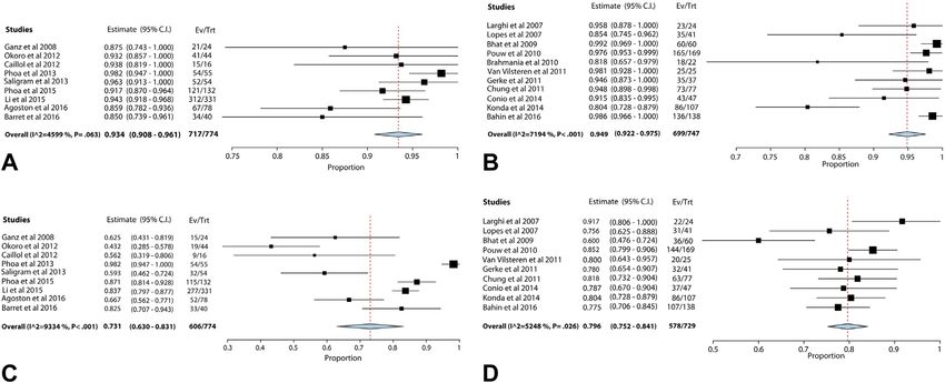
complete eradication rates of dysplasia/intramucosal EAC, reduce the demonstrated effect. The quality of evidence
recurrence rates, overall survival, EAC-related mortality, was very low for recurrence rates after EET and esophagec-
and adverse events. An existing systematic review and tomy (rated down for imprecision because only 1 recur-
meta-analysis by Wu et al46 was updated (794 studies rence was seen in the surgery group) and overall survival
were screened and 13 full-text articles were reviewed) to at 5 years (rated down for inconsistency). Similarly, the
inform this question (Table 8).23,47 Two studies were quality of evidence for EAC-related mortality was rated
added to the final analysis for the 5-year survival data. down to very low given the sparse events in both groups.
There was no difference between the 2 treatment modal- Considerations: This recommendation places a high
ities for the endpoint of complete eradication of HGD/in- value on the potential adverse effects associated with
tramucosal EAC (RR, .96; 95% CI, .91-1.01). Recurrence esophagectomy as compared with EET. Adverse events
rate of neoplasia was higher in the EET group (RR, 9.5; related to esophagectomy include bleeding, anastomotic
95% CI, 3.26-27.75). There was no difference between leakage, stenosis, prolonged hospitalization, and death.
the 2 groups with regard to overall survival (1-, 3-, and This analysis demonstrated a lower risk of major adverse
5-year survival) and EAC-related mortality. For the impor- events associated with EET (16 fewer per 100
tant outcome of 5-year survival, there was no difference be- adverse events compared with the esophagectomy). The
tween the 2 groups, with an RR of .88 (95% CI, .74-1.04; adverse events related to EET were mostly bleeding, perfo-
Fig. 7). Patients undergoing EET had significantly lower ration, and stricture formation, as discussed above. The
918 GASTROINTESTINAL ENDOSCOPY Volume 87, No. 4 : 2018 www.giejournal.orgGuidelines on endoscopic eradication therapies
TABLE 7. Continued
Summary of findings
Study event rates (%) Anticipated absolute effects
With EET With surveillance Relative effect (95% CI) Risk with surveillance Risk difference with EET
182/1854 (9.8%) 236/624 (37.8%) RR .22 Study population
10 per 100 8 fewer per 100
19/180 (10.6%) 24/91(26.4%) RR .42 (.24-.73) Study population
26 per 100 15 fewer per 100 (7 fewer to 20 fewer)
Overall adverse event rate across 37 studies was 8.8% (95% CI, 6.5%- 0 per 100 (rare) 9 more per 100 (7 more to 12 more)
11.9%). Treatment modality EET included RFA EMR. Stricture post- 0 per 100 (rare) 11 more per 100 (7 more to 12 more)
treatment most common adverse event 5.6% (95% CI, 4.2%-7.4%).
Overall adverse event rate across 7 studies was 10.6% (95% CI, 5.7%-
19.1%). Treatment modality EET included RFA EMR.
Study name Statistics for each study Risk ratio and 95% CI
Risk Lower Upper
ratio limit limit P value
Overholt 2005 0.46 0.259 0.806 .007
Shaheen 2009 0.13 0.015 1.050 .055
0.42 0.242 0.725 .002
0.01 0.1 1 10 100
Figure 4. Forest plot with 2 randomized controlled trials comparing endoscopic eradication therapies (radiofrequency ablation and photodynamic ther-
apy) with surveillance with the pooled relative risk of disease progression in Barrett’s esophagus patients with high-grade dysplasia. CI, Confidence
interval.
absolute risk of recurrence of neoplasia was minimally especially in patients in whom the cancer had not yet
higher in the EET group with 2 more recurrences of penetrated the muscularis mucosa, is associated with a
neoplasia per 100 patients compared with the esophagec- high 5-year survival rate.48 However, this treatment
tomy group. Limited data address the issue of patient approach is associated with an operative mortality of 2%
values, preferences, and burden associated with either of and a high morbidity rate seen even at high-volume cen-
these treatment approaches. As highlighted earlier, a study ters.48,49 The basic premise of EET is that BE patients
assessing the influence of EET and esophagectomy on with HGD and intramucosal EAC have a very low risk of
quality of life showed that patients undergoing EET re- lymph node metastasis (0% in patients with HGD and up
ported a greater fear of recurrence.29 to 2% in patients with intramucosal EAC).50 The
Discussion: Given the high tumor-free survival rates, effectiveness and safety profile of EET in BE-related
esophagectomy was the standard treatment for BE patients neoplasia has been well established.14-22 However, compar-
with HGD and intramucosal EAC, and all other therapies ative studies between esophagectomy and EET are limited.
were compared with this modality.23 Esophagectomy, Unfortunately, no RCT(s) was available to provide
www.giejournal.org Volume 87, No. 4 : 2018 GASTROINTESTINAL ENDOSCOPY 919Guidelines on endoscopic eradication therapies
Study name Subgroup within study Comparison Statistics for each study Ev ent rate and 95% CI
Ev ent Lower Upper
rate limit limit p-Value
Anders, 2014 EET Paper 0.063 0.016 0.218 0.000
Kommineni, 2015 EET Abstract 0.035 0.013 0.090 0.000
Nurkin, 2013 EET Paper 0.028 0.004 0.173 0.000
Oliphant, 2015 EET Abstract 0.181 0.108 0.287 0.000
Qumseya, 2013 EET Paper 0.019 0.003 0.124 0.000
Ramay, 2016 EET Abstract 0.032 0.005 0.196 0.001
Sharma, 2000 EET Paper 0.020 0.001 0.251 0.006
Velanovich, 2009 EET Paper 0.038 0.002 0.403 0.026
Wolf, 2015 EET Paper 0.084 0.068 0.103 0.000
Zemlyak, 2012 EET Paper 0.056 0.003 0.505 0.052
Lyday, 2010 EET Paper 0.012 0.001 0.164 0.002
Overholt, 2005 (PDT) EET Paper 0.130 0.084 0.198 0.000
Shaheen, 2009 (RFA) EET Paper 0.024 0.003 0.151 0.000
Verbeek, 2012 (RFA) EET Paper 0.221 0.175 0.276 0.000
0.074 0.045 0.117 0.000
Reid, 2000 Surveillance Paper 0.592 0.479 0.696 0.110
Weston, 2000 Surveillance Paper 0.267 0.104 0.533 0.083
Abela, 2008 Surveillance Paper 0.222 0.056 0.579 0.118
Ajumobi, 2009 Surveillance Paper 0.071 0.004 0.577 0.081
Alcedo, 2008 Surveillance Paper 0.333 0.043 0.846 0.571
Kahn, 2015 Surveillance Abstract 0.306 0.223 0.404 0.000
Overholt, 2005 (Surv) Surveillance Paper 0.286 0.192 0.402 0.001
Shaheen, 2009 (Surv) Surveillance Paper 0.190 0.073 0.412 0.009
Verbeek, 2012 (Surv) Surveillance Paper 0.399 0.347 0.453 0.000
0.340 0.255 0.438 0.002
0.199 0.153 0.256 0.000
-1.00 -0.50 0.00 0.50 1.00
Figure 5. Forest plot of studies providing an indirect comparison of cumulative rates of disease progression between Barrett’s esophagus patients with
high-grade dysplasia treated with radiofrequency ablation compared with those undergoing surveillance. CI, Confidence interval.
Study name Subgroup within study Comparison Time point Statistics for each study Event rate
and 95%CI
Event Lower Upper
rate limit limit p-Value
Gondrie, J.J._2 Prospective Paper 2008 0.091 0.013 0.439 0.028
Ganz, R.A. Retrospective Paper 2008 0.007 0.001 0.048 0.000
Pouw, R. Prospective Paper 2010 0.125 0.041 0.324 0.002
McEwan, H.C. Prospective Abstract 2012 0.088 0.047 0.161 0.000
Perry, K.A. Retrospective Paper 2014 0.028 0.002 0.322 0.013
Strauss, A.C. Retrospective Paper 2014 0.222 0.115 0.385 0.002
Phoa, K.Y._2 Prospective Paper 2015 0.189 0.131 0.265 0.000
0.146 0.111 0.189 0.000
-0.50 -0.25 0.00 0.25 0.50
Figure 6. Forest plot with studies reporting adverse events in Barrett’s esophagus patients with high-grade dysplasia/intramucosal esophageal adenocar-
cinoma treated with radiofrequency ablation with or without EMR. CI, Confidence interval.
conclusive evidence regarding superiority of 1 of these 2 the management of BE-related neoplasia patients. The panel
treatment modalities, and no such trial is expected in the members also accounted for the multiple endoscopy ses-
foreseeable future. sions required to achieve the goal of EET: complete eradica-
The effectiveness and safety of EET and adverse events tion of dysplasia and CE-IM.14,22,41 Most patients achieve CE-
associated with esophagectomy were important consider- IM within 3 ablative therapy sessions.53,54
ations for this recommendation. Another important determi- Esophagectomy is considered as the treatment of
nant was the higher recurrence rate of neoplasia associated choice for patients with submucosal cancer (T1b sm2-3 dis-
with EET compared with esophagectomy. Recurrence of in- ease), poorly differentiated cancer, and cancer associated
testinal metaplasia and neoplasia after EET was addressed with lymphatic or vascular infiltration given the high risk
in 2 recent systematic reviews and meta-analyses.51,52 These of lymph node metastasis (at least 20%).2,23,55 Long-term
studies demonstrated that >95% of all recurrences were suc- survival data in patients undergoing EET using contempo-
cessfully treated with EET, adding credence to use of EET for rary treatment modalities and identification of stage T1b
920 GASTROINTESTINAL ENDOSCOPY Volume 87, No. 4 : 2018 www.giejournal.orgGuidelines on endoscopic eradication therapies
EAC patients who may be able to undergo EET and achieve event rate of 1.5% (15 patients: major bleeding 14, perfora-
comparable outcomes with esophagectomy should be ad- tion 1, stricture 13), and all adverse events were managed
dressed in future studies. Studies identifying patient and endoscopically.57 The outcome of adverse events for
provider-specific determinants of optimal outcomes,14,41 patients undergoing EET was also addressed using a
and studies on patient preferences and quality of life recent systematic review and meta-analysis.22 Although
should be a research priority. the pooled rate of all adverse events with EET (RFA with
or without EMR) was 8.8% (95% CI, 6.5%-11.9%), in
Question 4: What is the role of EMR in BE patients studies that compared RFA with versus without EMR, the
with a visible lesion detected during screening or adverse event rate was significantly higher for RFA with
surveillance? EMR (RR, 4.4; P Z .015).
Recommendation: In BE patients referred for Certainty in the Evidence: For the outcome of
EET, we recommend endoscopic resection of all change in diagnosis in grade of dysplasia, the quality of ev-
visible lesions compared with no endoscopic resec- idence was rated up for large effect; the consensus
tion of visible lesions (strong recommendation, threshold was 20% among the panel members. There
moderate quality evidence). was no risk of bias, inconsistency, indirectness, impreci-
Summary of the Evidence: The patient-important sion, or publication bias.
outcomes for this clinical question were the difference in Considerations: This recommendation placed a high
progression rates to cancer among BE patients who under- value on the role of EMR in leading to change in diagnosis
went EMR of visible lesions (typically described as areas and the potential to impact patient management and out-
with nodularity, ulceration, plaques, areas of depression, comes. The benefits of EMR include assessment of histo-
or mucosal discoloration) compared with those who did logic depth of invasion to improve confidence of clinical
not undergo EMR of visible lesions (important outcome), decisions made for patients with BE-related dysplasia and
proportion of patients with a change in diagnosis in grade intramucosal EAC with EET.58 By altering the diagnosis,
of dysplasia and those with a change in management plan EMR has the potential to impact the management plan
as a result of EMR (critical outcome), and adverse events (although the impact of EMR on the management plan
related to EMR (critical outcome). There were no RCTs was not consistently reported across published studies).
that addressed these outcomes; hence, indirect compari- Under-diagnosis or over-diagnosis may have substantial
sons from observational studies were used to provide deleterious consequences for the patient. For example, a
this recommendation. The evidence profile for this clinical patient without cancer may undergo unnecessary esopha-
question is summarized in Supplementary Table 1 gectomy or patients with submucosal invasive EAC may
(available online at www.giejournal.org). incorrectly be treated with EET instead of surgery. Given
A systematic review and meta-analysis was conducted to the importance and magnitude of the effect on patient
address the question of change in diagnosis based on EMR. management and low risk of adverse events, the panel
Of the 1436 screened studies, 17 were reviewed in full-text members supported a strong recommendation for this
format and 14 were included in the final analysis. These clinical question.
studies included a total of 1116 patients with a total num- Discussion: EMR plays a critical role as a part of the
ber of 449 events. When a random-effects model was used, armamentarium for Barrett’s EET and has evolved into an
EMR resulted in a change in the pathologic diagnosis in important diagnostic/staging and therapeutic tool in the
39% (95% CI, 34%-45%) of all patients. Most of this change management of BE-related neoplasia patients.3,59 The value
was associated with upgrading of grade of dysplasia/ of EMR as a diagnostic/staging tool is enhanced by the pro-
neoplasia (Fig. 8). Review of published literature vision of larger and deeper tissue specimens (resection ex-
identified no studies comparing progression rates and tends to muscularis mucosa and submucosal level in most
adverse events between patients undergoing EMR of EMR specimens) compared with biopsy specimens, with
visible lesions with those not undergoing EMR for visible limited distortion allowing for an accurate assessment of
lesions. depth of neoplastic involvement and adequacy of resec-
The safety of EMR in BE patients has been established in tion.3 As highlighted above, a change in diagnosis in
multiple studies.22,56,57 In a cohort study of 681 patients nearly 40% of patients was noted in BE patients
treated at a tertiary care center, a total of 1388 endoscopic undergoing EMR for visible lesions. In addition to the
procedures were performed, and 2513 EMRs were per- change in diagnosis, provision of a larger specimen (with
formed using the cap or multiband mucosectomy tech- EMR) to pathology results in an improvement in
nique.56 No perforations were noted, bleeding post-EMR interobserver agreement among pathologists compared
was seen in 8 patients (1.2%: 7 treated endoscopically with biopsy specimens as demonstrated in at least 2
and 1 required surgery), and strictures were reported in studies.38,60 The recommendation of performing EMR of
7 patients (1%). Another large cohort study that included all visible lesions (no matter how subtle) is consistent
1096 consecutive patients with intramucosal EAC who un- with other recent guideline documents, quality indicator
derwent 2687 EMR procedures reported a major adverse documents, and current clinical practice.2,3,14,41,61
www.giejournal.org Volume 87, No. 4 : 2018 GASTROINTESTINAL ENDOSCOPY 921You can also read