Epigallocatechin-3-Gallate Prevents Acute Gout by Suppressing NLRP3 Inflammasome Activation and Mitochondrial DNA Synthesis - MDPI
←
→
Page content transcription
If your browser does not render page correctly, please read the page content below
molecules
Article
Epigallocatechin-3-Gallate Prevents Acute Gout by
Suppressing NLRP3 Inflammasome Activation and
Mitochondrial DNA Synthesis
Hye Eun Lee, Gabsik Yang, Youn Bum Park, Han Chang Kang , Yong-Yeon Cho, Hye Suk Lee
and Joo Young Lee *
 BK21 Plus, College of Pharmacy, The Catholic University of Korea, Bucheon 14662, Korea;
 esthel0513@catholic.ac.kr (H.E.L.); yangboncho@gmail.com (G.Y.); pyb12345678@naver.com (Y.B.P.);
 hckang@catholic.ac.kr (H.C.K.); yongyeon@catholic.ac.kr (Y.-Y.C.); sianalee@catholic.ac.kr (H.S.L.)
 * Correspondence: joolee@catholic.ac.kr; Tel.: +82-2-2164-4095; Fax: +82-2-2164-4059

 Academic Editors: Atanas G. Atanasov, Karel Šmejkal and Elke Heiss

 Received: 30 April 2019; Accepted: 3 June 2019; Published: 6 June 2019

 Abstract: Gout is a chronic inflammatory disease evoked by the deposition of monosodium urate
 (MSU) crystals in joint tissues. The nucleotide-binding oligomerization domain (NOD)-like receptor
 (NLR) family pyrin domain containing 3 (NLRP3) inflammasome is responsible for the gout
 inflammatory symptoms induced by MSU crystals. We investigated whether epigallocatechin-3-gallate
 (EGCG) suppresses the activation of the NLRP3 inflammasome, thereby effectively preventing gouty
 inflammation. EGCG blocked MSU crystal-induced production of caspase-1(p10) and interleukin-1β
 in primary mouse macrophages, indicating its suppressive effect on the NLRP3 inflammasome.
 In an acute gout mouse model, oral administration of EGCG to mice effectively alleviated gout
 inflammatory symptoms in mouse foot tissue injected with MSU crystals. The in vivo suppressive
 effects of EGCG correlated well with the suppression of the NLRP3 inflammasome in mouse foot
 tissue. EGCG inhibited the de novo synthesis of mitochondrial DNA as well as the production of
 reactive oxygen species in primary mouse macrophages, contributing to the suppression of the NLRP3
 inflammasome. These results show that EGCG suppresses the activation of the NLRP3 inflammasome
 in macrophages via the blockade of mitochondrial DNA synthesis, contributing to the prevention of
 gouty inflammation. The inhibitory effects of EGCG on the NLRP3 inflammasome make EGCG a
 promising therapeutic option for NLRP3-dependent diseases such as gout.

 Keywords: inflammasome; gout; mitochondria; reactive oxygen species; green tea; macrophages;
 innate immunity

1. Introduction
     Gout is a relatively common inflammatory arthritis that can cause pain in the joints and seriously
impair the quality of life of a patient. Gout is typically known to occur in middle-aged men; yet,
the incidence is increasing in the elderly population [1]. The precipitation of monosodium urate (MSU)
crystals in joints is a key factor in the initiation and development of gout [2].
     Accumulating evidence indicates that the MSU crystals-induced inflammatory responses and
gout pathogenesis are dependent on interleukin (IL)-1β [3]. It is now well established that the
nucleotide-binding oligomerization domain (NOD)-like receptor (NLR) family pyrin domain containing
3 (NLRP3) inflammasome senses MSU crystal deposition, subsequently activating the downstream
immune signals and inducing the production of IL-1β [3]. The NLRP3 inflammasome is a multiprotein
complex consisting of NLRP3, apoptosis-associated speck-like protein containing a CARD (ASC) as
an adaptor, and pro-caspase-1 as an effector enzyme [4]. Activation of the NLRP3 inflammasome

Molecules 2019, 24, 2138; doi:10.3390/molecules24112138                        www.mdpi.com/journal/molecules
Molecules 2019, 24, 2138                                                                             2 of 11

culminates in the activation of pro-caspase-1, leading to the cleavage of inactive pro-IL-1β and pro-IL-18
to the active form of IL-1β and IL-18. Mice deficient in NLRP3 fail to produce active IL-1β in the foot
pads in response to MSU crystals in an acute gout mouse model [5].
     Current therapeutic strategies to target IL-1β have proven successful for alleviating the symptoms
of gout in clinical studies, suggesting that targeting the NLRP3 inflammasome may have a critical
impact on gout treatment. Current clinical trials are underway for the development of IL-1 inhibitors
including anakinra (an IL-1 receptor antagonist) [6], rilonacept (IL-1 Trap, a soluble decoy receptor) [7],
and canakinumab (anti-IL-1β monoclonal antibody) [8]. Despite the efficacy of these inhibitors, there
are some issues in patients, limiting the extensive use of IL-1 inhibitors in the treatment of gout. These
include the high cost and the inconvenient administration route of IL-1 inhibitors [9].
     Therefore, we searched for orally available small-molecule drugs for gout treatment that inhibit
the NLRP3 inflammasome, thereby blocking the production of active IL-1β. Epigallocatechin-3-gallate
(EGCG) is an active component of green tea and is well known to have anti-inflammatory properties.
There is a previous report that EGCG reduces the messenger RNA (mRNA) and protein expression of
renal NLRP3 in a lupus nephritis mouse model [10]. Similarly, Gao et al. reported that EGCG decreases
the protein level of renal NLRP3 in a contrast-induced nephropathy rat model [11]. These reports
show that oral or intravenous infusion of EGCG suppresses the expression of tissue NLRP3 protein,
possibly leading to the reduction of NLRP3 inflammasome activation. However, it is not entirely clear
whether EGCG affects the activation of NLRP3 inflammasome induced by various NLRP3 inducers
in macrophages and in gouty inflammatory response. In this study, we investigated whether EGCG
could suppress the activation of the NLRP3 inflammasome in macrophages in order to pursue the
possibility of its therapeutic application for NLRP3-mediated diseases such as gout.

2. Results

2.1. EGCG Suppresses NLRP3 Inflammasome Activation in Primary Macrophages
     We investigated whether EGCG blocks the activation of the NLRP3 inflammasome induced by
various activators in primary mouse macrophages. To exclude the possibility that EGCG may affect the
priming activation step by lipopolysaccharide (LPS), macrophages were treated with EGCG after the
LPS was washed out. Cleavage of pro-caspase-1 and pro-IL-1β to caspase-1(p10) and IL-1β, respectively,
is considered a hallmark of inflammasome activation. EGCG inhibited the MSU crystal-induced
cleavage of pro-caspase-1 and pro-IL-1β to caspase-1(p10) and IL-1β in cell supernatants, as shown by
immunoblotting (Figure 1A). Consistently, EGCG reduced the MSU crystal-induced secretion of IL-1β
in a dose-dependent manner, as measured by enzyme-linked immunosorbent assay (ELISA) of the cell
culture medium (Figure 1B).
     We further examined whether EGCG inhibits NLRP3 inflammasome activation by other activators
such as adenosine triphosphate (ATP) and nigericin. EGCG suppressed ATP-induced cleavage of
pro-caspase-1 and pro-IL-1β to caspase-1(p10) and IL-1β in primary macrophages (Figure 1C). In
addition, ATP-induced IL-1β secretion was suppressed by EGCG (Figure 1D). Nigericin-induced the
cleavage of pro-caspase-1 and pro-IL-1β to caspase-1(p10) and IL-1β was blocked by EGCG (Figure 1E).
The nigericin-induced secretion of IL-1β was reduced by EGCG (Figure 1F).
     These results show that EGCG suppresses NLRP3 inflammasome activation induced by MSU
crystals, ATP, and nigericin, suggesting that EGCG inhibits NLRP3 inflammasome activation by
various stimuli.
Molecules 2019, 24, 2138 3 of 11
Molecules 2019, 24, x FOR PEER REVIEW 3 of 12
Figure 1. Epigallocatechin-3-gallate (EGCG) suppresses the activation of the (NOD)-like receptor (NLR)
Figure 1. Epigallocatechin-3-gallate (EGCG) suppresses the activation of the (NOD)-like receptor
family pyrin
(NLR) familydomain containing
pyrin domain3 (NLRP3)
containing inflammasome
3 (NLRP3) in primary macrophages.
inflammasome in primary Lipopolysaccharide
macrophages.
(LPS)-primed
Lipopolysaccharide (LPS)-primed bone marrow-derived primary macrophages werefor
bone marrow-derived primary macrophages were treated with EGCG 1 h and
treated then
with
stimulated with monosodium uric acid (MSU) crystals (500 µg/mL for 6 h),
EGCG for 1 h and then stimulated with monosodium uric acid (MSU) crystals (500 μg/mL for 6 h), adenosine triphosphate
(ATP) (5 mM triphosphate
adenosine for 1 h), or nigericin
(ATP) (5(10mMµM;for2 1h h),
for or
E and 16 h for
nigericin (10F) in the
μM; 2 h presence
for E andor16absence
h for F)ofinEGCG
the
as indicated.
presence or(A,C,E)
absenceCell culture
of EGCG as supernatants
indicated. (A),(SUP) and(E)cell
(C), and Celllysates
culture(LYS) were immunoblotted
supernatants (SUP) and cellfor
pro-caspase-1,
lysates (LYS) caspase-1(p10), pro-IL-1β,
were immunoblotted forand IL-1β. In thecaspase-1(p10),
pro-caspase-1, bar graph, the band density
pro-IL-1β, andofIL-1β.
caspase-1(p10)
In the
andbarcleaved IL-1β normalized to corresponding pro-caspase-1 and pro-IL-1β,
graph, the band density of caspase-1(p10) and cleaved IL-1β normalized to corresponding was expressed as means
± SD (n = 3). #, significantly different from vehicle group, p < 0.05; *, significantly
pro-caspase-1 and pro-IL-1β, was expressed as means ± SD (n = 3). #, significantly different from different from MSU,
ATP, or nigericin
vehicle < 0.05;p*,Immunohistochemical analysis of the mouse foot tissues with anti-caspase-1 and IL-1β antibody
staining showed that the levels of caspase-1 and IL-1β were increased in the foot tissues injected
with the MSU crystals, while oral administration of EGCG reduced the increased levels of caspase-1
and IL-1β in gouty foot tissues (Figure 3C).
These results demonstrate that oral administration of EGCG effectively alleviates the
Molecules 2019, 24, 2138
inflammatory symptoms of uric acid crystal-induced acute gout in mice, mediated by blockade4of of 11
NLRP3 inflammasome activation.
Figure
Figure Oral
2. 2. Oraladministration
administrationof ofEGCG
EGCG suppresses acute gout
suppresses acute goutsymptoms
symptomsinduced
inducedbybyMSU MSU crystal
crystal
injection in mice. Mice were orally administered EGCG (1, 10, and 30 mg/kg)
injection in mice. Mice were orally administered EGCG (1, 10, and 30 mg/kg) or vehicle (0.02% or vehicle (0.02% dimethyl
sulfoxide
dimethyl(DMSO)
sulfoxide in(DMSO)
water). inAfter 1 h,After
water). MSU crystals
1 h, (2 mg/0.1
MSU crystals mL ofmL
(2 mg/0.1 phosphate-buffered
of phosphate-buffered saline
(PBS)/mouse) or PBS was
saline (PBS)/mouse) or subcutaneously injected into
PBS was subcutaneously the footpad
injected into theoffootpad
the rightof hindfoot
the rightof each mouse.
hindfoot of
After
each24mouse.
h, the footpad
After 24tissues
h, the were
footpadcollected
tissuesfor further
were analysis.
collected (A) Time
for further course(A)
analysis. of footpad thickness
Time course of
gain compared
footpad with the
thickness gainfootpad
compared thickness at the
with the 0 h time
footpad point per
thickness at group.
the 0 h (B)
timeRepresentative picture
point per group. (B) of
hematoxylin
Representativeand picture
eosin staining of the hind
of hematoxylin andfeet.
eosinInfiltrating
staining of neutrophils
the hind feet.inInfiltrating
the hindfoot tissue appear
neutrophils in
as the hindfoot
purple dots. tissue appear as purple
(C) Representative dots. of
pictures (C)immunohistochemistry
Representative picturesstaining of immunohistochemistry
of foot tissues with
staining of foot (MPO)
myeloperoxidase tissues (200×).
with myeloperoxidase
(D) Supernatants (MPO)
of the(200×). (D) Supernatants
foot tissue homogenatesofwere the foot tissuefor
analyzed
homogenates
IL-6 by ELISA. The werevalues
analyzed forline
in the IL-6and
by ELISA. The represent
bar graphs values in the theline and±bar
means (n = 6 represent
SDgraphs the #,
mice/group).
means ± SDdifferent
significantly (n = 6 mice/group).
from vehicle#, group, p < 0.05;
significantly different from vehicle
*, significantly group,from
different p < 0.05;
MSU*,alone, p < 0.05.
significantly
different from MSU alone, p < 0.05.
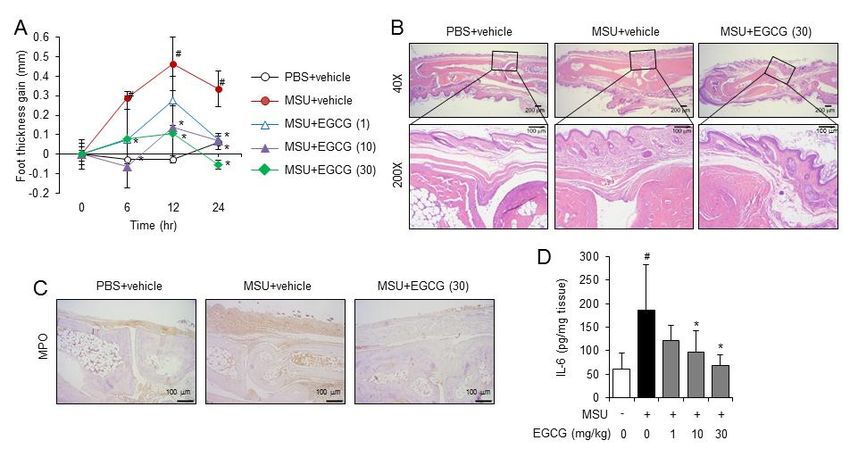
We examined whether EGCG suppresses NLRP3 inflammasome activation in foot tissues injected
with MSU crystals. Injection of MSU crystals induced the cleavage of pro-caspase-1 to caspase-1(p10) and
the cleavage of pro-IL-1β to IL-1β in the foot tissue homogenates (Figure 3A). The oral administration
of EGCG prevented the cleavage of pro-caspase-1 to caspase-1(p10) and of pro-IL-1β to IL-1β in the
foot tissues injected with MSU crystals (Figure 3A). EGCG decreased MSU crystal-induced IL-1β
production in foot tissue homogenates as determined by ELISA (Figure 3B). Immunohistochemical
analysis of the mouse foot tissues with anti-caspase-1 and IL-1β antibody staining showed that the
levels of caspase-1 and IL-1β were increased in the foot tissues injected with the MSU crystals, while
oral administration of EGCG reduced the increased levels of caspase-1 and IL-1β in gouty foot tissues
(Figure 3C).Molecules 2019, 24, 2138 5 of 11
Molecules 2019, 24, x FOR PEER REVIEW 5 of 12
Figure 3. Oral administration of EGCG attenuates NLRP3 inflammasome activation in an acute gout
Figure 3. Oral administration of EGCG attenuates NLRP3 inflammasome activation in an acute gout
mouse model. The foot tissue samples analyzed are the same as those used in Figure 2. (A) Supernatants
mouse model. The foot tissue samples analyzed are the same as those used in Figure 2. (A)
from the foot tissue homogenates were analyzed for pro-caspase-1, caspase-1(p10), pro-IL-1β, and IL-1β
Supernatants from the foot tissue homogenates were analyzed for pro-caspase-1, caspase-1(p10),
by immunoblotting. # indicates an individual sample. (B) The foot tissue homogenates were analyzed
pro-IL-1β, and IL-1β by immunoblotting. # indicates an individual sample. (B) The foot tissue
for IL-1β by ELISA. The values represent the means ± SD (n = 6 mice/group). #, significantly different
homogenates were analyzed for IL-1β by ELISA. The values represent the means ± SD (n = 6
from vehicle group, p < 0.05; *, significantly different from MSU alone, p < 0.05. (C) Representative
mice/group). #, significantly different from vehicle group, p < 0.05; *, significantly different from
pictures of immunohistochemistry staining of foot tissues for caspase-1 and IL-1β (200×).
MSU alone, p < 0.05. (C) Representative pictures of immunohistochemistry staining of foot tissues for
caspase-1
These anddemonstrate
results IL-1β (200×). that oral administration of EGCG effectively alleviates the inflammatory
symptoms of uric acid crystal-induced acute gout in mice, mediated by blockade of NLRP3
2.3. EGCG Inhibits the De Novo Synthesis of Mitochondrial DNA in Macrophages
inflammasome activation.
To investigate the mechanism by which EGCG suppresses NLRP3 inflammasome activation,
2.3.
we EGCG Inhibitswhether
investigated the De Novo
EGCG Synthesis of Mitochondrial
directly DNA in Macrophages
binds to the components of the NLRP3 inflammasome by
surface plasmon resonance
To investigate (SPR) analysis
the mechanism by whichwith a recombinant
EGCG suppressesprotein,
NLRP3 the PYD of NLRP3
inflammasome or ASC.
activation,
The SPR analysis demonstrated that EGCG did not directly associate with the
we investigated whether EGCG directly binds to the components of the NLRP3 inflammasome PYD of NLRP3 or ASC
by
(Figure 4A,B). We further investigated whether EGCG inhibits the enzymatic activity
surface plasmon resonance (SPR) analysis with a recombinant protein, the PYD of NLRP3 or ASC. of caspase-1 by
in vitro
The caspase-1
SPR analysis enzyme activity
demonstrated usingdid
that EGCG recombinant caspase-1.with
not directly associate EGCG did of
the PYD not suppress
NLRP3 or ASCthe
enzymatic activity of caspase-1, while a caspase inhibitor, Z-YVAD-FMK, decreased
(Figure 4A,B). We further investigated whether EGCG inhibits the enzymatic activity of caspase-1 by the caspase-1
inenzymatic activity
vitro caspase-1 (Figure
enzyme 4C).using
activity Theserecombinant
results show that EGCG
caspase-1. EGCG does
did not directly inhibit
not suppress NLRP3
the enzymatic
inflammasome
activity components
of caspase-1, while asuch as NLRP3,
caspase ASC,
inhibitor, and caspase-1.
Z-YVAD-FMK, decreased the caspase-1 enzymatic
activity (Figure 4C). These results show that EGCG does not directly inhibit NLRP3 inflammasome
components such as NLRP3, ASC, and caspase-1.Molecules 2019, 24, 2138 6 of 11
Molecules 2019, 24, x FOR PEER REVIEW 6 of 12
Figure 4. EGCG does not directly associate with NLRP3 and apoptosis-associated speck-like protein
Figure 4.aEGCG
containing CARDdoes(ASC)notor
directly
inhibitassociate
caspase-1with NLRP3activity.
enzyme and apoptosis-associated
(A,B) Sensorgrams speck-like proteinby
were obtained
containing a CARD (ASC) or inhibit caspase-1 enzyme activity. (A), (B) Sensorgrams
SPR analysis to determine the interaction between EGCG and recombinant (A) NLRP3-PYD or (B) were obtained
by Different
ASC. SPR analysis to determine
concentrations of the interaction
EGCG between
are presented EGCG
as an and plot
overlay recombinant
aligned at(A)
theNLRP3-PYD or
start of injection.
(B) of
Table ASC. Different
kinetic concentrations
parameters obtainedoffrom
EGCGthe are
SPRpresented as ancalculated
analysis and overlay plot aligned T200
by Biocore at theevaluation
start of
injection. Table of kinetic parameters obtained from the SPR analysis and calculated by Biocore T200
software. (C) An in vitro assay for caspase-1 enzymatic activity was performed using a fluorometric
evaluation software. (C) An in vitro assay for caspase-1 enzymatic activity was performed using a
caspase-1 assay kit with recombinant caspase-1 (rCaspase-1) in the presence or absence of EGCG or
fluorometric caspase-1 assay kit with recombinant caspase-1 (rCaspase-1) in the presence or absence
Z-YVAD-FMK. The values represent the means ± SD (n = 3). #, significantly different from vehicle,
of EGCG or Z-YVAD-FMK. The values represent the means ± SD (n = 3). #, significantly different
p < 0.05; *, significantly different from rCaspase-1 alone, p < 0.05.
from vehicle, p < 0.05; *, significantly different from rCaspase-1 alone, p < 0.05.
Activation of the NLRP3 inflammasome causes mitochondrial disruption, leading to the generation
Activation of the NLRP3 inflammasome causes mitochondrial disruption, leading to the
of reactive oxygen species (ROS) [12]. Therefore, we investigated whether EGCG reduces the generation
generation of reactive oxygen species (ROS) [12]. Therefore, we investigated whether EGCG reduces
of ROS induced by NLRP3 inflammasome activation in primary mouse macrophages. EGCG suppressed
the generation of ROS induced by NLRP3 inflammasome activation in primary mouse macrophages.
intracellular ROS production induced by the MSU crystals in macrophages (Figure 5A). Consistently,
EGCG suppressed intracellular ROS production induced by the MSU crystals in macrophages
ROS production
(Figure induced byROS
5A). Consistently, ATPproduction
or nigericin was significantly
induced decreased
by ATP or nigericin bysignificantly
was EGCG (Figure 5B,C).
decreased
ROS generated
by EGCG (Figure 5B,C). upon NLRP3 inflammasome activation induce the oxidation of mitochondrial
DNA (mtDNA),
ROS generatedand oxidized mtDNA
upon NLRP3 is released into
inflammasome the cytosol
activation to induce
induce the activation
the oxidation of the NLRP3
of mitochondrial
inflammasome
DNA (mtDNA), [13].andIt has been mtDNA
oxidized reportedisthat mtDNA
released intoisthe cytosol to de
synthesized novo the
induce during the process
activation of the of
NLRP3 inflammasome [13]. It has been reported that mtDNA is synthesized de novo duringdethe
NLRP3 inflammasome activation [13]. Therefore, we investigated whether EGCG affects the novo
synthesis
processofofmtDNA in macrophages.
NLRP3 inflammasome Relative[13].
activation totalTherefore,
mtDNA amounts were quantified
we investigated by quantitative
whether EGCG affects
PCRthewith primers
de novo specific
synthesis for theinmitochondrial
of mtDNA macrophages. D-loop
Relativeregion (D-loop)
total mtDNA or a region
amounts were of mtDNAby
quantified that
quantitative
is not PCR nuclear
inserted into with primers
DNAspecific
(non-NUMT)for the in
mitochondrial
macrophages. D-loop region (D-loop)
The amount or aDNAs
of nuclear region(Tert,
of
mtDNA
B2m) that is not to
was measured inserted into mtDNA
normalize nuclear DNA (non-NUMT)
production. in macrophages.
MSU crystals increasedThe
theamount
amountofofnuclear
mtDNA
DNAsnon-NUMT),
(D-loop, (Tert, B2m) was whilemeasured to normalize
EGCG suppressed MSU mtDNA production.
crystal-induced MSU crystals
mtDNA synthesis increased the
(Figure 5D,E).
amount of mtDNA (D-loop, non-NUMT), while EGCG suppressed MSU crystal-induced
These results show that EGCG blocks the de novo synthesis of mtDNA occurring upon NLRP3 activation mtDNA
synthesis (Figure
in macrophages. 5D,E).
These Thesesuggest
results results that
showEGCGthat EGCG
reduces blocks the de novoofsynthesis
the production oxidizedofmtDNAmtDNAby
occurring upon NLRP3 activation in macrophages.
suppressing both ROS production and mtDNA synthesis. These results suggest that EGCG reduces the
production of oxidized mtDNA by suppressing both ROS production and mtDNA
Collectively, these results show that EGCG suppresses the activation of the NLRP3 inflammasome synthesis.
Collectively, these results show that EGCG suppresses the activation of the NLRP3
in macrophages via the blockade of mtDNA synthesis, contributing to the prevention of
inflammasome in macrophages via the blockade of mtDNA synthesis, contributing to the prevention
gouty inflammation.
of gouty inflammation.Molecules 2019, 24, x FOR PEER REVIEW 7 of 12
Molecules 2019, 24, 2138 7 of 11
Figure 5. EGCG suppresses the production of reactive oxygen species and the de novo synthesis of
mitochondrial
Figure 5. EGCG DNA induced the
suppresses by NLRP3
productioninflammasome activators
of reactive oxygen in primary
species macrophages.
and the de novo synthesis(A–C) of
Bone marrow-derived primary mouse macrophages were pretreated with
mitochondrial DNA induced by NLRP3 inflammasome activators in primary macrophages. an oxidative stress(A)–(C)
indicator,
H2Bone
DCFDA (10 µM), for 1primary
marrow-derived h and further
mousestimulated
macrophages withwere
monosodium
pretreateduric acid
with an(MSU) crystals
oxidative (500
stress
µg/mL), ATP
indicator, H2(5 mM), (10
DCFDA or nigericin
μM), for 1(10
h µM) for 1 hstimulated
and further in the presence or absence of
with monosodium EGCG.
uric Reactive
acid (MSU)
oxygen species
crystals (ROS) levels
(500 μg/mL), ATP (5are presented
mM), as a(10
or nigericin percentage
μM) for 1 of the
h in thecontrol.
presence(D,E) LPS-primed
or absence of EGCG. bone
marrow-derived
Reactive oxygen primary mouse
species (ROS)macrophages were stimulated
levels are presented with MSU crystals
as a percentage (500 µg/mL)
of the control. (D), for
(E)6 h
in LPS-primed
the absence orbonepresence of EGCG. The
marrow-derived levels
primary of mitochondrial
mouse macrophagesDNA werewere determined
stimulated with MSUby quantitative
crystals
(500The
PCR. μg/mL)
valuesfor 6 h in the
represent theabsence
means ±orSD (n = 3); of
presence EGCG. The levels
#, significantly of mitochondrial
different DNA pwere
from vehicle group, < 0.05;
*, significantly different from MSU, ATP, or nigericin alone, p < 0.05.
determined by quantitative PCR. The values represent the means ± SD (n = 3); #, significantly
different from vehicle group, p < 0.05; *, significantly different from MSU, ATP, or nigericin alone, p <
3. Discussion
0.05.
EGCG, a green tea polyphenol component, is well known for its anti-inflammatory properties.
3. Discussion
Previous reports have shown that the inhibition of the NLRP3 inflammasome by EGCG may lead
EGCG, a green
to the suppression of tea polyphenol
lupus nephritiscomponent, is wellinknown
and peritonitis mousefor its anti-inflammatory
models properties.
[14,15]. Our results further
Previous reports have shown that the inhibition of the NLRP3 inflammasome
demonstrate that EGCG is effective in preventing acute gout by inhibiting NLRP3 inflammasome by EGCG may lead to
the suppression
activation, of lupus nephritis
as oral administration and reduced
of EGCG peritonitis in mouse models
inflammatory symptoms [14,15].
in anOur
MSUresults further
crystal-induced
demonstrate that EGCG is effective in preventing acute gout by inhibiting NLRP3
acute gout mouse model. The inhibitory effects of EGCG on gouty inflammation correlate well with inflammasome
activation,
decreased as oral
NLRP3 administration
inflammasome of EGCG
activity in goutyreduced
tissues. inflammatory
EGCG treatment symptoms in an secretion
reduced IL-1β MSU
crystal-induced acute gout mouse model. The inhibitory effects of EGCG on gouty
and neutrophil recruitment in foot tissues injected with MSU crystals. Gout is a form of inflammatory inflammation
correlate well with decreased NLRP3 inflammasome activity in gouty tissues. EGCG treatment
arthritis that is induced by the deposition of uric acid crystals, characterized by neutrophil infiltration
reduced IL-1β secretion and neutrophil recruitment in foot tissues injected with MSU crystals. Gout
into the inflammatory joints. Discovering the role of the NLRP3 inflammasome and the subsequent
is a form of inflammatory arthritis that is induced by the deposition of uric acid crystals,
release of IL-1β is important to elucidate the pathogenesis of this disease [16]. Our study suggests
characterized by neutrophil infiltration into the inflammatory joints. Discovering the role of the
that administration of EGCG could be extended to the treatment of other diseases relevant to uric acid
NLRP3 inflammasome and the subsequent release of IL-1β is important to elucidate the
crystal accumulation
pathogenesis of thisand the NLRP3
disease inflammasome.
[16]. Our study suggests that administration of EGCG could be
MSU crystals, ATP, and nigericin have
extended to the treatment of other diseases relevant different
to uricupstream
acid crystalsignaling pathways
accumulation and thetoNLRP3
activate
NLRP3. MSU
inflammasome. crystals are phagocytosed and destabilize the phagosome, which activates the NLRP3
inflammasome. ATP triggers NLRP3 inflammasome activation by binding to the P2X7 purinergic
receptor, thereby increasing potassium efflux. Nigericin, a microbial toxin derived from Streptomyces
hygroscopicus, decreases intracellular potassium levels by acting as a potassium ionophore. Our resultsMolecules 2019, 24, 2138 8 of 11
show that in addition to suppressing MSU-induced NLRP3 activation, EGCG treatment suppresses
various activators of the NLRP3 inflammasome, such as ATP and nigericin, suggesting that the
inhibitory target of EGCG may be common to these different activators. We previously reported
that the inhibitory effects of caffeic acid phenethyl ester are due to the direct binding of caffeic acid
phenethyl ester to ASC [5]. In contrast, the SPR analysis showed that EGCG does not directly bind
to ASC or NLRP3 PYD. EGCG also does not inhibit caspase-1 enzymatic activity. These results
demonstrate that the target of EGCG does not lie in the NLRP3 inflammasome complex itself but in the
upstream pathways.
Oxidized mtDNA is known to activate the NLRP3 inflammasome [17]. Zhong et al. showed
that NLRP3 priming induces de novo synthesis of mtDNA, which is further oxidized under oxidative
stress conditions [13]. Cytidine/uridine monophosphate kinase 2 (CMPK2) is an essential enzyme that
provides deoxyribonucleotides for mtDNA synthesis. CMPK2 expression is regulated by interferon
regulatory factor 1 (IRF1) transcription factor activated in an LPS-priming step [13]. CMPK2-dependent
mtDNA synthesis is required for the production of oxidized mtDNA fragments in response to NLRP3
activators [13]. Oxidized mtDNA associates with the NLRP3 inflammasome complex to activate NLRP3
inflammasome. We show that de novo synthesis of mtDNAs induced by MSU crystals is decreased
by EGCG in macrophages. This suggests that EGCG may regulate the upstream events to produce
mtDNA including CMPK2 activity or CMPK2 expression. The detailed mechanism for inhibition of de
novo synthesis of mtDNA by EGCG needs to be investigated further. Blockade of mtDNA synthesis
inhibited IL-1β production induced by NLRP3 activators such as ATP, nigericin, and MSU, while IL-1β
production by an AIM2 inflammasome activator, poly dA:dT, was not inhibited by blockade of mtDNA
synthesis [13]. These show that newly synthesized mtDNA is required for NLRP3 inflammasome
activation induced by various NLRP3 activators [13]. Therefore, it is likely that the common inhibitory
effect of EGCG on various NLRP3 inducers may be linked to the common suppression of mtDNA
synthesis in response to various NLRP3 inducers including ATP or nigericin. This needs to be further
elucidated in a future study. In addition, EGCG reduces the ROS levels increased by MSU crystal
stimulation in macrophages. These results suggest that the suppression of NLRP3 inflammasome
activation by EGCG is mediated by the blockade of mtDNA synthesis, which is the source of oxidized
mtDNA, an NLRP3 activator. EGCG appears to be effective in reducing the generation of oxidized
mtDNAs by blocking both new mtDNA synthesis and ROS production.
In conclusion, EGCG inhibits acute gout inflammation, including proinflammatory cytokine release
and neutrophil infiltration into the lesion site, mediated by the suppression of NLRP3 inflammasome
activation in macrophages. The inhibition of ROS production and mtDNA synthesis by EGCG
contributes to its suppressive effect on the NLRP3 inflammasome.
4. Materials and Methods
4.1. Animals and Cell Culture
Mice (C57BL/6) were obtained from RaOn Bio (Seoul, Korea). The mice were housed in a room
controlled for temperature (23 ± 3 ◦ C) and relative humidity (40–60%) under specific pathogen-free
conditions. Mice were acclimated in the animal facility for at least a week before the experiments.
Animal care and the experimental protocols were carried out in accordance with the guidelines
of the Institutional Animal Care and Use Committee (IACUC) of the Catholic University of Korea
(permission #2014-015). Bone marrow-derived primary macrophages (BMDMs) were prepared after
bone marrow was isolated from C57BL/6 mice as described previously [18]. Cell culture was performed
as previously described [19]. Briefly, BMDMs were cultured in Dulbecco’s modified eagle medium
(DMEM) containing 10% (v/v) fetal bovine serum (Invitrogen, Carlsbad, CA, USA), 10,000 units/mL of
penicillin, and 10,000 µg/mL of streptomycin (Invitrogen).Molecules 2019, 24, 2138 9 of 11
4.2. Reagents
Epigallocatechin-3-gallate (EGCG) was purchased from Sigma-Aldrich (St. Louis, MO, USA)
and the stock solution was prepared in DMSO. Purified LPS from Escherichia coli was obtained
from List Biological Laboratory Inc. (Campbell, CA, USA) and dissolved in endotoxin-free water.
Monosodium urate (MSU) and ATP were purchased from Invivogen (Carlsbad, CA, USA). Nigericin and
H2 DCFDA were purchased from Sigma-Aldrich. A caspase inhibitor, Z-YVAD-FMK, was obtained from
Calbiochem (Darmstadt, Germany). An antibody for mouse caspase-1 was obtained from Santa Cruz
Biotechnology (Santa Cruz, CA, USA). An antibody for interleukin-1β (IL-1β) was from R&D Systems
(Minneapolis, MN, USA). All other reagents, if not specified, were purchased from Sigma-Aldrich.
4.3. Analysis of Inflammasome Activation
This was performed as previously described [20]. BMDMs were plated in 6-well plates at a
density of 2 × 10⁶ cells/mL and primed with LPS (500 ng/mL for MSU crystals stimulation; 100 ng/mL
for ATP, and nigericin stimulation) for 4 h. To exclude the effect of EGCG on the LPS priming
step, EGCG was added after washing out the LPS with phosphate-buffered saline (PBS). The cells
were treated with EGCG for 1 h and stimulated with NLRP3 inflammasome activators such as MSU
crystals, ATP, and nigericin in the presence or absence of EGCG in serum-free medium. The cells were
lysed in radioimmunoprecipitation assay (RIPA) buffer (50 mM Tris-HCl, pH 7.4, 1% NP-40, 0.25%
sodium deoxycholate, 150 mM NaCl, 1 mM EGTA, 1 mM PMSF, 1 mM Na3 VO4 , 10 µg/mL aprotinin,
and 10 µg/mL leupeptin). The degradation of pro-caspase-1 to caspase-1(p10) and the cleavage of
pro-IL-1β to IL-1β in the supernatants were regarded as indicators of inflammasome activation and
were determined by immunoblot assays.
4.4. Enzyme-Linked Immunosorbent Assays
Enzyme-linked immunosorbent assays (ELISAs) were performed as previously described [21].
Levels of IL-1β and IL-6 in culture media or foot pad homogenates were determined using DuoSet
enzyme-linked immunosorbent assay (ELISA) kit (R&D Systems, Minneapolis, MN, USA).
4.5. An Acute Gout Mouse Model
An acute gout mouse model was performed as previously described [5]. Briefly, C57BL/6 mice
(7 to 8 weeks old) were orally administered with 0.5 mL sterilized water containing EGCG (1, 10,
30 mg/kg) or vehicle (0.02% DMSO). After 1 h, MSU crystals (2 mg in 0.1 mL of sterile, endotoxin-free
PBS) or PBS were subcutaneously injected under the plantar surface of the right paw. The foot thickness
was monitored over time. Twenty-four hours after injecting the MSU crystals, the foot tissues were
homogenized in RIPA buffer, and the supernatant was collected for ELISAs and immunoblot assays.
For histological analysis, sagittal sections of the footpads were fixed in 10% paraformaldehyde and
stained with hematoxylin and eosin. For immunohistochemistry, the sections were deparaffinized in
xylene and then rehydrated using alcohol series. Primary antibody was diluted 1:200. The sections
were then incubated with secondary antibody at room temperature for 30 min.
4.6. Caspase-1 Enzyme Activity Assay
The enzymatic activity of caspase-1 was determined using a Caspase-1 assay kit from Bio-vision
(Milpitas, CA, USA) according to the manufacturer’s instructions. Fluorescence was recorded at 400 nm
after excitation at 505 nm using a SpectraMaxM5 microplate reader (Molecular Devices, Sunnyvale,
CA, USA).
4.7. Reactive Oxygen Species (ROS) Measurement
ROS measurement was performed using H2 DCFDA according to the manufacturer’s instructions.
In brief, bone marrow-derived primary mouse cells were seeded in 96-well plates and incubatedMolecules 2019, 24, 2138 10 of 11
overnight. Macrophages were pre-treated with an oxidative stress indicator, H2 DCFDA (10 µM) for
1 h and further stimulated with monosodium uric acid (MSU) crystals (500 µg/mL), ATP (5 mM),
or nigericin (10 µM) for 1 h in the presence or absence of EGCG. Cells were washed with PBS and
fluorescence intensity (excitation = 485 nm; emission = 530 nm) were measured using a microtiter plate
reader (VICTORTM X3, PerkinElmer, MA, USA).
4.8. Quantitative Real-Time Polymerase Chain Reaction (qPCR) Analysis for Total Mitochondrial DNA
Total mitochondrial DNAs (mtDNAs) were isolated using G-spinTM DNA Mini Kit (iNtRON
Biotechnology, Korea) according to manufacturer’s instructions. qPCR was performed as
previously described [22]. mtDNAs were quantified by qPCR using primers specific for the
mitochondrial D-loop region (D-loop) or a specific region of mtDNA that is not inserted into
nuclear DNA (non-NUMT). Nuclear DNA encoding Tert or B2m was used for normalization.
Primer sequences are as follows: D-loop F: 50 -AATCTACCATCCTCCGTGAAACC-30 ; D-loop R:
50 -TCAGTTTAGCTACCCCCAAGTTTAA-30 ; Tert F: 50 -CTAGCTCATGTGTCAAGACCCTCTT-30 ;
Tert R: 50 -GCCAGCACGTTTCTCTCGTT-30 ; B2m F: 50 -ATGGGAAGCCGAACATACTG-30 ; B2m
R: 50 -CAGTCTCAGTGGGGGTGAAT-30 ; non-NUMT F: 50 -CTAGAAACCCCGAAACCAAA-30 ;
non-NUMT R: 50 -CCAGCTATCACCAAGCTCGT-30 . Specificity of the amplified PCR products
was assessed by melting curve analysis.
4.9. Statistical Analysis
Data are expressed as means ± SD. Comparisons of data between groups were performed by
one-way ANOVA followed by Tukey’s multiple range test. Values of p < 0.05 were considered
significant. Representative data are presented from two or three independent experiments.
Author Contributions: H.E.L. performed the experiments for acquisition and interpretation of data and wrote
a draft of the manuscript. G.Y. participated in designing research schemes, setting up animal experimental
procedures, and writing a draft of the manuscript. Y.B.P. participated in animal experiment. H.C.K., Y.-Y.C., and
H.S.L. participated in analyzing data and critically evaluated the experimental scheme. J.Y.L. conceived ideas of
the study, participated in research design and interpretation of data, and wrote and finalized the manuscript.
Funding: This study was supported by the grants, Health Fellowship Foundation (to H.E.L),
NRF-2017R1A2B2006281 (to J.Y.L.), NRF-2017M3A9F5028608 (to H.C.K.), and NRF-2017R1A4A1015036 (to
H.C.K.) from the National Research Foundation of Korea (NRF), funded by the Korean government (Ministry of
Science and ICT).
Conflicts of Interest: The authors declare no conflict of interest.
References
1. Wallace, K.L.; Riedel, A.A.; Joseph-Ridge, N.; Wortmann, R. Increasing prevalence of gout and hyperuricemia
over 10 years among older adults in a managed care population. J. Rheumatol. 2004, 31, 1582–1587. [PubMed]
2. Shi, Y.; Mucsi, A.D.; Ng, G. Monosodium urate crystals in inflammation and immunity. Immunol. Rev. 2010,
233, 203–217. [CrossRef] [PubMed]
3. Martinon, F.; Petrilli, V.; Mayor, A.; Tardivel, A.; Tschopp, J. Gout-associated uric acid crystals activate the
NALP3 inflammasome. Nature 2006, 440, 237–241. [CrossRef] [PubMed]
4. Martinon, F.; Burns, K.; Tschopp, J. The inflammasome: A molecular platform triggering activation of
inflammatory caspases and processing of proIL-beta. Mol. Cell 2002, 10, 417–426. [CrossRef]
5. Lee, H.E.; Yang, G.; Kim, N.D.; Jeong, S.; Jung, Y.; Choi, J.Y.; Park, H.H.; Lee, J.Y. Targeting ASC in NLRP3
inflammasome by caffeic acid phenethyl ester: A novel strategy to treat acute gout. Sci. Rep. 2016, 6, 38622.
[CrossRef]
6. McGonagle, D.; Tan, A.L.; Shankaranarayana, S.; Madden, J.; Emery, P.; McDermott, M.F. Management of
treatment resistant inflammation of acute on chronic tophaceous gout with anakinra. Ann. Rheum. Dis. 2007,
66, 1683–1684. [CrossRef] [PubMed]Molecules 2019, 24, 2138 11 of 11
7. Schlesinger, N.; Mysler, E.; Lin, H.Y.; De Meulemeester, M.; Rovensky, J.; Arulmani, U.; Balfour, A.;
Krammer, G.; Sallstig, P.; So, A. Canakinumab reduces the risk of acute gouty arthritis flares during initiation
of allopurinol treatment: Results of a double-blind, randomised study. Ann. Rheum. Dis. 2011, 70, 1264–1271.
[CrossRef]
8. Schumacher, H.R., Jr.; Sundy, J.S.; Terkeltaub, R.; Knapp, H.R.; Mellis, S.J.; Stahl, N.; Yancopoulos, G.D.;
Soo, Y.; King-Davis, S.; Weinstein, S.P.; et al. Rilonacept (interleukin-1 trap) in the prevention of acute
gout flares during initiation of urate-lowering therapy: Results of a phase II randomized, double-blind,
placebo-controlled trial. Arthritis Rheum. 2012, 64, 876–884. [CrossRef]
9. Schlesinger, N. Anti-interleukin-1 therapy in the management of gout. Curr. Rheumatol. Rep. 2014, 16, 398.
[CrossRef]
10. Tsai, P.Y.; Ka, S.M.; Chang, J.M.; Chen, H.C.; Shui, H.A.; Li, C.Y.; Hua, K.F.; Chang, W.L.; Huang, J.J.;
Yang, S.S.; et al. Epigallocatechin-3-gallate prevents lupus nephritis development in mice via enhancing the
Nrf2 antioxidant pathway and inhibiting NLRP3 inflammasome activation. Free Radic. Biol. Med. 2011, 51,
744–754. [CrossRef]
11. Gao, Z.; Han, Y.; Hu, Y.; Wu, X.; Wang, Y.; Zhang, X.; Fu, J.; Zou, X.; Zhang, J.; Chen, X.; et al. Targeting
HO-1 by Epigallocatechin-3-Gallate Reduces Contrast-Induced Renal Injury via Anti-Oxidative Stress and
Anti-Inflammation Pathways. PLoS ONE 2016, 11, e0149032. [CrossRef] [PubMed]
12. Jo, E.K.; Kim, J.K.; Shin, D.M.; Sasakawa, C. Molecular mechanisms regulating NLRP3 inflammasome
activation. Cell. Mol. Immunol. 2016, 13, 148–159. [CrossRef] [PubMed]
13. Zhong, Z.; Liang, S.; Sanchez-Lopez, E.; He, F.; Shalapour, S.; Lin, X.J.; Wong, J.; Ding, S.; Seki, E.; Schnabl, B.;
et al. New mitochondrial DNA synthesis enables NLRP3 inflammasome activation. Nature 2018, 560, 198–203.
[CrossRef] [PubMed]
14. Jhang, J.J.; Lu, C.C.; Yen, G.C. Epigallocatechin gallate inhibits urate crystals-induced peritoneal inflammation
in C57BL/6 mice. Mol. Nutr. Food Res. 2016, 60, 2297–2303. [CrossRef] [PubMed]
15. L’Allemain, G. Multiple actions of EGCG, the main component of green tea. Bulletin du Cancer 1999, 86,
721–724. [PubMed]
16. Martinon, F. Mechanisms of uric acid crystal-mediated autoinflammation. Immunol. Rev. 2010, 233, 218–232.
[CrossRef] [PubMed]
17. Shimada, K.; Crother, T.R.; Karlin, J.; Dagvadorj, J.; Chiba, N.; Chen, S.; Ramanujan, V.K.; Wolf, A.J.;
Vergnes, L.; Ojcius, D.M.; et al. Oxidized mitochondrial DNA activates the NLRP3 inflammasome during
apoptosis. Immunity 2012, 36, 401–414. [CrossRef] [PubMed]
18. Joung, S.M.; Park, Z.Y.; Rani, S.; Takeuchi, O.; Akira, S.; Lee, J.Y. Akt contributes to activation of the
TRIF-dependent signaling pathways of TLRs by interacting with TANK-binding kinase 1. J. Immunol. 2011,
186, 499–507. [CrossRef]
19. Yang, G.; Oh, J.W.; Lee, H.E.; Lee, B.H.; Lim, K.M.; Lee, J.Y. Topical Application of Dieckol Ameliorates
Atopic Dermatitis in NC/Nga Mice by Suppressing Thymic Stromal Lymphopoietin Production. J. Investig.
Dermatol. 2016, 136, 1062–1066. [CrossRef]
20. Yang, G.; Lee, H.E.; Lee, J.Y. A pharmacological inhibitor of NLRP3 inflammasome prevents non-alcoholic
fatty liver disease in a mouse model induced by high fat diet. Sci. Rep. 2016, 6, 24399. [CrossRef]
21. Yeon, S.H.; Yang, G.; Lee, H.E.; Lee, J.Y. Oxidized phosphatidylcholine induces the activation of NLRP3
inflammasome in macrophages. J. Leukoc. Biol. 2017, 101, 205–215. [CrossRef] [PubMed]
22. Yang, G.; Lee, H.E.; Shin, S.W.; Um, S.H.; Lee, J.D.; Kim, K.B.; Kang, H.C.; Cho, Y.Y.; Lee, H.S.; Lee, J.Y.
Efficient Transdermal Delivery of DNA Nanostructures Alleviates Atopic Dermatitis Symptoms in NC/Nga
Mice. Adv. Funct. Mater. 2018, 28, 1801918. [CrossRef]
Sample Availability: Not available.
© 2019 by the authors. Licensee MDPI, Basel, Switzerland. This article is an open access
article distributed under the terms and conditions of the Creative Commons Attribution
(CC BY) license (http://creativecommons.org/licenses/by/4.0/).You can also read