Essential role for paxillin tyrosine phosphorylation in LPS induced mitochondrial fission, ROS generation and lung endothelial barrier loss
←
→
Page content transcription
If your browser does not render page correctly, please read the page content below
www.nature.com/scientificreports

                OPEN              Essential role for paxillin tyrosine
                                  phosphorylation in LPS‑induced
                                  mitochondrial fission, ROS
                                  generation and lung endothelial
                                  barrier loss
                                  Panfeng Fu1,2*, Yulia Epshtein3, Ramaswamy Ramchandran1, Joseph B. Mascarenhas5,
                                  Anne E. Cress4,5, Jeffrey Jacobson3, Joe G. N. Garcia5 & Viswanathan Natarajan1,3*

                                  We have shown that both reactive oxygen species (ROS) and paxillin tyrosine phosphorylation
                                  regulate LPS-induced human lung endothelial permeability. Mitochondrial ROS (mtROS) is known
                                  to increase endothelial cell (EC) permeability which requires dynamic change in mitochondrial
                                  morphology, events that are likely to be regulated by paxillin. Here, we investigated the role of
                                  paxillin and its tyrosine phosphorylation in regulating LPS-induced mitochondrial dynamics, mtROS
                                  production and human lung microvascular EC (HLMVEC) dysfunction. LPS, in a time-dependent
                                  manner, induced higher levels of ROS generation in the mitochondria compared to cytoplasm or
                                  nucleus. Down-regulation of paxillin expression with siRNA or ecto-expression of paxillin Y31F
                                  or Y118F mutant plasmids attenuated LPS-induced mtROS in HLMVECs. Pre-treatment with
                                  MitoTEMPO, a scavenger of mtROS, attenuated LPS-induced mtROS, endothelial permeability and
                                  VE-cadherin phosphorylation. Further, LPS-induced mitochondrial fission in HLMVECs was attenuated
                                  by both a paxillin siRNA, and paxillin Y31F/Y118F mutant. LPS stimulated phosphorylation
                                  of dynamin-related protein (DRP1) at S616, which was also attenuated by paxillin siRNA, and
                                  paxillinY31/Y118 mutants. Inhibition of DRP1 phosphorylation by P110 attenuated LPS-induced
                                  mtROS and endothelial permeability. LPS challenge of HLMVECs enhanced interaction between
                                  paxillin, ERK, and DRP1, and inhibition of ERK1/2 activation with PD98059 blocked mitochondrial
                                  fission. Taken together, these results suggest a key role for paxillin tyrosine phosphorylation in LPS-
                                  induced mitochondrial fission, mtROS generation and EC barrier dysfunction.

                                  Abbreviations
                                  AJ	Adherens junction
                                  ARDS	Acute respiratory distress syndrome
                                  BCA	Bicinchoninic acid
                                  DTT	Dithiothreitol
                                  ETC	Electron transport chain
                                  FAK	Focal adhesion kinase
                                  HGF	Hepatocyte growth factor
                                  HRP	Horseradish peroxidase
                                  IgG	Immunoglobulin gamma
                                  IP	Immunoprecipitation
                                  LD	Leucine-rich domain

                                  1
                                   Department of Pharmacology, University of Illinois at Chicago, COMRB Room # 3137, 909, South Wolcott
                                  Avenue, Chicago, IL 60612, USA. 2The Affiliated Hospital of Medical School, Medical School of Ningbo University,
                                  247 Renmin Road, Ningbo, China. 3Department of Medicine, University of Illinois at Chicago, Chicago, IL,
                                  USA. 4Departments of Cellular and Molecular Medicine, University of Arizona Health Sciences, Tucson, AZ,
                                  USA. 5Department of Medicine, College of Medicine, University of Arizona Health Sciences, Tucson, AZ,
                                  USA. *email: fupanfeng@nbu.edu.cn; visnatar@uic.edu

Scientific Reports |   (2021) 11:17546               | https://doi.org/10.1038/s41598-021-97006-y                                               1

                                                                                                                                           Vol.:(0123456789)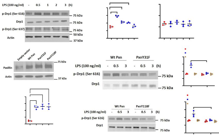
www.nature.com/scientificreports/
LIM Zinc-binding domain present in Lin-11, Isl-1, Mec-3
LPS Lipopolysaccharide
MtROS Mitochondrial ROS
NOX Nicotinamide adenine dinucleotide phosphate oxidase
PBS Phosphate buffered saline
ROS Reactive oxygen species
SDS Sodium dodecyl sulphate
TER Transendothelial electrical resistance
PAGE Polyacrylamide gel electrophoresis
SH3 SRC homology 3
TLR2 Toll-like receptor 2
TLR4 Toll-like receptor 4
VEGFR Vascular endothelial growth factor receptor
Paxillin is a multi-functional, multi-domain focal adhesion adaptor protein that serves as a scaffold for recruiting
and binding to structural and signaling m olecules1. The N-terminus of paxillin consists of 5 LD domains that are
protein recognition sites, and the C-terminus has four tandem LIM domains, which mediate protein–protein
interactions2. Also, the N-terminal of paxillin contains several proline-rich SH3 binding motifs with serine/
threonine and tyrosine phosphorylation sites2. Paxillin through its multiple domains binds to structural and
signaling proteins and regulates cell adhesion, migration, morphology and n eovascularization3–7. The multiple
serine/threonine and tyrosine phosphorylation sites in the paxillin are targeted by a diverse array of kinases
that are activated in response to various adhesion stimuli, cytokines and growth factors1,8. Phosphorylation of
tyrosine residues at the proline-rich SH3 motifs creates new binding sites for SH2-domain containing proteins,
while serine/threonine phosphorylation in LIM domains potentiates the localization of paxillin to a dhesions9.
Thus, phosphorylation of paxillin regulates its localization and function as an adaptor molecule.
Three major paxillin N-terminal tyrosine phosphorylation sites, Y31 and Y118, and Y181 have been identi-
fied with Y31 and Y118 phosphorylation modulating docking of SH2 domain-containing proteins such as Crk,
an adaptor molecule important for regulation of cell motility8,10. While Src and FAK are two prominent tyrosine
kinases that phosphorylate paxillin at Y31 and Y 11811,12, LPS-induced phosphorylation of paxillin at Y31 and
Y118 is mediated by c-Abl and not by Src or FAK in human lung microvascular endothelial cells (HLMVECs)13.
Further, c-Abl mediated tyrosine phosphorylation of Y31 and Y118 regulates LPS-induced pulmonary vascular
permeability and i njury13, and HGF- and S1P-induced reactive oxygen species (ROS) generation, lamellipodia
formation and endothelial p ermeability14.
LPS is a bacterial endotoxin that is extensively used to mimic sepsis-induced injury both in vivo and in vitro.
LPS induces secretion of pro-inflammatory cytokines, expression of adhesion molecules, endothelial permeability
changes, and apoptosis via TLR2 and TLR4 s ignaling15,16, and stimulates ROS production in macrophages and
lung endothelial cells17. ROS is known to play a role in LPS-induced endothelial permeability c hanges17–19 and
apoptosis20. In mammalian cells LPS-mediated ROS is mainly generated by NADPH Oxidase (NOX) family
proteins, and/or through mitochondrial electron t ransport21–23. Our previous study showed that c-Abl mediated
paxillin tyrosine phosphorylation at Y31 and Y118 regulates LPS-induced endothelial dysfunction and lung
injury13; however, the underlying molecular mechanism(s) by which paxillin regulates mitochondria (mt)-derived
ROS dependent endothelial dysfunction is unknown.
Here, using HLMVECs as a model system, we have investigated the role of paxillin and paxillin Y31 and
Y118 tyrosine phosphorylation in the regulation of LPS-mediated mtROS generation and endothelial dysfunc-
tion. Using ROS biosensors targeted to cytoplasm, mitochondria and nucleus, we show that LPS stimulation
of HLMVECs resulted in increased mtROS compared to the cytoplasm or nucleus. Further, we delineated the
pathway leading to LPS-mediated mtROS production via dynamin-related protein 1 (DRP1) activation and
mitochondrial fission that was dependent on paxillin tyrosine phosphorylation at Y31 and Y118, and interaction
between paxillin, DRP1 and ERK1/2. Blocking ERK1/2 activation by PD98059 attenuated mitochondrial fission
and mtROS. Our results provide insight into the role of tyrosine phosphorylation of paxillin in regulating mtROS
and endothelial barrier dysfunction via mitochondrial dynamics.
Results
LPS‑stimulated mtROS production in human lung endothelial cells is attenuated by paxillin
Y31F and Y118F mutants. ROS generated in cells by NOX proteins and mitochondrial electron trans-
port contributes to antimicrobial i mmunity25–27 and to intracellular signaling p
athways28,29. LPS stimulates ROS
production in macrophages and endothelial cells via Toll-like receptor 4 (TLR4) ligation with both, NOX and
mitochondria involved in this p rocess30,31. We have recently shown that paxillin and paxillin tyrosine phospho-
phox
rylation stimulates p47 -dependent ROS production by HGF and S1P in human lung ECs14. However, the
role of mitochondria in LPS-induced ROS generation via paxillin in the endothelium is unclear. To determine
the source of ROS after LPS challenge, HLMVECs were transfected with redox-sensitive biosensors that spe-
cifically target the cytoplasm, mitochondria, or the nucleus (pHyPer-cytosol, pHyPer-dMito or p-HyPer-nuc,
respectively). The validity of pHyPer ROS assay was verified by addition of exogenous hydrogen peroxide ( H2O2)
(5 µM) to HLMVECs transfected with pHyPer-cyto, pHyPer-mito or pHyPer-nuc for 1 h. Addition of exog-
enous hydrogen peroxide stimulated intensity of the biosensor fluorescence (pHyPer-cyto-control: 48 ± 15 vs.
H2O2 294 ± 38; pHyPer-mito-control: 54 ± 18 vs. H2O2 168 ± 28; pHyPer-nuc: control: 42 ± 14 vs. H2O2 80 ± 26).
As shown in Fig. 1A–D, LPS challenge of HLMVECs increased, in a time-dependent manner (6–24 h), mtROS
compared to cytoplasmic or nuclear ROS. Time points earlier to 6 h were not examined. These results with exog-
Scientific Reports | (2021) 11:17546 | https://doi.org/10.1038/s41598-021-97006-y 2
Vol:.(1234567890)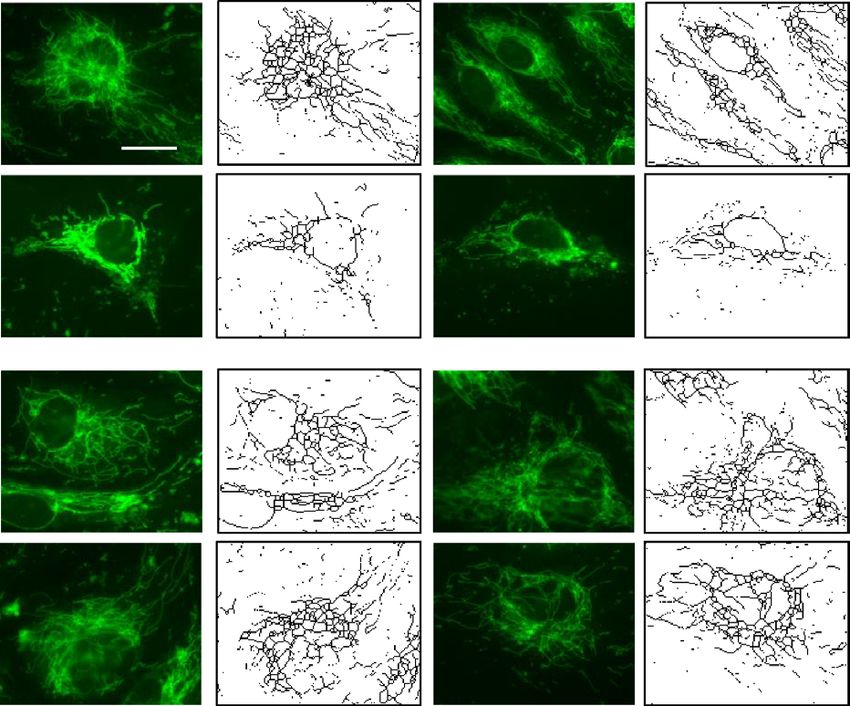
www.nature.com/scientificreports/
Vehicle LPS 6 h LPS 12 h LPS 24 h
A
pHyPer-cyto
pHyPer-dMito
pHyPer-nuc
B C D
ns
250 ns
✱✱✱ 60
60
200
ROS Intensity
ROS Intensity
✱✱
ROS Intensity
40 150 40
✱
100
20 20
50
0 0 0
l
6h
h
h
ol
6h
h
h
ro
ol
h
h
6h
12
24
12
24
tr
12
24
nt
tr
S
S
on
S
S
S
S
S
LP
on
LP
Co
S
S
LP
LP
LP
LP
LP
C
LP
LP
C
pHyPer-cyto pHyPer-mito pHyPer-nuc
Figure 1. LPS induces mitochondrial ROS generation. (A) HLMVECs were transfected with ROS detector
expressed in cytosol (pHyper Cyto), mitochondria (pHyper Mito), or nucleus (pHyper Nuclear). After 48 h of
transfection, cells were treated with 100 ng/ml LPS or PBS for various time points. ROS images were acquired
by fluorescent microscopy. Images were representative of 4–6 independent experiments. Scale bar: 5 μm. (B)
Quantification of cytosol ROS, LPS did not induce significant ROS generation in cytosol. Data were presented
as mean ± SD. (C) Quantification of mitochondrial ROS, data were presented as mean ± SD, *p < 0.05, LPS 6 h
versus PBS control; **p < 0.01, LPS 12 h versus PBS control; ***p < 0.005, LPS 24 h versus PBS control. (D)
Quantification of nuclear ROS, LPS did not induce significant nuclear ROS generation. Data were presented as
mean ± SD.
enous H2O2 show the functionality of the three pHyPer biosensors in determining LPS-induced ROS generation.
Next, we investigated the role of paxillin tyrosine phosphorylation on LPS-mediated mtROS generation by over-
expressing GFP-tagged wild type and paxillin Y31F and Y118F mutants in HLMVECs. LPS-increased mtROS
was attenuated by overexpression of paxillin mutants Y31F and Y118F (Fig. 2A,B) with ecto-expression of wild
type paxillin potentiating mtROS generation compared to non-transfected wild type cells. These data show that
LPS-mediated ROS generation between 6 and 24 h is predominantly of mitochondrial origin in HLMVECs.
However, the contribution of NOX enzymes in ROS generation earlier to 6 h in the cytoplasm is not ruled out.
Paxillin mutants Y31F and Y118F block LPS‑induced mitochondrial fragmentation in lung
endothelial cells. Recent studies have shown that LPS via TLR4 signaling alter mitochondrial dynamics
icroglia32 and m
(fusion vs. fission) in m acrophages33,34 that result in modulation of inflammatory responses
Scientific Reports | (2021) 11:17546 | https://doi.org/10.1038/s41598-021-97006-y 3
Vol.:(0123456789)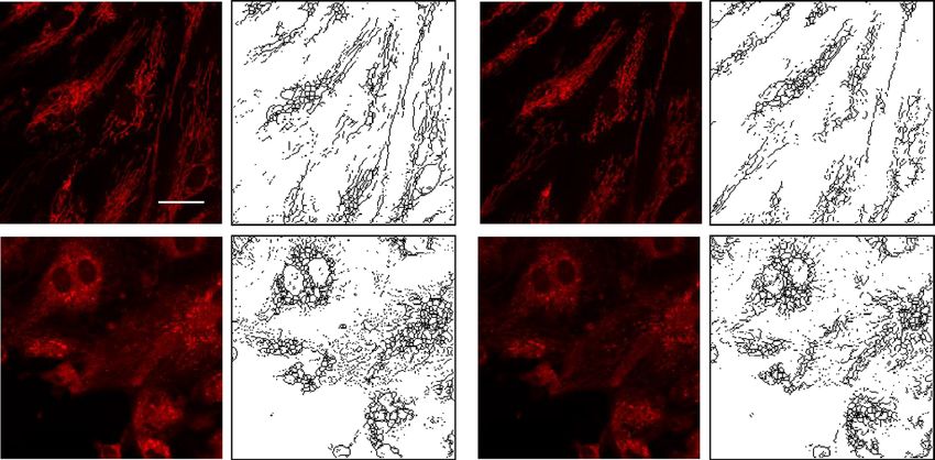
www.nature.com/scientificreports/
A Veh con LPS 100 ng/ml 12 h
B
mitoSOX GFP Merge mitoSOX GFP Merge
Vehicle ✱✱
GFP control ✱✱
LPS
250 ✱
200
ROS Intensity
Pxn wt 150
100 ns
50
Pxn Y31F
0
nY F
nY F
8F
8F
P x wt
P x wt
Px l
Px l
ro
ro
Px 3 1
Px 3 1
11
11
nt
nt
n
n
nY
nY
co
co
Pxn Y118F
PF
PF
G
G
Figure 2. Paxillin mediates LPS-induced mitochondrial ROS. (A) HLMVECs were transfected with GFP-
tagged control plasmid, wild type paxillin, paxillinY31F, or paxillin Y118F mutant plasmids before LPS
treatment. Mitochondrial ROS were detected by MitoSOX dye. At least 20 GFP positive cells were selected for
imaging. Images are representative of 5 independent experiments. Scale bar: 5 μm. (B) ROS were quantified by
measurement of fluorescence intensity by Image J software. Data were presented as mean ± SD, *p < 0.05, LPS
Pxn wt versus LPS control plasmid; **p < 0.01, LPS PxnY31F or LPS PxnY118 versus LPS control plasmid.
both in vivo and in vitro. LPS causes endothelial cell dysfunction and apoptosis; however, the role of paxillin
in LPS-induced alteration of mitochondrial morphology in lung ECs is unclear. To characterize mitochondrial
morphological changes induced by LPS, HLMVECs pre-treated with MitoTracker were challenged with LPS
(100 ng/ml), and mitochondrial morphological changes were monitored by confocal microscopy. As shown in
Fig. 3 A-E, cells challenged with LPS for 24 h exhibited characteristics of mitochondrial fission based on mito-
chondrial length/fragmented (1 µm), tubular (1–3 µm) and elongated (> 3 µm) phenotypes35. The total number
of fragmented structures was increased in LPS challenged HLMVECs whereas mitochondria with elongated
morphology were decreased. In addition, the average mitochondrial length was significantly reduced in LPS-
treated HLMVECs. Expression of the paxillin Y31F and Y118F mutants blocked LPS-induced mitochondrial
morphological changes from fusion to fission with increased elongated and branched mitochondria in HLM-
VECs. These results show that LPS-induced tyrosine phosphorylation of paxillin at Y31 and Y118 is essential for
switching mitochondrial morphology from fusion to fission in HLMVECs.
Paxillin Mutants Y31F and Y118F attenuate LPS‑induced DRP1 phosphorylation in lung
endothelial cells. Having demonstrated a role for paxillin Y31 and Y118 phosphorylation in LPS-mediated
changes in mitochondrial morphology, we next determined if paxillin tyrosine phosphorylation is essential
for DRP1 activation. DRP1 phosphorylation/dephosphorylation at Ser616 mediates mitochondrial fission36,37
itochondria38. LPS challenge of HLMVECs
while phosphorylation at Ser637 inhibits translocation of DRP1 to m
increased DRP1 phosphorylation at Ser616, but not Ser637, in a time-dependent manner without any change
in total DRP1 protein expression (Fig. 4A,B). Transfection of HLMVECs with paxillin wild type and paxil-
lin Y31F and Y118F mutants exhibited increased expression of the proteins compared to non-transfected cells
(Fig. 4C,D). Interestingly, LPS-mediated DRP1 Ser616 phosphorylation at 30 min was attenuated by Y31F and
Y118F mutants (Fig. 4E–H). Together, these results suggest a key role for paxillin Y31 and Y118 tyrosine phos-
phorylation in activation of DRP1 in lung ECs.
Inhibition of DRP1 activation by P110 reduces LPS‑induced mitochondrial fission and mtROS
production in human lung endothelial cells. To further determine the potential link between mito-
chondrial fission and mtROS production, we used a DRP1-specific peptide inhibitor P 11038,39 in LPS-challenged
lung endothelial cells. Pre-treatment of HLMVECs with P110 for 1 h followed by LPS challenge resulted in a sig-
nificant reduction in fragmented mitochondria with a concomitant increase in tubular and elongated mitochon-
dria, similar to control levels (Fig. 5A–E). Next, we investigated the relationship between mitochondrial fission
and mtROS production after LPS challenge. HLMVECs were challenged with LPS for 24 h with or without P110
and then with MitoSOX for 1 h to quantify mtROS by confocal microscopy. As shown in Fig. 5F,G, inhibition
of mitochondrial fission by P110 suppressed mitochondrial ROS generation. These results directly show that
increased mitochondrial fission induced by LPS stimulates mtROS production in lung endothelial cells.
ERK1/2 inhibition suppresses LPS‑induced DRP1 phosphorylation, mitochondrial fission and
mtROS production in lung endothelial cells. As ERK2-mediated DRP1 Ser616 phosphorylation drives
mitochondrial fission40,41, we investigated the role of ERK1/2 in LPS-induced DRP1 phosphorylation, mito-
Scientific Reports | (2021) 11:17546 | https://doi.org/10.1038/s41598-021-97006-y 4
Vol:.(1234567890)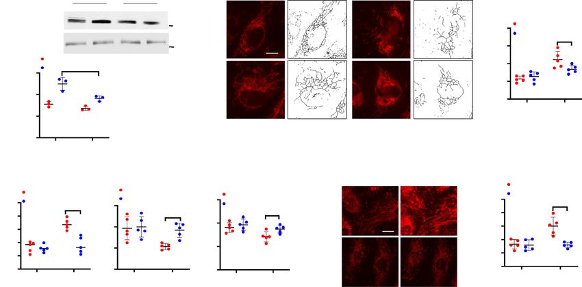
www.nature.com/scientificreports/
A Control plasmid Pxn wt
Veh con
LPS
PxnY31F PxnY118F
Veh con
LPS
B C D E
Control plasmid Control plasmid Control plasmid Control plasmid
✱
Pxn wt Pxn wt ✱ Pxn wt Pxn wt
✱
Number of networks (counts)
Number of individuals (counts)
PxnY31F 150 PxnY31F PxnY31F
100 PxnY31F ✱ 6 ✱
✱ PxnY118F
150
PxnY118F
PxnY118F
Rod/branch length ( m)
PxnY118F
80 ✱
Branches (counts)
100 ✱
60 4 100
✱✱
40
50
2 50
20
0 0 0 0
Control LPS Control LPS Control LPS Control LPS
Figure 3. Paxillin mediates LPS-induced HLMVECs mitochondrial fission. (A) HLMVECs were transfected
with Pxn wild type, PxnY31F, or PxnY118F plasmids. After 48 h of transfection, cells were stained with
MitoTracker (50 nM, 10 min) before termination of LPS challenge (100 ng/ml, 12 h). Images were acquired by
fluorescent microscopy and subjected to skeletonization by Image J software for the purpose of quantification
as described in the Methods. Scale bar: 5 μm. (B–E) Mitochondrial dynamics was assessed by four major
parameters of mitochondrial morphology, namely, number of individual fragments, number of networks, rod/
branch length, and branch counts. Data were presented as means ± SD, for number of individual fragments,
*p < 0.05 compared with LPS control plasmids; for number of networks, *p < 0.05, compared with LPS control
plasmid; for rod/branch length, *p < 0.05, **p < 0.01 compared with LPS control plasmid; for branch counts,
*p < 0.05 compared with LPS control plasmid.
chondrial fission and mtROS production. Pre-treatment of HLMVECs with PD98059, an inhibitor of MEK1/2,
reduced LPS-stimulated DRP1 Ser616 phosphorylation (Fig. 6A,B) and mitochondrial fission (Fig. 6C–G). Fur-
thermore, PD98059 inhibited LPS-induced mtROS production in HLMVECs (Fig. 6H,I). These results show a
role for ERK1/2 in LPS-mediated DRP1 phosphorylation, mitochondrial fission, and mtROS production in lung
endothelial cells.
Paxillin mutants attenuate LPS‑stimulated association between paxillin, DRP1 and ERK1/2
in lung endothelial cells. To further understand how paxillin modulates LPS-induced ERK and DRP1
activation to regulate mitochondrial fission and mtROS, we investigated the potential interaction between paxil-
lin, DRP1 and ERK1/2. HLMVECs transfected with vector control or paxillin Y31F and Y118F mutants were
exposed to either vehicle or LPS, and cell lysates were subjected to immunoprecipitation with anti-paxillin anti-
body and analyzed for the presence of either DRP1 or ERK1/2. As shown Fig. 7A–H, paxillin is constitutively
associated with DRP1, and ERK1/2 in control cells, with this association further increased by exposure to LPS
(30 min). In contrast, the presence of the paxillin mutant proteins, Y31F and Y118F, suppressed the association
of DRP1 and ERK1/2 in paxillin immunoprecipitates. These results suggest increased association between paxil-
lin, DRP1 and ERK1/2 after LPS challenge of HLMVECs.
Inhibition of DRP1 and mtROS production attenuates LPS‑mediated VE‑cadherin tyrosine
phosphorylation and endothelial dysfunction. We have shown that c-Abl-mediated tyrosine phos-
phorylation of paxillin regulates LPS-induced endothelial dysfunction and lung injury13. While LPS-induced
ROS also modulates endothelial cell permeability19, whether mitochondrial fission/mtROS play a role in LPS-
induced endothelial dysfunction is unknown. As shown in Fig. 8 A-D, LPS-induced tyrosine phosphorylation of
Scientific Reports | (2021) 11:17546 | https://doi.org/10.1038/s41598-021-97006-y 5
Vol.:(0123456789)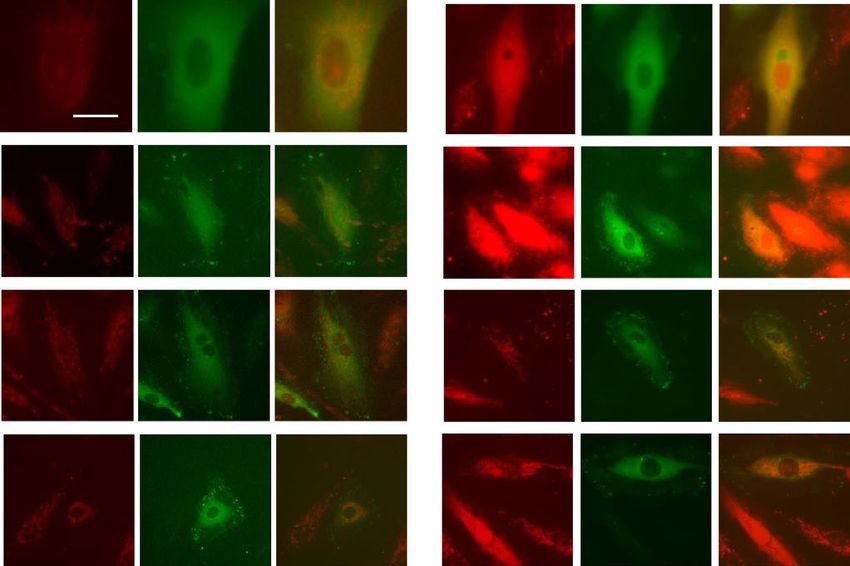
www.nature.com/scientificreports/
A B ✱✱
✱✱
1.5
4
✱✱✱
p-Drp1(637)/Drp1
p-Drp1(616)/Drp1
3 1.0
2
0.5
1
0 0.0
h
h
h
ol
h
h
h
h
h
ol
1
2
3
5
tr
1
2
3
5
tr
0.
0.
on
on
C
C
Control
C E F LPS 0.5 h
4 LPS 3 h
✱✱✱
p-Drp1(616)/Drp1
3
2
1
0
✱✱✱✱
Wt Pxn PxnY31F
D ✱✱✱✱
4
✱✱✱ G H Control
LPS 0.5 h
4
3 LPS 3 h
✱✱
Pxn/Actin
p-Drp1(616)/Drp1
2 3
1 2
0
1
xn
F
8F
or
31
ct
tP
11
nY
ve
nY
W
0
Px
y
Px
pt
Em
Wt Pxn PxnY118F
Figure 4. LPS-induced DRP1 phosphorylation requires both paxillin and ERK1/2. (A) HLMVECs were
treated with LPS (100 ng/ml) for the indicated periods, phosphorylation of DRP1 at Ser616 and Ser637 was
analyzed by Western blot. Equal protein loading was verified by probing with total actin and DRP1. (B) Results
of quantitative analysis of (A) are shown as ratio of phosphorylated DRP1 to total DRP1. Values are means ± SD
of 3 independent experiments. **p < 0.01, ***p < 0.005, compared with control. (C) HLMVECs were transfected
with wild type paxillin, mutant paxillinY31F plasmid or mutant paxillinY118F. After 48 h of transfection, cells
were lysed in cell lysis buffer, and cell lysates (20 µg protein) were subjected to SDS-PAGE and Western blotting
with anti-paxillin antibody. Shown is a representative blot of three independent experiments. (D) The paxillin
bands in (C) were quantified by image analysis. ***p < 0.005, compared to control cells transfected with empty
plasmid backbone. ****p < 0.001, compared to control cells transfected with empty plasmid backbone. (E,G)
HLMVECs were transfected with wild type paxillin, mutant paxillinY31F plasmid or mutant paxillinY118F.
After 48 h of transfection, cells were treated with vehicle or LPS (100 ng/ml) for 0.5 and 3 h. Cell lysates (20
µgs protein) were subjected to SDS-PAGE and Western blotting with phospho-Ser616 DRP1 and anti-DRP1
antibodies. Shown is a representative blot of three independent experiments. (F,H) Results of quantitative
analysis are shown as intensity ratio of phosphorylated DRP1 to total DRP1. Values are means ± SD of 3
independent experiments. ***p < 0.005, LPS 0.5 h wild type paxillin versus LPS 0.5 h mutant paxillinY31F.
**p < 0.01, LPS 0.5 h wild type paxillin versus LPS 0.5 h mutant paxillinY118F.
VE-cadherin at Y658 was suppressed by the DRP1 inhibitor, P110 and mtROS scavenger MitoTEMPO in HLM-
VECs. Next, we investigated the effect of P110 and MitoTEMPO on LPS-induced permeability determined by
TER and FITC-dextran method. Addition of LPS (1 µg/ml) to HLMVECs reduced TER (increased permeability)
starting at 4 h post LPS addition and decrease in TER was sustained up to 18 h, followed by a slight recovery
(Fig. 9A,D). Pre-treatment of HLMVECs with P110 or MitoTEMPO significantly reduced LPS-induced barrier
disruption as determined by restoration of TER (Fig. 9A–F) and inhibition of FITC-dextran leakage (Fig. 9G,H).
These results suggest that LPS-induced barrier disruption is mediated by DRP1 activation and mtROS genera-
tion.
Discussion
Paxillin plays an important role in cell morphology, migration, adhesion, and cell signal transduction. Our
earlier studies defined a key role of paxillin and paxillin tyrosine phosphorylation at Y31 and Y118 in HGF/S1P-
mediated ROS generation, lamellipodia formation and endothelial barrier function14. More recently, we showed
that paxillin and paxillin tyrosine phosphorylation are implicated in LPS-induced endothelial permeability and
lung injury13. Here, we now show a novel role for paxillin and paxillin tyrosine phosphorylation in LPS-induced
mitochondrial fission and mtROS production. Further, we observed that blocking mitochondrial fission and
mtROS reduced LPS-induced tyrosine phosphorylation of VE-cadherin and reversed endothelial dysfunction.
Scientific Reports | (2021) 11:17546 | https://doi.org/10.1038/s41598-021-97006-y 6
Vol:.(1234567890)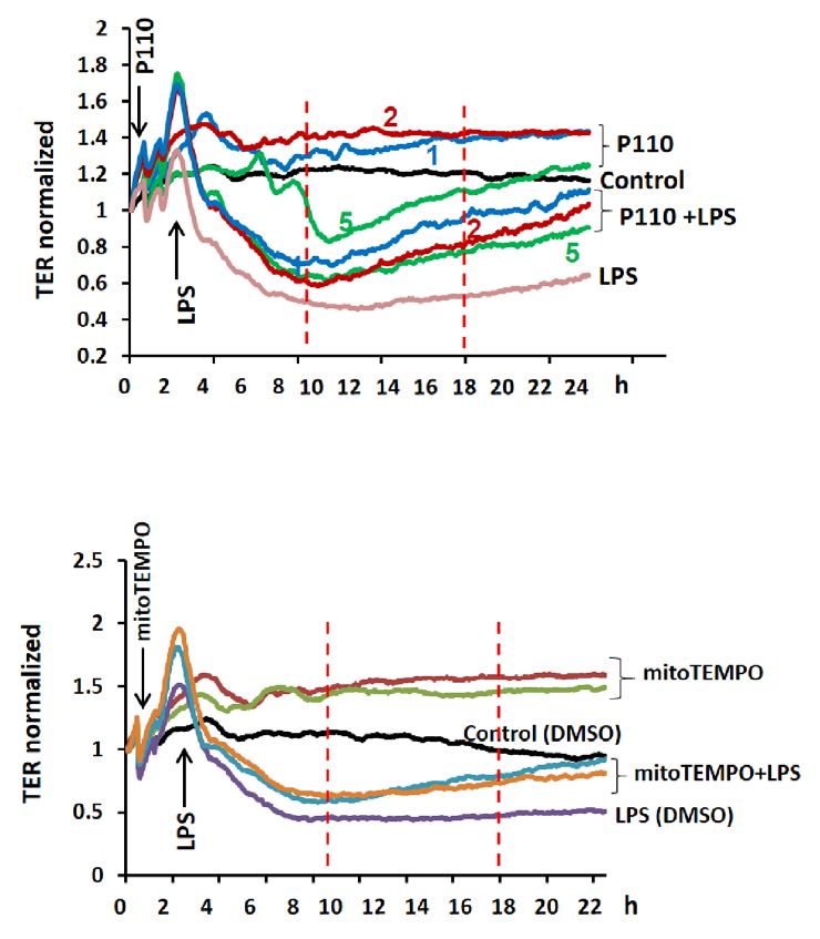
www.nature.com/scientificreports/
A Veh Skeletonized LPS Skeletonized
B Control C Control
P110 ✱✱ P110
Number of individuals (counts)
Number of networks (counts)
100 ✱✱
Con 200
80
150
60
100
40
20 50
P110 0 0
Vehicle LPS Vehicle LPS
MitoTracker
D Control E 150 Control F Veh LPS G
Control
P110
P110 ✱
6 ✱✱ P110 200
✱
Rod/branch length ( m)
Branches (counts)
MitoSox Intensity
100 Con 150
4
100
50
2
50
0
P110 0
0
Vehicle LPS Vehicle LPS Vehicle LPS
Figure 5. DRP1 inhibitory peptide blocks LPS-induced EC mitochondrial fission and ROS. (A) HLMVECs
were pretreated with DRP1 inhibitory peptide P110 (1 µM) for 1 h followed by adding of MitoTracker
10 min before imaging. Images were acquired at 5 min intervals. After 30 min imaging of the basal level of
mitochondrial morphological information, cells were treated with 100 ng/ml LPS followed by continuous
imaging of mitochondria at the same intervals. Presents are images acquired before LPS treatment (Veh column)
and after LPS treatment (LPS column). (B–E) Mitochondrial dynamics was assessed by number of individual
fragments, number of networks, rod/branch length, and branch counts. Data were presented as means ± SD, for
number of individual fragments, **p < 0.01, LPS alone versus LPS + P110; for number of networks, **p < 0.01,
LPS alone versus LPS + P110; for rod/branch length, **p < 0.01, LPS alone versus LPS + P110; for branch
counts, *p < 0.05, LPS alone versus LPS + P110. (F) HLMVECs were treated same as described above, instead
of using MitoTracker staining, cells were treated with 5 µm MitoSOX for 10 min before imaging. Shown are
representative of at least 5 images. Scale bar 10 μm. (G) MitoSOX intensity analysis, *p < 0.05, LPS alone versus
LPS + P110.
These findings suggest that paxillin tyrosine phosphorylation regulates mitochondrial dynamics, and mtROS,
which modulate LPS-induced lung endothelial permeability and barrier function.
As a multi-functional focal adhesion adaptor protein, paxillin exhibits LD motifs, LIM domains and SH2-
binding sites that facilitate recruiting and binding to structural and signaling p roteins1. These interactions play
an important role in cell migration and adhesion and require tyrosine phosphorylation at Y31, Y118 and Y131
sites. Paxillin is tyrosine phosphorylated by FAK, the Src family of kinases and c-Abl at Y31 and Y118, which
is critical for paxillin redistribution to focal adhesions and angiogenesis and endothelial barrier r egulation11,13.
Here we have demonstrated another novel role for paxillin in EC mitochondrial dynamics. The mitochondria
are now recognized to form dynamic networks and undergo cycles between fusion and fission depending upon
the external cues. The process of fusion is regulated by the transmembrane GTPases Mitofusin-1 and Mito-
fusin-2 and optic atrophy protein 1 (OPA1), while fission is mediated by DRP1 and Fission-1 (FIS1)40,41. Our
finding that LPS-induced mitochondrial fission is dependent on paxillin and paxillin tyrosine phosphorylation
in the ECs is novel. Ectopic expression of paxillin Y31F and Y118F mutants blocked LPS-induced DRP1 Ser
616 phosphorylation and mitochondrial fission. Further, the LPS-induced DRP1 Ser 616 phosphorylation and
mitochondrial fission was blocked by PD98059, an inhibitor of MEK that phosphorylates ERK1/2 (Fig. 6H);
thus, providing evidence for ERK1/2 involvement in mitochondrial fission elicited by LPS. Interestingly, LPS
stimulated DRP1 Ser 616 phosphorylation at 30 min that preceded the physiological responses such as mito-
chondrial fission, mtROS production, VE-cadherin phosphorylation, and permeability changes (Figs. 1, 3, 9).
Our co-immunoprecipitation studies show potential association between paxillin, DRP1 and ERK after LPS
challenge of HLMVECs and ecto-expression of paxillin Y31 or Y118 mutant reduced the association between
paxillin, DRP1 and ERK1. It is unclear if the increased association between paxillin, DRP1 and ERK1/2 is direct
or indirect via other proteins, including actin.
In addition to paxillin, cellular cytoskeletal components such as actin and cofilin, an actin-depolymerizing
factor, have been implicated in mitochondrial fi ssion42,43. Further, translocation of cofilin to mitochondria and
interaction with DRP1 was essential for erucin-induced mitochondrial fission and apoptosis in human breast
cancer cells44. DRP1 phosphorylation at Ser616 or dephosphorylation at Ser 637 regulates its translocation to
Scientific Reports | (2021) 11:17546 | https://doi.org/10.1038/s41598-021-97006-y 7
Vol.:(0123456789)
www.nature.com/scientificreports/
A DMSO PD98059
C
Veh Skeletonized LPS Skeletonized
LPS (100 ng/ml) - + - + D
p-Drp1 (Ser 616) 75 kDa Control
Number of individuals (counts)
Con 80
PD98059
✱
Drp1 75 kDa 60
B Control
LPS ✱✱
40
3
20
PD98059
p-Drp1/Drp1
2
0
Vehicle LPS
1
MitoTracker
0
DMSO PD98059
E F G H Veh LPS I
Control
Control Control
Control PD98059
PD98059 250
PD98059 150
Con ✱✱
Number of networks (counts)
100 PD98059
✱✱ 6
✱
Rod/branch length ( m)
✱✱✱ 200
Branches (counts)
MitoSox Intensity
80
100
4 150
60
100
40
2 50
50
20 PD98059
0
0 0 0
Vehicle LPS
Vehicle LPS Vehicle LPS Vehicle LPS
MitoSox
Figure 6. Erk mediates DRP1 phosphorylation and LPS-induced mitochondrial fission. (A) HLMVECs were
pretreated with PD98059 for 30 min followed by LPS for 30 min, phosphorylation of DRP1 was analyzed
by Western blot. (B) Results of quantitative analysis are shown as intensity ratio of phosphorylated DRP1
to total DRP1. Values are means ± SD of 3 independent experiments. **p < 0.01, LPS versus LPS + PD98059.
(C) HLMVECs were pretreated with PD98059 for 30 min followed by adding of MitoTracker 10 min before
imaging. (D–G) Mitochondrial dynamics was assessed by number of individual fragments, number of networks,
rod/branch length, and branch counts. Data were presented as means ± SD, for number of individual fragments,
*p < 0.05, LPS alone versus LPS + PD98059; for number of networks, **p < 0.01, LPS alone versus LPS + PD98059;
for rod/branch length, ***p < 0.005, LPS alone versus LPS + PD98059; for branch counts, *p < 0.05, LPS
along versus LPS + PD98059. Presents are representative of at least 5 images. Scale bar 5 μm. (H) HLMVECs
were treated same as described above and staining with MitoSOX for 10 min before imaging. Shown are
representative of at least 5 images. Scale bar 10 μm. (I) MitoSOX intensity analysis, *p < 0.05, LPS alone versus
LPS + PD98059.
mitochondria and mitochondria fission36. In the current study, LPS stimulated DRP1 Ser616 phosphoryla-
tion with no change in the status of DRP1 Ser 637 phosphorylation; however, dephosphorylation of DRP1Ser
637 in response to erucin regulated DRP1 translocation to m itochondria44. Further, mitochondrial fission and
mitophagy are dependent on cofilin-mediated actin depolymerization activity at the mitochondrial fission site43.
Interestingly, suppression of NADPH oxidase mediated ROS production led to elevated actin polymerization
(F-actin) in neutrophils45. Thus, it is likely that elevated ROS production could lead to actin depolymerization.
However, a plausible link between paxillin-mediated mitoROS in cofilin phosphorylation and actin remodeling
is unclear. In addition to DRP1, cofilin is also translocated to mitochondrial outer membrane in breast cancer
cells; however, it is unclear if paxillin is translocated to mitochondria in response to LPS.
The two major sources of cellular ROS are NOX family proteins and mitochondria. The NOX2- and NOX4-
derived ROS in ECs are known to regulate vascular function; however, excess accumulation of ROS leads to
vascular dysfunction46,47. In human lung ECs, the effect of HGF- or S1P-induced p 47phox activation and ROS accu-
mulation on lamellipodia formation is known to be regulated by paxillin tyrosine phosphorylation indicating a
OX214. Moreover, interaction between TLR4 and NOX4 was shown to be essential for LPS-induced ROS
role for N
in ECs and smooth muscle cells48. In addition to NOX, several studies have shown that mitochondria and mtROS
are immunomodulators49–51 and suppression of mtROS alleviated i nflammation52. However, the role of mtROS in
regulating endothelial barrier function is unclear. Here, we have demonstrated for the first time that LPS-induced
paxillin tyrosine phosphorylation regulates mtROS production in HLMVECs, and down-regulation of paxil-
lin with siRNA or paxillin function with the Y31 and Y118 mutants reduces LPS-induced mtROS generation.
Furthermore, LPS-induced mtROS was dependent on the mitochondrial fission. Here, we have demonstrated
that inhibition of mitochondrial fission with a DRP1 specific inhibitor, P11038 markedly suppressed mtROS gen-
eration in LPS-stimulated HLMVECs. A similar inhibition of LPS-induced mtROS generation by Mdivi-1 and
DRP1 knockdown attenuating the production of pro-inflammatory mediators was reported in microglial c ells53.
Additionally, LPS-induced mitochondrial fission and mtROS production was attenuated by PD98059, an inhibitor
of MEK suggesting a role for ERK1/2. While this study shows mitochondrial fission as an upstream regulator of
mtROS generation, in certain cell types such as primary hippocampal neurons mtROS seems to regulate beta-
amyloid (Aβ) induced mitochondrial morphological changes. Aβ-mediated mitochondrial granular shape in
Scientific Reports | (2021) 11:17546 | https://doi.org/10.1038/s41598-021-97006-y 8
Vol:.(1234567890)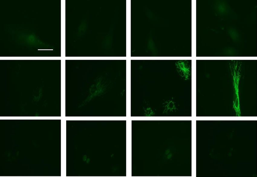
www.nature.com/scientificreports/
A B C D Control
Control LPS ✱✱
1.5 0.6
LPS
✱✱✱
IP: Pxn IP: Pxn
Wt Pxn PxnY31F 0.4
Erk1/2/Pxn
Wt Pxn PxnY31F 1.0
Drp1/Pxn
LPS (100 ng/ml) - + - + LPS (100 ng/ml) - + - +
50 kDa 0.2
0.5
IB: Drp1 75 kDa IB: Erk1/2
75 kDa 75 kDa
IB: Pxn IB: Pxn 0.0
0.0 Wt Pxn PxnY31F
Wt Pxn PxnY31F
E F G H
IP: Pxn Control IP: Pxn Control
1.5
LPS 1.0 LPS ✱✱
Wt Pxn PxnY118F ✱✱✱ Wt Pxn PxnY118F
LPS (100 ng/ml) - + - + LPS (100 ng/ml) - + - + 0.8
1.0 50 kDa
Drp1/Pxn
Erk1/2/Pxn
IB: Drp1 75 kDa IB: Erk1/2 0.6
75 kDa 75 kDa 0.4
IB: Pxn 0.5 IB: Pxn
0.2
0.0 0.0
Wt Pxn PxnY118F Wt Pxn PxnY118F
Figure 7. Phosphorylation of paxillin Y31 and Y118 is required for paxillin interactions with Erk1/2 and DRP1.
(A–D) HLMVECs were transfected with wild type paxillin or paxillinY31F mutant plasmids, interactions of
paxillin with DRP1 and Erk1/2 was assessed by IP experiment. Data were presented as means ± SD, **p < 0.01,
LPS wild type paxillin versus LPS paxillinY31F mutant; ***p < 0.005, LPS wild type paxillin versus LPS
paxillinY31F mutant, n = 3. E–H, HLMVECs were transfected with wild type paxillin or paxillinY118F mutant
plasmids, interactions of paxillin with DRP1 and Erk1/2 was assessed by IP experiment. Data were presented
as means ± SD, **p < 0.01, LPS wild type paxillin versus LPS paxillinY118F mutant; ***p < 0.005, LPS wild type
paxillin versus LPS paxillinY181F mutant, n = 3.
A B
Vehicle
✱✱✱
P110
Vehicle P110 1.0
✱
p-VE-Cad/Total VE-Cad
LPS - 3 6 24 - 3 6 24 h 0.8
150 kDa
p-VE-CadY658 0.6
100 kDa
0.4
150 kDa
VE-Cad 0.2
100 kDa
0.0
PBS LPS 3h LPS 6h LPS 24h
C D
DMSO
mitoTEMPO
DMSO 0.6 ✱
MitoTEMPO ✱
p-VE-Cad/Total VE-Cad
✱✱✱
LPS - 3 6 24 - 3 6 24 h
0.4 ✱
150 kDa
p-VE-CadY658
100 kDa
0.2
150 kDa
VE-Cad
100 kDa 0.0
PBS LPS 3h LPS 6h LPS 24h
Figure 8. Inhibition of mitochondrial ROS dampens LPS-induced VE-Cadherin phosphorylation. HLMVECs
were pretreated with P110 (A) or MitoTEMPO (C) to block mitochondrial ROS, cells were then treated with LPS
for various time points. Phosphorylation of VE-Cadherin at Y658 was detected by Western blot. Two independent
experiments were performed. (B,D) Results of quantitative analysis are shown as intensity ratio of phosphorylated
VE-Cadherin to total VE-Cadherin. Values are means ± SD of 2 independent experiments. *p < 0.05, vehicle versus
P110 for 3 h of LPS treatment (B) and vehicle versus MitoTEMPO for 3 h and 6 h (D) of LPS treatment; ***p < 0.005,
vehicle versus P110 for 6 h of LPS treatment (B) and vehicle versus MitoTEMPO for 24 h of LPS treatment (D).
Scientific Reports | (2021) 11:17546 | https://doi.org/10.1038/s41598-021-97006-y 9
Vol.:(0123456789)www.nature.com/scientificreports/
A B C G
10 h 18 h
PBS PBS
LPS ✱✱✱ PBS LPS
1.5 2.0 ✱✱
LPS 3000
✱✱✱
FITC Fluorescence, AU
✱✱
TER normalized
TER normalized
1.5
1.0
2000
✱✱
1.0
0.5
0.5 1000
0.0 0.0
0 M 1 M 2 M 5 M 0 M 1 M 2 M 5 M 0
Vehicle P110
P110 concentration P110 concentration
D E 10 h
F
18 h H PBS
PBS PBS
1.5 LPS LPS ✱✱✱
LPS 2.0 3000
✱
FITC Fluorescence, AU
✱✱✱
TER normalized
TER normalized
✱
1.5
1.0 2000
✱✱✱
1.0
0.5 1000
0.5
0.0 0.0 0
0 M 1 M 5 M 0 M 1 M 5 M DMSO MitoTEMPO
MitoTEMPO concentration MitoTEMPO concentration
Figure 9. Inhibition of mitochondrial ROS attenuated LPS-induced HLMVEC barrier permeability. The
effects of mitochondrial ROS on endothelial barrier function were tested by measurements of trans-endothelial
electrical resistance (TER) (A–F) and FITC-dextran leakage (G,H). HLMVECs were pretreated with
different concentrations of P110 (1–5 µM) or MitoTEMPO (1 or 5 µM) for 30 min followed by LPS (100 ng/
ml) challenge. TER was recorded continuously for 24 h (A,D). Shown are representative tracings from 4
independent experiments. The TERs at 10 h and 18 h time points were used for statistical analysis (B,C & E,F).
(B,C) Values are means ± SD. **p < 0.01, P110 2 μM versus LPS control, n = 4; ***p < 0.005, P110 5 μM versus
LPS, n = 4. (E,F) Values are means ± SD. *p < 0.05, MitoTEMPO 1 or 5 μM versus LPS; ***p < 0.005, MitoTEMPO
1 or 5 μM versus LPS, n = 4. (G,H), HLMVECs monolayer permeability was assessed by FITC-dextran leakage
as describe in METHODS. Values are means ± SD. **p < 0.01, P110 + LPS versus LPS, n = 4; ***p < 0.005,
MitoTEMPO + LPS versus LPS, n = 4.
hippocampal neurons was dependent on mtROS as mitoTEMPO restored tubular mitochondrial morphology
suggesting a reciprocal relationship between mtROS and mitochondrial dynamics in n eurodegeneration38. The
present study does not address which of the pathways in the mtROS production are modulated by paxillin and
paxillin tyrosine phosphorylation. In the mitochondria, escape of electrons from the mitochondrial electron
transport chain (ETC) results in the formation of superoxide (O2.-)54, while NOX4 localized in the mitochondria
generates hydrogen peroxide ( H2O2)55,56. While we were able to demonstrate LPS-induced mtROS between 6 and
24 h post-LPS challenge, there was no increase of ROS in the cytoplasm and nuclear compartments as determined
by targeted pHyPer-cytosolic and pHyPer-nuclear ROS sensors (Fig. 1). It is conceivable that LPS stimulated ROS
production in the cytoplasmic compartment earlier to 6 h, and we did not determine ROS generation induced
by LPS between 1 and 5 h, and therefore cannot rule out ROS production in the cytoplasm. Further studies are
necessary to address the precise role of paxillin and paxillin tyrosine phosphorylation in ETC and/or NOX4
dependent mtROS production in response to a pro-inflammatory stimulus in the endothelium.
Maintenance of endothelial barrier integrity is crucial for vessel wall homeostasis and lung function under
normal and pathological conditions such as sepsis, ARDS, bacterial lung infection and ventilator-induced lung
injury. Increased ROS production mediated by NOX2/NOX4 has been linked to endothelial barrier disruption
and pulmonary edema, a hallmark of ARDS or sepsis-induced lung inflammatory i njury46. Earlier studies have
suggested that mtROS regulate VEGFR transactivation and down-stream s ignaling57, endothelial s prouting58,59,
TNF-α-mediated apoptosis60, and endothelial inflammation61. However, the role of mtROS in endothelial
barrier dysfunction in lung inflammatory injury is unclear. Our study suggests a causal relationship between
LPS-induced mitochondrial fission, mtROS and endothelial permeability. The LPS-induced endothelial bar-
rier dysfunction, as determined by TER and FITC-dextran techniques, was blunted by paxillin mutants, DRP1
inhibition with P110 and scavenging mtROS with mitoTEMPO. One potential mechanism of LPS-induced
endothelial hyper-permeability is by disassembly of adherens junction proteins including claudin, occludin,
and VE-cadherin62. LPS and other inflammatory mediators stimulate VE-cadherin phosphorylation at Y658,
Y685 and Y731 that induces vascular permeability and leukocyte extravasation. Inhibition of DRP1 and scaveng-
ing mtROS attenuated LPS-induced VE-cadherin Y658 phosphorylation and endothelial permeability. These
results demonstrate for the first time a causal link between mitochondrial fission, and mtROS in modulating
VE-cadherin phosphorylation at the adherens junction (AJ) by LPS and modulating endothelial permeability.
In addition to endothelial permeability, mtROS has been shown to regulate pro-inflammatory responses of
macrophages to bacterial infection through disulfide linkage of nuclear factor kB (NF-kB) essential modulator
(NEMO)63, expression of pro-inflammatory mediators in microglial c ells64, and VEGF-induced endothelial
Scientific Reports | (2021) 11:17546 | https://doi.org/10.1038/s41598-021-97006-y 10
Vol:.(1234567890)www.nature.com/scientificreports/
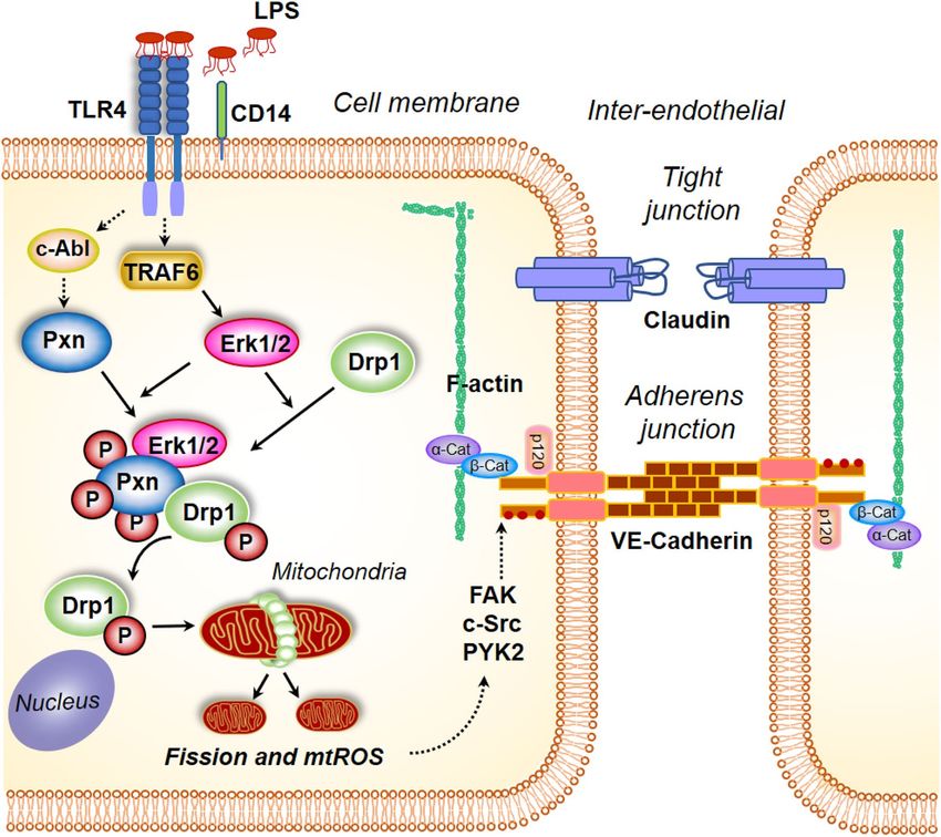
Figure 10. Model of paxillin and DRP1 activation by LPS in mitochondrial fission, mitochondrial ROS
generation and endothelial permeability. Stimulation of endothelial cells with lipopolysaccharide (LPS) results
in changes in paxillin phosphorylation, which is essential for DRP1 phosphorylation by ERK. Activation of
DRP1 results in its translocation to mitochondria resulting in mitochondrial fission accompanied by enhanced
mitochondrial ROS. Blocking paxillin or DRP1 phosphorylation attenuates mitochondrial ROS generation
and VE-cadherin phosphorylation status. Similarly, blocking mitochondrial ROS attenuated LPS-induced
VE-cadherin phosphorylation and endothelial permeability.
migration through R ac165. Further, a recent study suggests a role for NOX-derived ROS in the cytosol in the
generation of mtROS in coronary endothelium59; however, in our study, LPS challenge between 6–24 h stimulated
predominantly mtROS and not the cytosolic or nuclear ROS in HLMVECs, which was regulated by paxillin tyros-
ine phosphorylation dependent DRP1 activation. While the mtROS dependent signaling pathway(s) involved
in VE-cadherin Y658 phosphorylation/dephosphorylation is yet to be defined, several tyrosine phosphatases
including VE-PTP66, SHP267, and PTPN1468 have been implicated in dephosphorylation of VE-cadherin at AJs
to maintain a non-leaky endothelial barrier. It has been shown that ROS inhibits phosphatases especially protein
tyrosine phosphatases69 and it is possible that mtROS generated by LPS inhibits VE-PTP, SHP2 or PTPN14, and
maintains VE-cadherin tyrosine phosphorylation that results in delayed recovery of AJs and barrier integrity.
Further studies are necessary to determine mechanism(s) involved in mtROS mediated regulation of VE-cadherin
phosphorylation/dephosphorylation status in the AJs of endothelial cells exposed to inflammatory mediators
such as LPS or thrombin.
In conclusion, in this study we have defined a novel pathway of paxillin tyrosine phosphorylation mediated
mitochondrial fission, which regulates LPS-mediated mtROS production and endothelial barrier dysfunction
(Fig. 10). Further, we have demonstrated that the mtROS-mediated endothelial barrier dysfunction is due to
aberrant dephosphorylation of VE-cadherin at AJs. Thus, our results suggest that regulating mitochondrial
dynamics and/or mtROS may be a novel therapeutic approach to ameliorate pulmonary leak in inflammatory
lung pathologies such as ARDS, sepsis and ventilator-induced lung injury.
Scientific Reports | (2021) 11:17546 | https://doi.org/10.1038/s41598-021-97006-y 11
Vol.:(0123456789)www.nature.com/scientificreports/
Materials and methods
Materials. Human lung microvascular endothelial cells (HLMVECs) (catalog: cc-2583) and endothelial
basal media (EBM-2) (catalog: 3162) were obtained from Lonza (San Diego, CA). FuGENE HD (catalog: E2311)
transfection reagent was from Promega Corporation (Madison, WI). PD98059 (catalog: P215-1MG) was pur-
chased from Sigma-Aldrich (Saint Louis, MO). Bovine serum albumin (catalog: sc-2323) was obtained from
Santa Cruz Biotechnology (Dallas, TX). Thirty-five mm poly-D-lysine-coated glass-bottomed dishes were from
MatTek (Ashland, MA, USA). MitoSOX and MitoTracker Red were from Fisher Scientific (Waltham, MA). Pax-
illin, Y31, Y118 phosphor-paxillin, Erk1/2, phosphor-Erk, DRP1 and S616 phospho-DRP1 antibodies were from
Cell Signaling Technology (Danvers, MA).
Endothelial cell culture. HLMVECs cultured in complete media (EBM-2) containing 10% fetal bovine
serum (FBS), growth factors (provided as a kit from Lonza (San Diego, CA) and 1% Penicillin/Streptomycin
were maintained at 37 °C and 5% CO2 and grown to contact-inhibited monolayers that revealed typical cob-
blestone morphology. Cells were then detached with 0.05% trypsin and resuspended in fresh complete EBM-2
medium, and cultured on gold electrodes for electrical resistance determinations (transendothelial electrical
resistance TER), or glass coverslips for fluorescent microscopy studies, or on 35, 60 or 100-mm culture dishes
for preparation of cell lysates and Western blot analysis.
Measurement of transendothelial electrical resistance. Transendothelial electrical resistance
(TER) was measured in an electrical cell-substrate impedance sensing system (Applied Biophysics, Troy, NY) as
described previously14. Briefly, HLMVECs were grown to ∼ 95% confluence in polycarbonate wells containing
gold electrodes connected to a phase-sensitive lock-in amplifier. Electrodes containing cells were placed in an
electrical cell-substrate impedance incubator for 1 h to stabilize basal electrical resistance. The total electrical
resistance across the endothelial monolayer was determined by the combined resistance between the basal sur-
face of the cell and the electrode, providing a measure of alterations in cell–cell or cell–matrix adhesion. In the
experiments assessing the time course of the response, TER is expressed as normalized resistance (i.e., ratio of
resistance at a given time to resistance at “zero” time).
Plasmids and transfection. Enhanced green fluorescent protein (EGFP)-tagged wild type paxillin and
Y31F, Y118F mutants were prepared as described previously13. ROS biosensors targeted to cytosol, mitochon-
dria, and nucleus were obtained from Evrogen (Moscow, Russia). Constructs were added to HLMVECs grown
to ∼ 80% confluence in EBM-2-MV growth media (Lonza) supplemented with 10% FBS. After overnight cul-
ture, the medium was replaced with fresh complete medium and cells were cultured for additional 48 h before
experimentation.
Live cell ROS imaging. Live cell ROS detection was performed with a Carl Zeiss 780 Confocal microscopy
with cells either transfected with different ROS biosensors localized at cytosol, mitochondria, or nucleus, or cells
stained with mitochondrial ROS detector, MitoSOX. Forty eight hours after transfection, HLMVECs cultured
in MatTek (Ashland, MA, USA) 35 mm poly-D-lysine-coated glass-bottomed dish were changed with fresh
medium containing 2% FBS, and the cell culture dish was mounted onto con-focal microscopy equipped with
a temperature-controlled chamber supplied with humidified 5% CO2. Thirty minutes after the cells adapted to
the chamber environment, basal ROS images were acquired with 63 × /1.40 oil objective at excitation wavelength
of 488 nm or 587 nm for detection of ROS biosensor or MitoSOX signal, respectively. After basal ROS acquired,
LPS was added into cell culture medium, followed by image acquisition at different time points. Quantification
of ROS was performed as previously described.
Live cell mitochondria visualization and mitochondrial morphology quantification. Mitochon-
dria visualization was carried out by live cells labelled with MitoTracker Red. This was achieved by incubating
the cells with culture media containing 20 nM dye for 30 min within the culture incubator, followed by washing
3 times with pre-warmed phosphate-buffered saline. Images were obtained with Carl Zeiss 780 Confocal micros-
copy as described above with excitation wavelength of 587 nm and 590–660 emission filter set. Analysis of image
was carried out with ImageJ software. Analysis of mitochondrial network morphology was preceded with image
pre-processing steps including unsharp mask, enhancement of local contrast, binary making and skeletoniza-
tion. These processes were integrated into an Image J Macro file and were applied for all images processing. All
pixels within a skeleton are then grouped into three categories: end point pixels, slab pixels, and junction pixels.
Pixels spatial relationships were used to measure the length of each branch and the number of branches in each
skeletonized feature. To simplify the analysis, numbers of individual structures (rod, punctate, large/round),
networks and branches, and length of rod and branch, were used for quantification to define mitochondrial
morphology.
Immunoblotting and immunoprecipitation. Immunoblotting and immunoprecipitation (IP) studies
were performed as described p reviously14,70. In brief, after appropriate treatments, cells were pelleted in ice-cold
PBS, lysed in standard lysis buffer (Cell Signaling, Beverly, MA), and sonicated. Lysates were then centrifuged at
1,000×g for 10 min at 4 °C, supernatants were collected, and protein assayed using BCA protein assay kit. For IP
experiments, equal amounts of protein (1 mg) from each sample were pre-cleared with control IgG conjugated
to Protein A/G agarose beads at 4 °C for 1 h, supernatants were collected and incubated overnight with primary
antibody coupled with Protein A/G agarose beads at 4 °C. Next day, the samples were centrifuged at 1,000×g for
Scientific Reports | (2021) 11:17546 | https://doi.org/10.1038/s41598-021-97006-y 12
Vol:.(1234567890)www.nature.com/scientificreports/
1 min in a microfuge centrifuge and the pellet containing the agarose beads were washed three times with lysis
buffer at room temperature. After centrifugation at 1,000×g for 1 min, the beads were collected by removing
supernatant buffer, and 40 µl of SDS sample buffer (100 mM Tris–HCl pH 6.8, 4% SDS, 0.1% bromophenol blue,
20% glycerol, 200 mM DTT] were added to the beads and boiled. Lysates were then subjected to 10% SDS-PAGE
followed by Western blotting. Proteins were detected by immunoblotting using appropriate primary antibodies,
and HRP-conjugated anti-rabbit or anti-mouse secondary antibodies. Band intensities were quantified by den-
sitometry using Image J software.
Statistical analysis. Results are expressed as means ± SD of 3–5 independent experiments. Statistical sig-
nificance was assessed by ANOVA followed by multiple comparisons with Bonferroni corrections. Statistical
significance was defined at p < 0.05.
Received: 8 October 2020; Accepted: 12 August 2021
References
1. Brown, M. C. & Turner, C. E. Paxillin: adapting to change. Physiol. Rev. 84, 1315–1339. https://doi.org/10.1152/physrev.00002.
200484/4/1315[pii] (2004).
2. Turner, C. E. Paxillin interactions. J. Cell Sci. 113(Pt 23), 4139–4140 (2000).
3. Iwasaki, T. et al. Involvement of phosphorylation of Tyr-31 and Tyr-118 of paxillin in MM1 cancer cell migration. Int. J. Cancer
97, 330–335. https://doi.org/10.1002/ijc.1609[pii] (2002).
4. Petit, V. et al. Phosphorylation of tyrosine residues 31 and 118 on paxillin regulates cell migration through an association with
CRK in NBT-II cells. J. Cell Biol. 148, 957–970 (2000).
5. Salgia, R. et al. Expression of the focal adhesion protein paxillin in lung cancer and its relation to cell motility. Oncogene 18, 67–77
(1999).
6. Turner, C. E. et al. Paxillin LD4 motif binds PAK and PIX through a novel 95-kD ankyrin repeat, ARF-GAP protein: A role in
cytoskeletal remodeling. J. Cell Biol. 145, 851–863 (1999).
7. Teranishi, S., Kimura, K. & Nishida, T. Role of formation of an ERK-FAK-paxillin complex in migration of human corneal epithelial
cells during wound closure in vitro. Invest. Ophthalmol. Vis. Sci. 50, 5646–5652 (2009).
8. Schaller, M. D. Paxillin: A focal adhesion-associated adaptor protein. Oncogene 20, 6459–6472 (2001).
9. Webb, D. J. et al. FAK-Src signalling through paxillin, ERK and MLCK regulates adhesion disassembly. Nat. Cell Biol. 6, 154–161
(2004).
10. Yano, H. et al. Paxillin alpha and Crk-associated substrate exert opposing effects on cell migration and contact inhibition of growth
through tyrosine phosphorylation. Proc. Natl. Acad. Sci. USA 97, 9076–9081 (2000).
11. Brown, M. C., Cary, L. A., Jamieson, J. S., Cooper, J. A. & Turner, C. E. Src and FAK kinases cooperate to phosphorylate paxillin
kinase linker, stimulate its focal adhesion localization, and regulate cell spreading and protrusiveness. Mol. Biol. Cell 16, 4316–4328
(2005).
12. Roy, S., Ruest, P. J. & Hanks, S. K. FAK regulates tyrosine phosphorylation of CAS, paxillin, and PYK2 in cells expressing v-Src,
but is not a critical determinant of v-Src transformation. J. Cell Biochem. 84, 377–388 (2002).
13. Fu, P. et al. c-Abl mediated tyrosine phosphorylation of paxillin regulates LPS-induced endothelial dysfunction and lung injury.
Am. J. Physiol. Lung Cell Mol. Physiol. 308, L1025-1038. https://doi.org/10.1152/ajplung.00306.2014 (2015).
14. Fu, P. et al. Role played by paxillin and paxillin tyrosine phosphorylation in hepatocyte growth factor/sphingosine-1-phosphate-
mediated reactive oxygen species generation, lamellipodia formation, and endothelial barrier function. Pulm Circ. 5, 619–630.
https://doi.org/10.1086/683693 (2015).
15. Choy, K. W., Lau, Y. S., Murugan, D., Vanhoutte, P. M. & Mustafa, M. R. Paeonol attenuates LPS-induced endothelial dysfunction
and apoptosis by inhibiting BMP4 and TLR4 signaling simultaneously but independently. J. Pharmacol. Exp. Ther. 364, 420–432.
https://doi.org/10.1124/jpet.117.245217 (2018).
16. Pawar, R. D. et al. Bacterial lipopeptide triggers massive albuminuria in murine lupus nephritis by activating Toll-like receptor 2
at the glomerular filtration barrier. Immunology 128, e206-221. https://doi.org/10.1111/j.1365-2567.2008.02948.x (2009).
17. Kratzer, E. et al. Oxidative stress contributes to lung injury and barrier dysfunction via microtubule destabilization. Am. J. Respir.
Cell Mol. Biol. 47, 688–697. https://doi.org/10.1165/rcmb.2012-0161OC (2012).
18. Gandhirajan, R. K. et al. Blockade of NOX2 and STIM1 signaling limits lipopolysaccharide-induced vascular inflammation. J.
Clin. Invest. 123, 887–902. https://doi.org/10.1172/JCI65647 (2013).
19. Fu, P. et al. Amifostine reduces lung vascular permeability via suppression of inflammatory signalling. Eur. Respir. J. 33, 612–624.
https://doi.org/10.1183/09031936.00014808 (2009).
20. Qi, D. et al. Vaspin protects against LPSinduced ARDS by inhibiting inflammation, apoptosis and reactive oxygen species genera-
tion in pulmonary endothelial cells via the Akt/GSK3beta pathway. Int. J. Mol. Med. 40, 1803–1817. https://doi.org/10.3892/ijmm.
2017.3176 (2017).
21. Vieira, L. D. et al. Oxidative stress induced by prenatal LPS leads to endothelial dysfunction and renal haemodynamic changes
through angiotensin II/NADPH oxidase pathway: Prevention by early treatment with alpha-tocopherol. Biochim. Biophys. Acta
Mol. Basis Dis. 3577–3587, 2018. https://doi.org/10.1016/j.bbadis.2018.09.019 (1864).
22. Han, J. E. & Choi, J. W. Control of JNK for an activation of NADPH oxidase in LPS-stimulated BV2 microglia. Arch. Pharm. Res.
35, 709–715. https://doi.org/10.1007/s12272-012-0415-1 (2012).
23. Check, J. et al. Src kinase participates in LPS-induced activation of NADPH oxidase. Mol. Immunol. 47, 756–762. https://doi.org/
10.1016/j.molimm.2009.10.012 (2010).
24. Singel, K. L. & Segal, B. H. NOX2-dependent regulation of inflammation. Clin. Sci. (Lond) 130, 479–490. https://doi.org/10.1042/
CS20150660 (2016).
25. Rada, B. & Leto, T. L. Oxidative innate immune defenses by Nox/Duox family NADPH oxidases. Contrib. Microbiol. 15, 164–187.
https://doi.org/10.1159/000136357 (2008).
26. Leto, T. L. & Geiszt, M. Role of Nox family NADPH oxidases in host defense. Antioxid. Redox. Signal 8, 1549–1561. https://doi.
org/10.1089/ars.2006.8.1549 (2006).
27. Yang, C. S. et al. NADPH oxidase 2 interaction with TLR2 is required for efficient innate immune responses to mycobacteria via
cathelicidin expression. J. Immunol. 182, 3696–3705. https://doi.org/10.4049/jimmunol.0802217 (2009).
28. Li, Q., Li, J., Liu, Y., Zhang, M. & Chen, C. Anagliptin prevents apoptosis of human umbilical vein endothelial cells by modulating
NOX-4 signaling pathways. Biomed. Pharmacother. 103, 1623–1631. https://doi.org/10.1016/j.biopha.2018.04.187 (2018).
Scientific Reports | (2021) 11:17546 | https://doi.org/10.1038/s41598-021-97006-y 13
Vol.:(0123456789)You can also read


