Estrogen disorders: Interpreting the abnormal regulation of aromatase in granulosa cells (Review) - Spandidos Publications
←
→
Page content transcription
If your browser does not render page correctly, please read the page content below
INTERNATIONAL JOURNAL OF MOlecular medicine 47: 73, 2021

                  Estrogen disorders: Interpreting the abnormal
                regulation of aromatase in granulosa cells (Review)
                                             TING LIU1, YIFEI HUANG2 and HUI LIN1

                   1
                    Department of Pathophysiology, School of Basic Medicine Sciences, Nanchang University;
                   2
                    First Clinical Medical School, Nanchang University, Nanchang, Jiangxi 330006, P.R. China

                                        Received October 2, 2020; Accepted January 27, 2021

                                                      DOI: 10.3892/ijmm.2021.4906

Abstract. Ovarian granulosa cells (GCs) are the most                     which eventually create an imbalance in the estrogen secre‑
important source of estrogen. Therefore, aromatase (estrogen             tion by the target tissues. The present review highlights these
synthase), which is the key enzyme in estrogen synthesis,                useful factors as potential inhibitors for target therapy.
is not only an important factor of ovarian development, but
also the key to estrogen secretion by GCs. Disorders of the
ovarian estrogen secretion are more likely to induce female              Contents
estrogen‑dependent diseases and fertility issues, such as
ovarian cancer and polycystic ovary syndrome. Hence, aroma‑              1. Introduction
tase is an important drug target; treatment with its inhibitors          2. Promoter regulation
in estrogen‑dependent diseases has attracted increasing                  3. Aromatase expression localization
attention. The present review article focuses on the regulation          4.	CYP19A1 is dependent on FSH/cAMP
and mechanism of the aromatase activity in the GCs, as well              5. Regulation of aromatase
as the specific regulation of aromatase promoters. In GCs,               6. Regulatory pathways of aromatase in GCs
follicle‑stimulating hormone (FSH) is dependent on the cyclic            7.	Changes in aromatase activity of GCs induce diseases
adenosine monophosphate (cAMP) pathway to regulate the                   8.	Current and future perspectives
aromatase activity, and the regulation of this enzyme is related         9.	Conclusions
to the activation of signaling pathways, such as phosphati‑
dylinositol 3‑kinase (PI3K) and extracellular signal‑regulated
kinase (ERK). In addition, endocrine‑disrupting substance                1. Introduction
and other related factors affect the expression of aromatase,
                                                                         Aromatase is a cytochrome P450 monooxygenase that
                                                                         encodes CYP19A1. Its expression is regulated via differential
                                                                         promoter activation in a tissue‑specific manner. Aromatase
                                                                         mainly occurs in the ovaries (1), brain cells (2) and testes of
Correspondence to: Dr Hui Lin, Department of Pathophysiology,            rodents (3), as well as in human fat cells (4) and placental
School of Basic Medicine Sciences, Nanchang University, 461 Bayi
                                                                         cells (5). Under the catalytic action of this enzyme, testos‑
Avenue, Nanchang, Jiangxi 330006, P.R. China
E‑mail: huilin88@ncu.edu.cn                                              terone and androstenedione become demethylated, causing
                                                                         the A ring to be aromatized, to finally produce estrone and
Abbreviations: AC, adenylate cyclase; AREG, amphiregulin; AMH,           estradiol. Moreover, the transforming aromatase is localized
Anti‑Müllerian hormone; BMP‑2, bone morphogenetic protein‑2;             in granulosa cells (GCs) of the ovarian follicles (6). Aromatase
BPA, bisphenol‑A; cAMP, cyclic adenosine monophosphate; DES,             plays an important role in GCs. GCs and theca cells synthesize
diethylstilbestrol; EGF, epidermal growth factor; E2, estradiol; E3,     estrogen under the synergy of luteinizing hormone (LH) and
estriol; ESR1, estrogen receptor 1; FSH, follicle‑stimulating hormone;   follicle‑stimulating hormone (FSH). In this process, first, the
HCG, human chorionic gonadotropin; HGF, hepatocyte growth factor;        theca cells synthesize androgens and transfer them to ovarian
IGF1, insulin‑like growth factor 1; LH, luteinizing hormone; MEHP,       GCs through the basement membrane, after which the andro‑
mono‑(2‑ethylhexyl) phthalate; CB NPs, carbon black nanoparticles,
                                                                         gens are converted into estrogen through the catalytic action
PRL, prolactin; PI3K, phosphatidylinositol 3‑kinase; PCOS, polycystic
                                                                         of aromatase. The theory that the two types of cells together
ovary syndrome; PNE, prenatal nicotine exposure; SF‑1, steroidogenic
factor‑1; TGF‑β, transforming growth factor‑β; TNF, tumor                with LH and FSH function together in estrogen synthesis is
necrosis factor; TCDD, 2,3,7,8‑tetrachlorodibenzo‑p‑dioxin; TZDs,        collectively termed as ‘2‑cell, 2‑gonadotropin hypothesis’ (7).
thiazolidinediones; TLR4, Toll‑like receptors 4; TH, thyroid hormone     Subsequently, most of the resultant hormone enters the blood‑
                                                                         stream and acts on the target organs, such as the breast, while
Key words: granulosa cells, aromatase, FSH, cAMP, inhibitor              only a small amount of the hormone participates in the ovarian
                                                                         development. GCs are thus extremely important in the process
                                                                         of reproduction; their proliferation and growth determine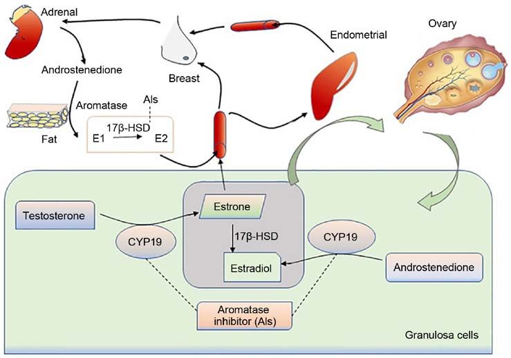
2                    LIU et al: EXPLORING THE REGULATION OF AROMATASE IN OVARIAN GRANULOSA CELLS

the maturation of follicles and the production of estrogen. In       ovarian specific I.3 and II can be activated by the majority
healthy females prior to menopause, human estrogen is mainly         of phytoestrogens, thereby enhancing the expression level of
derived from ovarian GCs and the placenta, and the expres‑           CYP19A1 mRNA (17). The combination of Jun protein with
sion of P450 aromatase is significantly higher in the GCs than       the CRE near the aromatase promoter PII results in changes
in other tissues (8). Hence, GCs are considered as powerful          in the CRE sites and inhibits the cAMP transcription acti‑
models for studying aromatase and their mechanisms of action.        vation, thereby inhibiting aromatase gene transcription in
    Estrogen plays an important role in the female health and        GCs (18). In addition, in pig GCs, Smad4 combines with
fertility status. It is mainly derived from 3 sources: Estrone is    the Smad4‑binding site (SBE) in the aromatase promoter
mainly converted from androstenedione of the adrenal gland           region as a transcription factor to enhance the expression
through the skin and adipose tissue (9,10); estradiol (E2), the      of CYP19A1 (19). Notably, in the absence of the nuclear
most widely effective estrogen, is mainly produced by GCs            receptor element site (NREA) in rabbit GCs, efficient aroma‑
in the ovaries, and is the main estrogen product synthesized         tase gene transcription can be performed (20). Previous
before menopause; estriol (E3), the weakest estrogen, is             research has also reported the phenomenon of a novel P450
mainly synthesized in the placenta. Since the ovaries are the        aromatase promoter in the luteinization of horse GCs treated
main organs which secrete estrogen, the normal expression of         with human chorionic gonadotropin (HCG), which mainly
aromatase is of utmost importance. Presently, the association        resulted in the transcriptional upregulation of If promoter
between aromatase and the ovarian GCs, as well as the regula‑        and the downregulation of II promoter (21).
tory mechanisms of this enzyme in GCs remain undefined.
The present review thus focused on aromatase expression              3. Aromatase expression localization
and the molecular regulatory mechanisms in ovarian GCs in
order to help interpret estrogen disorders. Potential aromatase      As mentioned above, the transcription of the aromatase gene
inhibitors (AIs) are also discussed an effort to open new            is regulated by tissue‑specific promoters. This point raises
research avenues for hormone‑dependent diseases and fertility        the question of how aromatase is expressed and localized
treatment influenced by estrogen‑secretion disorders (Table I).      in GCs. The ability of GCs to produce steroids is mainly
                                                                     controlled by FSH. FSH binds to its receptor, and this receptor
2. Promoter regulation                                               interacts with adenylate cyclase (AC) to regulate aromatase
                                                                     through the protein kinase A (PKA) pathway (22). In the
The expression of aromatase in different tissues is controlled by    presence of steroids, the expression of P450arom mRNA in
distinct promoters, and there are >10 promoters that function        GCs is increased rapidly under the effect of FSH/cAMP (23).
for selective purposes and tissue specificity. These promoters       The literature confirms that the addition of the C19 steroid
control the expression of aromatase by recruiting different          substrates (androstenedione and testosterone) to the culture
transcription factors and combining with the cis‑regulatory          medium of GCs can accelerate the synthesis of estradiol in a
elements (Fig. 1) (11). The transcription of its genes is mainly     concentration‑ and time‑dependent manner (24).
controlled by the distal promoter I.1 of the placenta (at 40‑kb          Velthut‑Meikas et al for instance, reported 2 origins of
upstream of the translation start site) and the proximal             introns from the sequencing of mural GCs (MGCs) and
promoter II of the ovary. The aromatase promoter I.4 plays an        cumulus GCs (CGCs) miRNA; one from the FSH receptor
important role in regulating aromatase expression in the skin;       gene and the other from CYP19A1, which implies the
aromatase expression in the adipose tissue is controlled by the      genome‑wide expression of miRNAs in GCs (25). The immu‑
ovarian promoter II, as well as by the promoters between I.1         nohistochemical localization of aromatase on the ovaries
and II (i.e., I.4 and I. 3) (8). The ovaries (promoter II), brain,   of fillies aged 6‑18 months detected aromatase in the mural
tissue‑specific promoter (If) and bone (promoter I.4 and I.6)        granulosa of follicles. Moreover, the immunoreactivity of
are mainly involved in the transcription of the aromatase            P450arom changes with the size of follicles and disappears
gene (12,13).                                                        in the atretic follicles (26). Observation under an electron
    The abnormal regulation of promoters can result in the           and light microscope to locate the aromatase of human ovary
development of various diseases. For example, promoter               and placenta detected that aromatase immune activity occurs
transition from I.4 to I.3/II causes the excessive production        mainly in the ovarian granulosa and lutein cells, as well as in
of estrogen in the breast, rendering breast epithelial cells         a portion of the cytotrophoblast and syncytiotrophoblast of
cancerous, thereby causing breast cancer (11). Similarly,            the placenta (27). In addition, aromatase, similar to the locally
immoderate estrogen secretion due to the mutation in the             produced estrogen and androgen, plays an important role in
promoter I.3/II results in endometrial cancer and ovarian            follicular maturation, and the highest expression of aromatase
cancer, and a cAMP‑responsive element (CRE) binding                  in the GCs occurs in the pre‑estrus cycle period (28).
protein (CREB) between promoter I.3/II regulates aromatase
through the cAMP/PKA‑dependent pathway, which results in             4. CYP19A1 is dependent on FSH/cAMP
the disordered expression of aromatase (14,15).
    The transcription of the CYP19A1 promoter in GCs                 FSH can regulate the maturation and proliferation of ovarian
is affected by various factors. Sharma et al reported that,          GCs and binds to the specific receptor FSHR of Sertoli cells
in the buffalo, PII plays a major role in the regulation of          and GCs (29). Previous studies have reported that FSH can
CYP19A1 of ovarian GCs, while PI.1 plays a minor role,               activate cAMP signaling in vitro or in vivo to enhance the
although PI.1 is transcriptionally upregulated in the lutein‑        expression of aromatase in ovarian GCs (30‑32). FSH is
ization of GCs (16). Previous research has confirmed that            the main factor that stimulates the expression of aromatase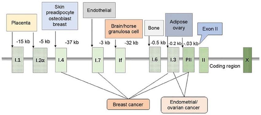
INTERNATIONAL JOURNAL OF MOlecular medicine 47: 73, 2021                                                                3

Table I. Classification of aromatase inhibitors.

Generation                    Type I (steroidal)                    Type II (non‑steroidal)                                     Shortcomings

First  None                                                          Aminoglutethimide                            Low potency, lack of specificity
Second Formestane                                                    Fadrozol, Collettiside                       Obvious side‑effects, drug resistance
Third  Exemestane                                                    Letrozole (Femara)                           Less side‑effects, (Aromasin)
		                                                                   Anastrazole (Armida)                         Easy to induce osteoarthropathy
		                                                                   Vorozole (Rivizor)

Figure 1. Regulation of aromatase promoters. The expression of aromatase is regulated by tissue‑specific promoters, and the figure illustrates the distance
between each specific promoter and the first exon II. Breast adipose stromal cells mainly express aromatase through the promoter I.4, while breast cancer
tissue use 4 promoters (I.4, I.7, I.3 and II) to regulate aromatase, significantly increasing the expression level of aromatase mRNA and promoting local estrogen
production. Endometrial cancer and ovarian cancer are mainly caused by PI.3 and PII, which also lead to a disruption in the expression of aromatase.

CYP19A1. In GCs, it can upregulate the expression of                              the expression of FSH receptor, while BMP‑2 can increase
CYP19A1 by activating the transcription factor GATA4.                             the expression of aromatase in GC cells.
Simultaneously, the activation of GATA4 requires the                                  Cyclic adenosine monophosphate (cAMP) can tran‑
activation of other kinases, such as ERK1/2, PKA and                              scriptionally regulate aromatase. The inhibitory effect of
PI3K (33). Aromatase activity of immature rat GCs increases                       progesterone and R5020 on GCs aromatase in rat ovaries
under the action of FSH and LH (32). E2 can enhance this                          has been found to be dose‑dependent; moreover, HCG
effect of FSH, which supports the ‘2‑cell, 2‑gonadotropin                         combined with FSH can significantly increase the amount of
hypothesis’ of ovarian synthesis of estrogen. In addition,                        cAMP and aromatase activity is enhanced at the appropriate
FSH not only enhances the activity of aromatase in a                              concentrations (41). After adenylate cyclase is activated by
time‑dependent manner, but also enhances the aromatase                            forskolin, it rapidly, but transiently induces LH‑responsive
complex component‑flavin protein NADPH‑cytochrome                                 genes [such as the nuclear receptor 4A subfamily; e.g.,
P‑450 reductase (34). Previous research has reported that                         nerve growth factor IB (NGFI‑B), nuclear receptor‑related 1
Anti‑Müllerian hormone (AMH) (35) and virus analog (36)                           (NURR1) and neuron‑derived orphan receptor 1 (NOR‑1)].
inhibit the increase in aromatase expression and estrogen                         However, the ectopic expression of NURR1 or NGFI‑B can
production induced by FSH. The presence of diethylstil‑                           attenuate the cAMP‑responsive activation of the aroma‑
bestrol and androstenedione can enable FSH to increase                            tase promoter in KGN cells. Therefore, the expression of
the expression of aromatase in a dose‑dependent manner,                           aromatase mRNA may be closely related to the induc‑
leading to increased estrogen secretion. According to the                         tion of nuclear receptor subfamily 4A (42). In addition,
stimulating effect of FSH on the aromatase activity of rat                        luteolin‑7‑methylether (XLY29) can inhibit the phosphory‑
GCs, it can be used as a biological assay for GC aroma‑                           lation of the cAMP response element binding protein by
tase (37). Moreover, the bone morphogenetic protein (BMP)                         regulating the expression of aromatase promoter 1.3/II,
family plays an important role in human ovarian develop‑                          thereby reducing mouse serum 17β ‑estradiol levels (43).
ment; for example, BMP‑2 (38), BMP‑6 (39) and BMP‑7 (40)                          The expression of CYP19A1 is thus closely related to the
can reduce the quantity of LH receptor in hGCs and increase                       regulation of FSH/cAMP.
4                    LIU et al: EXPLORING THE REGULATION OF AROMATASE IN OVARIAN GRANULOSA CELLS

5. Regulation of aromatase                                           Endocrine‑disrupting substances. An increasing number
                                                                     of studies have indicated that GCs are easily affected by
MicroRNAs (miRNAs or miRs). In recent years, miRNAs                  environmental pollutants in their ability to secrete estrogen.
have become increasingly popular and discovered as drug              Bisphenol A (BPA), which is industrially used as a plasticizer,
targets. KGN cells treated with FSH siRNA can induce the             has been reported to decrease the aromatase expression
differential expression of miR‑329‑3p, miR‑1261, miR‑144‑5p,         in GC cell‑line KGN stimulated by FSH to reduce the
miR‑130a‑3p, miR‑185‑5p and miR‑4463, among which                    estradiol secretion, which may function by inducing peroxi‑
miR‑4463 has been found to target the expression of ESR1             some proliferator‑activated receptor‑ γ (PPARγ) (62,63).
and CYP19A1 to influence estradiol secretion (44). In addition,      In rats, 2,3,7,8‑tetrachlorodibenzo‑p‑dioxin (TCDD) may
miR‑378 (45), miR‑10b (19)and miR‑1275 (46) combine with             reduce the aromatase mRNA expression in GCs through the
the 3' untranslated region (3'‑UTR) to downregulate the effec‑       AHR/ARNT signaling pathway, causing a decrease in the
tive transcription of CYP19A1 in ovarian GCs. miR‑764‑3p can         estradiol secretion (64,65). TCDD may also directly or indi‑
inhibit the expression of CYP19A1 by targeting steroidogenic         rectly block the endocrine function of human luteinized GCs
factor‑1 (SF‑1) to reduce the production of 17β‑estradiol (47).      (hLGCs) through the interaction of PTK/MAP2K and PKA
Through in vitro and in vivo verifications, miR‑326 has been         signaling (66,67). Plasticizer mono‑(2‑ethylhexyl) phthalate
found to activate C/EBP‑β by upregulating CREB, thereby              and its metabolite mono‑(2‑ethylhexyl) phthalate (MEHP)
inhibiting the expression of CYP19A1 in buffalo GCs and              also reduce the transcription of aromatase in human and rat
causing a decrease in the 17β‑estradiol levels (48). Moreover,       GCs, and thereby reduce the secretion of estradiol (68‑70).
miR‑214‑3p has been found to upregulate the expression of cell       Recently, it was reported that carbon black nanoparticles (CB
cycle genes in porcine GCs, but to downregulate the mRNA             NPs) can inhibit the transcription level of aromatase in GCs by
expression levels of CYP19A1 and steroidogenic acute regula‑         activating the ERK1/2 pathway and influencing E2 secretion.
tory protein (StAR) (49). The overexpression of miR‑29a in           In addition, PD98059 signals can inhibit the ERK1/2 signaling
cov434 and KGN cells can cause the expression of aromatase           pathway to reduce the adverse effects of CB NPs (71). In addi‑
to decrease, affecting the estradiol secretion and inhibiting the    tion, prenatal nicotine exposure (PNE) can reduce histone 3
cell proliferation (50). In a previous study, a GC model treated     lysine 27 acetylation (H3K27ac) and H3K9ac of P450arom in
with DOX led to the increased expression of miR‑132‑3p in a          the ovarian GC cell line, KGN, through nicotine acetylcholine
dose‑dependent manner, as well as in a change in the expres‑         receptor (nAChR), which consequently results in the reduc‑
sion of CYP19A1 and the secretion of 17β‑estradiol (51).             tion of the aromatase expression and estradiol production in
                                                                     the ovaries (72). In GCs, lead may affect female fertility by
Growth factors. Aromatase activity in GCs is regulated by            downregulating the transcription of estrogen receptor β (ERβ)
various growth factors. Cell location studies have indicated         and p450arom (73).
that the insulin‑like growth factor‑I (IGF1) gene and its                Morinaga et al also evaluated aromatase activity in KGN
receptor gene are highly expressed and translated in ovarian         cells and screened 55 endocrine‑disrupting chemicals (74).
GCs (52,53). IGF1 can increase the production of P‑450arom in        For the first time, they proved that benomyl and its metabolite
a concentration‑dependent manner and can increase the activity       carbendazim can enhance aromatase activity through the regu‑
of aromatase. In addition, IGF1 can cooperate with FSH and is        lation of the transcription level, possibly through a microtubule
more obvious than any hormone alone (54). However, epidermal         interference mechanism rather than through the cAMP‑PKA
growth factor (EGF) can inhibit the synthesis of P450 and            pathway (74). In addition, 2,2‑bis‑(p‑hydroxyphenyl)‑1,1,1‑tri‑
antagonize FSH. These changes are likely to be related to the        chloroethane (HPTE) has been reported to inhibit the increase
mRNA encoding level of aromatase (55). In addition, amphi‑           in the expression of P450arom mRNA, P450scc and 3β‑HSD
regulin (AREG) in the EGF‑like growth factor induced by HCG          in FSH‑cultured GCs, albeit it had no effect on StAR. HPTE
can activate the AKT signaling pathway and upregulate the            also affected the cAMP site of action and may therefore involve
expression of aromatase and estrogen in human granulosa lutein       a broad steroid regulation of the cAMP‑PKA pathway (75).
(hGL) cells (56). Moreover, fibroblast growth factor (FGF) and       Endocrine‑disrupting substances are thus likely to affect
platelet‑derived growth factor (PDGF) can reduce the effect of       human reproduction and sexual differentiation by modifying
cAMP and may act based on the corresponding tyrosine kinase          the steroid secretion properties of GCs.
activity to regulate the synthesis of cytochrome P450 in GCs (57).
Some growth factors, such as vascular endothelial growth             Insulin sensitizers. Metformin is an insulin sensitizer with
factor A (VEGF‑A) and FGF2 can synergistically promote the           direct function in the ovaries, which may downregulate the
expression of CYP19A1 in buffalo GCs and enhance the extent          expression of aromatase promoter I.4, I.3 and II through the
of estradiol secretion (58). Hepatocyte growth factor (HGF) can      MEK/ERK pathway, thereby reducing the aromatase activity
also inhibit the expression of FSH‑induced cAMP‑dependent            in GCs (76). In addition, the expression of aromatase mRNA
P450arom and 17β ‑hydroxysteroid dehydrogenase (HSD),                decreases in human luteinized GCs cultured with metformin,
thereby inhibiting GC steroid production in ovarian GCs (59).        although it can significantly increase the effect of insulin on
Transforming growth factor‑β (TGF‑β) has also been reported          IGF1R mRNA and IR (77). Pioglitazone or rosiglitazone can
to enhance the expression of FSH‑induced P450arom mRNA               inhibit estrogen production in human ovarian GCs by inter‑
in rat GCs (60), as well as accelerate the production of E1 and      fering with the combination of aromatase and androgens, and
E2 by 1.45‑ and 2.7‑fold. However, leptin has been reported to       the effect of thiazolidinediones (TZDs) in reducing the activity
antagonize this effect of TGF‑β, which reduces the expression        of aromatase may explain the application of TZD in the treat‑
of GC aromatase (61).                                                ment of estrogen‑dependent diseases and polycystic ovary
INTERNATIONAL JOURNAL OF MOlecular medicine 47: 73, 2021 5
syndrome (PCOS) (78). Troglitazone is also a compound of P450 aromatase (CYP19) and c‑fos also decreased (87). Hence,
TZDs that is likely to exert a direct inhibitory effect on aroma‑ c‑fos is likely to be downstream of the MEK/ERK pathway,
tase in GCs through the nuclear receptor system PPARγ/RXR and CYP19 is upregulated and CYP17 is downregulated in
heterodimer (79). Insulin sensitizers thus have a probable GCs, which together inhibit the production of estradiol (87).
potential as effective inhibitors of aromatase that warrant
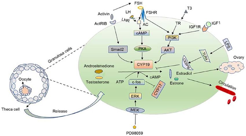
further investigation (Table II). Others. Activin can enhance the aromatase activity of FSH
in the rat GCs (88). Nomura et al confirmed the mechanisms
6. Regulatory pathways of aromatase in GCs action in KGN cells (89). Moreover, Smad2 is downstream
of activin‑type IB receptor (ActRIB); it participates in the
cAMP/PKA pathway. In GCs, cAMP is the second messenger activin‑signaling pathway to regulate the expression of P450
of FSH, and cAMP inducers and cAMP analogs can induce aromatase, thereby regulating the follicular development (89).
multiple functions of GCs in vitro (80). LH receptor expres‑ In addition, LPS‑induced Toll‑like receptor 4 (TLR4) signaling
sion is regulated by the combined action of estradiol and FSH; can downregulate the expression of CYP19A1 and 17β‑estradiol
therefore, in the cultured rat GCs, the induction of FSH can in bovine GCs under the action of CCAAT/enhancer binding
make the cells respond to LH, thereby enhancing the overall protein beta (CEBPB) (90). Notably, Notch signaling affects
aromatase activity (81). LH and FSH exert their biological the secretion of estrogen by affecting the expression of the
functions through the mediation of G protein‑coupled recep‑ upstream transcription factors, Wt1, SF‑1, GATA4 and GATA6,
tors LHCGR and FSHR, thereby causing the activation of and that of downstream‑related enzymes related to steroid
several signaling cascades, such as the cyclic AMP/protein production (91). The research on the mechanism of aromatase
kinase A (cAMP/PKA) pathway (82). On binding with the regulation in GCs is thus ongoing and not yet concrete (Fig. 2).
corresponding receptors in GCs, LH and FSH activate the Gs
protein on the cell membrane, resulting in the production of 7. Changes in aromatase activity of GCs induce diseases
cAMP. Therefore, they activate the cAMP‑dependent PKA
signaling pathway and act on the expression of CYP19A1, Aromatase is the only enzyme in vertebrates that catalyzes the
which is a classic PKA‑dependent aromatase regulation production of estrogen. The disruption of estrogen secretion
pathway (13). can thus easily cause cancer of the breast, endometrium or
ovaries, which are together referred to as ‘estrogen‑dependent
PI3K. The rapid activation of the PKA signaling pathway tumors’ (92). These tumors can also cause female reproduc‑
also induces other cascading reactions. For example, FSH tive issues. GCs are the main source of ovarian estrogen
can promote the activation of PI3K signaling in the rat GCs secretion, which not only induces ovarian development but
and cause the rapid phosphorylation of its downstream branch also releases estrogen into the blood and supply it to various
point, AKT; as a result, the expression of ovarian genes such as organs. Therefore, whether the secretion of GCs aromatase
CYP19A1 increases (80). Moreover, it can enhance the activity is normal directly affects the health status of various organs.
of HIF‑1 downstream of the PI3K/AKT/Raheb/mammalian Nowadays, the inhibition of aromatase‑induced estrogen
target of rapamycin (mTOR) pathway in GCs (83). IGF1 has biosynthesis is considered to be the main strategy for the
been confirmed to be an important molecule for hormone therapy of estrogen‑dependent diseases. Considering that the
synthesis in GCs (76); it can activate PI3K/Akt, which upregu‑ aromatase status of patients with recurrent granulosa cell
lates FSH receptor. When Akt and PKA are activated, they tumors remains unaltered, the detection of the expression of
regulate the expression of CYP19A1 (84). It has previously been aromatase can provide more accurate information for serum
confirmed that the molecular mechanism of 3,5,3'‑tri‑iodothy‑ E2 in the primary tumor as a relapse marker (93).
ronine (T3) regulates the expression of CYP19A1 in GC and Ovarian GC tumors are one of the most common ovarian
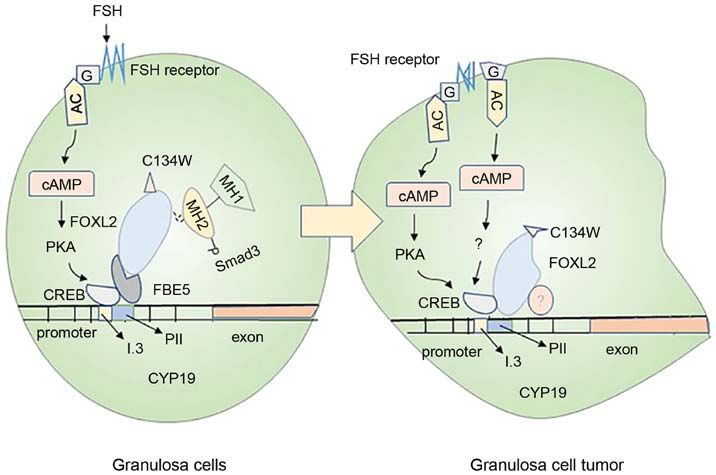
promotes follicle development. The combination of FSH and tumors, mainly causing abnormal estrogen secretion (94)
T3 requires the activation of the PI3K/Akt pathway to regulate (Fig. 3). In normal GCs, FSH binds to its receptor to activate
CYP19A1. Therefore, CYP19A1 is likely to be a downstream Gs protein and upregulate the aromatase expression through
effector of the PI3K/Akt pathway that is activated by FSH and the PKA pathway. However, mutations in the Gs protein
T3, which acts as a new mediator for FSH and T3 to induce on the cell membrane are likely to induce the production
the development of GCs and follicles, while regulating FSH of cancerous cells (95). The forkhead family FOXL2 is an
and TH (85). evolutionarily conserved member of the transcription factor;
its mutation is closely related to the generation of ovarian GC
ERK. The surge of LH prior to ovulation can rapidly inhibit tumor (96). Smad3 can cooperate with FOXL2:C134W in
the proliferation of GCs and differentiate them into luteal combination with the CYP19A1 promoter FBE5 of the human
cells. However, the proper dose of LH can also cause the rapid GC line (HGrC1) to enhance the expression of aromatase,
activation of ERK1/2 when acting on GCs, while FSH also which is probably the key reason for the large amount of
have the same effect (86). ERK, similar to PKA, is a down‑ estrogen secretion by the human GC line (97). The mutation
stream signaling molecule involved in the action of LH and of FOXL2:C134W is the main reason for the formation of
FSH. This point raises a question of whether aromatase is adult ovarian granulosa cell tumor (GCT); it can enhance the
regulated as a downstream molecule of this signaling pathway. induction of aromatase by FOXL2, and aromatase is the direct
Huang et al used PD98059 to block the hormone‑stimulated target of FOXL2:C134W (98). This information deepens our
MEK1 in KGN cells and found that 17‑hydroxylase/17,20 understanding of the research on the pathogenesis of the adult
lyase (CYP17) expression increased, and that the expression of GC tumor.6 LIU et al: EXPLORING THE REGULATION OF AROMATASE IN OVARIAN GRANULOSA CELLS
Table II. Potential aromatase inhibitors.
Classification Molecules Mechanism of action Species (Refs.)
MicroRNAs miR‑4463 Target the expression of ESR1 and CYP19A1 Human (42)
miR‑378 Downregulate the transcription of CYP19A1 Pig (45)
miR‑10b Combines with 3'‑UTR Pig (45)
miR‑1275 Combines with 3'‑UTR Pig (45)
miR‑764‑3p By targeting SF‑1 Mouse (47)
miR‑326 Activate C/EBP‑β by upregulating CREB Buffalo (48)
miR‑214‑3p Downregulated CYP19A1 and StAR Pig (48)
miR‑29a Decrease aromatase expression Human (50)
Hormones AMH Inhibit the effect of FSH on estrogen production Pig (42)
R5020 Dose‑dependent inhibition Rat (41)
Progesterone Dose‑dependent inhibition Rat (41)
Leptin Antagonism TGFβ Rat (61)
Growth factor EGF Suppress the effects of FSH and cAMP Human (116)
FGF Reduce the role of cAMP Human (57)
PDGF Reduce the role of cAMP Human (57)
HGF Destruction of FSH‑induced cAMP‑mediated Rat (59)
Insulin sensitizers Metformin Downregulate the aromatase promoter Human (76)
Rosiglitazone Reduce the effect of aromatase Human (78)
Pioglitazone Reduce the effect of aromatase Human (78)
Troglitazone Through nuclear receptor system Human (79)
ESR1, estrogen receptor 1; SF‑1, steroidogenic factor‑1; StAR, steroidogenic acute regulatory protein; AMH, anti‑Müllerian hormone;
EGF, epidermal growth factor; FGF, fibroblast growth factor; PDGF, platelet‑derived growth factor; HGF, Hepatocyte growth factor.
Figure 2. Regulatory mechanism of aromatase and ‘2‑cell, 2‑gonadotropin hypothesis’. The left panel illustrates that follicles are mainly composed of oocytes,
granulosa cells and theca cells. Androstenedione and androsterone secreted by theca cells penetrate into granulosa cells and are converted into estrone and
estradiol by the aromatase in granulosa cells under the stimulation of FSH and LH hormones. A small part acts on the ovaries, and the majority is released
into the blood, collectively referred to as the ‘2‑cell, 2‑gonadotropin hypothesis’. The right panel mainly introduces the regulatory mechanism of aromatase
in granulosa cells. FSH and LH bind to receptors and regulate CYP19A1 through PKA pathway. At the same time, IGF‑1 binds to its receptor to activate
PI3K/Akt, which upregulates the expression of CYP19; T3 cooperates with FSH also to acts on aromatase via PI3K/AKT pathway. The inhibition of the
hormone‑stimulated ERK with PD98059 can act on c‑fos, causing the upregulation of CYP19 and the downregulation of CYP17. LH, luteinizing hormone;
FSH, follicle‑stimulating hormone; ERK, extracellular signal‑regulated kinase; PI3K, phosphoinositide 3‑kinase; PKA, protein kinase A; cAMP, cyclic
adenosine monophosphate; CEBPB, CCAAT/enhancer‑binding protein beta.INTERNATIONAL JOURNAL OF MOlecular medicine 47: 73, 2021 7
Figure 3. Mechanisms of ovarian granulosa cell carcinogenesis. The normal granulosa cells shown in the left panel and the cancerous granulosa cells shown in
the right panel mainly introduce the carcinogenesis mechanism of the cells. In normal granulosa cells, FSH binds to the receptor and acts through the trimer
G protein (G) and adenylate cyclase, then, combine to the CRE binding site of CYP19A1 through the protein kinase A pathway, causing increased expression
of aromatase. In the granulosa cells on the right, the FSH may not depend on the activation of this pathway, and the cancerization mainly originates from the
mutation of the trimer G protein (G) and the activation of the FSH receptor. In addition, Smad3 in normal granulosa cells cooperates with FOXL2:C134W and
FBE5 to act on CYP19A1 gene. The mutant FOXL2:C134W in granulosa cell tumors binds to another specific site of CYP19 and recruits unknown proteins,
causing cell mutations. These are likely to cause abnormal expression of aromatase in granulosa cells and cancerization of cells. FSH, follicle‑stimulating
hormone; cAMP, cyclic adenosine monophosphate; PKA, protein kinase A; CREB, cAMP‑response element binding protein.
The abnormal secretion of estradiol from the GCs that the highly expressed lncRNA HUPCOS of granulosa cells
can result in abnormalities in the target organs, such as in patients with PCOS can interact with RNA‑binding protein
the breast and uterus (Fig. 4). Based on the literature, the with multiple splicing (RBPMS) and inhibit the expression of
expression levels of aromatase in the diseased tissues of the aromatase, thereby mediating the excess of androgen in the
endometrium and breast are significantly higher than those in follicular fluid of PCOS patients Therefore, the decrease in the
normal tissues (99), with adult ovarian cancer patients being aromatase activity conversely helps maintain high androgen
more prone to endometrial and breast cancer (100). In addi‑ levels (102,107). In addition, CYP19A1 and A‑Kinase anchor
tion, the increased expression of aromatase can also induce protein 95 (AKAP95) levels are significantly decreased in
endometriosis and uterine fibroids in females. In granulosa human luteinized GCs of patients with PCOS. AKAP95 is
cells, SF‑1 can bind to the cAMP response element and act thus likely to act on FSH‑stimulated CYP19A1 (108). All of
on aromatase promoter II, thereby causing estrogen produc‑ these participate in the pathological mechanism of PCOS.
tion (101). Abnormally expressed SF‑1 in endometriosis will Moreover, the abnormal expression of aromatase mRNA in
compete with the aromatase expression inhibitor‑chicken oval‑ GCs can also induce obesity in women, which is an important
bumin upstream promoter‑transcription factor (COUP‑TF) factor affecting female fertility. It has been reported that leptin
for the same binding site, thereby stimulating the activity of can induce neuropeptide cocaine‑ and amphetamine‑regulated
aromatase P450 (102). Moreover, glucocorticoids and IL1βcan transcript (CART) in GCs, thereby indirectly inhibiting the
affect the secretion of estrogen by regulating the expression of cAMP levels and the aromatase expression in GCs, causes
aromatase promoter 1.4, thereby affecting the occurrence of ovarian dysfunction and reduced fertility (109).
uterine fibroids (103).
The estrogen secretion disorder, which induces increase 8. Current and future perspectives
in the incidence of PCOS, is the main cause of female infer‑
tility (104). PCOS is mainly manifested as a disease of the Environmental pollution and modification in diets are consid‑
reproductive dysfunction and endocrine disorders, which ered to be important elements affecting female fertility and
is mainly characterized by the high levels of androgens and health issues. Several diseases have been confirmed in rela‑
anovulation. The hyperandrogenic state of the ovaries of tion to estrogen‑secretion disorders in the epidemiology and
patients with PCOS is the main risk factor for the decline of experimental studies. In some patients with estrogen‑depen‑
aromatase production in luteinized GCs (105). It has been dent diseases, high levels of estrogen are accompanied by
reported that androgens can enhance the FSH effect in granu‑ the overexpression of aromatase (15). For example, in breast
losa cells by increasing FSHR. When androgen secretion is cancer treatment, multiple AIs have been developed, such as
excessive, the high sensitivity of GCs to FSH causes a large exemestane, anastrozole, letrozole and vorozole. The current
increase in AMH, and AMH in turn inhibits the effect of FSH first‑line treatment for breast cancer mainly uses third‑gener‑
on aromatase in follicles (106). Recently, Che et al (107) found ation AIs (110,111). In addition, AIs have begun to be used in8 LIU et al: EXPLORING THE REGULATION OF AROMATASE IN OVARIAN GRANULOSA CELLS
Figure 4. Source of estrogen production and the site of aromatase inhibitor action. In granulosa cells, under the action of aromatase, androstenedione and
androsterone can be converted into estradiol and estrone. A small part of the secreted estrogen acts on the development of the ovaries; the majority is released
into the blood circulation and enter the uterus, breast, kidney and other target tissues. In addition to the estrogen source of granulosa cells, androstenedione
secreted by the adrenal glands can be converted to E1 in tissues such as fat, then it is converted into E2 under the action of 17‑HSD, and finally acts on each
target tissue from the blood circulation. The uncontrolled regulation of aromatase will cause estrogen secretion disorder, and even cause endocrine disorders of
ovarian, uterine, breast and other target tissues, and even cancer. However, aromatase inhibitors can downregulate the highly expressed CYP19A1 and interrupt
the conversion of androgens and androstenedione to estrogen, thus exerting a therapeutic effect on some estrogen‑dependent diseases.
the treatment of estrogen‑regulated diseases, such as ovarian placenta (at 40 kb upstream of the translation start site) and
cancer and endometrial cancer, as well as inducing ovula‑ the proximal promoter II of the ovary (8). FSH can regulate
tion. Presently, AIs combined with progesterone and GnRH the expression of aromatase by activating the PKA signaling
agonists are mainly used to treat women with endometriosis, pathway, while simultaneously activating the PI3K (85),
along with reducing the risk of ovarian cysts induced by AIs ERK (87) and other signaling pathways, thereby upregulating
alone. However, symptoms, such as pelvic pain may recur the expression of CYP19A1. The disadvantage of this approach
following AI treatment. For women with polycystic ovary is the lack of substantial research supporting whether the tran‑
syndrome and obesity, letrozole has a higher live birth rate sient activation of P38MAPK caused by FSH can also regulate
compared with clomiphene citrate, and it has been regarded as the expression of aromatase. In addition, owing to biodiversity,
the first‑line therapy of inducing ovulation. Moreover, AIs are individual differences exist in the regulation of aromatase due
also a good first choice for women with infertility due to the to different factors, which may present new areas of interest in
presence of uterine fibroids, the wish to preserve the uterus, or future research. Understanding the molecular modifications
being unsuitable for surgery (112). and the mechanisms of action in GCs that can be targeted
Although AIs are effective in the treatment of in the disorders of aromatase secretion for the treatment of
estrogen‑dependent diseases and ovulation induction, osteo‑ diseases caused by ovarian hormones is expected to develop
arthropathy, menopausal symptoms, intestinal discomfort and high‑efficiency, low‑toxic, and side effects of specific drug
drug resistance, which are easily induced following treatment targets.
with AIs, remain concerns which require resolutions. A recent
study designed and synthesized a novel aromatase inhibitor 9. Conclusions
based on triazole and imidazole (113). However, the current
inhibitors are mainly used in the treatment of breast cancer, The present article summarized and discussed the regulatory
albeit for other steroid‑dependent diseases and female infer‑ characteristics of the CYP19A1 promoter in ovarian GCs, as
tility, and there are a relatively few effective and specific drugs well as the regulation of aromatase as a downstream effector
available (114,115), which indicates that the search for novel through multiple signaling pathways. The expression of
AIs and other effective drug targets is crucial. aromatase in GCs is affected by various factors, and it is one
GCs, which are the main site of ovarian estrogen produc‑ of the main causes of estrogen‑dependent diseases and PCOS.
tion, also generate a large amount of aromatase. Aromatase Endocrine‑disrupting substances in the environment can cause
can promote the biosynthesis of estrogen in GCs, and the alteration in the expression of aromatase and affect the normal
production of estrogen can promote the follicular development reproduction and sexual differentiation in the human body.
of GCs and inhibit the apoptosis of GCs. The transcription of Furthermore, some insulin sensitizers have been developed as
genes is mainly controlled by the distal promoter I.1 of the AIs for the clinical treatment. Other substances, such as FSH,INTERNATIONAL JOURNAL OF MOlecular medicine 47: 73, 2021 9
IGF‑1 and TGFβ have been proven to promote the expression 7. Shoham Z, Jacobs HS and Insler V: Luteinizing hormone: Its
role, mechanism of action, and detrimental effects when hyper‑
of aromatase mRNA and protein, although miRNAs, HGF and secreted during the follicular phase. Fertil Steril 59: 1153‑1161,
leptin can partially inhibit or specifically identify the aroma‑ 1993.
tase promoter and downregulate the aromatase transcription, 8. Nelson LR and Bulun SE: Estrogen production and action. J Am
Acad Dermatol 45 (Suppl 3): S116‑S124, 2001.
leading to the disturbance in estrogen secretion. Thus, the 9. Slominski A, Zbytek B, Nikolakis G, Manna PR, Skobowiat C,
discovery of potential aromatase inhibitor targets is expected Zmijewski M, Li W, Janjetovic Z, Postlethwaite A, Zouboulis CC
to provide new directions for the treatment of estrogen‑depen‑ and Tuckey RC: Steroidogenesis in the skin: Implications for
local immune functions. J Steroid Biochem Mol Biol 137:
dent diseases and PCOS. 107‑123, 2013.
10. Bulun SE, Chen D, Moy I, Brooks DC and Zhao H: Aromatase,
Acknowledgements breast cancer and obesity: A complex interaction. Trends
Endocrinol Metab 23: 83‑89, 2012.
11. Zhao H, Zhou L, Shangguan AJ and Bulun SE: Aromatase
Not applicable. expression and regulation in breast and endometrial cancer.
J Mol Endocrinol 57: R19‑R33, 2016.
12. Shozu M, Zhao Y and Simpson ER: TGF‑beta1 stimulates expres‑
Funding sion of the aromatase (CYP19) gene in human osteoblast‑like
cells and THP‑1 cells. Mol Cell Endocrinol 160: 123‑133, 2000.
The present study was supported by grants from the National 13. Stocco C: Aromatase expression in the ovary: Hormonal and
molecular regulation. Steroids 73: 473‑487, 2008.
Natural Science Foundation of China (no. 31900852 to HL), 14. Bulun SE, Chen D, Lu M, Zhao H, Cheng Y, Demura M,
Nanchang University (no. PY201801 to HL) and the Natural Yilmaz B, Martin R, Utsunomiya H, Thung S, et al: Aromatase
Science Foundation of Jiangxi Province (nos. 2018BAB215012 excess in cancers of breast, endometrium and ovary. J Steroid
Biochem Mol Biol 106: 81‑96, 2007.
and 20192ACB21026 to HL). 15. Bulun SE and Simpson ER: Aromatase expression in women's
cancers. Adv Exp Med Biol 630: 112‑132, 2008.
Availability of data and materials 16. Sharma D, Ghai S and Singh D: Different promoter usage for
CYP19 gene expression in buffalo ovary and placenta. Gen
Comp Endocrinol 162: 319‑328, 2009.
Not applicable. 17. Solak KA, Wijnolts FMJ, Nijmeijer SM, Blaauboer BJ, van den
Berg M and van Duursen MBM: Excessive levels of diverse
phytoestrogens can modulate steroidogenesis and cell migration
Authors' contributions of KGN human granulosa‑derived tumor cells. Toxicol Rep 1:
360‑372, 2014.
HL, TL and YH designed and wrote the manuscript. All 18. Ghosh S, Wu Y, Li R and Hu Y: Jun proteins modulate the
ovary‑specific promoter of aromatase gene in ovarian granulosa
authors have read and approved the final manuscript. cells via a cAMP‑responsive element. Oncogene 24: 2236‑2246,
2005.
Ethics approval and consent to participate 19. Li Q, Du X, Pan Z, Zhang L and Li Q: The transcription factor
SMAD4 and miR‑10b contribute to E2 release and cell apoptosis
in ovarian granulosa cells by targeting CYP19A1. Mol Cell
Not applicable. Endocrinol 476: 84‑95, 2018.
20. Andrieu T, Féral C, Joubert M, Benhaim A and Mittre H: The
absence of a functional nuclear receptor element A (NREA) in
Patient consent for publication the promoter II of the aromatase P450 gene in rabbit granulosa
cells. J Steroid Biochem Mol Biol 101: 127‑135, 2006.
Not applicable. 21. Boerboom D, Kerban A and Sirois J: Dual regulation of
promoter II‑ and promoter 1f‑derived cytochrome P450 aroma‑
tase transcripts in equine granulosa cells during human chorionic
Competing interests gonadotropin‑induced ovulation: A novel model for the study of
aromatase promoter switching. Endocrinology 140: 4133‑4141,
1999.
The authors declare that they have no competing interests. 22. Simpson ER: Sources of estrogen and their importance. J Steroid
Biochem Mol Biol 86: 225‑230, 2003.
References 23. Miyoshi T, Otsuka F and Shimasaki S: GRK‑6 mediates FSH
action synergistically enhanced by estrogen and the oocyte in rat
granulosa cells. Biochem Biophys Res Commun 434: 401‑406,
1. Mendelson CR, Jiang B, Shelton JM, Richardson JA and 2013.
Hinshelwood MM: Transcriptional regulation of aromatase in 24. Czajka‑Oraniec I and Simpson ER: Aromatase research and its
placenta and ovary. J Steroid Biochem Mol Biol 95: 25‑33, 2005. clinical significance. Endokrynol Pol 61: 126‑134, 2010.
2. Li J and Gibbs RB: Detection of estradiol in rat brain 25. Velthut‑Meikas A, Simm J, Tuuri T, Tapanainen JS, Metsis M
tissues: Contribution of local versus systemic production. and Salumets A: Research resource: Small RNA‑seq of human
Psychoneuroendocrinology 102: 84‑94, 2019. granulosa cells reveals miRNAs in FSHR and aromatase genes.
3. Lambard S, Silandre D, Delalande C, Denis‑Galeraud I, Mol Endocrinol 27: 1128‑1141, 2013.
Bourguiba S and Carreau S: Aromatase in testis: Expression and 26. Mlodawska W and Slomczynska M: Immunohistochemical
role in male reproduction. J Steroid Biochem Mol Biol 95: 63‑69, localization of aromatase during the development and atresia
2005. of ovarian follicles in prepubertal horses. Theriogenology 74:
4. Mahendroo MS, Mendelson CR and Simpson ER: Tissue‑specific 1707‑1712, 2010.
and hormonally controlled alternative promoters regulate aroma‑ 27. Naganuma H, Ohtani H, Harada N and Nagura H: Immunoelectron
tase cytochrome P450 gene expression in human adipose tissue. microscopic localization of aromatase in human placenta and
J Biol Chem 268: 19463‑19470, 1993. ovary using microwave fixation. J Histochem Cytochem 38:
5. Wang Y, Pan P, Li X, Zhu Q, Huang T and Ge RS: Food compo‑ 1427‑1432, 1990.
nents and environmental chemicals of inhibiting human placental 28. Shaikh AA: Estrone and estradiol levels in the ovarian venous
aromatase. Food Chem Toxicol 128: 46‑53, 2019. blood from rats during the estrous cycle and pregnancy. Biol
6. Ai A, Tang Z, Liu Y, Yu S, Li B, Huang H, Wang X, Cao Y and Reprod 5: 297‑307, 1971.
Zhang W: Characterization and identification of human immor‑ 29. Szymańska K, Kałafut J, Przybyszewska A, Paziewska B,
talized granulosa cells derived from ovarian follicular fluid. Exp Adamczuk G, Kiełbus M and Rivero‑Müller A: FSHR trans‑activa‑
Ther Med 18: 2167‑2177, 2019. tion and oligomerization Front Endocrinol (Lausanne) 9: 760, 2018.10 LIU et al: EXPLORING THE REGULATION OF AROMATASE IN OVARIAN GRANULOSA CELLS
30. Jiang C, Hou X, Wang C, May JV, Butnev VY, Bousfield GR and 50. Li Y, Liu YD, Zhou XY, Chen SL, Chen X, Zhe J, Zhang J,
Davis JS: Hypoglycosylated hFSH has greater bioactivity than Zhang QY and Chen YX: MiR‑29a regulates the proliferation,
fully glycosylated recombinant hFSH in human granulosa cells. aromatase expression, and estradiol biosynthesis of human
J Clin Endocrinol Metab 100: E852‑E860, 2015. granulosa cells in polycystic ovary syndrome. Mol Cell
31. Hobeika E, Armouti M, Kala H, Fierro MA, Winston NJ, Scoccia B, Endocrinol 498: 110540, 2019.
Zamah AM and Stocco C: Oocyte‑secreted factors synergize with 51. Al‑Kawlani B, Murrieta‑Coxca JM, Chaiwangyen W, Fröhlich K,
FSH to promote aromatase expression in primary human cumulus Fritzsche A, Winkler S, Markert UR and Morales‑Prieto DM:
cells. J Clin Endocrinol Metab 104: 1667‑1676, 2019. Doxorubicin induces cytotoxicity and miR‑132 expression in
32. Parakh TN, Hernandez JA, Grammer JC, Weck J, Hunzicker- granulosa cells. Reprod Toxicol 96: 95‑101, 2020.
Dunn M, Zeleznik AJ and Nilson JH: Follicle‑stimulating 52. Ogo Y, Taniuchi S, Ojima F, Hayashi S, Murakami I, Saito Y,
hormone/cAMP regulation of aromatase gene expression requires Takeuchi S, Kudo T and Takahashi S: IGF‑1 gene expression is
beta‑catenin. Proc Natl Acad Sci USA 103: 12435‑12440, 2006. differentially regulated by estrogen receptors α and β in mouse
33. Kwintkiewicz J, Cai Z and Stocco C: Follicle‑stimulating endometrial stromal cells and ovarian granulosa cells. J Reprod
hormone‑induced activation of Gata4 contributes in the Dev 60: 216‑223, 2014.
up‑regulation of Cyp19 expression in rat granulosa cells. Mol 53. Zhou J, Chin E and Bondy C: Cellular pattern of insulin‑like
Endocrinol 21: 933‑947, 2007. growth factor‑I (IGF‑I) and IGF‑I receptor gene expression in
34. Hong Y, Li H, Yuan YC and Chen S: Molecular characterization the developing and mature ovarian follicle. Endocrinology 129:
of aromatase. Ann N Y Acad Sci 1155: 112‑120, 2009. 3281‑3288, 1991.
35. Li Y, Gao D, Xu T, Adur MK, Zhang L, Luo L, Zhu T, Tong X, 54. Mani AM, Fenwick MA, Cheng Z, Sharma MK, Singh D and
Zhang D, Wang Y, et al: Anti‑Müllerian hormone inhibits lutein‑ Wathes DC: IGF1 induces up‑regulation of steroidogenic
izing hormone‑induced androstenedione synthesis in porcine and apoptotic regulatory genes via activation of phosphati‑
theca cells. Theriogenology 142: 421‑432, 2020. dylinositol‑dependent kinase/AKT in bovine granulosa cells.
36. Fang Y, Wang B, Lyu S, Zhang K, Cheng Q and Zhu Y: Virus Reproduction 139: 139‑151, 2010.
analog decreases estradiol secretion in FSH‑treated human 55. Herrmann M, Scholmerich J and Straub RH: Influence of
ovarian granulosa cells. Gynecol Endocrinol 36: 346‑350, 2020. cytokines and growth factors on distinct steroidogenic enzymes
37. Kajitani T, Liu S, Maruyama T, Uchida H, Sakurai R, Masuda H, in vitro: A short tabular data collection. Ann NY Acad Sci 966:
Nagashima T, Ono M, Arase T and Yoshimura Y: Analysis of 166‑186, 2002.
serum FSH bioactivity in a patient with an FSH‑secreting pitu‑ 56. Fang L, Yu Y, Li Y, Wang S, Zhang R, Guo Y, Li Y, Yan Y and
itary microadenoma and multicystic ovaries: A case report. Hum Sun YP: Human chorionic gonadotropin‑induced amphiregulin
Reprod 23: 435‑439, 2008. stimulates aromatase expression in human granulosa‑lutein cells:
38. Shi J, Yoshino O, Osuga Y, Koga K, Hirota Y, Nose E, Nishii O, A mechanism for estradiol production in the luteal phase. Hum
Yano T and Taketani Y: Bone morphogenetic protein‑2 (BMP‑2) Reprod 34: 2018‑2026, 2019.
increases gene expression of FSH receptor and aromatase and 57. Mendelson CR, Merrill JC, Steinkampf MP and Simpson ER:
decreases gene expression of LH receptor and StAR in human Regulation of the synthesis of aromatase cytochrome P‑450 in
granulosa cells. Am J Reprod Immunol 65: 421‑427, 2011. human adipose stromal and ovarian granulosa cells. Steroids 50:
39. Shi J, Yoshino O, Osuga Y, Koga K, Hirota Y, Hirata T, Yano T, 51‑59, 1987.
Nishii O and Taketani Y: Bone morphogenetic protein‑6 stimu‑ 58. Mishra SR, Bharati J, Rajesh G, Chauhan VS, Taru Sharma G,
lates gene expression of follicle‑stimulating hormone receptor, Bag S, Maurya VP, Singh G and Sarkar M: Fibroblast growth
inhibin/activin beta subunits, and anti‑Müllerian hormone in factor 2 (FGF2) and vascular endothelial growth factor A
human granulosa cells. Fertil Steril 92: 1794‑1798, 2009. (VEGFA) synergistically promote steroidogenesis and survival
40. Shi J, Yoshino O, Osuga Y, Nishii O, Yano T and Taketani Y: of cultured buffalo granulosa cells. Anim Reprod Sci 179: 88‑97,
Bone morphogenetic protein 7 (BMP‑7) increases the expression 2017.
of follicle‑stimulating hormone (FSH) receptor in human granu‑ 59. Zachow RJ, Ramski BE and Lee H: Modulation of estrogen
losa cells. Fertil Steril 93: 1273‑1279, 2010. production and 17beta‑hydroxysteroid dehydrogenase‑type 1,
41. Overes HW, de Leeuw R and Kloosterboer HJ: Regulation of cytochrome P450 aromatase, c‑met, and protein kinase Balpha
aromatase activity in FSH‑primed rat granulosa cells in vitro messenger ribonucleic acid content in rat ovarian granulosa cells
by follicle‑stimulating hormone and various amounts of human by hepatocyte growth factor and follicle‑stimulating hormone.
chorionic gonadotrophin. Hum Reprod 7: 191‑196, 1992. Biol Reprod 62: 1851‑1857, 2000.
42. Wu Y, Ghosh S, Nishi Y, Yanase T, Nawata H and Hu Y: The 60. Chen YJ, Hsiao PW, Lee MT, Mason JI, Ke FC and
orphan nuclear receptors NURR1 and NGFI‑B modulate Hwang JJ: Interplay of PI3K and cAMP/PKA signaling, and
aromatase gene expression in ovarian granulosa cells: A possible rapamycin‑hypersensitivity in TGFbeta1 enhancement of
mechanism for repression of aromatase expression upon lutein‑ FSH‑stimulated steroidogenesis in rat ovarian granulosa cells.
izing hormone surge. Endocrinology 146: 237‑246, 2005. J Endocrinol 192: 405‑419, 2007.
43. Du BW, Zhang XJ, Shi N, Peng T, Gao JB, Azimova B, Zhang R, 61. Zachow RJ, Weitsman SR and Magoffin DA: Leptin impairs the
Pu DB, Wang C, Abduvaliev A, et al: Luteolin‑7‑methylether synergistic stimulation by transforming growth factor‑beta of
from Leonurus japonicus inhibits estrogen biosynthesis in human follicle‑stimulating hormone‑dependent aromatase activity and
ovarian granulosa cells by suppression of aromatase (CYP19). messenger ribonucleic acid expression in rat ovarian granulosa
Eur J Pharmacol 879: 173154, 2020. cells. Biol Reprod 61: 1104‑1109, 1999.
44. Lee SY, Kang YJ, Kwon J, Nishi Y, Yanase T, Lee KA and 62. Kwintkiewicz J, Nishi Y, Yanase T and Giudice LC: Peroxisome
Koong MK: miR‑4463 regulates aromatase expression and proliferator‑activated receptor‑gamma mediates bisphenol A
activity for 17β‑estradiol synthesis in response to follicle‑stimu‑ inhibition of FSH‑stimulated IGF‑1, aromatase, and estradiol in
lating hormone. Clin Exp Reprod Med 47: 194‑206, 2020. human granulosa cells. Environ Health Perspect 118: 400‑406,
45. Xu S, Linher‑Melville K, Yang BB, Wu D and Li J: Micro‑RNA378 2010.
(miR‑378) regulates ovarian estradiol production by targeting 63. Bloom MS, Mok‑Lin E and Fujimoto VY: Bisphenol A and
aromatase. Endocrinology 152: 3941‑3951, 2011. ovarian steroidogenesis. Fertil Steril 106: 857‑863, 2016.
46. Liu J, Li X, Yao Y, Li Q, Pan Z and Li Q: miR‑1275 controls 64. Dasmahapatra AK, Wimpee BA, Trewin AL and Hutz RJ:
granulosa cell apoptosis and estradiol synthesis by impairing 2,3,7,8‑Tetrachlorodibenzo‑p‑dioxin increases steady‑state
LRH‑1/CYP19A1 axis. Biochim Biophys Acta Gene Regul estrogen receptor‑beta mRNA levels after CYP1A1 and CYP1B1
Mech 1861: 246‑257, 2018. induction in rat granulosa cells in vitro. Mol Cell Endocrinol 182:
47. Wang L, Li C, Li R, Deng Y, Tan Y, Tong C and Qi H: 39‑48, 2001.
MicroRNA‑764‑3p regulates 17β ‑estradiol synthesis of mouse 65. Dasmahapatra AK, Wimpee BA, Trewin AL, Wimpee CF,
ovarian granulosa cells by targeting steroidogenic factor‑1. Ghorai JK and Hutz RJ: Demonstration of 2,3,7,8‑tetrachloro‑
In Vitro Cell Dev Biol Anim 52: 365‑373, 2016. dibenzo‑p‑dioxin attenuation of P450 steroidogenic enzyme
48. Chaurasiya V, Kumari S, Onteru SK and Singh D: miR‑326 mRNAs in rat granulosa cell in vitro by competitive reverse
down‑regulate CYP19A1 expression and estradiol‑17b produc‑ transcriptase‑polymerase chain reaction assay. Mol Cell
tion in buffalo granulosa cells through CREB and C/EBP‑ β. Endocrinol 164: 5‑18, 2000.
J Steroid Biochem Mol Biol 199: 105608, 2020. 66. Enan E, Moran F, VandeVoort CA, Stewart DR, Overstreet
49. Shi S, Zhou X, Li J, Zhang L, Hu Y, Li Y, Yang G and Chu G: JW and Lasley BL: Mechanism of toxic action of 2,3,7,8‑tetra‑
MiR‑214‑3p promotes proliferation and inhibits estradiol synthesis chlorodibenzo‑p‑dioxin (TCDD) in cultured human luteinized
in porcine granulosa cells. J Anim Sci Biotechnol 11: 94, 2020. granulosa cells. Reprod Toxicol 10: 497‑508, 1996.INTERNATIONAL JOURNAL OF MOlecular medicine 47: 73, 2021 11
67. Baldridge MG, Marks GT, Rawlins RG and Hutz RJ: Very 84. Zhou Y, Zeng C, Li X, Wu PL, Yin L, Yu XL, Zhou YF and
low‑dose (femtomolar) 2,3,7,8‑tetrachlorodibenzo‑p‑dioxin Xue Q: IGF‑I stimulates ERβ and aromatase expression via
(TCDD) disrupts steroidogenic enzyme mRNAs and steroid IGF1R/PI3K/AKT‑mediated transcriptional activation in endo‑
secretion by human luteinizing granulosa cells. Reprod metriosis. J Mol Med (Berl) 94: 887‑897, 2016.
Toxicol 52: 57‑61, 2015. 85. Liu J, Han Y, Tian Y, Weng X, Hu X, Liu W, Heng D, Xu K,
68. Lovekamp TN and Davis BJ: Mono‑(2‑ethylhexyl) phthalate Yang Y and Zhang C: Regulation by 3,5,3'‑tri‑iodothyronine
suppresses aromatase transcript levels and estradiol production and FSH of cytochrome P450 family 19 (CYP19) expression in
in cultured rat granulosa cells. Toxicol Appl Pharmacol 172: mouse granulosa cells. Reprod Fertil Dev 30: 1225‑1233, 2018.
217‑224, 2001. 86. Cottom J, Salvador LM, Maizels ET, Reierstad S, Park Y,
69. Reinsberg J, Wegener‑Toper P, van der Ven K, van der Ven H Carr DW, Davare MA, Hell JW, Palmer SS, Dent P, et al:
and Klingmueller D: Effect of mono‑(2‑ethylhexyl) phthalate Follicle‑stimulating hormone activates extracellular signal‑regu‑
on steroid production of human granulosa cells. Toxicol Appl lated kinase but not extracellular signal‑regulated kinase
Pharmacol 239: 116‑123, 2009. kinase through a 100‑kDa phosphotyrosine phosphatase. J Biol
70. Davis BJ, Weaver R, Ga i nes L J a nd Hei ndel JJ: Chem 278: 7167‑7179, 2003.
Mono‑(2‑ethylhexyl) phthalate suppresses estradiol production 87. Huang X, Jin J, Shen S, Xia Y, Xu P, Zou X, Wang H, Yi L, Wang Y
independent of FSH‑cAMP stimulation in rat granulosa cells. and Gao Q: Modulation of expression of 17‑Hydroxylase/17,20
Toxicol Appl Pharmacol 128: 224‑228, 1994. lyase (CYP17) and P450 aromatase (CYP19) by inhibition
71. Simon V, Avet C, Grange‑Messent V, Wargnier R, Denoyelle C, of MEK1 in a human ovarian granulosa‑like tumor cell line.
Pierre A, Dairou J, Dupret JM and Cohen‑Tannoudji J: Carbon Gynecol Endocrinol 32: 201‑205, 2016.
black nanoparticles inhibit aromatase expression and estradiol 88. Findlay JK: An update on the roles of inhibin, activin, and
secretion in human granulosa cells through the ERK1/2 pathway. follistatin as local regulators of folliculogenesis. Biol Reprod 48:
Endocrinology 158: 3200‑3211, 2017. 15‑23, 1993.
72. Fan G, Zhang Q, Wan Y, Lv F, Chen Y, Ni Y, Zou W, Zhang W 89. Nomura M, Sakamoto R, Morinaga H, Wang L, Mukasa C
and Wang H: Decreased levels of H3K9ac and H3K27ac in and Takayanagi R: Activin stimulates CYP19A gene expres‑
the promotor region of ovarian P450 aromatase mediated low sion in human ovarian granulosa cell‑like KGN cells via the
estradiol synthesis in female offspring rats induced by prenatal Smad2 signaling pathway. Biochem Biophys Res Commun 436:
nicotine exposure as well as in human granulosa cells after 443‑448, 2013.
nicotine treatment. Food Chem Toxicol 128: 256‑266, 2019. 90. Yenuganti VR, Ravinder and Singh D: Endotoxin induced
73. Taupeau C, Poupon J, Treton D, Brosse A, Richard Y and TLR4 signaling downregulates CYP19A1 expression through
Machelon V: Lead reduces messenger RNA and protein levels of CEBPB in buffalo granulosa cells. Toxicol In Vitro 42: 93‑100,
cytochrome p450 aromatase and estrogen receptor beta in human 2017.
ovarian granulosa cells. Biol Reprod 68: 1982‑1988, 2003. 91. Wang Y, Lu E, Bao R, Xu P, Feng F, Wen W, Dong Q, Hu C,
74. Morinaga H, Yanase T, Nomura M, Okabe T, Goto K, Harada N Xiao L, Tang M, et al: Notch signalling regulates steroidogen‑
and Nawata H: A benzimidazole fungicide, benomyl, and its esis in mouse ovarian granulosa cells. Reprod Fertil Dev 31:
metabolite, carbendazim, induce aromatase activity in a human 1091‑1103, 2019.
ovarian granulose‑like tumor cell line (KGN). Endocrinology 145: 92. Manna PR, Molehin D and Ahmed AU: Dysregulation of aroma‑
1860‑1869, 2004. tase in breast, endometrial, and ovarian cancers: An overview of
75. Zachow R and Uzumcu M: The methoxychlor metabolite, therapeutic strategies. Prog Mol Biol Transl Sci 144: 487‑537,
2,2‑bis‑(p‑hydroxyphenyl)‑1,1,1‑trichloroethane, inhibits 2016.
steroidogenesis in rat ovarian granulosa cells in vitro. Reprod 93. Kato N, Uchigasaki S, Fukase M and Kurose A: Expression of
Toxicol 22: 659‑665, 2006. P450 aromatase in granulosa cell tumors and sertoli‑stromal cell
76. Rice S, Pellatt L, Ramanathan K, Whitehead SA and Mason HD: tumors of the ovary: Which cells are responsible for estrogen‑
Metformin inhibits aromatase via an extracellular signal‑regu‑ esis? Int J Gynecol Pathol 35: 41‑47, 2016.
lated kinase‑mediated pathway. Endocrinology 150: 4794‑4801, 94. Kitamura S, Abiko K, Matsumura N, Nakai H, Akimoto Y,
2009. Tanimoto H and Konishi I: Adult granulosa cell tumors of the
77. Fuhrmeister IP, Branchini G, Pimentel AM, Ferreira GD, ovary: A retrospective study of 30 cases with respect to the
Capp E, Brum IS and von Eye Corleta H: Human granulosa cells: expression of steroid synthesis enzymes. J Gynecol Oncol 28:
Insulin and insulin‑like growth factor‑1 receptors and aromatase e31, 2017.
expression modulation by metformin. Gynecol Obstet Invest 77: 95. Hsueh AJ, Adashi EY, Jones PB and Welsh TH Jr: Hormonal
156‑162, 2014. regulation of the differentiation of cultured ovarian granulosa
78. Seto‑Young D, Avtanski D, Parikh G, Suwandhi P, Strizhevsky M, cells. Endocr Rev 5: 76‑127, 1984.
Araki T, Rosenwaks Z and Poretsky L: Rosiglitazone and piogli‑ 96. Cocquet J, Pailhoux E, Jaubert F, Servel N, Xia X, Pannetier M,
tazone inhibit estrogen synthesis in human granulosa cells by De Baere E, Messiaen L, Cotinot C, Fellous M and Veitia RA:
interfering with androgen binding to aromatase. Horm Metab Evolution and expression of FOXL2. J Med Genet 39: 916‑921,
Res 43: 250‑256, 2011. 2002.
79. Mu YM, Yanase T, Nishi Y, Waseda N, Oda T, Tanaka A, 97. Belli M, Iwata N, Nakamura T, Iwase A, Stupack D and
Takayanagi R and Nawata H: Insulin sensitizer, troglitazone, Shimasaki S: FOXL2C134W‑induced CYP19 expression via
directly inhibits aromatase activity in human ovarian granulosa cooperation with SMAD3 in HGrC1 cells. Endocrinology 159:
cells. Biochem Biophys Res Commun 271: 710‑713, 2000. 1690‑1703, 2018.
80. Gonzalez‑Robayna IJ, Falender AE, Ochsner S, Firestone GL 98. Fleming NI, Knower KC, Lazarus KA, Fuller PJ, Simpson ER
and Richards JS: Follicle‑Stimulating hormone (FSH) stimulates and Clyne CD: Aromatase is a direct target of FOXL2: C134W
phosphorylation and activation of protein kinase B (PKB/Akt) in granulosa cell tumors via a single highly conserved binding
and serum and glucocorticoid‑lnduced kinase (Sgk): Evidence site in the ovarian specific promoter. PLoS One 5: e14389, 2010.
for A kinase‑independent signaling by FSH in granulosa cells. 99. Leung K: (S)‑6‑[(4‑Chlorophenyl)(1H‑1,2,4‑triazol‑1‑yl)
Mol Endocrinol 14: 1283‑1300, 2000. methyl]‑1‑[(11)C]methyl‑1H‑benzotriazole. In: Molecular
81. Donadeu FX and Ascoli M: The differential effects of the gonado‑ imaging and contrast agent database (MICAD). National Center
tropin receptors on aromatase expression in primary cultures of for Biotechnology Information, Bethesda, MD, 2004.
immature rat granulosa cells are highly dependent on the density 100. Moro F, Leombroni M, Pasciuto T, Trivellizzi IN, Mascilini F,
of receptors expressed and the activation of the inositol phosphate Ciccarone F, Zannoni GF, Fanfani F, Scambia G and Testa AC:
cascade. Endocrinology 146: 3907‑3916, 2005. Synchronous primary cancers of endometrium and ovary vs
82. Riccetti L, Sperduti S, Lazzaretti C, Casarini L and Simoni M: endometrial cancer with ovarian metastasis: An observational
The cAMP/PKA pathway: Steroidogenesis of the antral follic‑ study. Ultrasound Obstet Gynecol 53: 827‑835, 2019.
ular stage. Minerva Ginecol 70: 516‑524, 2018. 101. Michael MD, Kilgore MW, Morohashi K and Simpson ER:
83. Alam H, Maizels ET, Park Y, Ghaey S, Feiger ZJ, Chandel NS Ad4BP/SF‑1 regulates cyclic AMP‑induced transcription from
and Hunzicker‑Dunn M: Follicle‑stimulating hormone activa‑ the proximal promoter (PII) of the human aromatase P450
tion of hypoxia‑inducible factor‑1 by the phosphatidylinositol (CYP19) gene in the ovary. J Biol Chem 270: 13561‑13566, 1995.
3‑kinase/AKT/Ras homolog enriched in brain (Rheb)/mamma‑ 102. Panghiyangani R, Soeharso P, Andrijono, Suryandari DA,
lian target of rapamycin (mTOR) pathway is necessary for Wiweko B, Kurniati M and Pujianto DA: CYP19A1 gene
induction of select protein markers of follicular differentiation. expression in patients with polycystic ovarian syndrome. J Hum
J Biol Chem 279: 19431‑19440, 2004. Reprod Sci 13: 100‑103, 2020.You can also read