EUROPEAN JOURNAL OF ENTOMOLOGY
←
→
Page content transcription
If your browser does not render page correctly, please read the page content below
EUROPEAN JOURNAL OF ENTOMOLOGY
                        ISSN (online): 1802-8829                                                                    Eur. J. Entomol. 115: 268–295, 2018
                        http://www.eje.cz                                                                                     doi: 10.14411/eje.2018.027
                                                                                                                                   ORIGINAL ARTICLE

Ants of the genus Protalaridris (Hymenoptera: Formicidae),
more than just deadly mandibles
JOHN E. LATTKE 1, THIBAUT DELSINNE 2, GARY D. ALPERT 3 and ROBERTO J. GUERRERO 4

1
 Departamento de Zoologia, Universidade Federal do Paraná, Caixa Postal 19020, CEP 81531-980, Curitiba, PR, Brazil;
e-mail: piquihuye@gmail.com
2
    Société d’Histoire Naturelle Alcide-d’Orbigny, 57 rue de Gergovie, 63170 Aubière, France; e-mail: tdelsinne@shnao.eu
3
    Museum of Northern Arizona, 3100 North Fort Valley Rd., Flagstaff, AZ 86001, USA; e-mail: garydalpert@gmail.com
4
 Grupo Insectos Neotropicales, Programa de Biología, Facultad de Ciencias Básicas, Universidad del Magdalena, Carrera 32,
N° 22-08, Santa Marta, Magdalena, Colombia; e-mail: rguerrero@unimagdalena.edu.co

Key words. Formicidae, Attini, Protalaridris, taxonomy, morphology, mandible, distribution, predation, Haidomyrmecini

Abstract. The ants of the genus Protalaridris are revised based upon their morphology. Seven species are recognized; the type
species (P. armata Brown, 1980) and six species described as new: P. aculeata Lattke & Alpert, sp. n., P. arhuaca Guerrero,
Lattke & Alpert, sp. n., P. bordoni Lattke, sp. n., P. leponcei Delsinne & Lattke, sp. n., P. loxanensis Lattke, sp. n., and P. punctata
Lattke, sp. n. The genus is patchily distributed in mesic forested areas from western Panama to northern Venezuela and along the
Andes to the Amazon watershed of southwestern Peru. The generic description is modified to accommodate a short-mandibulate
species. Sporadic biological observations of one long-mandibulate species suggest they are sit-and-wait ambush predators that
open their jaws to approximately 180° when stalking. All species are described and imaged, an identification key and a distribution
map is provided. Comparing the mandibular morphology of long-mandibulate Protalaridris with other extant and extinct ants bear-
ing elongate, dorsoanterior arching mandibles suggests the supposed mandibular apex in these taxa is actually a hypertrophied,
preapical tooth and their supposed basal mandibular tooth is the main mandibular shaft.

ZooBank Article LSID: 327B98BF-55DA-4BBB-9BAE-DE3FF3A14CE3

INTRODUCTION
                                                                                  tral America. Amongst ants, most Protalaridris are easily
   The leaf litter of the American tropics harbors several                        recognizable due to their dramatic jaws: slender and wide-
rare ant genera, known from scant specimens, and virtually                        ly separated at the base with one or two massive ventral
lacking natural history data. One such genus is Protalarid-                       preapical teeth and an exquisitely pointed apical tooth that
ris, described in 1980 by William Brown, Jr. from workers                         make for a sophisticated tridimensional trapping structure.
taken from humid forests in Ecuador and Colombia. It was                          But mandibles alone do not make a genus, as has been
considered a member of the Tribe Basicerotini, along with                         demonstrated in Strumigenys F. Smith, 1860, the senior
5 other genera: Basiceros Schulz, 1906; Eurhopalothrix                            synonym of close to 30 names that were each considered
Brown & Kempf, 1961; Octostruma Forel, 1912; Rhopalo-                             a proper genus some 30–40 years ago (Bolton, 2000). Dif-
thrix Mayr, 1870, and Talaridris Weber, 1941, until the                           ferences in mandibular morphology were the main argu-
revisionary work of Ward et al. (2015) transferred these                          ment for proposing most of these genera but subsequent
genera to the Tribe Attini. We will refer to these genera as                      work has shown that long-mandibulate species are nested
the Basiceros group. The genus Octostruma was revised                             within the short-mandibulate groups (Ward et al., 2015).
by Longino (2013a), the Central American and Caribbe-                             The discovery of a new species with all the characteris-
an species of Eurhopalothrix were reviewed by Longino                             tics of the traditional Protalaridris habitus, except for the
(2013b), and Rhopalothrix was reviewed by Longino &                               elongate mandibles, obliges a reconsideration of the genus.
Boudinot (2013). The increasing use of leaf-litter sifting                        This revision redescribes the genus as well as the type and
for both taxonomic/faunistic collecting as well as ecologi-                       the, until now, only known species, Protalaridris armata
cal studies, has enabled the recovery of additional species                       Brown, 1980, and describes an additional six species. All
belonging to Protalaridris, extending its geographic dis-                         species are imaged, an identification key and distribution
tribution south to Peru and north to Venezuela and Cen-                           map are included as well as a discussion of their natural

    Final formatted article © Institute of Entomology, Biology Centre, Czech Academy of Sciences, České Budějovice.
    An Open Access article distributed under the Creative Commons (CC-BY) license (http://creativecommons.org/licenses/by/4.0/).

268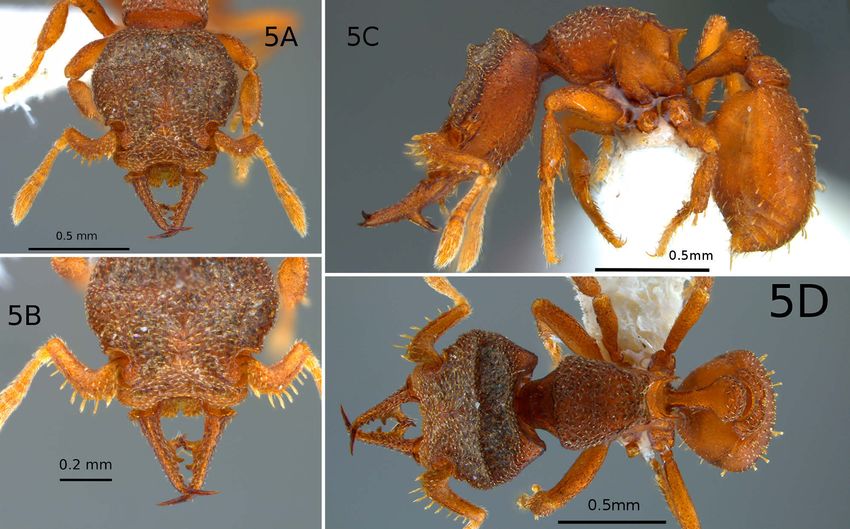
Lattke et al., Eur. J. Entomol. 115: 268–295, 2018                                                          doi: 10.14411/eje.2018.027

history. Considerations of the unusually shaped, dorsally              tain teeth and hairs) are best seen by using reflected background
arching mandibles of the long-mandibulate species of Pro-              lighting. For recognizing species, we used discontinuities in the
talaridris, and comparisons with similarly shaped mandi-               variation of a set of morphological characters between groups of
bles in both extant and extinct ant taxa, lead to a reinterpre-        ants that share distinctive characters and continuous variation for
                                                                       said character, or others, as indicative of reproductive isolation
tation of their mandibular morphology.
                                                                       between groups. The characters we found most useful for alpha
MATERIALS AND METHODS                                                  taxonomy are listed in the results.

Generalities                                                           Specimen repositories

   The alpha taxonomy is based upon direct observation of speci-          Specimens from the following collections were studied:
mens and comparative study of their morphology. Most morpho-              ARCE – Ant Reference Collection Ecuador, Instituto de Cien-
logical terms used are standard for ant taxonomic descriptions, as     cias Biológicas, Escuela Politécnica Nacional, Quito, Ecuador.
defined in Hölldobler & Wilson (1990), Bolton (1994), and Shat-           CAS – California Academy of Sciences, Dept. of Entomology,
tuck (1999). Descriptive terms for cuticular sculpturing follow        San Francisco, California, U.S.A.
Harris (1979), pilosity stature follows Wilson (1955), and wing           DZUP – Coleção Entomológica Pe. Jesus Santiago Moure, De-
venation follows Yoshimura & Fisher (2012) for vein and cell           partamento de Zoologia, Universidade Federal do Paraná, Curi-
names and Mason (1986) for vein development terms. Tubular             tiba, Brazil.
veins are sclerotized and raised on both the ventral and dorsal           FMNH – Field Museum of Natural History, Chicago, Illinois,
wing surfaces, while sclerotized nebulous veins and unsclerotized      USA.
spectral veins are raised on the dorsal surface only. For describing      ICN – Instituto de Ciencias Naturales Insects Collection, Uni-
hair shapes, the terms defined by Bolton (2000) are used as well       versidad Nacional de Colombia, Bogotá, Colombia.
as some botanical terms used for describing leaf shapes (Harris &         JTLC – John T. Longino Collection, Department of Biology,
Harris, 2001). Despite their origins in botanical morphology they      University of Utah, Salt Lake City, Utah, U.S.A.
are readily applicable to ant hairs. The following terms describe         MCZC – Museum of Comparative Zoology, Harvard Univer-
hair shapes:                                                           sity, Cambridge, Massachusetts, U.S.A.
   clavate – club-shaped, with a cylindrical basal section and a          MIZA – Museo del Instituto de Zoología Agrícola, Universidad
swollen, but not flattened distal section. Similar in outline to a     Central de Venezuela, Maracay, Venezuela.
spatulate hair, but the latter is flattened.                              NHMUK – The Natural History Museum, London, England,
   lanceolate – lance-shaped, with the widest part basad (Fig. 1G).    U.K.
   linear – long and narrow, much more so than oblong, with par-          QCAZ – Museo de Zoología, Pontificia Universidad Católica
allel to subparallel sides (Fig. 1B).                                  del Ecuador, Quito, Ecuador.
   oblong – flattened, two to four times longer than broad with           RBINS – Royal Belgian Institute of Natural Sciences, Ento-
parallel or subparallel sides (Fig. 1C).                               mology Collection, Brussels, Belgium.
   ovate – egg-shaped, flattened, with the widest part basad (Fig.        USNM – National Museum of Natural History, Washington,
1E).                                                                   D.C., U.S.A.
   reniform – kidney-shaped, flattened and widest close to mid-           UTPL – Universidad Técnica Particular de Loja, Loja, Ecua-
length, the base is between two shallow convex lobes and the           dor.
lateral and apical margins describe a broad convexity (Fig. 1F).       Images
   spatulate – elongate and flattened, gradually tapering basad           High resolution digital images of P. aculeata sp. n. and P. ar-
with the widest part close to the apex (Fig. 1D).                      huaca sp. n. were taken using a Leica DFC290 camera attached
   subspatulate – similar to spatulate but with a lesser degree of     to a Leica Z6APO stereomicroscope. Image stacks were taken
tapering, not exactly oblong but not spatulate (Fig. 1A).              using Leica Application Suite v3.8 (2003–2011) and united with
   Many of the descriptions and diagnoses describe outlines of         Combine ZP (http://combinezp.software.informer.com/). The im-
particular body part margins; these (in particular the shape of cer-   ages of P. armata were taken with a motorized Leica M16Z im-
                                                                       aging system using a Canon 7D camera mounted on the scope.
                                                                       LED dome lights were used for illumination, image stacks were
                                                                       processed with Helicon Focus Pro 6.7.1 and final rendering with
                                                                       Photoshop CS6. The P. loxanensis sp. n. wing images were taken
                                                                       with a Leica DFC 500 camera mounted on a motorized Leica
                                                                       MZ1 stereoscope and processed with Leica LAS 3D Viewer and
                                                                       Leica LAS Montage 4.7 with final rendering using GIMP 2.8. All
                                                                       other images were taken with a Leica MC170HD camera attached
                                                                       to a Leica S8APO stereomicroscope. Each image stack was taken
                                                                       using Leica Application Suite ver. 4.3.0 (2003–2013) and united
                                                                       with Combine ZP. Final editing was done with Adobe Photoshop
                                                                       CS5. The distribution map was created using QGIS v2.14 (Quan-
                                                                       tum GIS Development Team, 2016).
                                                                       Measurements and indices
                                                                         Most morphological measurements were made using a ste-
                                                                       reoscopic microscope with an ocular micrometer. The following
                                                                       measurements are adopted from Brown & Kempf (1960) and are
                                                                       expressed in millimeters:
Fig. 1. Hair shape terms. A – subspatulate; B – linear; C – oblong;      HL – Head length: mid-line length of the cephalic capsule,
D – spatulate; E – ovate; F – reniform; G – lanceolate.                measured in full-face (dorsal) view, from the anterior margin of

                                                                                                                                     269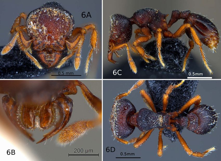
Lattke et al., Eur. J. Entomol. 115: 268–295, 2018                                                                            doi: 10.14411/eje.2018.027

the clypeus to the midpoint of a line drawn across the occipital        P. armata Brown, 1980 (central Ecuador to western Pan-
margin.                                                                   ama)
   HW – Head width: maximum width of head, measured in the              P. bordoni Lattke, sp. n. (northcentral Venezuela)
same plane as HL.                                                       P. loxanensis Lattke, sp. n. (southcentral Ecuador)
   ML – Mandible length: straight-line maximum length of a
                                                                        P. punctata Lattke, sp. n. (northcentral Venezuela)
mandible, measured from the base at the insertion into the head
capsule, to the apex. Not measured in the same plane as HL, but         leponcei group
in a dorsal view of the mandible itself (in P. leponcei sp. n., the
                                                                        P. leponcei Delsinne & Lattke, sp. n. (southcentral Ecua-
mandible was measured in the same plane as HL as it is also its
dorsal view).
                                                                          dor)
   EL – Eye length: maximum length of the compound eye, meas-           Identification key for workers of Protalaridris
ured perpendicular to the eye.
                                                                        1   Mandible long and slender. Dorsal margin in lateral view
   SL – Scape length: maximum length of the first antennal seg-
                                                                            concave, forming obtuse angle with clypeal plane. In frontal
ment, as measured from the anteriormost margin of the basal lobe
                                                                            view, first basal tooth of mandible separated from basal rim
or angle to the apex.
                                                                            by diastema. Eye separated from dorsal cephalic surface by at
   PW – Pronotal width: maximum width of pronotum in dorsal
                                                                            most one its diameter (armata group) .................................. 2
view.
                                                                        –   Mandible short, stout, and triangular. Dorsal margin in lateral
   WL – Weber’s length of the mesosoma: diagonal length, meas-
                                                                            view strongly bowed with basal portion in same plane as cl-
ured in lateral view from the anterior margin of the pronotum (ex-
                                                                            ypeus and apical portion strongly bent downward. In frontal
cluding the collar) to the posterior extremity of the metapleural
                                                                            view, first basal tooth of mandible continuous with basal rim.
lobe.
                                                                            Eye well separated from dorsal cephalic surface by twice its
   PH – Petiole height: height of the petiole measured in profile
                                                                            diameter (Fig. 6c) (leponcei group) ................. leponcei sp. n.
from the ventralmost point of the petiole vertically to a line inter-
                                                                        2   Cephalic dorsum bears prominently elevated frontovertexal
secting the dorsal-most point of the node.
                                                                            ridge that is separated from occipital carina by broad and con-
   PL – Petiole length: the length of the petiole measured in pro-
                                                                            cave sulcus, especially evident in lateral view of head (Figs
file from the anterior process to the posteriormost point of the
                                                                            2c, 5c) ................................................................................... 3
tergite, where it surrounds the postpetiolar articulation.
                                                                        –   Frontovertexal ridge variably developed, but never so el-
   DPW – The maximum width of the petiole in dorsal view.
                                                                            evated that it forms a distinct concavity with the occipital
   PPL – The maximum length of the postpetiole in dorsal view.
                                                                            carina when seen laterally. Cephalic margin posterad of ridge
   PPW – The maximum width of the postpetiole in dorsal view.
                                                                            straight to convex in lateral view (Figs 4c, 7c, 9c) ............... 4
   The following indices were calculated from the preceding
                                                                        3   Large ventral mandibular tooth situated closer to mandibu-
measurements: CI – Cephalic index: HW/HL; MI – Mandibular
                                                                            lar base than to its mid-length; anterior margin of scape with
index: ML/HW; OI – Ocular index: EL/HW; SI – Scape index:
                                                                            6–7 erect hairs; eye with 3–4 relatively indistinct ommatidia;
SL/HW; LPI – Lateral Petiole Index: PH/PL; DPI – Dorsal Peti-
                                                                            dorsal surface of mandible and lateral surface of tibiae with
ole Index: DPW/PL.
                                                                            abundant tubercles ........................................... aculeata sp. n.
                                                                        –   Large ventral mandibular tooth situated closer to mandibular
RESULTS
                                                                            mid-length than to its base; anterior margin of scape with 8–9
Characters                                                                  erect hairs; eye reduced to 1 apparent ommatidium; dorsal
                                                                            surface of mandible and lateral surface of tibiae rugulose at
  In this study the following characters proved to be the
                                                                            most, lacking tubercles ..................................... bordoni sp. n.
most helpful in separating species. Head: General shape,                4   Each mandible with single prominent and massive preapical
sculpture of dorsal surface, presence or absence of trans-                  tooth that crosses that of opposing mandible, tooth closer to
verse carina, shape of antennal fossa. Compound eye: Gen-                   mandibular base than mandibular mid-length and easily vis-
eral shape, number of ommatidia, relative distance from                     ible in lateral view; one or two more slender preapical teeth
cephalic dorsum and antennal fossa. Mandible: General                       may overlap (Figs 3b, 5b, 7b, 9b), but these teeth are not vis-
shape, angle formed with dorsal cephalic surface, relative                  ible in lateral view; eye separated from dorsal cephalic sur-
length, number of teeth/denticles along dorsal and ventral                  face by at least half a diameter ............................................. 5
preapical margins, number of large ventral teeth; general               –   Each mandible with two prominent and massive preapical
                                                                            teeth, proximal tooth situated close to mandibular mid-length
sculpture pattern; shape and orientation of setae along
                                                                            and distal tooth closer to mandibular apex; both teeth cross
internal mandibular border. Antenna: Shape of scape and                     their opposite counterpart and are visible in lateral view (Fig.
number of standing hairs along anteroventral margin of                      4b); eye borders on dorsal cephalic margin ..... armata Brown
scape. Labrum: General shape, development of anterome-                  5   Anterior labral margin in dorsal view weakly sinuate, median
dian notch, number and shape of labral hairs, geometry of                   emargination not deeper than one-fourth of labral length; la-
hairs. Mesosoma: General shape of dorsal margin in lateral                  brum relatively narrow, its lateral basal margins clearly vis-
view, curvature of propodeal declivity anterad of tooth;                    ible in full face view, uncovered by mandibles (Fig. 7a) .......
shape of propodeal tooth.                                                    ...................................................................... loxanensis sp. n.
                                                                        –   Anterior labral margin medially cleft; median notch deeper
Species list                                                                than one-fourth of labral length; lateral basal margins of la-
                                                                            brum in full-face view totally covered by mandibles ........... 6
armata group                                                            6   Labrum with angular to bluntly angular anterolateral lobes;
P. aculeata Lattke & Alpert, sp. n. (southwestern Peru)                     mandible apicad of large ventral tooth with series of short
P. arhuaca Guerrero, Lattke & Alpert, sp. n. (northeast Co-                 teeth, none crossing or touching their opposite counterparts
   lombia)                                                                  (Fig. 9b) ...........................................................punctata sp. n.

270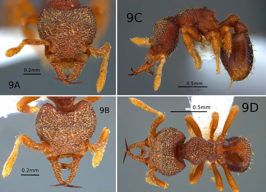
Lattke et al., Eur. J. Entomol. 115: 268–295, 2018                                                           doi: 10.14411/eje.2018.027

–   Labrum with rounded anterolateral lobes; mandible apicad             sal surface; mandibular dorsum strongly sculpted, ventral
    of large ventral tooth with single large tooth that touches or       surface weakly sculpted; apical tooth and apex of massive
    crosses its opposite counterpart (Fig. 3b) .........arhuaca sp. n.   ventral tooth/teeth smooth. In lateral view mandibular dor-
Genus Protalaridris Brown, 1980
                                                                         sal margin concave or forms obtuse angle with main longi-
                                                                         tudinal axis of cephalic capsule. Mandible generally with
Protalaridris Brown, 1980: 36. Type-species: Protalaridris ar-           two internal rows of preapical teeth, one ventral another
  mata Brown, 1980, by original designation.                             dorsal; dorsal teeth sometimes absent or reduced in number
Baroni Urbani & De Andrade, 2007: 88 (as junior synonym of
                                                                         (P. armata). Dorsal preapical teeth numbering 1–7 pointing
  Basiceros Schulz, 1906).
Ward et al., 2015: 77 (transferred from Basicerotini to Attini).         mesad, relatively small, never crossing each other; ventral
                                                                         preapical teeth numbering 4–5, of varying size with at least
   Worker. Head in dorsal view about as long as wide, wid-               some teeth that cross, including one or two massive ven-
est posterior to eye; cephalic dorsum with transverse blunt              tromedially directed teeth. Mandibular dorsum with abun-
frontovertexal ridge, posteriorly broad convex; occasion-                dant appressed and elongate lanceolate to simple hairs that
ally ridge sharp and narrow (P. aculeata sp. n. & P. bor-                arch anterad. 2. leponcei species-group: mandible short,
doni sp. n.). Vertex broad and convex when ridge is blunt,               stout, and triangular; dorsal margin in lateral view strongly
mostly concave when ridge is sharp with narrow flat strip                bowed with basal portion in same plane as clypeus and api-
bordering the occipital carina. Brief median longitudinal                cal portion strongly bent downward. Masticatory margin
carina present on frontal area, extending posterad from                  with single row of 9 preapical teeth of irregular size, pro-
posteromedian clypeal margin. Epistomal suture well de-                  truding dorsomedially. Apical and longest preapical teeth
fined, shaped as open inverted V; clypeus posterolaterally               crossed with mandibles closed. In frontal view first basal
forms anterior part of antennal fossa, anterior margin of                tooth of mandible continuous with basal rim.
frontal lobe meets posterior clypeal margin through nar-                    Labrum extended, shape varying from rounded to rec-
rowly arched space. Vertex both posterior and anterior to                tangular with an anteromedian notch ranging in depth from
frontovertexal ridge feebly but broadly impressed on each                shallow to over half the length of the labrum, six to 32 usu-
side; anterior clypeal margin generally with broad median                ally flattened hairs present along margins, the longest an-
concavity. Head in lateral view with broadly convex pos-                 terolaterally placed and the shortest posterolaterally placed
terodorsal margin that meets dorsal margin at an angle; ce-              and within the anterior notch. With head in dorsal view
phalic dorsal margin straight to broadly concave.                        the labrum mostly visible with mandibles closed in armata
   Compound eye relatively small, directed anterolater-                  group but entirely hidden from view in the leponcei group.
ally, with 1–12 usually indistinct ommatidia; eye separat-                  Mesosoma subpyriform to pyriform in dorsal view,
ed from dorsal cephalic surface by at least one diameter,                broadest across anterior pronotum, cervix marked off by
sometimes less but never directly bordering the dorsal ce-               a blunt arcuate margin. Mesosomal dorsal margin con-
phalic surface. No erect pilosity on cephalic dorsum but P.              tinuously convex in lateral view, curvature ranging from
leponcei sp. n. bears one erect spatulate hair on each side              weakly convex to strongly convex; no sutures evident
of head, posterior to compound eye and lateral to frontal                across dorsum, except for shallow narrow furrow usually
carina; medially pointing subspatulate to lanceolate ap-                 obscured by particulate matter, immediately anterad of
pressed hairs generally present. Antenna issues forth on                 transverse carina marking top of concave dorsal part of the
each side through a deep semicircular notch in the dorso-                propodeal concavity, and presumably marking the anterior
lateral cephalic margin; antennal fossa shaped as sinuous                margin of propodeum. Propodeal spiracle surrounded by
emargination along anterolateral cephalic margin in full-                elevated ring of cuticle, opening faces posterolaterally, lo-
face view, interior of fossa mostly finely areolate; antennal            cated approximately at half length of declivity less than
scrobe very shallow and broad, extending posterad of com-                one diameter from tooth; dorsal propodeal margin very
pound eye. Antenna 9-segmented with 2-segmented club,                    brief; declivity finely areolate ventrad of upper margin of
pedicel suboval, segments 3–7 short and transverse, apical               teeth, rugulose dorsad, separated from dorsum by distinct
segment longer than preceding seven segments. Scape flat-                transverse carina, not covered by debris or encrustations.
tened, robust with flat to convex dorsal surface, ventral sur-           Petiole relatively short with poorly developed node, node
face finely areolate, anterior basal lobe slightly expanded,             transverse in dorsal view, obliquely subtruncate in lateral
except in P. aculeata sp. n. where the lobe is lamellate,                view, postpetiole twice as wide as petiole, rounded above,
anterior margin with 5–10 spatulate to reniform hairs.                   with trace of median longitudinal sulcus posterad, and
   Mandible: 1. armata species-group: mandibles long and                 weak median posterior emargination. Underside of petiole
slender, in full length view straight to slightly arched, in-            biconvex, anteroventral process shaped as discrete angle
sertions remote, shafts crossing at apices when completely               or lacking. No erect hairs on dorsum of mesosoma, peti-
closed, each tapering apicad towards medially directed api-              ole, and postpetiole; mesosomal lateral surface without
cal tooth, tooth dark brown, shining, and acutely pointed.               standing pilosity. Encrustations present on dorsal surface
Mandibles form complex cradle or cage mainly bound                       of petiolar node and postpetiole.
by the main axis of each mandible as well as one, or two,                   Abdominal tergite IV with broadly convex main dorsal
prominent ventral teeth. Base of mandible at cephalic inser-             surface, dorsolaterally bordered by longitudinal blunt ridge
tion expands into flange with smooth rim and areolate dor-               that defines elongate lateral vertical region of tergite, trans-

                                                                                                                                    271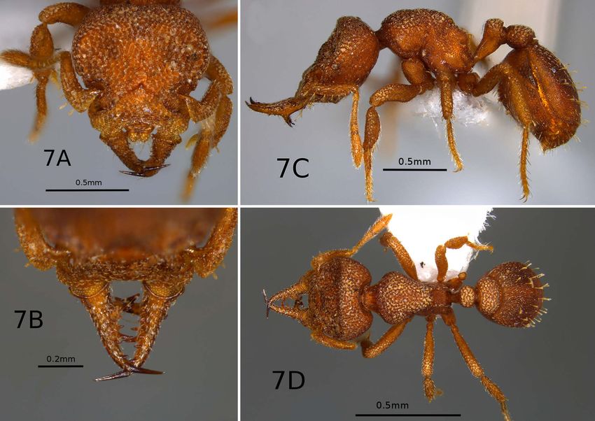
Lattke et al., Eur. J. Entomol. 115: 268–295, 2018                                                doi: 10.14411/eje.2018.027

verse ridge present along anterior margin; gastral tergum         Only a few winged individuals are known for the genus.
with 20–50, sometimes more, erect to suberect spatulate         The following discusses differences and similarities be-
hairs arranged in at least four longitudinal rows; antero-      tween the wing venation of Protalaridris and some of the
ventral gastral process lacking. Legs short and compact,        genera of the Basiceros group. The fore wing of P. bordoni
femora gradually thickened apicad, and tibiae even thicker,     sp. n. and P. loxanensis sp. n. is very much like that of
tarsi more slender; 1–2 spatulate hairs present on apex of      Basiceros scambognathus (Brown, 1949) as illustrated in
tibiae, meso- and metatarsal segments with pairs of erect       Brown & Kempf (1960: 173), but the Cubital vein sepa-
spatulate hairs in V; tarsal claws small. Venation (for P.      rates from M-Cu at the junction with cu-a in B. scambog-
bordoni sp. n. and P. loxanensis sp. n. gynes). (Fig. 8) Fore   nathus whereas in Protalaridris the separation of Cu oc-
wing with 4 closed cells (costal, basal, submarginal, and       curs at a distance from cu-a. In Protalaridris 1A continues
subbasal); veins C, Sc+R, M+Cu, Cu, cu-a, and 1A tubular;       briefly apicad of cu-a but it stops at cu-a in Basiceros. The
Cu tubular for a distance; Rs partly spectral between M and     fore wing of P. loxanensis sp. n. has the costal cell well-
2rs-m; separation of Cu from M-Cu occurs at a distance          defined between tubular veins C and Sc+R as in Rhopalo-
from cu-a; 1A continues briefly apicad of cu-a. Hind wing       thrix Mayr, 1870; basal cell well-defined between tubular
with tubular C+Sc+R, Sc+R, and Rs+M; 1A briefly tubular         veins Sc+R and M+Cu (similar to Rhopalothrix but M+Cu
and M+Cu partially tubular.                                     darker, better defined in Protalaridris); submarginal cell
   Comments. The inclusion of Protalaridris leponcei            mostly well-defined though Rs is partly spectral between
sp. n., with its strikingly different mandibles, obliged a      M and 2rs-m; subbasal cell closed as M+Cu, 1A, and cu-a
redefinition of the genus, but most changes are limited to      markedly tubular; Cu tubular for a distance. Fore wing
mandibular morphology, as the majority of other charac-         with 4 closed cells in Protalaridris and Basiceros (costal,
ters are shared, including the number of antennal segments.     basal, submarginal, and subbasal); in Rhopalothrix with 2
The labral margin hairs are very short in P. leponcei sp. n.    (costal and basal). The hind wing in P. loxanensis sp. n.
with none longer than 1/4 the labral width while in the other   has tubular C+Sc+R, Sc+R, and Rs+M; 1A is briefly tubu-
species some hairs will always exceed half the labral width     lar and M+Cu is also partially tubular. In contrast the fore
in length. A monotypic genus could have been proposed           wing venation of Rhopalothrix subspatulata Longino &
but there is a long history of ant genera established be-       Boudinot, 2013 is relatively reduced, with Rs+M, M, and
cause of variances in mandibular morphology, with further       Rs apicad of M all reduced to spectral veins, cu-a is absent
studies forcing a taxonomic weeding out of excess names.        and A has a vestigial tubular section (Longino & Boudinot,
Differences in mandibular morphology and antennal seg-          2013). Additionally the hind wing of R. subspatulata has
mentation have traditionally been important in separating       only C+Sc+R developed as tubular veins whilst Sc+R and
genera within the Basiceros and Strumigenys groups, but         Rs+M are spectral. As in Protalaridris, 1A is briefly tubu-
recent work has gathered convincing evidence to the con-        lar but M+Cu is totally spectral.
trary, especially in the latter genus. It was only after the
                                                                SPECIES ACCOUNTS
extensive work of several myrmecologists (Baroni Urbani
& De Andrade, 1994; Bolton, 1999; Ward et al., 2015) that       Protalaridris aculeata Lattke & Alpert, sp. n.
many genus-group names were sunk as junior synonyms             (Figs 2a–d)
of Strumigenys. A tipping point was finally reached when        ZooBank taxon LSID:
J. Longino and M. Branstetter graciously shared the pre-        067C6EC4-5150-4464-A544-FCFC2C1FC104
liminary results of their UCE molecular analysis of the
                                                                  Worker diagnosis. Mandibular shaft with massive tooth
Basiceros group of genera and these clearly indicate the
                                                                projecting ventromesad close to mandibular base, mandi-
inclusion of P. leponcei sp. n. as an ingroup within Prota-
                                                                ble with abundant tubercles; labrum in frontal view rectan-
laridris.
                                                                gular and narrow, anterior margin with six long hairs. Basal
   The two strikingly different mandibular morphologies
                                                                angle of scape well-developed, lamellate; scape anterior
permit easy recognition of two informal species groups
                                                                margin with 6 spatulate hairs. Cephalic dorsum with me-
within the genus: (1) The armata group, with elongate
                                                                dian rectangular raised area. External tibial surfaces with
mandibles, and (2) the leponcei group, with short, triangu-
                                                                abundant tubercles.
lar mandibles. Most species of the armata group have the
frontovertexal ridge arching laterally until it either meets      Holotype measurements. HL 0.67, HW 0.70, ML 0.37, EL
the eye or misses it by an ocular diameter, whereas in P.       0.03, SL 0.35, PW 0.38, WL 0.60, PH 0.18, PL 0.25, DPW 0.18
leponcei sp. n. the ridge is separated from the eye by sev-     mm. CI 1.05, MI 0.52, OI 0.05, SI 0.50, LPI 0.73, DPI 0.73.
eral times its diameter. This latter condition is also found      Worker description. With head in frontal view ante-
in the ascrobicula group of Octostruma (Longino, 2013a).        rior clypeal margin with broad median concavity, laterally
Within the armata group P. aculeata sp. n. and P. bordoni       broadly convex; lateral cephalic margin posterad of anten-
sp. n. share the prominent frontovertexal ridge and a most-     nal fossa convex, head widest at posterior two-thirds, lat-
ly concave vertex except for a narrow flat strip that borders   eral cephalic margins mostly converging anterad; posterior
the occipital carina, in sharp contrast with the blunt fron-    cephalic margin with medial emargination, posterolaterally
tovertexal ridge with a mostly convex vertex of the other       convex with inconspicuous occipital lobe; cephalic dorsum
known species.                                                  posterad of antennal fossa with well-defined, carinate bor-

272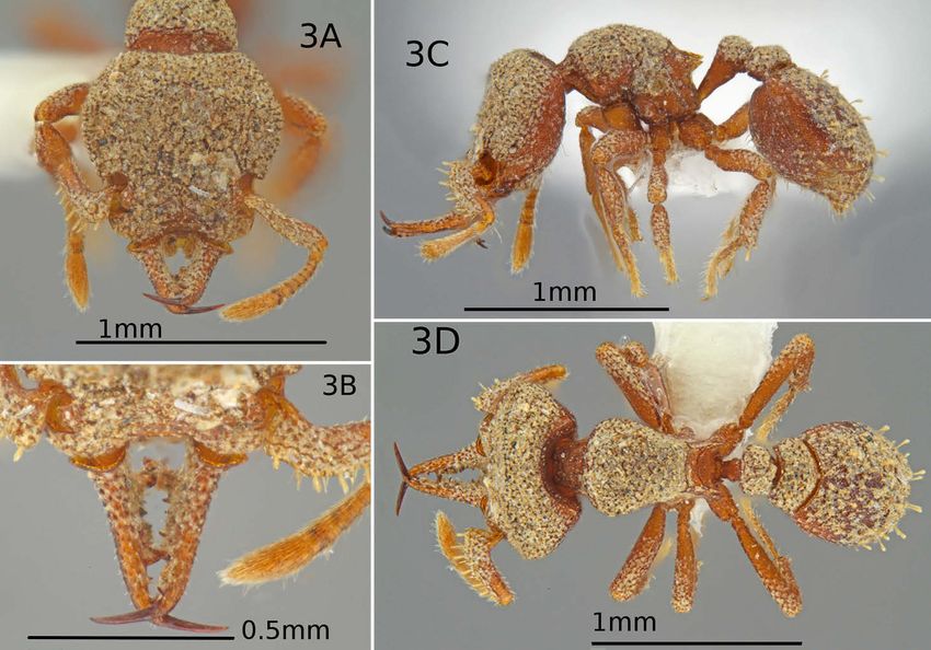
Lattke et al., Eur. J. Entomol. 115: 268–295, 2018                                                          doi: 10.14411/eje.2018.027

Fig. 2. P. aculeata sp. n. Worker holotype: A – head dorsal view; B – mandible, full length view; C – body lateral view; D – body, dorsal
view. Images by G. Alpert.

ders. Frontal lobe dorsally convex, anterior margin forms             ated closer to mandibular base than apex in mandibular full
narrow angle with posterior clypeal margin, width less than           length view; ventral tooth in dorsal view tapering apicad,
half that of fossa. Eye with 3–4 relatively indistinct omma-          internal margin edentate, apically broadly concave. Internal
tidia, facing anterolaterally, separated from antennal fossa          surface of ventral tooth mostly smooth and shining. With
along lateral cephalic margin by not more than 3 diameters,           mandibles closed the apices of each ventral tooth cross;
separated from dorsal face less than one diameter in lateral          apical and ventral tooth ferruginous brown, smooth and
cephalic view.                                                        shining. Mandibular dorsum with low but distinct, blunt
   Cephalic dorsum mostly rugulose reticulate, rugulae di-            tubercles and rugulae, ventral surface anterior of ventral
rected longitudinally posteriorly and obliquely laterally,            tooth rugulose and with appressed pilosity directed apicad;
space between rugulae shining; cephalic dorsum divided                smooth and shining sculpture limited to posteroventral
into elevated central area, convex transverse posterior               area, ventral tooth, and mandibular apex.
area and vertical lateral convex area. Central elevation                In cephalic lateral view mandibular dorsal margin forms
rectangular, extending posterad approximately two-thirds              broad concavity with dorsal cephalic surface, ventral tooth
of cephalic length; lateral margin of elevation vertical,             tapers posteroventrally, mostly straight, its length about
extends from frontal carina, elevation bordered posterad              1/3 that of lateral mandibular width. Mandibular dorsum
by transverse carina, carina broadly concave medially, ru-            with short, arched, slender hairs pointing apicad or mesad.
gulose reticulate with longitudinal elevated median area.             Dorsal mandibular margin with 5–6 preapical denticles,
Cephalic dorsum between transverse carina and posterior               besides medium sized tooth set at mid-length between
cephalic carina convex in lateral cephalic view. Cephalic             massive ventral tooth and base of apical tooth; dorsal
dorsum with mesially pointing subspatulate to lanceolate              mandibular margin with decumbent slender hairs, directed
appressed hairs; occiput areolate, posterolateral region              anteromesad. Ventral mandibular margin with 8 denticles
areolate-rugulose. Cephalic ventrum mostly areolate to                between base of apical tooth and ventral massive tooth.
areolate-rugulose, posterolaterally densely punctulate and              Labrum rectangular and narrow, lateral margins visible
shining. Antennal scrobe shallow but distinct, posteriorly            in dorsal view with mandibles closed, basal ridge broadly
ending just beyond eye.                                               arched; apically rounded with distinct but shallow median
   Mandible in full length view with both shafts converg-             notch. Median ventral surface with abundant hairs, dorsal
ing until just anterad of mandibular mid-length, then each            surface sculpted, not smooth and shining. Anterior margin
is weakly convex, bending at base of apical tooth. Man-               of labrum with six long hairs, third and second hairs from
dible with prominent ventromedially directed tooth, situ-             labral cleft the longest, slender and weakly lanceolate. In-

                                                                                                                                    273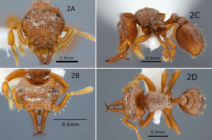
Lattke et al., Eur. J. Entomol. 115: 268–295, 2018 doi: 10.14411/eje.2018.027
ternal hairs weakly spatulate with rounded apex. Labral CA-879 leg. Plot IE20. Matute Tierra Firme Forest, ‘Burhenuvia’
cleft with 2 very short hairs. Palpal formula unknown. [Buchenavia] fruit fall berlesate.
Scape in dorsal view with weak basal lobe, anterior mar- Type locality. Madre de Dios, Peru.
gin broadly convex and lamellate; scape in general broadly Etymology. The species epithet is derived from the Latin neu-
ter plural of acūleātus “having stingers or spines”, also “sting-
arched. External margin dorsad of lamella with 6 elongate
ing”, and alludes to the numerous tubercles on the mandibles and
spatulate hairs, ventral margin with 10 slender hairs; an- tibiae of this species.
terior scape face rugulose and shining, anterobasal lobe
weakly expanded anterad, dorsum with very fine short Distribution. Only known from type locality.
hairs. Cross-section of scape at mid-length triangular, ante- Biology. It is the only Protalaridris found outside of pre-
rior convex margin, dorsal margin broadly convex, ventral montane and montane rainforest, in a lowland site of the
margin mostly straight. Amazon basin relatively close to the Andes. It represents
Mesosomal dorsal margin in lateral view convex, prome- the southernmost known limits of the distribution range for
sonotal suture marked by shallow concavity, mesonotum the genus, separated by a gap of close to 1500 km from the
with narrow anterior margin, mesonotal dorsal margin closest known Protalaridris in southern Ecuador. Buche-
mostly straight, dorsal propodeal margin brief; declivitous navia Eichler, 1866 is a tree genus of the Combretaceae
margin covered by triangular tooth; base of tooth broadly family with several species known from the Amazonian
concave, apex pointed, posterior margin lamellate, briefly forest.
concave. Mesosomal dorsum and dorsolateral half of pro- Comments. Even though this species is described from
notum rugulose, with decumbent and arched weakly lan- a single specimen, its morphology is very distinct within
ceolate hairs, mostly directed posteromesad. Half of lateral the genus, particularly the tuberculate sculpturing on the
pronotum, part of mesopleuron, and propodeum punctulate mandibular and lateral tibial surfaces. Most Protalaridris
to areolate, wanting pilosity, metapleuron punctulate; mes- have 8 or more spatulate hairs along the anterior margin of
opleuron with rugulae. Pronotum with anterior transverse the scape but there are only 5 or 6 hairs in P. aculeata sp.
areolate strip with scattered rugulae, not marked off by n. and P. loxanensis sp. n. A crust of dirt or debris covers
transverse carina from dorsal surface; promesonotal suture most of the body, ventrolateral cephalic surface, and most
marked as shallow transverse trough. of abdominal tergum IV, though the tergal crust is appar-
Petiolar node strongly convex in lateral view, anterior ently thinner than that of the head. The occiput, propodeal
petiolar margin mostly straight to broadly concave, an- declivity, and a transverse strip anterad to the anterior ca-
teroventral process triangular small, postpetiolar dorsal rina on abdominal tergum IV are contrastingly clean and
margin in lateral view broadly convex. Petiole with long devoid of any debris. The number and arrangement of erect
straight hair on ventrolateral surface, posterolaterally di- hairs on the gastral tergum should be considered provision-
rected. Postpetiole transverse in dorsal view, anterior mar- al as it can vary in other species.
gin concave and shorter than convex posterior margin, A label accompanying the specimen discloses it was rec-
dorsum of petiolar node and postpetiole areolate-rugulose ognized as an undescribed species by B.H. Dietz in 2002.
with posteriorly directed appressed, arched hairs. Dorsal It is probably a rare ant as a two week long ant taxonomy
margin of abdominal tergum IV broadly convex in lateral course in July of 2012 involving over 25 students and in-
view, ventral margin markedly convex, with greatest height structors collecting intensively at a locality (Sachavacayoc
just anterad of gastral mid-length; abdominal tergum IV Centre, –12.8587° –69.3552°, 235 m) just 38 km SW of the
with transverse carina along anterodorsal margin that type locality of P. aculeata sp. n. failed to detect this ant.
separates dorsum from brief transverse anterior surface; The 2013 edition of Ant Course was held at a site 251 km
dorsal pilosity consisting of sparse arched subdecumbent EEW (Villa Carmen Station, –12.8947° –71.4031°) of the
lanceolate hairs, and some 22–26 suberect, spatulate hairs. type locality, closer to the Andes, with a large group of stu-
Abdominal sternite IV densely areolate anterad becoming dents and instructors intensively collecting ants between
punctate posterad, sparsely clothed by arched decumbent the altitudes of 500–700 m, but no specimens were found.
hairs pointing posterad. Tarsal claws simple, long and The type locality is 55 km W from the Bolivian border and
slender; legs stout, not elongate; protibial apex with lateral under 200 km S from the Brazilian border, suggesting the
spatulate hair. Meso- and metatibial apices each with an- presence of this genus in these countries.
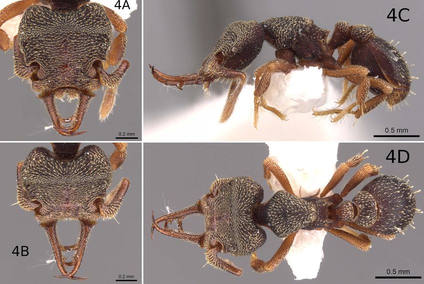
terolateral and posterolateral spatulate hairs, anterior hairs Protalaridris arhuaca Guerrero, Lattke & Alpert,
largest. External surface of tibiae with numerous low tu- sp. n.
bercles. Body mostly ferruginous, darker tint on transverse (Figs 3a–d)
cephalic carina and cephalic dorsum posterad of carina,
ZooBank taxon LSID:
apex of mandible and ventral mandibular tooth, mesoso- ADBBC186-0CD1-4DB3-8951-746FC140DF4E
mal dorsum and propodeal lamella, including tooth, and
gaster throughout. Worker diagnosis. Cephalic vertex with median sub-
Queen and male. Unknown. quadrate elevation. Labrum wider than long, deeply cleft
with 8 large anterolateral hairs and no hairs in labral cleft.
Type material. Holotype worker (MCZC). Peru. Madre Overlapping preapical mandibular teeth between mid-
de Dios: Cuzco Amazónico, 15 km NE Puerto Maldonado length of mandible and base of apical tooth belong to
[–12.5312° –69.0713°], 200 m, 16.VII.1989, SP Cover, JE Tobin
dorsal row. Frontovertexal ridge with abundant small pro-
274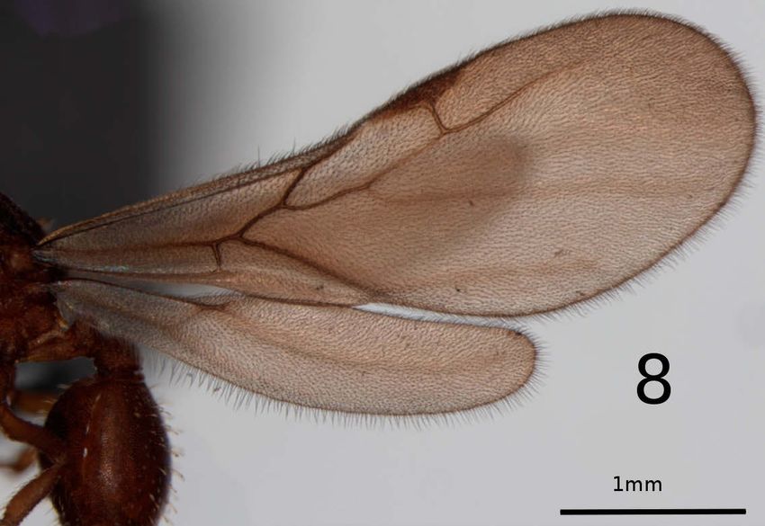
Lattke et al., Eur. J. Entomol. 115: 268–295, 2018                                                            doi: 10.14411/eje.2018.027

Fig. 3. P. arhuaca sp. n. Worker paratype: A – head dorsal view; B – mandible full length view; C – body lateral view; D – body dorsal view.
Specimen CASENT0633240. Images by G. Alpert.

tuberances. Dorsum of head, most of mesosoma, petiolar                  margin straight, internal margin sinuous; anterior margin
node, and postpetiole torose-rugulose.                                  of fossa brief and transverse, frontal carina with anterior
  Measurements of type specimens. Holotype (paratypes,                  half convex and posterior half shaped as subquadrate to
n = 3): HL 0.84 (0.82–0.87); HW 0.87 (0.86–0.90); ML 0.53               convex emargination. Internal surface of the fossa densely
(0.53–0.57); EL 0.04 (0.05–0.06); SL 0.49 (0.47–0.51); PW 0.49          foveolate, without pilosity. Cephalic dorsal surface irregu-
(0.48–0.52); WL 0.72 (0.73–0.79); PH 0.20 (0.20–0.22); PL 0.37          larly rugulose from frontovertexal ridge to anterior clypeal
(0.36–0.43); PPL 0.20 (0.19–0.24); PPW 0.38 (0.34–0.39); DPW            margin; head posterad of ridge with abundant rounded,
0.26 (0.25–0.27) mm. CI 1.04 (1.05–1.07); MI 0.61 (0.60–0.63);          protuberances connected by short and thick sharply-crest-
OI 0.04 (0.06–0.07); SI 0.56 (0.54–0.56); LPI 0.54 (0.51–0.58);         ed rugulae, sculpture more accentuated posterolaterally;
DPI 70 (63–72).                                                         bunched series of longitudinal rugulae extend posteriorly
   Worker description. Head slightly wider than long                    from frontal carina forming low, broad crest that almost
in full-face view; posterior margin broadly concave to                  touches frontovertexal ridge. Ill-defined longitudinal me-
straight with small median emargination; lateral cephalic               dian ridge present, extending posterad from posterior cl-
margin posterior to antennal fossa forming blunt obtuse                 ypeal margin. Area between longitudinal and lateral cari-
angle with straight to weakly convex sides, widest at fron-             nae strongly concave, area posterad to frontovertexal ridge
tovertexal ridge, lateral margin forms discrete angle with              with median, subquadrate-shaped raised area (best seen
posterior margin of antennal fossa; lateral cephalic margin             in dorsal view of the posterior cephalic margin) laterally
briefly concave along posterior half of antennal fossa, then            bound by ovoid depression. Cephalic dorsum with short
gradually curves convex towards clypeus. Anterior clypeal               and apically acute, appressed hairs, each hair separated by
margin medially concave with lateral convex lobe next to                distance equal to or greater than its length.
basal mandibular flange. Cephalic lateral margin bordered                 Mandible elongate, external and internal margins in
by raised, blunt carina that curves medially to form strong-            full-length view mostly parallel, cross-section at one-third
ly raised frontovertexal ridge shaped as broad, anteriorly              anterior-length is convex externally and straight (vertical)
facing concavity, continuously arching except for brief me-             internally; mandible tapers to medially directed, acutely
dian concavity that forms anterior margin of median ver-                pointed apical tooth, as long as 1/3 mandibular length.
texal protuberance. Antennal fossa conspicuous, as long as              Mandibular internal margin with longitudinal dorsal row of
wide, reaching barely half the length of the head, anterior             4 teeth and 3–4 denticles: teeth situated apicad of massive

                                                                                                                                       275
Lattke et al., Eur. J. Entomol. 115: 268–295, 2018 doi: 10.14411/eje.2018.027
ventral tooth, from the base apicad, teeth 1–3 relatively dorsum and lateral surfaces of pronotum and mesonotum
small and non-overlapping; tooth 4 large, approximately torose-rugulose. Propodeal declivity finely areolate; nar-
twice as long and as wide at base than preceding teeth. row posteroventral strip of pronotum, mesometapleuron,
Tooth 4 situated anterad of mandibular mid-length and and lateral propodeal face pruinose with shallow foveolae.
overlapping with large tooth of other mandible, 2–3 den- Anteroventral margin of pronotum with shallow, roughly
ticles situated apicad of tooth, 1 or two denticles situated scrobiculate sulcus. Mesosomal dorsum lacking standing
basad of tooth 1; mandibular internal margin with ventral hairs. Petiole as long as propodeal declivity in lateral view,
row of 5–6 denticles best seen ventrally, posteriormost dorsal margin straight, ventral margin slightly sinuous;
denticle largest in size and situated on base of large ventral node discrete, rectangular in dorsal view, wider than long;
tooth. Ventral tooth placed closer to mandibular base than postpetiole twice as wide as long, anterior margin strongly
to mid-length, crossing opposing tooth from other mandi- concave, posterior margin very convex, lateral margins
ble, internal ridge with low triangular flange. Mandibular posteriorly diverging. Most of petiole densely foveolate,
dorsum mostly rugulose-torulose, ventrally mostly smooth dorsum of petiolar node and postpetiole torose-rugulose.
and shining, including ventral tooth and apical tooth; man- Fourth abdominal tergite anteriorly concave in dorsal view,
dibular basal flange finely areolate. Mandible with sparse complementing posterior convex margin of postpetiolar
curved decumbent hairs along lateral margin, apex of each tergite; fourth abdominal segment makes up 3/4 of gaster,
hair contacting mandibular surface; ventral mandibular dorsum of tergite foveolate-rugose; sides of fourth abdomi-
surface with 2 rows of very short erect hairs between base nal tergite, all gastral sterna, and rest of tergites densely
of ventral tooth and base of apical tooth; 6–8 obliquely po- foveolate; gastral dorsum with more than 10 erect, sub-
sitioned, long hairs present from mandibular base to basal spatulate hairs; sparse pubescence also present; sternites
half of ventral tooth. with sparse erect hairs.
Labrum wider than long, anterolaterally bluntly angular Coxae finely areolate. Femora with anterior, posterior,
and lateral margin convex, anteromedially with narrow V- and ventral surfaces relatively flat, dorsally convex; erect
shaped incision as long or longer than labral mid-length; and semierect hairs present on both internal and external
dorsal surface shining and rugulose with fine punctae; la- femoral surfaces; tibiae laterally compressed, dorsal margin
brum anterolaterally with 4 thick, cylindrical hairs, 2 hairs in lateral view straight and ventral margin convex; meso-
on anterior margin and 2 hairs on anterolateral margin, and metatibia longer than protibia. First tarsomere of each
lateralmost hair the shortest, no hairs in median emargina- leg half as long as respective tibia, tarsomeres 2–4 each
tion, apex of some hairs flattened; ventral surface of la- slightly longer than wide. Coxae and lateral tibial surfaces
brum with erect pubescence and subspatulate hairs, some densely foveolate; dorsum and apical-lateral surface of
apically cleft. profemur mostly torulose-rugulose, meso- and metafemora
Scape barely reaches frontovertexal ridge; in dorsal view apically torulose-rugulose, basally foveolate. Tibiae with
with two oblique surfaces separated by longitudinal ca- thick flattened hairs on ventral surface, hairs as long as
rina, internal surface with bead-like projections arranged half tibial width. Lateral apex of tibiae each with flattened
in rows and low carinae, external surface with two longi- spatulate hair, mesotibial apex with small flattened hairs on
tudinal concavities separated by median lamella; external posterior side. Head, scape, mandible, promesonotum and
margin with 2 rows of hairs, dorsal row with 6 lamellate most of dorsal propodeal face and rest of abdomen brown;
thick hairs, ventral row of 9–10 cylindrical thick hairs, nar- funiculus, mesometapleuron, lateral and declivitous propo-
rower than dorsal hairs. Scape opaque, funiculus shining. deal faces, and most of legs yellowish-brown; mandibular
Antenna with pedicel as wide as long, campaniform; seg- apical tooth reddish-brown.
ment 3 trapezoid, slightly wider than long, segments 4–8 Queen and male. Unknown.
trapezoid, serially increasing in length, 2–8 punctulate, Type material. Holotype worker (point-mounted; ICN): CO-
apical segment smooth; scape apex with semierect hairs, LOMBIA. Magdalena: 9 km SE Minca, 11.08972° –74.06021°
following segments with abundant decumbent to subde- ±100 m, 1650 m, 28 May 2017, R. Guerrero, J. Longino#9848-
cumbent hairs as long as pedicel. Eye reduced to single s. Taken in secondary growth montane wet forest from sifted
ommatidium, separated from dorsal cephalic surface by leaf litter. Holotype bears red label with unique specimen iden-
distance equal to less than one diameter. tifier ‘ICN 093589’. Paratypes: Three point-mounted workers
Dorsal margin of mesosoma in lateral view mostly con- with same data as holotype: One with unique specimen identi-
vex, dorsal propodeal margin concave to apex of tooth; fier CASENT0644163 and a Q code, deposited in DZUP. One
with unique specimen identifier CASENT0644170 and a Q
pronotal collar opaque and finely punctate; propleuron
code, deposited in JTLC. One with unique specimen identifier
finely areolate. Mesometapleuron briefly cleft along ven- CASENT0644171 and a Q code, deposited in JTLC.
tral margin, mesopleuron mostly flat, rugulose ventrally; Additional material. COLOMBIA. Magdalena: 4 km SE La
metapleuron mostly flat except for anterodorsal depres- Minca, 11.12257° –74.08508°, ± 20 m, 1180 m, 4.xii.2014, MG
sion next to propodeum, depression and propodeum joined Branstetter 2452, CASENT0633240, ADMAC DNA voucher, 1
by carina; mesopleuron posteroventrally convex. Spiracle worker (JTLC).
rounded, posteriorly facing; dorsal propodeal face sepa- Type locality. Magdalena, Colombia.
rated from declivity by transverse carina. Mesosoma trap- Etymology. The species epithet is in recognition of the Arhua-
ezoid in dorsal view, widest anterad, most of mesosomal co people, an indigenous group with ancestral lands close to the
type locality of the species.
276Lattke et al., Eur. J. Entomol. 115: 268–295, 2018 doi: 10.14411/eje.2018.027
Distribution. Only known from the Sierra Nevada of mandibles, when closed, cover the lateral margins of the
Santa Marta, Magdalena, Colombia labrum in P. arhuaca sp. n., but in P. loxanensis sp. n. these
Biology. All specimens of P. arhuaca have been col- margins are visible despite full mandibular closure. The la-
lected from sifted leaf-litter in premontane rain forest be- brum in P. loxanensis sp. n. has a weak anterior concavity
tween 1180 and 1650 m on the northwestern slopes of the but in P. arhuaca sp. n. the labrum is deeply cleft. P. punc-
Sierra Nevada de Santa Marta. The holotype and specimen tata sp. n. also has a relatively wide labrum with a deep
CASENT0644163 were found in the lower site, a dense emargination, as in P. arhuaca sp. n., but this cleft forms
forest with closed canopy and abundant leaf-litter. Speci- an acute angle in P. punctata sp. n. and in P. arhuaca sp. n.
mens CASENT0644170 & CASENT0644171 were taken it forms an obtuse angle.
from secondary growth forest with an open canopy and
Protalaridris armata Brown, 1980
some scattered large trees. The site is surrounded by pas-
tures and crossed by trails. The litter in this site was taken (Figs 4a–d)
from the base of the largest trees. Protalaridris armata Brown, 1980: 37, Figs 1–8 (worker).
Comments. P. arhuaca sp. n. is similar to P. loxanen- Basiceros armata: Baroni Urbani & De Andrade, 2007: 90.
sis sp. n., also having a large apical tooth that overlaps its
counterpart on the opposing mandible. In P. arhuaca sp. Worker diagnosis. Mandibular shaft with two massive
n. this tooth is part of the dorsal row of dentition, in con- overlapping ventromesially projecting teeth, dorsal man-
trast with P. loxanensis sp. n. where the tooth is part of dibular margin lacking preapical teeth or denticles except
the ventral row of preapical mandibular dentition. Their for a single denticle at base of apical tooth; mandibular
respective head lengths and labral configuration also per- shaft slender, in full-length view very gradually tapering
mit separation. The head length of P. arhuaca sp. n. is the apicad. Lateral margin of labrum with three anterolaterally
longest in Protalaridris, including P. loxanensis sp. n. (HL projecting hairs; abdominal tergite IV with 12–16 erect
> 0.80 mm vs. 0.76–0.78 mm, respectively). P. arhuaca sp. hairs.
n. has a subquadrate head compared with the subrectan- Worker measurements. (n = 10): HL 0.60–0.87; HW 0.69–
gular shape in P. loxanensis sp. n. (CI ≤ 1.07 vs. ≥ 1.10). 0.93; ML 0.40–0.58; EL 0.04–0.09; SL 0.36–0.47; PW 0.40–0.64;
Although other Protalaridris may have quadrate to sub- WL 0.64–0.87; PH 0.21–0.29; PL 0.22–0.27; DPW 0.11–0.27
quadrate heads, their respective widths and lengths are mm. CI 0.95–1.15; MI 0.53–0.65; OI 0.06–0.11; SI 0.50–0.53;
less than in P. arhuaca sp. n. In a dorsal cephalic view the LPI 0.83–1.20; DPI 0.42–1.10.
Fig. 4. P. armata Brown 1980. Worker paratype: A – head dorsal view; B – mandible, full length view; C – body lateral view; D – body dorsal
view. Specimen CASENT0900943. Images by Will Ericsson, Antweb.
277Lattke et al., Eur. J. Entomol. 115: 268–295, 2018 doi: 10.14411/eje.2018.027 Worker description. Head slightly wider than long in Labrum entirely visible in frontal view, basal margin dorsal view; posterior margin mostly broadly convex to posteriorly convex, labrum wider posterad than anterad; bluntly angular, occipital lobe projects posterad as blunt bilobed with apically blunt median cleft almost extend- angle, lateral cephalic margin posterior to eye broadly con- ing to labral mid-length, lateral margin weakly concave. vex to bluntly angular. Cephalic lateral margins anterad of Ventral surface mostly shining, dorsal surface with scat- maximum head width converge anteriorly; anterior clypeal tered punctulae, not as shining. Labral margins with lan- margin medially concave, anterolaterally forming blunt ceolate flattened hairs; lateral margin with 3 anterolater- angle, clypeus bordered posterolaterally by antennal fossa. ally directed hairs, progressively becoming longer anterad, Eye distinct, partially visible in frontal view, oval, appar- anterior margin with 2 hairs, and internal margin with 2–3 ently 4–5 partially fused ommatidia, facing anterolaterally, anteromedially directed hairs. Palpal formula unknown. separated from antennal fossa by one diameter or less in Scape in dorsal view with longitudinal rugulae anterad, lateral view, dorsal ocular margin at same level as dor- posterad sparsely punctate, subparallel anterior and poste- sal cephalic margin in lateral cephalic view. Head widest rior margins, slightly wider basad than apicad, posterior along dorsolateral ridge. Cephalic lateral surface elongate, margin broadly concave, anterior margin broadly convex, facing lateroventrad in lateral view, mostly occupied by anterobasal lobe weakly expanded anterad, thin longitudi- broad and shallow antennal scrobe that fades posteriorly nal lamella usually present between anterobasal lobe and at same distance as dorsal transverse carina; head widest apex; dorsum with arched hairs particularly anterad. Cross- along dorsolateral ridge. Cephalic dorsum mostly densely section of scape at mid-length subrectangular, dorsal mar- punctate with abundant brief longitudinal rugae, commonly gin broadly convex, ventral margin mostly straight, ante- encrusted with dirt and debris that obscure cuticle; dorsum rior margin concave to convex, posterior margin straight to traversed at widest point by broadly concave to bluntly weakly concave; scape anteroventral margin bears 8 prom- angular carina that forms elevated crest dividing cephalic inent apically truncate hairs; basal hair simple and longest, dorsum into two sloping surfaces; frontovertexal ridge al- other hairs spatulate, weakly arching anterad, apical-most ways obvious but may vary in vertical development. Low 4 hairs smallest of all. but distinct longitudinal carina present between posterior Mesosomal dorsum in lateral view weakly convex, dor- clypeal midpoint and frontovertexal ridge. Cephalic dor- sal propodeal margin very brief; propodeal tooth broadly sum with short appressed, mostly transverse lanceolate triangular, posterior base prolonged ventrally as broadly ground hairs; posterolateral and ventral cephalic surfaces concave lamella, in dorsal view relatively thick not la- densely punctate, pilosity sparse. mellate. Pronotum, mesosomal dorsal and dorsolateral Mandibles form cradle mainly bound by the main axis surfaces rugulose, including anepisternum and dorsal ex- of each mandible as well as two prominent ventral teeth. tremes of metapleuron and lateral propodeum. Numerous Mandible in full length view progressively tapers apicad, decumbent ground hairs on lateral pronotum, mesonotum mostly straight, bending mesad just before base of apical and propodeum, mostly directed postero to posteromesad; tooth, in lateral view dorsal mandibular margin sinuous, no decumbent hairs on mesometapleuron; no erect hairs convex up to level of basal ventral tooth and concave api- on dorsum of head, mesosoma, petiole, and postpetiole. cad. Base of mandible at cephalic insertion expands into Katepisternum, most of metapleuron, and base of pro- flange with smooth rim and areolate dorsal surface. Man- podeal tooth punctate to areolate; katepisternum lacking dibular dorsum mostly punctate with scattered rugulae transverse rugulae. Pronotum with defined anterior face basad, abundant appressed and elongate to lanceolate or separated from dorsal face by abrupt curvature, collar with linear hairs that arch anterad present throughout mandibu- few longitudinal carinae; promesonotal suture barely dis- lar shaft, densest on basal half; ventral mandibular surface tinguishable. mostly shining, with scattered punctulae. Mandibular dor- Petiolar node subquadrate with anterior petiolar margin sal margin sinuous in lateral view, forms weak obtuse angle evenly and broadly concave in lateral view, dorsal margin with dorsal cephalic surface, mandibular base emarginate straight to weakly convex, posterior margin brief and ver- at junction with clypeus; massive ventral teeth pointing tical, length less than half that of dorsal margin. Petiolar mostly mesad, ventral projection of tooth in lateral view anteroventral process shaped as weak angle to absent, ven- less than contiguous thickness of mandibular shaft. Dor- tral margin posterad of process broadly concave to sinuate; sal mandibular margin lacking preapical dentition except postpetiolar dorsal margin in lateral view mostly broadly for denticle close to base of apical tooth. Mandibular ven- convex, curvature more pronounced posterad. Petiolar tral margin with 4 preapical teeth: teeth 1 and 3 massive, node and postpetiole transverse in dorsal view; postpetiole mesoventrally directed and overlapping with their coun- broadly concave anteriorly and convex posteriorly, dorsum terparts, basal tooth largest, apically cleft; tooth 3 simple; and dorsolateral surfaces of petiolar node and postpetiolar tooth 2 small, closer to tooth 3 than tooth 1, tooth 4 at base tergum areolate-rugulose with posteriorly directed ap- of apical tooth. Ventral tooth in dorsal view tapering api- pressed, linear to lanceolate ground hairs. Petiolar anterior cad; in anterior view relatively straight with decumbent surface smooth and shining with abundant punctae, lateral- tooth along anterodorsal margin. Mandibular apical tooth ly and ventrally opaque and areolate; postpetiolar ventrum dark brown shining and sharply pointed. transverse, opaque and areolate. 278
You can also read