Evidence for the cure of HIV infection by CCR5 32/ 32 stem cell transplantation
←
→
Page content transcription
If your browser does not render page correctly, please read the page content below
CLINICAL TRIALS AND OBSERVATIONS

Evidence for the cure of HIV infection by CCR5⌬32/⌬32 stem
cell transplantation
Kristina Allers,1 Gero Hütter,2 Jörg Hofmann,3 Christoph Loddenkemper,4 Kathrin Rieger,2 Eckhard Thiel,2 and
Thomas Schneider1
1Department of Gastroenterology, Infectious Diseases, and Rheumatology, Medical Clinic I, Campus Benjamin Franklin, Charité-University Medicine Berlin,

Berlin, Germany; 2Department of Hematology, Oncology, and Transfusion Medicine, Medical Clinic III, Campus Benjamin Franklin, Charité-University Medicine
Berlin, Berlin, Germany; 3Institute of Medical Virology, Helmut-Ruska-Haus, Campus Mitte, Charité-University Medicine Berlin, Berlin, Germany; and 4Institute of
Pathology/Research Center ImmunoSciences (RCIS), Campus Benjamin Franklin, Charité-University Medicine Berlin, Berlin, Germany

HIV entry into CD4ⴙ cells requires inter-                 immune reconstitution. In the present               tible to productive infection with CXCR4-
action with a cellular receptor, generally                study, we demonstrate successful recon-             tropic HIV. Furthermore, during the pro-
either CCR5 or CXCR4. We have previ-                      stitution of CD4ⴙ T cells at the systemic           cess of immune reconstitution, we found
ously reported the case of an HIV-infected                level as well as in the gut mucosal im-             evidence for the replacement of long-
patient in whom viral replication remained                mune system after CCR5⌬32/⌬32 stem                  lived host tissue cells with donor-derived
absent despite discontinuation of antiret-                cell transplantation, while the patient re-         cells, indicating that the size of the viral
roviral therapy after transplantation with                mains without any sign of HIV infection.            reservoir has been reduced over time. In
CCR5⌬32/⌬32 stem cells. However, it was                   This was observed although recovered                conclusion, our results strongly suggest
expected that the long-lived viral reser-                 CD4ⴙ T cells contain a high proportion              that cure of HIV has been achieved in this
voir would lead to HIV rebound and dis-                   of activated memory CD4ⴙ T cells, ie, the           patient. (Blood. 2011;117(10):2791-2799)
ease progression during the process of                    preferential targets of HIV, and are suscep-

Introduction
Destruction of the immune system by the HIV is driven by the loss                 emphasizes the importance for continuing research in the field of
of CD4⫹ T cells in the peripheral blood and lymphoid tissues. Viral               CCR5-targeted treatment strategies, but uncertainty has remained
entry into CD4⫹ cells is mediated by the interaction with a cellular              over whether a cure for HIV infection has been achieved in this
chemokine receptor, the most common of which are CCR5 and                         patient.
CXCR4.1 Because subsequent viral replication requires cellular                        In the setting of HIV infection, the effects of pretransplantation
gene expression processes, activated CD4⫹ cells are the primary                   conditioning do not allow the complete elimination of HIV, as
targets of productive HIV infection. Consequently, HIV infection                  demonstrated by previous studies in which researchers demon-
leads predominantly to the depletion of activated memory CD4⫹                     strated that HIV-infected patients who undergo stem cell transplan-
T cells, most of which reside in the gastrointestinal (GI) mucosa.2-4             tation generally experience a viral rebound when ART is discontin-
Although therapeutic control of HIV replication allows the immune                 ued.12-17 For this reason, together with the fact that CXCR4-tropic
system to partially restore and delays disease progression, the cure              HIV variants (X4 HIV) were present within the patient’s pretrans-
of HIV infection remains still unachievable with use of the                       plantation HIV population, it was reasonable to hypothesize that
currently available antiretroviral drugs. The major barrier to viral              HIV from the viral reservoir may reseed the body once the immune
eradication in patients receiving antiretroviral therapy (ART) is the             system has efficiently been restored with X4 HIV-susceptible target
establishment of HIV reservoirs, including low-level productively                 cells.18,19
and latently infected cells.5-7 Thus, maintenance of replication-                     Accordingly, key questions that remain to be answered are
competent HIV in long-lived cells and distinct anatomical sanctuar-               (1) whether CD4⫹ T cells have been efficiently restored throughout
ies allows the virus to reseed the body once ART is discontinued.8                the body; (2) whether or not the patient’s immune system includes
    Cells of persons homozygous for the CCR5 gene variant ⌬32                     HIV-susceptible target cells; and (3) how stable the size of the HIV
(CCR5⌬32/⌬32) are naturally resistant to infection with CCR5-                     reservoir is during the process of immune reconstitution after
tropic HIV strains (R5 HIV) because of the lack of CCR5                           CCR5⌬32/⌬32 SCT.
cell-surface expression.9 Previously, we demonstrated the feasibil-                   Here, to address these questions, we extend our previous study
ity of hematopoietic stem cell transplantation (SCT) with CCR5⌬32/                to improve our knowledge about the curative potential of CCR5⌬32/
⌬32 donor cells (CCR5⌬32/⌬32 SCT) in an HIV-infected patient                      ⌬32 SCT for HIV infection. We evaluated the reconstitution of
with relapsed acute myeloid leukemia (AML) and documented                         CD4⫹ T cells at the systemic level as well as in the mucosal
absent viremia during the first 20 months of remission, during                    immune system during the posttransplantation period of more than
which time the patient did not receive ART.10,11 This case clearly                3.5 years. To verify the ability of the recovered CD4⫹ T cells to act

Submitted September 23, 2010; accepted December 2, 2010. Prepublished             The publication costs of this article were defrayed in part by page charge
online as Blood First Edition paper, December 8, 2010; DOI 10.1182/               payment. Therefore, and solely to indicate this fact, this article is hereby
blood-2010-09-309591.                                                             marked ‘‘advertisement’’ in accordance with 18 USC section 1734.

An Inside Blood analysis of this article appears at the front of this issue.      © 2011 by The American Society of Hematology

BLOOD, 10 MARCH 2011 䡠 VOLUME 117, NUMBER 10                                                                                                                2791
2792 ALLERS et al BLOOD, 10 MARCH 2011 䡠 VOLUME 117, NUMBER 10
as HIV target cells, their activation status, CXCR4 expression heat-inactivated fetal calf serum (Sigma-Aldrich), 100 U/mL of penicillin,
profile, and susceptibility to productive HIV infection was ana- and 100 g/mL streptomycin (both from Biochrom) before flow cytometric
lyzed. Moreover, because the absence of the CCR5 wild-type gene analysis.
variant in donor cells provided us with the possibility to discrimi-
nate between donor- and host-derived immune cells, we were able Flow cytometric analysis and cell sorting
to examine the persistence of potential viral reservoirs, in addition
Flow cytometric analysis was performed by the use of antibodies against
to the detection of viral sequences, in distinct tissue compartments.
CD3 (clone UCHT1; BD Biosciences), CD4 (SK3; BD), CD31 (WM59;
BD), CD38 (HIT2; BD), CD45RO (UCHL1; BD), CD49d (9F10; BD),
CD62L (Dreg-56; BD), CXCR4 (12G5; BD), HLA-DR (Immu357; Beck-
Methods man Coulter), and Ki67 (Ki67; DAKO). Absolute numbers of CD4⫹ T cells
were determined in fresh whole blood by the use of TruCount tubes and
Subjects CD3/CD4/CD8 TriTest (BD) according to the manufacturer’s protocol.
Data were acquired on the FACSCalibur flow cytometer (BD) and analyzed
In February 2007, an HIV-infected patient underwent SCT because of a with CellQuest software (BD). Lymphocytes were gated on the basis of
relapse of AML with a graft consisting of CCR5⌬32/⌬32 donor cells. The characteristic forward and sideward scatter properties. Central memory
pretransplantation conditioning regimen included 100 mg/m2 amsacrine, CD4⫹ T cells were classified by coexpression of CD45RO and CD62L,
30 mg/m2 fludarabine, 2 g/m2 cytarabine (day ⫺12 until ⫺9); 60 mg/kg and effector memory CD4⫹ T cells were classified by lack of CD62L.
cyclophosphamide (days ⫺4 and ⫺3); 5.5 mg/kg rabbit antithymocyte Recent thymic emigrants were identified by coexpression of CD31 and
globuline (in 3 doses between day ⫺3 and ⫺1); and a 400-cGy total body CD62L on CD45RO⫺ CD4⫹ T cells and central naive CD4⫹ T cells by lack
irradiation (TBI; day ⫺5). ART was discontinued on the day of transplanta- of CD31.22 CXCR4 expression density on CD4⫹ T cells was evaluated
tion, and 13 months later the patient received a second transplantation with as the mean fluorescence intensity (MFI) of CXCR4 expression divided by
CCR5⌬32/⌬32 stem cells from the same donor because of a second relapse the MFI value obtained with the corresponding isotype control (BD) and is
of AML. The conditioning regimen consisted of 100 mg/m2 cytarabine expressed as the MFI ratio.
(day ⫺7 until day ⫺1), 6 mg/m2 gemtuzumab (day ⫺7 and day ⫺1), and a For mucosal cell sorting, the following additional antibodies were used:
200-cGy TBI (day ⫺1). For clinical data and further details, see Hütter et anti-CD33 (AC104.3E3; Miltenyi Biotec) and anti-CD68 (Kim7; BD).
al.10 At 5.5, 24, and 29 months after the first CCR5⌬32/⌬32 SCT, the Cell-sorting procedures were performed by customer service of the Flow
patient underwent colonoscopy, and biopsy specimens were taken as the Cytometry Core Facility at the Berlin-Brandenburg Center for Regenerative
result of suspected intestinal graft-versus-host disease (GVHD) while Therapies, Germany, with the use of the FACSAriaII flow cytometer (BD)
tapering immunosuppressive treatment. With the patient’s informed consent and FACSDiva software (BD). Mucosal CD4⫹ T cells were identified by
for this procedure, 10-13 additional colon biopsy specimens were collected their coexpression of CD3 and CD4 in the lymphocyte gate, and mucosal
at each time point for research purpose of the present study. Examination macrophages were selected by their coexpression of CD33 and CD68 in
of histologic colon sections excluded the diagnosis of intestinal GVHD. the CD3⫺ macrophage gate.23 Antibodies were conjugated to fluorescin,
Twelve months after transplantation, the patient underwent a liver biopsy, phycoerythrin, peridinin chlorophyll protein, or allophycocyanin.
and histologic examination confirmed GVHD grade 1, which was con-
trolled with adaption of immunosuppressive therapy (ie, cyclosporine A,
methylprednisolone, mycophenolate mofetil). At 17 months after transplan- HIV-susceptibility assay
tation, the patient presented with neurologic disorders. Magnetic resonance
CCR5-tropic HIV-1 strain JR-CSF (obtained from the EVA Center for AIDS
imaging of the brain identified signal abnormalities compatible with
Reagents, National Institute for Biological Standards and Control [NIBSC])
leukoencephalopathy. For further evaluation, cerebrospinal fluid (CSF)
was propagated in PBMC. A stock of CXCR4-tropic HIV-1 strain NL4-3
samples were collected repeatedly, and, in addition, a brain biopsy was
was generated from the HIV-1 molecular clone pNL4-3 (obtained from the
performed. Polymerase chain reaction (PCR) detection of JC virus was
EVA Center for AIDS Reagents) and then propagated in PBMC. Virus-
negative in all samples. Histologic evaluation revealed astrogliosis with
containing cell culture supernatants were passed through a 22-m pore-size
microglial activation. The cumulative effect of initial AML treatment
filter (BD) to remove cell debris and then treated with Dnase (Boehringer
chemotherapy and salvage chemotherapy after relapse of AML, as well as
Mannheim) in the presence of 1mM MgCl2 for 30 minutes at 37°C to
pretransplantation conditioning regimen, including TBI, were assumed as
remove contaminating DNA. Virus stocks were stored at ⫺80°C. The
the cause of leukoencephalopathy,20 which turned out to be self-limiting.
infectious titer of thawed viral stocks was determined by tissue culture
Immunosuppressive treatment has been stopped 38 months after CCR5⌬32/
infectious dose 50% assays in PBMC. Before infection, PBMCs or MMCs
⌬32 SCT without recurrence of GVHD.
were activated with PHA and IL-2 for 48 hours. Cells were washed and
In addition, 10 HIV-uninfected SCT patients were included into this
cultivated with virus at a multiplicity of infection of 0.001 in RPMI1640
study (SCT controls). Four of these patients underwent colonoscopy,
medium supplemented with 20 U/mL of IL-2. Viral stocks diluted in
and intestinal GVHD was histologically excluded in all cases. A total of
cell-free medium served as background control, the patient’s cells alone as a
15 HIV-uninfected patients served as healthy controls; 5 of them underwent
mock control, and cell-free virus suspensions as a control for background
colonoscopy for cancer preventive examination. The study was approved
corrections. Supernatants were removed from cell cultures and cell-free
by the Charité-University Medicine Berlin institutional review board,
controls as indicated and were replaced by fresh medium and stored at
and all participants provided informed consent to study participation in
⫺80°C until analysis for viral replication by quantitative measurement of
accordance with the Declaration of Helsinki.
the HIV-1 core protein p24 production with the HIV-1 p24 ELISA assay
(XpressBiotech) according to the manufacturer’s protocol.
Cell preparation and activation
Peripheral blood mononuclear cells (PBMCs) were isolated from heparin- Immunohistochemistry and immunofluorescence staining
ized venous blood by standard Ficoll gradient centrifugation, and mucosal
mononuclear cells (MMCs) were isolated from colon biopsy specimens Immunostaining on paraffin sections was performed as described previ-
by collagenease type II (Sigma-Aldrich) digestion.21 Cells were either ously.24 Primary antibodies were mouse anti-CD4 (1F6; Novocastra),
immediately used for subsequent analysis or cryoconserved until HIV mouse anti-CD68 (PGM1; DAKO), or goat anti-CCR5 (CKR-5 [C20];
susceptibility assays. For some experiments, PBMCs were activated for Santa Cruz Biotechnology). For detection of CD4 labeling, the Streptavi-
2 days with 3 g/mL of phythemagglutinin (PHA; Sigma-Aldrich) and dine Alkaline Phosphatase-kit (DAKO) was used. Positive cells within
50 U/mL of recombinant interleukin-2 (IL-2; R&D Systems) in RPMI the mucosa of colon tissue were quantified per high-power field (hpf,
1640 ⫹ GlutaMAX cell culture medium (Invitrogen) containing 10% 0.237 mm2), and 10 hpf were averaged in each case. Per sampling at leastBLOOD, 10 MARCH 2011 䡠 VOLUME 117, NUMBER 10 CURE OF HIV INFECTION BY CCR5⌬32/⌬32 SCT 2793
Figure 1. Peripheral CD4ⴙ T cells have been efficiently restored and contain an increased proportion of activated/effector memory CD4ⴙ T cells compared with
healthy control patients. CD4⫹ T-cell numbers and frequencies of effector memory cells (EM), central memory cells (CM), recent thymic emigrants (RTE), and central naive
cells (CN) within CD4⫹ T cells (A) during the course of immune reconstitution after CCR5⌬32/⌬32 SCT and (B) in SCT controls (27.5 ⫾ 7 months after transplantation)
compared with healthy patients were determined in fresh whole blood. Median CD4⫹ T-cell numbers of healthy patients is indicated by the thick horizontal line, and the dashed
horizontal lines denote the normal 25th and 75th percentiles in panel A. The horizontal lines in panel B denote the median values of each group. Statistical significances are
given for comparisons between healthy control values and SCT control values (*P ⬍ .05, **P ⬍ .01, ***P ⬍ .001). (C) CD4⫹ T-cell expression of the activation markers CD38,
HLA-DR, and CD49d and the proliferation marker Ki67 at 9.5 and 24 months after CCR5⌬32/⌬32 SCT in comparison with SCT control and healthy control patients. Data are
representative for 5 SCT control and 4 healthy control patients.
3 sections were analyzed. Immunohistochemical evaluations were per- Statistical analysis
formed in a blinded manner, ie, the researcher was unaware of the patient’s
Data are represented as medians and were analyzed with the use of 2-tailed
clinical characteristics. For CD4/CCR5 or CD68/CCR5 double immunoflu-
Student t test with Prism software Version 4.0 (Graph Pad Inc). Significance
orescence labeling, Alexa-Fluor 488–conjugated antimouse was used in
is denoted with asterisks (ie, *P ⬍ .05, **P ⬍ .01, ***P ⬍ .001).
combination with Alexa-Fluor 555–conjugated antigoat (Invitrogen). Im-
ages were acquired by the use of a fluorescence microscope (AxioImager
Z1) equipped with a charged-coupled-device camera (AxioCam MRm) and
processed with Axiovision software (Carl Zeiss MicroImaging). Negative Results
controls were performed by omitting the primary antibodies, and unspecific
staining of the antibodies was excluded by use of isotype control antibodies. Efficient recovery of CD4ⴙ T cells was associated with a
characteristic enrichment of activated/effector memory CD4ⴙ
T cells
CCR5 genotyping
To study the CCR5 gene variant in HIV target cells, genomic DNA was After CCR5⌬32/⌬32 SCT, chimerism analysis as well as genotyp-
extracted from sorted mucosal CD4⫹ T cells or macrophages with the use ing of CCR5 alleles suggested that host T cells were completely
of the NucleoSpin TissueXS (Macherey & Nagel) according to the eliminated from the periphery.10 Numbers of donor-derived periph-
manufacturer’s protocol. DNA was then subjected to PCR amplification eral CD4⫹ T cells increased continuously and, after 2 years,
with primers for the CCR5 gene spanning the ⌬32-region from nucleotide reached levels within the normal range of age-matched healthy
826 to 1138 on the chromosome 3p21.31 (accession no: NM_000579). The patients (Figure 1A). Further phenotypic analysis revealed an
expected fragments were 312 bp for the CCR5 wild-type and 280 bp for the increase of memory CD4⫹ T-cell numbers, with a parallel, but low,
CCR5⌬32 variant. increase of CD4⫹ recent thymic emigrant as well as central naive
CD4⫹ T-cell numbers. In both the CCR5⌬32/⌬32 SCT patient and
Detection of HIV and HIV-specific antibodies the SCT control patients, the proportion of central memory CD4⫹
T cells was within the normal range, whereas effector memory
Viral RNA was isolated from plasma or CSF and the long terminal repeat CD4⫹ T cells remained markedly enriched within the CD4⫹ T-cell
and gag regions were amplified and detected with the use of the COBAS
compartment compared with healthy control values (Figure 1A-B).
AmpliPrep/COBAS TaqMan HIV-1 Test v1.0 (Roche). Total DNA was
This cellular composition indicates a proliferative expansion of
isolated from PBMCs, tissue biopsy specimens, and sorted cells with the
use of the QIAamp DNA Blood Mini Kit, the Allprep DNA/RNA Mini Kit
mature CD4⫹ T cells. In accordance, the frequency of cells
(both from QIAGEN), and the NucleoSpin Tissue XS, respectively, expressing the activation markers CD38, CD49d, and HLA-DR
following the manufacturer’s directions and the long terminal repeat and and the proliferation marker Ki67 was greater within CD4⫹ T cells
env regions were detected as described previously.10 Antibodies directed from CCR5⌬32/⌬32 SCT and control SCT patients than from
against HIV antigens in serum samples were detected with immunoblot healthy control patients (Figure 1C). Thus in both cases, CD4⫹
(Abbott) as described previously.10 T cells recovered primarily through homeostatic proliferation of2794 ALLERS et al BLOOD, 10 MARCH 2011 䡠 VOLUME 117, NUMBER 10
SCT were negative for the CCR5 wild-type gene (Figure 2B).
This demonstrates that increased numbers of mucosal CD4⫹
T cells were exclusively derived from donor hematopoietic
cells. Taken together, these results reveal that circulating
donor-derived CD4⫹ T cells were efficiently recruited to the GI
tract and have repopulated the mucosal CD4⫹ T-cell compart-
ment after CCR5⌬32/⌬32 SCT.
CXCR4 surface availability is not impaired on recovered
CD4ⴙ T cells
Reconstitution of the CD4⫹ T-cell compartment after CCR5⌬32/
⌬32 SCT was associated with an expansion of activated memory
cells (Figures 1 and 2), the preferential targets of productive HIV
infection. Donor-derived CD4⫹ T cells are naturally resistant to
CCR5-tropic HIV infection because of the lack of CCR5 surface
expression. We were interested in whether recovered CCR5⌬32/
⌬32 CD4⫹ T cells might also exhibit reduced CXCR4 surface
availability. Therefore, we analyzed fresh whole blood cells and
MMC for CXCR4 surface expression on CD4⫹ T cells in compari-
son with cells obtained from CCR5 wild-type patients. As shown in
Figure 3A, both the frequency of CXCR4-expressing cells within
memory CD4⫹ T cells as well as CXCR4 surface expression
density at the single cell level (expressed as the MFI ration) were
Figure 2. The mucosal immune system has been efficiently repopulated with comparable with those of CCR5 wild-type control patients
donor-derived CD4ⴙ T cells. (A) Immunohistochemical quantification of CD4⫹ (80.8% ⫾ 2.0% and 6.6% ⫾ 1.0%, respectively). This was also
T cells in colon tissue of the CCR5⌬32/⌬32 SCT patient, SCT control patients observed for the peripheral naive CD4⫹ T-cell compartment (not
(27 ⫾ 9 months after transplantation), and healthy control patients. The horizontal
lines denote the median values of each group. (B) Genomic DNA was extracted from shown).
mucosal CD4⫹ T cells and subjected to CCR5-specific PCR spanning the ⌬32 region.
memory CD4⫹ T cells, confirming previous reports of posttransplan-
tation immune reconstitution.25,26 These results demonstrate that
the CCR5⌬32/⌬32 SCT patient experienced a regular reconstitu-
tion of the peripheral CD4⫹ T-cell compartment after CCR5⌬32/
⌬32 SCT, including the characteristic enrichment of activated/
effector memory CD4⫹ T cells.
Donor-derived CD4ⴙ T cells have efficiently repopulated the gut
mucosal immune system
Most of the body’s CD4⫹ T cells reside in the GI tract. To assess the
recovery of CD4⫹ T cells in the gut mucosal immune system,
CD4⫹ T cells were immunohistochemically quantified in colon
tissue sections at 3 time points after CCR5⌬32/⌬32 SCT and were
compared with SCT control and healthy control patients. The
number of mucosal CD4⫹ T cells increased during the posttrans-
plantation period, and at 29 months after CCR5⌬32/⌬32 SCT,
the density of CD4⫹ T cells in the GI mucosa was similar to that of
the SCT control patients (162 vs 180 ⫾ 33 cells/hpf; Figure 2A).
Thus, no lack of immune reconstitution could be noted in the
mucosal immune system. Interestingly, compared with healthy
control patients there was a more than 2-fold increased fre-
quency of mucosal CD4⫹ T cells in all SCT patients (60 ⫾ 12 vs
162 ⫾ 29 cells/hpf), demonstrating that treatment with condition-
ing followed by SCT triggers the enrichment of HIV target cells in
the gut mucosal immune system (Figure 2A).
To confirm the donor-origin of mucosal CD4⫹ T cells, we
performed additional phenotypic and genotypic analysis. In situ Figure 3. CXCR4 surface expression on peripheral and mucosal CD4ⴙ T cells is
detection of CCR5 by immunofluorescence staining at 5.5 and not impaired in the CCR5⌬32/⌬32 SCT patient. CD4⫹ T cells in (A) fresh whole
24 months after CCR5⌬32/⌬32 SCT revealed no CCR5 expression blood, MMC (5.5 months after transplantation), or (B) ex vivo PHA/IL-2–activated
on mucosal CD4⫹ T cells (not shown), which corroborates our PBMCs were analyzed for the frequency of CXCR4 surface-expressing cells and
the CXCR4 expression density. CXCR4 expression density on CD4⫹ T cells was
previous finding from flow cytometric analysis.10 Moreover, CD4⫹ evaluated as the MFI of CXCR4 expression divided by the MFI value obtained with
T cells sorted from MMC at 24 and 29 months after CCR5⌬32/⌬32 the corresponding isotype control and is expressed as the MFI ratio.BLOOD, 10 MARCH 2011 䡠 VOLUME 117, NUMBER 10 CURE OF HIV INFECTION BY CCR5⌬32/⌬32 SCT 2795
We investigated the presence of residual host immune cells after
CCR5⌬32/⌬32 SCT by in situ immunofluorescence detection of
cellular CCR5 expression. Clinical samples from the liver, the
brain, and the colon could be used for research purposes in the
present study after a diagnosis was given. Brain tissue speci-
mens were available from the white matter and the cortex. From
the colon, 3 separate biopsy specimens were available from each
of 3 time points during the course of immune reconstitution. In
the liver, 12 months after CCR5⌬32/⌬32 SCT, CCR5-expressing
CD4⫹ T cells or macrophages/Kupffer cells were not detectable
(Figure 5A). Likewise, 17 months after CCR5⌬32/⌬32 SCT, no
CCR5-expressing macrophages/microglia were found in the brain
(Figure 5B).
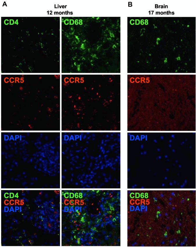
In the colon, there was no evidence of residual host CD4⫹
T cells after CCR5⌬32/⌬32 SCT, as already described previously
(Figure 2B). However, in situ immunofluorescence staining re-
vealed the presence of CCR5-expressing macrophages at 5.5 months
Figure 4. Recovered peripheral and mucosal CD4ⴙ T cells are susceptible to after CCR5⌬32/⌬32 SCT, which is in agreement with our previous
productive X4 HIV infection. PBMC and MMC obtained 24 months after CCR5⌬32/
flow cytometric data10 and demonstrates the persistence of host
⌬32 SCT were activated with PHA and IL-2 and then incubated with the CCR5-tropic
HIV-1 strain JR-CSF or the CXCR4-tropic HIV-1 strain NL4-3 at a multiplicity of macrophages during the first months after CCR5⌬32/⌬32 SCT
infection of 0.001. Viral replication was quantified by measuring the amount of HIV (Figure 6A). Importantly, later in the course of immune reconstitu-
core protein p24 in the cell-free supernatants of cultures. No virus production was tion, CCR5 expression on macrophages became undetectable
observed in the mock controls. Similar results were obtained with peripheral
lymphocytes purified at 9.5 and 34.5 and months after CCR5⌬32/⌬32 SCT. indicating their replacement with donor-derived cells (Figure 6A).
To further prove the origin of mucosal macrophages, we performed
additional genotypic analysis of sorted mucosal macrophages. As
Because the level of CXCR4 expression may vary with cell shown in Figure 6B, 24 and 29 months after CCR5⌬32/⌬32 SCT,
activation, we next analyzed CXCR4 expression on CD4⫹ T cells mucosal macrophages were negative for the CCR5 wild-type gene.
upon ex vivo activation and found efficient expression of CXCR4 The absence of host’s genomic DNA in mucosal macrophages at
on CCR5⌬32/⌬32 CD4⫹ T cells (Figure 3B). These data demon- these time points confirms the phenotypic results and suggests that
strate that the CCR5⌬32/⌬32 SCT was not associated with an host macrophages have been replaced with donor-derived cells
impaired CXCR4 expression on recovered CD4⫹ T cells. In vivo, during the posttransplantation period.
the availability of CXCR4 may be affected by the chemokine
CXCL12, the physiologic ligand of CXCR4.27 During the immune HIV remains undetectable in distinct tissue compartments
reconstitution period, CXCL12 plasma levels in the CCR5⌬32/⌬32
The presence of HIV RNA and HIV DNA was examined in distinct
SCT patient remained within the normal range of healthy patients
tissue compartments during the course of 45 months after CCR5⌬32/
(not shown), indicating that the in vivo availability of CXCR4
⌬32 SCT. Viral sequences were not detectable in all the samples
was not impaired by naturally occurring receptor occupation.
tested (Table 1).
Altogether, these results indicate that recovered CD4⫹ T cells are
not protected against X4 HIV entry.
Antibodies against HIV decrease over time
Recovered CD4ⴙ
T cells are susceptible to productive Previously, we reported the loss of antibodies directed against
X4 HIV infection the HIV polymerase as well as a decrease of HIV envelope and
core-specific antibodies during the first 20 months after CCR5⌬32/
Susceptibility of recovered CD4⫹ T cells in the central as well as
⌬32 SCT.10 Immunoblot analysis revealed a continuing decline of
the mucosal immune system to productive HIV infection was
HIV specific antibodies thereafter demonstrating the process of
studied by ex vivo infections of PBMC and MMC obtained after
serodeconversion: whereas HIV core-directed antibodies (p17,
CCR5⌬32/⌬32 SCT. As shown in Figure 4, cells from both
p24) disappeared completely, the serum level of antibodies against
compartments were susceptible to productive infection by X4 HIV.
the HIV envelope (gp41, gp120) further decreased. Today, the
Consistent with our previous observation, virus production of the
patient has only HIV envelope-specific antibodies.
PBMC-propagated X4 HIV strain was greater in peripheral than in
mucosal CD4⫹ T cells.28 As expected, because of the lack of CCR5
surface expression on donor-derived cells, both peripheral and
mucosal CD4⫹ T cells were resistant to R5 HIV infection. Discussion
Long-lived HIV target cells of host origin were replaced with Immune reconstitution is critical to the long-term success of the
donor-derived cells during the posttransplantation period SCT, and, in HIV-infected patients, also provides a prerequisite for
viral rebound and HIV disease progression. Progressive infection
Because of the fact that recovered CD4⫹ T cells are susceptible to in turn impairs the reconstitution of CD4⫹ T cells after SCT. Our
productive X4 HIV infection, long-lived HIV-infected host cells results show that systemic recovery of CD4⫹ T cells after CCR5⌬32/
that survive chemo- and irradiation therapies represent potential ⌬32 SCT and discontinuation of ART was not impaired compared
sources from which HIV to emerge. Noncirculating immune cells with that of SCT control patients. In accordance with previous
such as tissue CD4⫹ T cells or macrophages are virtually chemo/ studies,25,26 repopulation of the CD4⫹ T-cell compartment was
radio-resistant and, therefore, represent possible viral reservoirs. associated with peripheral expansion of donor-derived memory2796 ALLERS et al BLOOD, 10 MARCH 2011 䡠 VOLUME 117, NUMBER 10
Figure 5. No evidence for residual HIV target cells of
host origin in the liver and the brain. CCR5-expressing
CD4⫹ T cells or macrophages were detected (A) in liver
and (B) in brain tissue sections obtained 12 and 17 months
after CCR5⌬32/⌬32 SCT, respectively, by in situ immuno-
fluorescence double staining for CD4 (green) or CD68
(green) and CCR5 (red). Original magnification ⫻400.
Images were acquired by use of the AxioImager Z1
fluorescence microscope (Carl Zeiss MicroImaging)
coupled to the AxioCam MRm digital camera (Carl Zeiss).
Acquisition software: Axiovision (Carl Zeiss). Software
used for image processing: Adobe Photoshop CS (Adobe
Systems).
CD4⫹ T cells, that probably occurs to compensate for the limited or lymph nodes regardless of the infection route, and even with
thymic capacity in adults.29-31 Generally, this homeostasis-driven complete suppression of viremia for many years, residual low-level
expansion of activated memory CD4⫹ T cells leads to an enrich- replication in the GI tract prevents full recovery of mucosal CD4⫹
ment of the preferential targets for productive infection with both T cells in ART-treated HIV-infected patients.2,37-39 Poor recovery of
R5 HIV and X4 HIV32 and likely contributes to the rapid dynamic CD4⫹ T cells in the mucosal immune system is therefore an
of HIV rebound after conventional SCT in HIV-infected pa- important risk factor for the development of HIV disease progres-
tients.12,14,15,17 Viral tropism analysis was not in the focus of sion. After CCR5⌬32/⌬32 SCT, we found that the process of
previous reports of HIV-infected patients with conventional SCT immune reconstitution included a gradual increase of donor-
and would be an interesting issue to address in future studies. derived CD4⫹ T cells in the GI mucosa. Compared with HIV-
In the CCR5⌬32/⌬32 SCT patient, CD4⫹ T-cell numbers have
uninfected SCT control patients, mucosal CD4⫹ T-cell numbers
even returned to the normal range of healthy patients whereas HIV
normalized whereas HIV remained undetectable in gut tissue
RNA and HIV DNA remain continuously undetectable in plasma
specimens as well as in mucosal HIV target cell populations. These
and PBMC, respectively. Today, by monitoring the most common
findings argue for the absence of HIV disease progression in the
prognostic markers, ie plasma viral load and CD4⫹ T-cell counts in
the peripheral blood, HIV disease cannot be assessed in this patient. largest component of the lymphoid organ system. Surprisingly,
However, observations from the central immune compartment compared with healthy control patients, mucosal CD4⫹ T-cell
need not be representative for distinct tissue compartments through- numbers in both the CCR5⌬32/⌬32 SCT patient and the SCT
out the body. Only 1%-2% of the body’s total CD4⫹ T cells reside control patients were increased. This finding may likely be
in the peripheral blood, whereas the majority of immune cells are explained by the high prevalence of activated/effector memory
located in the GI tract.33 Containing most of the body’s activated CD4⫹ T cells in the circulation, for which we have previously
memory CD4⫹ T cells with high expression of cellular receptors, found enhanced gut-homing capacity.40 In addition, the normalized
the mucosal immune system is highly prone to productive infection frequency of central memory cells within circulating CD4⫹ T cells
with both R5 HIV and X4 HIV.3,28,34-36 In fact, profound depletion suggests that recovered CD4⫹ T cells have been efficiently directed
of CD4⫹ T cells in the GI mucosa occurs earlier than that in blood to peripheral lymph nodes.41,42 Furthermore, the decline of HIV-BLOOD, 10 MARCH 2011 䡠 VOLUME 117, NUMBER 10 CURE OF HIV INFECTION BY CCR5⌬32/⌬32 SCT 2797
CD4⫹ T cells are susceptible to productive infection with X4 HIV,
demonstrating that the CCR5⌬32/⌬32 SCT has not provided
protection against X4 HIV infection. Consequently, the patient’s
risk of exogenous HIV reinfection is not completely eliminated.
Altogether, our results demonstrate that the process of immune
reconstitution has successfully restored both the central and the
mucosal immune system with CD4⫹ T cells that lack CCR5 surface
expression but have susceptibility to productive X4 HIV infection.
Consequently, host cells that survived the chemoirradiation thera-
pies represent potential sources for X4 HIV rebound. Host-
originating CD4⫹ T cells appear to be completely removed from
the patient’s immune system; however, in particular tissue macro-
phages may play a critical role as viral reservoir because they are
virtually resistant to conditioning procedures and less prone to the
cytopathic effects of HIV infection.45 HIV became undetectable in
the brain during a neuropathologic episode, although the associated
microglia activation and astrogliosis may support reactivation of
viral replication from latently infected cells. This provides indirect
evidence for the absence of replication-competent HIV in cells of
the brain. Furthermore, in brain as well as in liver tissue sections,
no CCR5 expression on macrophages was detectable, indicating
the replacement of host microglial cells and Kupffer cells by
donor-derived cells. Because CCR5 is not constitutively expressed
on tissue macrophages,46 the limited sample availability did not
allow us to extend the phenotypic results to cell-specific genomic
analysis, and also, because the analyzed sections are representative
only for a very limited area of the respective organ, these findings
cannot definitely exclude the presence of residual, potentially
infected, host cells.
However, there is convincing evidence from studies in mice to
suggest that host tissue macrophages were efficiently replaced with
donor-derived cells during the course of immune reconstitution.
For example, although it is generally accepted that microglia under
steady-state conditions are very slowly renewed by cells of
hematopoietic origin, it has been demonstrated that the condition-
ing procedure efficiently enhances this process after stem cell
transplantation.47,48 Moreover, the majority of Kupffer cells are
replaced already early after SCT49 and, importantly, increasing
conversion rates of tissue macrophages over time after transplanta-
tion has been demonstrated in distinct tissue compartments through-
out the whole body.50,51 Evidence in support of the conclusion that
conversion from host to donor tissue macrophages took place in the
patient after CCR5⌬32/⌬32 SCT comes from our serial analysis in
colon tissue. Here, phenotypic results revealed that residual host
cells were present within the mucosal macrophage population
during the first months after CCR5⌬32/⌬32 SCT. Later in the
course of immune reconstitution, host-originating macrophages
Figure 6. Host macrophages were replaced with donor-derived cells during the
course of immune reconstitution. (A) CCR5-expressing macrophages were
detected by in situ immunofluorescence double staining for CD68 (green) and CCR5 Table 1. Detection time points of HIV RNA and HIV DNA after
(red) in colon tissue sections obtained 5.5 or 24 months after CCR5⌬32/⌬32 SCT. CCR5⌬32/⌬32 SCT
CCR5-expressing macrophages are indicated by yellow arrows. (B) At 24 and
HIV RNA LTR and gag HIV DNA LTR and env
29 months after CCR5⌬32/⌬32 SCT, macrophages were sorted from mucosal cells
(in months after (in months after
and genotyped by CCR5 variant-specific PCR.
transplantation) transplantation)
Plasma 0-45 (each month)
specific antibodies after CCR5⌬32/⌬32 SCT indicates the continu- PBMC 0-45 (each month)
ous absence of HIV gene expression in lymphoid tissues after BMMC 3, 12, 16.5, 40
discontinuation of ART. CSF 14, 14.5, 15.5, 17
In addition to their natural protection from R5 HIV infection, Brain 17
CCR5⌬32/⌬32 CD4⫹ T cells of some persons have been suggested Colon 5.5, 24, 29
to be less susceptible to X4 HIV entry as a result of down-regulated Mucosal CD4⫹ T cells 24, 29
CXCR4 expression.43,44 However, in the patient described here, we Mucosal macrophages 24, 29
found no evidence for an abnormal CXCR4 expression on recov- BMMC indicates bone marrow mononuclear cells; CSF, cerebrospinal fluid; and
ered CD4⫹ T cells. Moreover, the patient’s peripheral and mucosal PBMC, peripheral blood mononuclear cell.2798 ALLERS et al BLOOD, 10 MARCH 2011 䡠 VOLUME 117, NUMBER 10
became undetectable in the GI mucosa by both phenotypic and This work was supported by a research funding from the
genotypic analysis. These findings suggest that the replacement of German Research Foundation (DFG KFO104) to K.A. and T.S.
host tissue cells with donor-derived cells has reduced the size of the The HIV-1 molecular clone pNL4-3 from Dr Malcolm Martin was
viral reservoir during the course of immune reconstitution and, provided by the EU Program EVA Center for AIDS Reagents,
consequently, has lowered the risk of HIV rebound over time. Cell NIBSC (AVIP Contract Number LSHP-CT-2004-503487). HIV-1
replacement in tissues under posttransplantation conditions may JR-CSF from Dr Isy Chen was provided from the WHO-UNAIDS
even allow for complete eradication of HIV; however, the unfeasi- Virus Network through the Center for AIDS Reagents.
bility to analyze every single cell in living humans rules out the
possibility to positively prove viral eradication in this patient.
In summary, our results demonstrate successful CD4⫹ T-cell Authorship
reconstitution at the systemic level as well as in the largest
immunologic organ after CCR5⌬32/⌬32 SCT and in addition Contribution: K.A. designed experiments; K.A., J.H., and C.L.
provide evidence for the reduction in the size of the potential HIV performed experiments and analyzed data; K.A. and C.L. com-
reservoir over time. Although the recovered CD4⫹ T cells are posed the figures; K.A., G.H., J.H., and T.S. interpreted and
susceptible to infection with X4 HIV, the patient remains without discussed the data; G.H., K.R., and E.T. collected data; E.T.
any evidence of HIV infection for more than 3.5 years after critically revised the manuscript for important intellectual content;
discontinuation of ART. From these results, it is reasonable to T.S. supervised the research; K.A. wrote the manuscript; and all
conclude that cure of HIV infection has been achieved in this authors read and approved the manuscript.
patient. The current affiliation for G.H. is the Institute of Transfusion
Medicine and Immunology, University Heidelberg, Germany. The
current affiliation for C.L. is the Department of Pathology, Technis-
Acknowledgments che Universität München, Munich, Germany.
Conflict-of-interest disclosure: The authors declare no compet-
We are grateful to the patients for their participation in this project. ing financial interests.
We thank Diana Bösel and Simone Spiekermann for excellent Correspondence: Kristina Allers, Department of Gastroenterol-
technical assistance and Désirée Kunkel from the Berlin- ogy, Infectious Diseases, and Rheumatology, Medical Clinic I,
Brandenburg Center for Regenerative Therapies for technical Campus Benjamin Franklin, Charité-University Medicine, Hinden-
support with cell sorting. burgdamm 30, 12203 Berlin, Germany; e-mail: kristina.allers@charite.de.
References
1. Moore JP, Kitchen SG, Pugach P, Zack JA. The gene. Hemophilia Growth and Development 18. Levy JA. Not an HIV cure, but encouraging new
CCR5 and CXCR4 coreceptors—central to un- Study, Multicenter AIDS Cohort Study, Multicenter directions. N Engl J Med. 2009;360(7):724-725.
derstanding the transmission and pathogenesis Hemophilia Cohort Study, San Francisco City Co- 19. De Mendoza C. Is HIV Eradication Feasible?
of human immunodeficiency virus type 1 infec- hort, ALIVE Study. Science. 1996;273(5283): AIDS Rev. 2009;1152-53.
tion. AIDS Res Hum Retroviruses. 2004;20(1): 1856-1862.
20. Soussain C, Ricard D, Fike JR, Mazeron JJ,
111-126. 10. Hütter G, Nowak D, Mossner M, et al. Long-term Psimaras D, Delattre JY. CNS complications of
2. Mehandru S, Poles MA, Tenner-Racz K, et al. control of HIV by CCR5 Delta32/Delta32 stem- radiotherapy and chemotherapy. Lancet. 2009;
Primary HIV-1 infection is associated with prefer- cell transplantation. N Engl J Med. 2009;360(7): 374(9701):1639-1651.
ential depletion of CD4⫹ T lymphocytes from ef- 692-698.
21. Shacklett BL, Yang O, Hausner MA, et al. Optimi-
fector sites in the gastrointestinal tract. J Exp 11. Hütter G, Schneider T, Thiel E. Transplantation of zation of methods to assess human mucosal
Med. 2004;200(6):761-770. selected or transgenic blood stem cells—a future T-cell responses to HIV infection. J Immunol
3. Schneider T, Jahn HU, Schmidt W, Riecken EO, treatment for HIV/AIDS? J Int AIDS Soc. 2009; Methods. 2003;279(1-2):17-31.
Zeitz M, Ullrich R. Loss of CD4 T lymphocytes in 12(1):10.
22. Kohler S, Thiel A. Life after the thymus: CD31⫹
patients infected with human immunodeficiency 12. Avettand-Fenoel V, Mahlaoui N, Chaix ML, et al.
and CD31⫺ human naive CD4⫹ T-cell subsets.
virus type 1 is more pronounced in the duodenal Failure of bone marrow transplantation to eradi-
Blood. 2009;113(4):769-774.
mucosa than in the peripheral blood. Berlin Diar- cate HIV reservoir despite efficient HAART. AIDS.
rhea/Wasting Syndrome Study Group. Gut. 1995; 2007;21(6):776-777. 23. Rogler G, Hausmann M, Vogl D, et al. Isolation
37(4):524-529. and phenotypic characterization of colonic mac-
13. Polizzotto MN, Skinner M, Cole-Sinclair MF,
rophages. Clin Exp Immunol. 1998;112(2):205-
4. Veazey RS, DeMaria M, Chalifoux LV, et al. Gas- Opat SS, Spencer A, Avery S. Allo-SCT for hema-
215.
trointestinal tract as a major site of CD4⫹ T cell tological malignancies in the setting of HIV. Bone
depletion and viral replication in SIV infection. Marrow Transplant. 2010;45(3):584-586. 24. Allers K, Loddenkemper C, Hofmann J, et al. Gut
Science. 1998;280(5362):427-431. mucosal FOXP3⫹ regulatory CD4⫹ T cells and
14. Schlegel P, Beatty P, Halvorsen R, McCune J.
Nonregulatory CD4⫹ T cells are differentially af-
5. Finzi D, Blankson J, Siliciano JD, et al. Latent in- Successful allogeneic bone marrow transplant in
fected by simian immunodeficiency virus infection
fection of CD4⫹ T cells provides a mechanism for an HIV-1–positive man with chronic myelogenous
in rhesus macaques. J Virol. 2010;84(7):3259-
lifelong persistence of HIV-1, even in patients on leukemia. J Acquir Immune Defic Syndr. 2000;
3269.
effective combination therapy. Nat Med. 1999; 24(3):289-290.
15. Sora F, Antinori A, Piccirillo N, et al. Highly active 25. Roux E, Helg C, Dumont-Girard F, Chapuis B,
5(5):512-517.
antiretroviral therapy and allogeneic CD34(⫹) Jeannet M, Roosnek E. Analysis of T-cell repopu-
6. Wong JK, Hezareh M, Gunthard HF, et al. Recov- lation after allogeneic bone marrow transplanta-
peripheral blood progenitor cells transplantation
ery of replication-competent HIV despite pro- tion: significant differences between recipients of
in an HIV/HCV coinfected patient with acute my-
longed suppression of plasma viremia. Science. T-cell depleted and unmanipulated grafts. Blood.
eloid leukemia. Exp Hematol. 2002;30(3):279-
1997;278(5341):1291-1295. 1996;87(9):3984-3992.
284.
7. Chun TW, Carruth L, Finzi D, et al. Quantification 26. Almeida AR, Borghans JA, Freitas AA. T cell
16. Holland HK, Saral R, Rossi JJ, et al. Allogeneic
of latent tissue reservoirs and total body viral load homeostasis: thymus regeneration and peripheral
bone marrow transplantation, zidovudine, and
in HIV-1 infection. Nature. 1997;387(6629):183- T cell restoration in mice with a reduced fraction
human immunodeficiency virus type 1 (HIV-1)
188. of competent precursors. J Exp Med. 2001;
infection. Studies in a patient with non-Hodgkin
8. Chun TW, Davey RT Jr, Engel D, Lane HC, lymphoma. Ann Intern Med. 1989;111(12):973- 194(5):591-599.
Fauci AS. Re-emergence of HIV after stopping 981. 27. Bleul CC, Farzan M, Choe H, et al. The lympho-
therapy. Nature. 1999;401(6756):874-875. 17. Wolf T, Rickerts V, Staszewski S, et al. First case cyte chemoattractant SDF-1 is a ligand for
9. Dean M, Carrington M, Winkler C, et al. Genetic of successful allogeneic stem cell transplantation LESTR/fusin and blocks HIV-1 entry. Nature.
restriction of HIV-1 infection and progression to in an HIV-patient who acquired severe aplastic 1996;382(6594):829-833.
AIDS by a deletion allele of the CKR5 structural anemia. Haematologica. 2007;92(4):e56-58. 28. Aziz S, Fackler OT, Meyerhans A, Muller-Lantzsch N,BLOOD, 10 MARCH 2011 䡠 VOLUME 117, NUMBER 10 CURE OF HIV INFECTION BY CCR5⌬32/⌬32 SCT 2799
Zeitz M, Schneider T. Replication of M-tropic HIV-1 in preponderance of CCR5(⫹) CXCR4(⫹) mononu- 44. Agrawal L, Lu X, Qingwen J, et al. Role for
activated human intestinal lamina propria lympho- clear cells enhances gastrointestinal mucosal CCR5Delta32 protein in resistance to R5, R5X4,
cytes is the main reason for increased virus load in susceptibility to human immunodeficiency virus and X4 human immunodeficiency virus type 1 in
the intestinal mucosa. J Acquir Immune Defic Syndr. type 1 infection. J Virol. 2001;75(18):8390-8399. primary CD4⫹ cells. J Virol. 2004;78(5):2277-
2005;38(1):23-30. 37. Chun TW, Nickle DC, Justement JS, et al. Persis- 2287.
29. Roux E, Dumont-Girard F, Starobinski M, et al. tence of HIV in gut-associated lymphoid tissue 45. Swingler S, Mann AM, Zhou J, Swingler C,
Recovery of immune reactivity after T-cell– despite long-term antiretroviral therapy. J Infect Stevenson M. Apoptotic killing of HIV-1–infected
depleted bone marrow transplantation depends Dis. 2008;197(5):714-720. macrophages is subverted by the viral envelope
on thymic activity. Blood. 2000;96(6):2299-2303. 38. Mehandru S, Poles MA, Tenner-Racz K, et al. glycoprotein. PLoS Pathog. 2007;3(9):1281-1290.
30. Mackall CL, Hakim FT, Gress RE. Restoration of Lack of mucosal immune reconstitution during 46. Wang J, Crawford K, Yuan M, Wang H, Gorry PR,
T-cell homeostasis after T-cell depletion. Semin prolonged treatment of acute and early HIV-1 in- Gabuzda D. Regulation of CC chemokine recep-
Immunol. 1997;9(6):339-346. fection. PLoS Med. 2006;3(12):e484. tor 5 and CD4 expression and human immuno-
39. Guadalupe M, Sankaran S, George MD, et al. deficiency virus type 1 replication in human mac-
31. Mackall CL, Fleisher TA, Brown MR, et al. Age,
Viral suppression and immune restoration in the rophages and microglia by T helper type 2
thymopoiesis, and CD4⫹ T-lymphocyte regen-
gastrointestinal mucosa of human immunodefi- cytokines. J Infect Dis. 2002;185(7):885-897.
eration after intensive chemotherapy. N Engl
J Med. 1995;332(3):143-149. ciency virus type 1-infected patients initiating 47. Simard AR, Rivest S. Bone marrow stem cells
therapy during primary or chronic infection. J Virol. have the ability to populate the entire central ner-
32. Gondois-Rey F, Grivel JC, Biancotto A, et al. Seg- 2006;80(16):8236-8247. vous system into fully differentiated parenchymal
regation of R5 and X4 HIV-1 variants to memory microglia. FASEB J. 2004;18(9):998-1000.
T cell subsets differentially expressing CD62L in 40. Allers K, Kunkel D, Moos V, et al. Migration pat-
ex vivo infected human lymphoid tissue. AIDS. terns of nonspecifically activated versus nonacti- 48. Mildner A, Schmidt H, Nitsche M, et al. Microglia
2002;16(9):1245-1249. vated nonhuman primate T lymphocytes: prefer- in the adult brain arise from Ly-6ChiCCR2⫹
ential homing of activated autologous CD8⫹ T monocytes only under defined host conditions.
33. Mowat AM, Viney JL. The anatomical basis of cells in the rectal mucosa. J Immunother. 2008; Nat Neurosci. 2007;10(12):1544-1553.
intestinal immunity. Immunol Rev. 1997;156:145- 31(4):334-344. 49. Klein I, Cornejo JC, Polakos NK, et al. Kupffer cell
166.
41. Iezzi G, Scheidegger D, Lanzavecchia A. Migra- heterogeneity: functional properties of bone mar-
34. Mattapallil JJ, Douek DC, Hill B, Nishimura Y, tion and function of antigen-primed nonpolarized row derived and sessile hepatic macrophages.
Martin M, Roederer M. Massive infection and loss T lymphocytes in vivo. J Exp Med. 2001;193(8): Blood. 2007;110(12):4077-4085.
of memory CD4⫹ T cells in multiple tissues dur- 987-993. 50. Priller J, Flugel A, Wehner T, et al. Targeting
ing acute SIV infection. Nature. 2005;434(7037):
42. Sallusto F, Lenig D, Forster R, Lipp M, gene-modified hematopoietic cells to the central
1093-1097.
Lanzavecchia A. Two subsets of memory T lym- nervous system: use of green fluorescent protein
35. Veazey RS, Tham IC, Mansfield KG, et al. Identi- phocytes with distinct homing potentials and ef- uncovers microglial engraftment. Nat Med. 2001;
fying the target cell in primary simian immunodefi- fector functions. Nature. 1999;401(6754):708- 7(12):1356-1361.
ciency virus (SIV) infection: highly activated 712. 51. Kennedy DW, Abkowitz JL. Kinetics of central
memory CD4(⫹) T cells are rapidly eliminated in 43. Agrawal L, Jin Q, Altenburg J, et al. CCR5Delta32 nervous system microglial and macrophage en-
early SIV infection in vivo. J Virol. 2000;74(1):57- protein expression and stability are critical for re- graftment: analysis using a transgenic bone mar-
64. sistance to human immunodeficiency virus type 1 row transplantation model. Blood. 1997;90(3):
36. Poles MA, Elliott J, Taing P, Anton PA, Chen IS. A in vivo. J Virol. 2007;81(15):8041-8049. 986-993.You can also read