Evolution of Sexes from an Ancestral Mating-Type Specification Pathway
←
→
Page content transcription
If your browser does not render page correctly, please read the page content below
Evolution of Sexes from an Ancestral Mating-Type
Specification Pathway
Sa Geng1, Peter De Hoff2¤, James G. Umen1*
1 Donald Danforth Plant Science Center, St. Louis, Missouri, United States of America, 2 The Salk Institute for Biological Studies, La Jolla, California, United States of
America

     Abstract
     Male and female sexes have evolved repeatedly in eukaryotes but the origins of dimorphic sexes and their relationship to
     mating types in unicellular species are not understood. Volvocine algae include isogamous species such as Chlamydomonas
     reinhardtii, with two equal-sized mating types, and oogamous multicellular species such as Volvox carteri with sperm-
     producing males and egg-producing females. Theoretical work predicts genetic linkage of a gamete cell-size regulatory
     gene(s) to an ancestral mating-type locus as a possible step in the evolution of dimorphic gametes, but this idea has not
     been tested. Here we show that, contrary to predictions, a single conserved mating locus (MT) gene in volvocine algae—
     MID, which encodes a RWP-RK domain transcription factor—evolved from its ancestral role in C. reinhardtii as a mating-type
     specifier, to become a determinant of sperm and egg development in V. carteri. Transgenic female V. carteri expressing male
     MID produced functional sperm packets during sexual development. Transgenic male V. carteri with RNA interference
     (RNAi)-mediated knockdowns of VcMID produced functional eggs, or self-fertile hermaphrodites. Post-transcriptional
     controls were found to regulate cell-type–limited expression and nuclear localization of VcMid protein that restricted its
     activity to nuclei of developing male germ cells and sperm. Crosses with sex-reversed strains uncoupled sex determination
     from sex chromosome identity and revealed gender-specific roles for male and female mating locus genes in sexual
     development, gamete fitness and reproductive success. Our data show genetic continuity between the mating-type
     specification and sex determination pathways of volvocine algae, and reveal evidence for gender-specific adaptations in the
     male and female mating locus haplotypes of Volvox. These findings will enable a deeper understanding of how a master
     regulator of mating-type determination in an ancestral unicellular species was reprogrammed to control sexually dimorphic
     gamete development in a multicellular descendant.

  Citation: Geng S, DeHoff P, Umen JG (2014) Evolution of Sexes from an Ancestral Mating-Type Specification Pathway. PLoS Biol 12(7): e1001904. doi:10.1371/
  journal.pbio.1001904
  Academic Editor: Nick H. Barton, Institute of Science and Technology Austria, Austria
  Received March 24, 2014; Accepted May 30, 2014; Published July 8, 2014
  Copyright: ß 2014 Geng et al. This is an open-access article distributed under the terms of the Creative Commons Attribution License, which permits
  unrestricted use, distribution, and reproduction in any medium, provided the original author and source are credited.
  Data Availability: The authors confirm that all data underlying the findings are fully available without restriction. All relevant data are within the paper and its
  Supporting Information files.
  Funding: This work was supported by grant NIH R01 GM078376 to J.G.U. The funders had no role in study design, data collection and analysis, decision to
  publish, or preparation of the manuscript.
  Competing Interests: The authors have declared that no competing interests exist.
  Abbreviations: BFP, blue fluorescent protein; DIC, differential interference contrast; HA, hemagglutinin; IF, immunofluorescence; MT, mating type locus; RNAi,
  RNA interference; RT, reverse transcription; SVM, standard volvox medium.
  * Email: jumen@danforthcenter.org
  ¤ Current address: Synthetic Genomics, Inc., La Jolla, California, United States of America

Introduction                                                                            proposed by Parker and colleagues [7] modeled the evolution of
                                                                                        anisogamy (asymmetric-sized gametes) from a starting population
   In many unicellular and simple multicellular eukaryotes sexual                       of isogametes (i.e., mating types) and identified the evolutionary
interactions are governed by mating types. Among sexually                               forces that might cause a mating-type system to evolve into
reproducing organisms mating types are thought to have evolved                          anisogamy (large and small gamete types) or oogamy (eggs and
before gamete size differences and separate sexes evolved [1,2].                        sperm). Additional theories and modifications to the original ideas
Mating types (defined below) control sexual differentiation and                         of Parker and colleagues have been proposed (reviewed in [1,8–
specialized roles of cells that function as gametes in a diverse range                  10]), but very little attention has been given to the mechanism
of taxa including fungi, algae, ciliates, and cellular slime molds [3–                  through which natural selection might act on a mating-type system
6]. In mating-type systems gametes can be isomorphic but can                            to drive the transition to anisogamy or oogamy. One model
only mate with partners that express a different mating type than                       involves the establishment of genetic linkage between a polymor-
their own. Male and female gametes, on the other hand, are a                            phic locus that affects gamete size and a mating-type locus [11].
hallmark of multicellular organisms such as metazoans and land                          However, the genetic basis for the evolution of anisogamy/
plants. Males and females have developmentally specialized                              oogamy has not been determined in any experimental system, and
gamete types: large immotile eggs that are produced by females                          it is not known whether it requires the addition of size control
or female reproductive organs, and small motile sperm produced                          genes or other genes to an ancestral mating locus as the model
by males or male reproductive organs. The groundbreaking theory                         proposes.

PLOS Biology | www.plosbiology.org                                                  1                                July 2014 | Volume 12 | Issue 7 | e1001904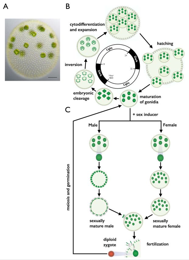
Sexes Evolved from Ancestral Mating-Type Specification Pathway

  Author Summary                                                           and repression of the minus program. A second MT2 gene, MTD1,
                                                                           also contributes to MT2 gametic differentiation but is not essential
  Sexual differentiation in eukaryotes is manifested in two                for it [26]. MID is a rapidly evolving gene [27], but orthologs have
  fundamentally different ways. Unicellular species may have               been found in MT2 strains or in males of all volvocine algae
  mating types where gametes are morphologically identical                 examined to date including VcMID in V. carteri (Figure S1A)
  but can only mate with those expressing a different                      [20,21,27–30]. However, the role of MID in sex determination has
  mating type than their own, while multicellular species                  not been investigated outside of Chlamydomonas.
  such as plants and animals have male and female sexes or                    Volvox carteri f. nagariensis (hereafter V. carteri) is a spheroidal
  separate reproductive structures that produce sperm and                  multicellular alga whose vegetative form is identical for males and
  eggs. The relationship between mating types and sexes                    females (Figure 1A). Each vegetative spheroid contains ,2,000
  and whether or how an ancestral mating-type system
                                                                           sterile flagellated somatic cells on the periphery that provide
  could have evolved into a sexually dimorphic system are
                                                                           motility, while inside the spheroid are ,16 large immotile
  unknown. In this study we investigated sex determination
  in the multicellular green alga Volvox carteri, a species with           reproductive cells called gonidia. All of the cells are embedded
  genetic sex determination; we established the relationship               within a clear extracellular matrix that comprises most of the
  of V. carteri sexes to the mating types of its unicellular               spheroid volume. The two-day vegetative reproductive cycle
  relative, Chlamydomonas reinhardtii. Theoretical work has                begins with mature gonidia undergoing embryogenesis to form
  suggested that sexual dimorphism could be acquired by                    new miniature juvenile spheroids. During embryogenesis a pro-
  linkage of gamete size-regulatory genes to an ancestral                  grammed series of symmetric and asymmetric cleavage divisions
  mating-type locus. Instead, we found that a single                       occurs to produce a hollow ball of 2,000 small cells with 12–16
  ancestral mating locus gene, MID, evolved from its role                  large cells on the anterior surface. The process of inversion then
  in determining mating type in C. reinhardtii to determine                turns the embryo inside out so that the large cells end up on the
  either spermatogenesis or oogenesis in V. carteri. Our                   interior of the spheroid where they will differentiate into new
  findings establish genetic and evolutionary continuity                   gonidia, and the small cells end up oriented with their basal bodies
  between the mating-type specification and sex determi-                   facing outward, and will begin to grow flagella as they undergo
  nation pathways of unicellular and multicellular volvocine               somatic differentiation. Over the next 1.5 days the juveniles grow,
  algae, and will enable a greater understanding of how a                  mature into adults, hatch, and begin the cycle again (reviewed in
  transcriptional regulator, MID, acquired control over a                  (Figure 1B) [31,32]). Unlike C. reinhardtii that uses a nutrient trigger
  complex developmental pathway.                                           for gametogenesis, sexual differentiation in V. carteri is triggered by
                                                                           a diffusible glycoprotein hormone called sex-inducer that is active
   Volvocine algae are an excellent model for investigating the            on both sexes [33–35]. In response to sex-inducer, gonidia from
evolution of sexual dimorphism. They form a monophyletic clade             vegetative females and males undergo modified embryogenesis
encompassing a progression from unicellular species to multicel-           programs to produce sexual spheroids (Figure 1C) [36,37].
lular forms with increasing organismal size and cell-type special-         Sexually induced female spheroids have ,2,000 somatic cells
ization [12,13]. Volvocine algae all have a haploid vegetative             similar to vegetative females, but inside contain 32–48 large egg
reproductive cycle, but under specific conditions can be induced to        cells that are formed during embryogenesis through altered timing
undergo sexual differentiation and mating to form dormant                  of asymmetric cell divisions. Sexually induced male spheroids
diploid zygospores. Zygospores undergo meiosis and produce                 develop with 128 somatic cells and 128 large cells called
haploid progeny that reenter the vegetative phase [4,14].                  androgonidia that are also produced through modification of
Chlamydomonas and smaller colonial volvocine genera are isoga-             asymmetric embryonic division patterning. The day after male
mous, while larger colonial forms are anisogamous or oogamous as           sexual embryogenesis each androgonidial cell undergoes addition-
is the case with the genus Volvox [15,16]. Some species of Volvox          al cleavage divisions to form a packet of 64 or 128 sperm cells.
and other anisogamous volvocine algae are heterothallic with               Sperm packets hatch and swim together to a sexual female where
genetically determined male and female sexes, while others are             they break apart into individual sperm that enter the female
homothallic with a single clone producing a mixture of all-male            through a fertilization pore. Sperm swim within the female until
and all-female colonies (dioecy), or homothallic with a single clone       they find an egg and then fuse with it to form a diploid zygospore.
producing colonies containing both male and female gametes                 Upon germination a single vegetative meiotic progeny is formed
(monoecy) (reviewed in [16]). Previous studies have made use of            while the remaining three meiotic products are discarded as polar
volvocine algae to evaluate theories relating to the evolution of          bodies (Figure 1C) [38].
anisogamy and oogamy [13,17–19], but the genetic basis for                    Sexual differentiation in V. carteri is controlled by a dimorphic
sexual dimorphism in this clade is still unclear [4,20,21].                sex-determining locus (MT) with haplotypes designated MTM
   In C. reinhardtii, the two genetically determined mating types,         (male) and MTF (female). V. carteri MT occupies an equivalent
plus and minus, are morphologically similar, but express mating-           chromosomal position to C. reinhardtii MT based on flanking
related genes that allow fusion with a partner of the opposite             syntenic gene content, but is at least 5-fold larger. Compared with
mating type [14,22]. Gametic differentiation in C. reinhardtii is          C. reinhardtii MT V. carteri MT contains more sequence rearrange-
triggered by absence of nitrogen (2N) and is governed by a mating          ments between haplotypes, more repeat sequences, and has
locus (MT) whose two haplotypes, MT+ and MT2, are large,                   gametolog pairs (genes with an allele in both MT haplotypes) that
rearranged multigenic regions, which are suppressed for recom-             are far more differentiated from each other [20,24]. V. carteri MTF
bination and therefore segregate as Mendelian alleles [23,24]. The         and MTM haplotypes can thus be considered a UV sex
C. reinhardtii gene MID (CrMID) is present only in the MT2                 chromosome pair [39]. As described above, it has been proposed
haplotype and encodes a putative RWP-RK family transcription               that anisogamy or oogamy could evolve through a size-regulatory
factor whose expression is induced by 2N and that governs                  gene becoming linked to an ancestral mating locus [11]. Both
gametic differentiation [25]. The presence of MID activates the            MTM and MTF haplotypes contain a putative cell-size regulatory
minus differentiation program and represses the plus program,              gene, MAT3, whose alleles are highly dimorphic in sequence and
while the absence of MID causes activation of the plus program             expression between the sexes [20]. However, it is now apparent

PLOS Biology | www.plosbiology.org                                     2                            July 2014 | Volume 12 | Issue 7 | e1001904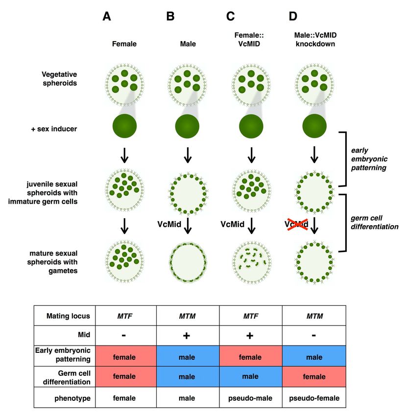
Sexes Evolved from Ancestral Mating-Type Specification Pathway

PLOS Biology | www.plosbiology.org   3                     July 2014 | Volume 12 | Issue 7 | e1001904
Sexes Evolved from Ancestral Mating-Type Specification Pathway

Figure 1. V. carteri vegetative and sexual cycles. (A) Color DIC image of vegetative V. carteri spheroid with large reproductive cells (gonidia) on
the interior and somatic cells on the exterior. Scale bar = 50 mm. (B) Key stages of the two-day vegetative reproductive cycle are depicted with relative
timing indicated by the interior clock diagram showing the 16 h light and 8 h dark phases. Starting from ,6:00 and going clockwise vegetative
gonidia undergo embryonic cleavage followed by inversion to make new juvenile spheroids. Juveniles grow and eventually hatch and mature into
the next generation of parental spheroids. The vegetative cycle is identical for males and females. (C) Key stages of the sexual cycle are depicted top
to bottom starting with vegetative male or female gonidia that have been exposed to sex-inducer. Sexually induced gonidia undergo modified
embryogenesis to produce sexual males with 128 large androgonidia and 128 somatic cells, or sexual females with 32–48 eggs. Subsequent cleavage
of androgonidia produces sperm packets that are released and swim to a female whereupon they dissolve into single sperm and enter the female
spheroid to fertilize eggs. Diploid zygotes differentiate into environmentally resistant, orange-pigmented, dormant zygospores that when
germinated undergo meiosis and produce three polar bodies plus a single haploid vegetative progeny that can reenter the vegetative reproductive
cycle.
doi:10.1371/journal.pbio.1001904.g001

that anisogamy and oogamy in volvocine algae predate the                       (Figure 2B). When wild-type vegetative male gonidia are exposed
appearance of MAT3 allelic dimorphism in the lineage meaning                   to sex inducer they undergo modified embryogenesis and develop
that other mating locus genes probably underlie the origins of                 with 128 sperm packets and 128 somatic cells (Figure 2C). When
anisogamy and oogamy [40]. Although MTM contains a MID                         vegetative gonidia from Eve::VcMID-BH lines were exposed to sex
homolog, VcMID (Figure S1A), its role in sexual differentiation is             inducer they developed into progeny spheroids with a novel
unclear because VcMID mRNA is expressed constitutively in both                 pseudo-male sexual phenotype: They produced ,2,000 somatic
vegetative and sexual stages of males [20]. The apparent                       cells and 32–48 sexual germ-cell precursors in a pattern similar to
uncoupling of VcMID expression from the sexual cycle suggests                  that of female eggs; but each of the 32–48 germ-cell precursors in
that the VcMid protein might have a function outside of the sexual             the Eve::VcMID-BH lines underwent additional cleavage divisions
cycle or that its function might be regulated differently than that of         like male androgonidia to produce sperm packets (Figure 2D).
CrMID whose expression is induced by 2N.                                       Moreover, the sperm produced in Eve::VcMID-BH lines were
   In this study, we tested the role of VcMID in V. carteri sex                capable of fertilizing wild-type female eggs to produce character-
determination by making transgenic females that express VcMid                  istically orange-pigmented, thick-walled zygotes (Figure 2E).
protein or by knocking down its expression in males using RNAi.                However, male function was incomplete as fertility defects were
We found that expression of VcMID in females is sufficient to                  noted (see next section). Similar to wild-type sperm (and unlike
convert eggs to sperm packets, while its absence in males causes               wild-type female eggs) the Eve::VcMID-BH sperm cells were
androgonidial cells to differentiate into eggs. However, alteration            terminally differentiated and could not revert back to vegetative
of VcMid expression did not affect female or male early embryonic              growth if left unfertilized. Another male-specific phenotype
patterning during which the number and location of germ-cell                   exhibited by Eve::VcMID-BH lines was frequent spontaneous
precursors is established. We found that VcMID mRNA is                         occurrence of sexual differentiation in vegetative cultures [36], a
expressed in all cell types, but VcMid protein accumulation is                 trait whose underlying basis is not clear, but which appears to be
regulated by cell type and its subcellular localization is restricted to       under the control of VcMID.
nuclei of differentiating and mature male gametes. Swapping
experiments with CrMid demonstrated that the VcMid DNA                         Fertility Defects in Females Expressing VcMid
binding domain and N-terminal domain are both required for its                    Although some of the Eve::VcMID-BH sperm were functional
function in directing spermatogenesis in V. carteri. Crosses with sex-         and could fertilize wild-type eggs, the sperm packets and sperm
reversed strains revealed sexually antagonistic interactions be-               cells from these strains had multiple defects including hetero-
tween genes in MT and the sexual development pathway                           chronic delays in maturation and hatching defects (Figure S3A).
controlled by VcMid that negatively impacted reproductive fitness              The sperm packets were four times larger than those from wild-
when gamete type did not match the mating locus genotype.                      type males and contained about four times as many sperm cells
                                                                               (256 sperm/packet) (Figure 2F and 2G), some of which were
Results                                                                        aberrantly formed in contrast with wild-type sperm cells that had
                                                                               uniform morphology (Figures 2H, 2I, and S3B–S3F). Nonetheless
VcMid Controls Spermatogenesis                                                 the crosses between Eve::VcMID-BH pseudo-males and wild-type
   We tested the role of VcMid protein in sexual differentiation by            females produced MTF/MTF diploid zygotes (Figure 2E) that
generating female transgenic lines with an autosomally integrated              could germinate and produce haploid progeny. Forty-one progeny
VcMID transgene (pVcMID-BH) expressed under its own                            from one such cross were genotyped, half of which (19/41)
promoter and fused to a blue fluorescent protein (BFP) and a                   inherited the VcMID-BH transgene and developed as pseudo-
hemagglutinin (HA) epitope tag at its C-terminus to detect                     males, and half of which lacked the transgene and developed as
expression (Eve::VcMID-BH) (Figures 2A and S1B). Female                        normal females (22/41).
transformants carrying an untagged version of VcMID (Figure
S1C) had identical phenotypes as those carrying the tagged                     Expression and Localization of VcMid Are under Post-
version, and all subsequent work was done with tagged strains and              transcriptional Control
untagged transformants as a negative control for detection of                     The absence of a vegetative phenotype in Eve::VcMID-BH
VcMid protein. Eve::VcMID-BH lines showed a normal vegetative                  transgenic lines despite constitutive expression of the VcMID-BH
phenotype and constitutively expressed the mRNA for the VcMID-                 mRNA (Figure S2) suggested that VcMid protein expression or
BH transgene, an expression pattern identical to the endogenous                localization might be under posttranscriptional control. Although
VcMID mRNA in males (Figure S2) [20]. As described above,                      we could not detect BFP fluorescence in Eve::VcMID-BH strains,
when wild-type vegetative female gonidia are exposed to sex                    we could detect the HA epitope tag by Western blotting at all
inducer they undergo modified embryogenesis and develop into                   stages of vegetative and sexual development (Figures 3A and S4).
sexual spheroids with 32–48 eggs and ,2,000 sexual somatic cells               Using immunofluorescence (IF) a nuclear-localized signal was

PLOS Biology | www.plosbiology.org                                         4                            July 2014 | Volume 12 | Issue 7 | e1001904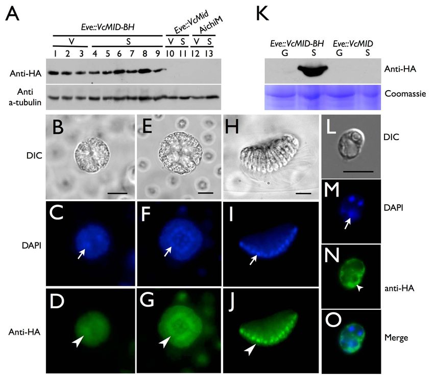
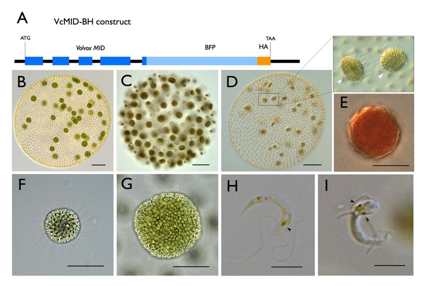
Sexes Evolved from Ancestral Mating-Type Specification Pathway

Figure 2. Ectopic Expression of VcMID converts egg precursors to sperm packets. (A) Diagram of VcMID-BH expression construct. Dark blue
boxes depict VcMID exons; black lines depict introns and intergenic regions; light blue box depicts BFP; orange box depicts HA epitope. Start (ATG)
and stop (TAA) codon positions are shown along with scale bar in black. (B) Wild-type mature sexual Eve (female) spheroid with ,32 large green
eggs. (C) Wild-type mature sexual AichiM (male) spheroid with ,128 sperm packets. (D) Mature sexual Eve::VcMID-BH pseudo-male with ,32 sperm
packets, two of which are magnified in expanded box to the right. Scale bars for (B–D) = 50 mm. (E) Mature zygote from Eve::VcMID-BH6Eve. (F) Sperm
packet from wild-type AichiM. (G) Sperm packet from pseudo-male Eve::VcMID-BH. Scale bars for (E–G) = 25 mm. (H) Sperm cell from wild-type male
with a single eyespot indicated by a black arrowhead. (I) Aberrant sperm cell from Eve::VcMID-BH pseudo-male with two eyespots indicated by black
arrowheads. Scale bars for (H) and (I) = 5 mm.
doi:10.1371/journal.pbio.1001904.g002

detected for VcMid-BH protein in cleaving androgonidia and in               that restricts its accumulation to somatic cells during vegetative
mature sperm packets (Figures 3B–3J, S5A–S5R, and S6C–S6H),                 growth (Figures 3K and S6B). However, unlike the case for
a result similar to earlier findings of Mid protein localization in         androgonidia and sperm cells, the VcMid-BH protein signal in
sperm nuclei of Pleodorina [21]. However, nuclear VcMid-BH was              vegetative somatic cells was excluded from the nucleus and was
not detected during early stages of sexual male embryogenesis               instead observed only in the cytosol and peri-nuclear region
prior to androgonidia cleavage (Figure S7).                                 (Figures 3L–3O, S6I–S6P, and S9C–S9J). Together these data
   We also examined VcMid-BH expression and localization in                 suggest that cell-type–limited expression and regulated nuclear
gonidia and somatic cells from vegetative spheroids. RNA was                localization of VcMid restrict its function to developing and
prepared from purified gonidial or somatic cells and reverse                mature sexual male germ cells in V. carteri.
transcription and PCR (RT-PCR) detected VcMID-BH mRNA
at similar levels in both cell types from transgenic females                VcMid Is Required for Spermatogenesis and Represses
(Eve::VcMID-BH) and males (AichM::VcMID-BH) (Figure S8B and                 Oogenesis in Males
S8D). The endogenous VcMID transcript from wild-type males was                 In order to test whether VcMID is necessary for sexual
also expressed in both vegetative cell types (Figure S8C and S8D).          differentiation of males we developed a new strategy for gene
Whole cell extracts were prepared from purified Eve::VcMID-BH               knockdown on the basis of RNAi-inducing hairpin constructs. The
or AichM::VcMID-BH somatic cells and gonidia and subjected to               hairpin-forming portion of the construct corresponding to VcMID
SDS-PAGE and Western blotting to detect VcMid-BH protein                    sequences was inserted directly into the 39 UTR of the nitA
(Figures 3K, S9A, and S9B). In contrast to VcMID-BH mRNA that               selectable marker gene to allow direct and simultaneous selection
was present in both vegetative cell types, VcMid-BH protein was             of both NitA+ and hairpin expression (Figures 4A and S10; Text
only detected in vegetative somatic cells indicating that there is          S1). This strategy has been successful for other loci besides VcMID
cell-type–specific regulation of VcMid protein synthesis or stability       (unpublished data), but only the results for VcMID are presented

PLOS Biology | www.plosbiology.org                                      5                           July 2014 | Volume 12 | Issue 7 | e1001904
Sexes Evolved from Ancestral Mating-Type Specification Pathway

Figure 3. Cell-type restricted expression and sex-regulated nuclear localization of VcMid. (A) Immunoblot of SDS-PAGE fractionated
protein extracts from HA-tagged pseudo-male strain Eve::VcMID-BH (lanes 1–9), untagged pseudo-male strain Eve::VcMid (lanes 10, 11), and wild-type
male strain AichiM (lanes 12, 13). Lanes 1–3 contain extracts from vegetative spheroids at adult stage, mid-cleavage stage, and unhatched juvenile
stage respectively. Lanes 4–9 contain extracts from spheroids undergoing sexual development with pre-cleavage stage, mid-cleavage stage,
unhatched juvenile stage, cleaving androgonidia stage, and mature sexual adult stage, respectively. See also Figure S2B and S2C. Lanes 10 and 12
contain extracts from adult vegetative-stage spheroids. Lanes 11 and 13 contain extracts from mature sexual-stage spheroids. The bands in the upper
panel are VcMid-BH protein detected with an anti-HA antibody. The bands in the lower panel come from the same blot re-probed with an anti-
tubulin antibody as a loading control. (B–J) DIC (B, E, H) or false-colored IF images of cleaving androgonidia from Eve::VcMID-BH sexual germ cells at
the two-cell stage (B–D), sixteen-cell (E–G) stage, and from a mature sperm packet (H–J). IF samples were stained with DAPI shown in blue (C, F, I) or
with anti-HA shown in green (D, G, J). Arrows and arrowheads show locations of a representative nucleus from each image. Scale bars = 10 mm. (K)
Upper panel, anti-HA immunoblot of SDS-PAGE fractionated protein extracts of purified vegetative gonidia (G) or somatic (S) cells from Eve::VcMID-BH
and Eve::VcMID spheroids. Lower panel, Coomassie-stained gel used as a loading control. (L–O) IF detection of VcMid-BH protein from a
representative vegetative somatic cell of Eve:: VcMID-BH stained as in (B–J). The arrows in (M, O) show the nucleus, while the chloroplast nucleoids
(smaller DAPI-stained spots) are unlabeled. The VcMid-BH signal in (N, O) is excluded from the nucleus as evident in the merged image (O). Scale
bar = 7.5 mm.
doi:10.1371/journal.pbio.1001904.g003

here. We also note that the VcMID knockdown phenotype was                     Materials and Methods for details). All transgenic male strains had
gene specific and did not occur when hairpins targeting other loci            normal vegetative phenotypes, and both hairpin constructs
were introduced into V. carteri (unpublished data). Two hairpins              reduced VcMID expression; but AichiM::VcMID-hp1 lines had
targeting VcMID (VcMID-hp1 and VcMID-hp2) were introduced                     lower VcMID transcript levels than AichiM::VcMID-hp2 lines
into a V. carteri wild-type male strain AichiM to generate                    (Figure S2). We note that unlike wild-type males or pseudomales
AichiM::VcMID-hp1 and AichiM::VcMID-hp2 transgenic lines (see                 (see above), vegetative cultures of AichiM::VcMID-hp1 lines did not

PLOS Biology | www.plosbiology.org                                        6                           July 2014 | Volume 12 | Issue 7 | e1001904
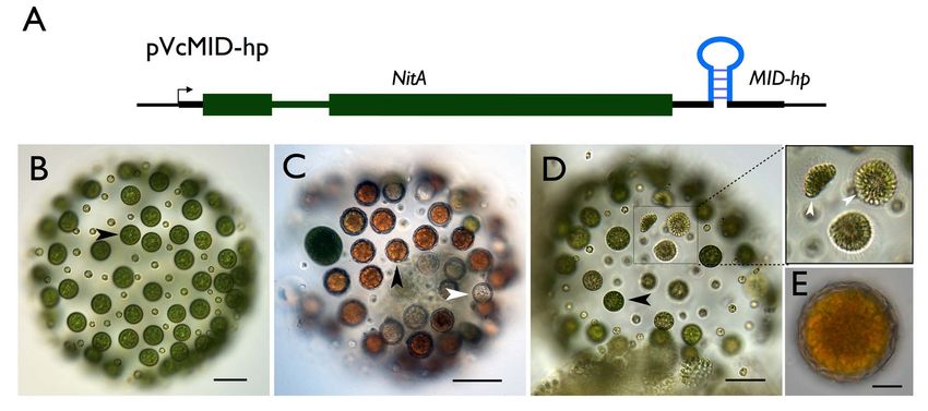
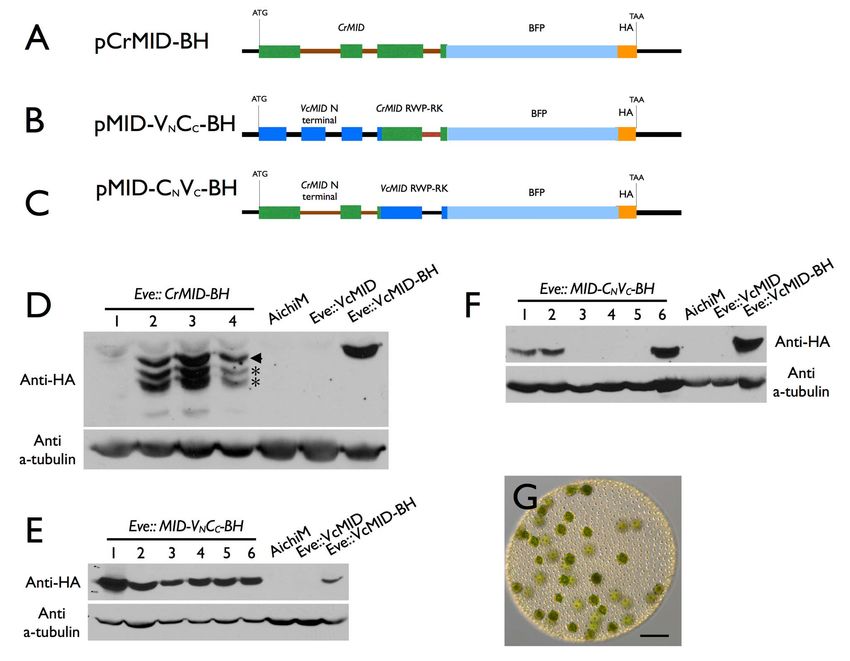
Sexes Evolved from Ancestral Mating-Type Specification Pathway

Figure 4. Conversion of male germ cells to eggs by knockdown of VcMID. (A) Diagram of VcMID hairpin construct. Green boxes, NitA exons;
medium green line, intron; thick black lines, UTRs; VcMID hairpin structure inserted in 39 UTR is shown in its approximate location. (B) Sexually
induced AichiM::VcMID-hp1 pseudo-female with eggs in place of sperm packets. Black arrowhead shows an egg. (C) AichiM::VcMID-hp1 pseudo-female
fertilized by wild-type male sperm. Black arrowhead shows a normal-appearing developed zygote; white arrowhead shows an aborted zygote. (D)
Hermaphrodite phenotype of mature sexually induced AichiM::VcMID-hp2 spheroid with eggs and sperm packets. Black arrowhead shows egg; boxed
region has three sperm packets that are magnified in the expanded view to the right. Scale bars for (B–D) = 50 mm. (E) Zygote produced by self-
fertilization of AichiM::VcMID-hp2 spheroid. Scale bar = 10 mm.
doi:10.1371/journal.pbio.1001904.g004

undergo spontaneous sexual induction. Sexually induced Ai-                 Partial VcMID Knockdown Generates Self-Fertile
chiM::VcMID-hp1 lines with strong knockdowns showed a novel                Hermaphrodites
phenotype: Their early sexual development proceeded as it would               AichiM::VcMID-hp2 lines were not as severely knocked down for
for a wild-type male strain and resulted in spheroids that contained       VcMID expression as AichiM::VcMID-hp1 lines (Figure S2), and had
128 small somatic cells and 128 large cells that resembled                 a distinct hermaphrodite phenotype in which sexual spheroids
uncleaved androgonidia (Figure 4B), but the large cells never              developed with a mixture of normal-looking male sperm packets
underwent further cleavage into sperm packets. Instead, many of            and pseudo-female eggs (Figure 4D). The hermaphrodite lines
them could be successfully fertilized with wild-type male sperm to         exhibited self-fertility as evidenced by zygospores that formed in
make MTM/MTM diploid zygospores (Figure 4C). The ability to                sexually induced monocultures (Figure 4E). These results indicate
differentiate as zygospores when fertilized indicates that the             that V. carteri sex determination is highly sensitive to VcMid dosage
presumptive androgonidia in AichiM::VcMID-hp1 strains were                 where either a male or female fate is established depending on the
converted to functional eggs and that these strains were behaving          level of VcMid.
as pseudo-females. However, unlike normal zygotes from a wild-
type cross, 30%–50% of the zygotes from AichiM6AichiM::Vc-
MID-hp1 pseudo-female crosses died and bleached shortly after              Volvox and Chlamydomonas Mid Proteins Are
fertilization (Figure 4C), a phenotype that depended on addition           Functionally Distinct
of exogenous sperm. The surviving zygotes from these crosses                  In several instances genes from C. reinhardtii have been shown to
produced some viable meiotic progeny, but germination and                  function interchangeably with their V. carteri orthologs in
survival of the progeny were reduced compared with normal                  developmental processes such as inversion and asymmetric cell
wild-type zygotes (Table S1). 29/43 viable progeny inherited the           division (reviewed in [42,43]). Mid proteins have at least two
VcMID-hp1 transgene and developed as pseudo-females, while                 domains: The C-terminal region has a predicted RWP-RK motif
14/43 lacked the transgene and developed as normal males. The              DNA binding domain, while the N-terminal region does not show
apparent deviation from a 1:1 inheritance pattern of the                   similarity to characterized protein domains from other organisms
transgene was noted but was not pursued further in this study.             (Figure S1) [28]. We tested whether either of the domains from
The high mortality of pseudo-female eggs—whose mating loci                 CrMid could substitute for those of VcMid to control spermato-
are genetically male—suggest that MTF contains genes that                  genesis in V. carteri. To do so we generated three constructs in
promote female gamete and/or zygote fitness that are absent                which all or part of the CrMID genomic coding region was
from MTM. If left unfertilized, the eggs from AichiM::VcMID-hp1            substituted for VcMID sequences in pVcMID-BH. One construct
lines could de-differentiate and reenter the vegetative reproduc-          contained the entire CrMID gene (pCrMID-BH) (Figure 5A) while
tive cycle as do unfertilized female eggs. A similar phenotype as          the other two contained the CrMID N-terminal domain fused to
our pseudo-male strain was reported previously for a male                  the VcMID DNA binding domain (pMID-VNCC-BH) (Figure 5B),
mutant [41], but the mutant strain is no longer available for              or the VcMID N-terminal domain fused to the CrMID DNA
characterization.                                                          binding domain (pMID-CNVC-BH) (Figure 5C). All three

PLOS Biology | www.plosbiology.org                                     7                           July 2014 | Volume 12 | Issue 7 | e1001904
Sexes Evolved from Ancestral Mating-Type Specification Pathway

Figure 5. Chlamydomonas MID cannot substitute for Volvox MID. (A–C) Diagrams of constructs pCrMID-BH (A), pMid-VNCC-BH (B), and pMid-
CNVC-BH (C). Dark blue boxes depict VcMID exons; black lines depict VcMID introns and intergenic regions; green boxes depict CrMID exons; brown
lines depict CrMID introns; light blue boxes depict BFP; Orange box depicts HA epitope. Start (ATG) and stop (TAA) codons are shown along with scale
bar in black. (D–F) Immunoblots of SDS-PAGE fractionated protein extracts from Eve transformants or control strains probed with anti-HA antibodies
(upper panels) or re-probed with tubulin antibodies (lower panels). Numbered lanes indicate independent transformants, some of which express the
tagged Mid constructs. Control strains are wild-type male AichiM, Eve transformed with untagged VcMID, and Eve transformed with tagged VcMID-BH
shown in Figure 1A. (D) Eve::Mid-CrMID-BH transformants. Arrowhead indicates predicted full length CrMid-BH and asterisks represent breakdown or
alternative processing products. (E) Eve::Mid-VNCC-BH transformants. (F) Eve:: Mid-CNVC-BH transformants. (G) Color DIC image of sexually induced
Eve::Mid-CNVC-BH transformant number 6 showing typical arrangement of eggs similar to a wild-type female. Scale bar = 200 mm.
doi:10.1371/journal.pbio.1001904.g005

constructs as well as pVcMID-BH were introduced into wild-type               Discussion
females, and transformants that expressed the different predicted
proteins were identified (Figures 5D–5F). Unlike Eve::VcMID-BH               Deep Homology between Mating Types and Sexes
transformants that showed a pseudo-male phenotype (Figure 1D),                  Although it was reasonable to predict that the route for
none of the transformants that expressed CrMid or Mid chimeras               evolving sexual dimorphism would be through addition of new
had this phenotype, but instead always developed as wild-type                genetic functions and pathways to a core mating locus as
females (Figure 5G; Table S2). The CrMid-BH protein was                      proposed originally by Charlesworth for the evolution of
expressed as a full-length form and two shorter isoforms                     anisogamy [11], we found instead that a single conserved
(Figure 5D), possibly due to proteolytic cleavage or incorrect               volvocine algal mating locus gene, MID, is largely responsible for
pre-mRNA processing. However, lines with either of the two                   controlling male versus female sexual differentiation in V. carteri.
chimeric constructs expressed only full-length predicted proteins at         It is notable that VcMID controls multiple sexual traits in V. carteri
levels comparable to VcMid-BH (Figure 5E and 5F). We conclude                that have no analogs in C. reinhardtii. The male traits controlled
from these experiments that CrMid and VcMid are not                          by VcMID include specialized cleavage divisions of androgonidia,
functionally interchangeable, and that both the N-terminal and               sperm packet inversion, sperm packet hatching, specialized
RWP-RK domains of VcMid are required to activate spermato-                   sperm cell morphology, and gamete recognition without flagellar
genesis in sexual germ cells.                                                adhesion.

PLOS Biology | www.plosbiology.org                                       8                           July 2014 | Volume 12 | Issue 7 | e1001904
Sexes Evolved from Ancestral Mating-Type Specification Pathway

   The term deep homology is used to describe ancient and                     Evolution of Mid and Sexual Cycles in the Volvocine
conserved genetic mechanisms that control traits that, on the                 Lineage
surface, appear disparate or have no obvious homology relation-                  As described in the Introduction, volvocine algae exhibit wide
ship [44]. Metazoan eye development is a classic example; across              diversity in their sexual cycles: There are isogamous, anisogamous,
phyla with very different eye architecture, it is controlled by the           and oogamous species with sexual cycles that can be heterothallic
conserved transcription factor, eyeless/Pax6 [45]. In the case of             or homothallic. Among homothallic species some are dioecious
volvocine algae Mid proteins control two very different manifes-              (producing a mixture of all male and all female sexual offspring) or
tations of sexual reproduction in C. reinhardtii and V. carteri whose         monoecious (producing sexual offspring containing gametes of
MID orthologs have been diverging for as long as 200 million years            both sexes in one individual) [16,55].
[46]. Sexually dimorphic gametes have evolved from isogamous                     V. carteri is a heterothallic species with genetically determined
ancestors several times in independent multicellular taxa, and the            sexes; but, a remarkable phenotype was produced by a partial
mechanism could be similar to, or different from, that in volvocine           RNAi knockdown of VcMID in males (Figure 4D). Rather than
algae where a master regulatory gene acquired the ability to direct           developing as male or female, the partial-knockdown male
sperm-egg dimorphism. Oogamy was likely established very early                spheroids became self-fertile monoecious hermaphrodites that
in the Streptophyte lineage before the split between Charophyte               produced sperm and eggs in a single individual. On the basis of
algae and land plants, but the origins and bases of oogamy in                 this observation, it can be inferred that sexual differentiation in V.
Charophytes remain unclear [47]. The recent identification of an              carteri is bi-stable and highly sensitive to initial MID dosage. Once
anisogamous sexual cycle in a Choanoflagellate—a unicellular                  the Mid-sensitive step of development is initiated (which may
relative of metazoans—introduces the potential for investigating              coincide with nuclear translocation of VcMid protein), positive
the early evolution of gamete size dimorphism in animals [48].                and negative feedback loops may be used to lock the sex
                                                                              determination program into a male or female state. This state
MID Is a Master Regulator of Sex Determination in                             could be achieved either independently of VcMid concentration,
Volvocine Algae                                                               or through positive/negative reinforcement of initial expression
   Our results demonstrate for the first time a function for Mid              states. The phenotype of hermaphroditic development by partial
protein in a volvocine algal sexual cycle outside of the genus                VcMID knockdown may be relevant to the evolution of homo-
Chlamydomonas and suggest that Mid could be the master regulator              thallism, which appears to have arisen in all three major clades of
of mating type, gamete size, and gender throughout the volvocine              Volvox [16,55,59]. We speculate that naturally evolved homothallic
lineage where MID genes have been identified in most genera                   volvocine algae possess a MID gene whose expression is insufficient
[21,28,30]. Although MID sequences were previously shown to                   to specify 100% male gamete production—much like our VcMID-
evolve rapidly, the finding that Mid protein from C. incerta (now             hp2 strains. Moreover, the timing of MID expression in homothallic
reclassified as C. globosa [49]) can substitute for C. reinhardtii Mid        species of Volvox could play a role in determining monoecious versus
indicates that functional conservation can be retained after                  dioecious reproductive development: If the Mid-sensitive develop-
speciation [27]. However, C. reinhardtii Mid protein could not                mental switch is triggered relatively late in development after germ-
substitute for the V. carteri ortholog, which appears to require both         cell precursors are formed then a mixture of male and female
its native DNA binding and N-terminal domains to function in sex              gametes could develop within a single spheroid as we found with
determination (Figure 5). Future work using cross-species comple-             VcMID-hp2 strains (i.e., homothallism, monoecy). In contrast, if the
mentation will help clarify whether Mid protein function co-                  Mid-sensitive step occurred very early in development before
evolved with sexual dimorphism in volvocine algae as our data                 individual germ cells were established, then the fate of all germ cells
suggest might be the case.                                                    within a mature spheroid might be locked into a male or female
   Transcriptional regulatory network evolution has been studied              program and the resulting population would produce a mixture of
in various developmental contexts [50–53], but very little is known           all-female or all-male spheroids (i.e., homothallism, dioecy).
about how regulatory networks are modified or coopted during                     The bi-stability of sex determination at intermediate levels of
unicellular to multicellular transitions [54]. The Mid system in              MID expression in V. carteri is also reminiscent of the iso1 mutant
volvocine algae represents a new opportunity for understanding                phenotype in C. reinhardtii where MT2 iso1 cells differentiate into a
how a cell-type specification pathway in a unicellular ancestor               mixture of plus and minus gametes that iso-agglutinate but cannot
evolved to control a complex developmental program in a                       self-fertilize [60]. The self-infertility of iso1 MT2 strains is due to
multicellular descendant. While mating-type differentiation in                the absence of FUS1, a gene from the MT+ haplotype that is
Chlamydomonas appears to involve differential expression of a small           required for gamete fusion [61]. In contrast, there are no essential
number of genes between the plus and minus gametes [14,22], a                 genes in MTF of V. carteri that are absolutely required for
larger set of differentially expressed genes might be expected to             fertilization and subsequent germination of progeny from matings
specify sperm and eggs, which are developmentally very different              between pseudo-females (VcMID knockdown strains) and males.
from each other [37,55]. An important future goal will be to                  This lack of essential female MT genes for completing the sexual
identify and compare the direct and indirect targets of Mid                   cycle may have facilitated transitions from heterothallism to
proteins in both C. reinhardtii and V. carteri that are predicted to be       homothallism in volvocine algae.
more numerous and diverse in V. carteri.
   Another interesting question is whether MID-like genes function            V. carteri Has Evolved New Regulatory Inputs for Mid
in sex determination in green algae outside of the volvocine                     In volvocine genera other than Volvox—including the anisoga-
lineage. The molecular bases for sex determination in most green              mous genus Pleodorina— sexual differentiation and MID expression
algae and protists are poorly understood, but RWP-RK family                   are both triggered by the absence of nitrogen (2N) [21,25,28,62].
proteins are found throughout the green eukaryotic lineage [56],              In contrast, V. carteri f. nagariensis and other Volvox species use
including small Mid-like proteins in Prasinophyte algae [57], and             species-specific pheromones called sex inducers to trigger sexual
even in distantly related Cryptophyte algae [58]. It remains to be            differentiation [16,55,63]. A seemingly parsimonious evolutionary
seen whether these Mid-like proteins in non-volvocine species                 route for rewiring input into the Mid pathway in V. carteri would
function in sex determination.                                                simply place VcMID transcription under the control of sex inducer

PLOS Biology | www.plosbiology.org                                        9                           July 2014 | Volume 12 | Issue 7 | e1001904
Sexes Evolved from Ancestral Mating-Type Specification Pathway
instead of nitrogen availability. Unexpectedly, however, VcMID sex, but harm the other [39,65], and should accumulate repeat
mRNA and VcMid protein are both expressed constitutively at all sequences, as appears to have occurred in both Volvox and in the
life cycle stages (Figures 3A and S4) [20], and unlike the case in C. bryophyte Marchantia [20,66]. Accumulation of sexually antagonistic
reinhardtii, VcMid appears to be under at least three types of alleles has not been tested for haploid sex chromosomes, but the
posttranscriptional control: (i) Although VcMID mRNA is present extensive divergence between V. carteri MT gametologs suggests that
in both vegetative cell types (somatic and gonidial cells), VcMid this phenomenon may contribute to the developmental and fitness
protein is only translated or stably produced in somatic cells and is defects we observed in pseudo-male and pseudo-female strains.
absent from vegetative gonidia and vegetative embryos (Figures 3K Although none of the sex-limited MT genes besides VcMID
and S6B). (ii) The VcMid protein produced in somatic cells is appear to be essential for V. carteri sex determination and
excluded from the nucleus (Figures 3L–3O, S6I–S6P, and S9C– completion of the sexual cycle, they clearly impact sexual
S9J). (iii) In response to the presence of sex inducer, VcMid protein development and reproductive fitness. A striking phenotype for
accumulates in the nuclei of cleaving androgonidial cells and in the both the pseudo-male and pseudo-female strains was the
nuclei of sperm cells where it is presumed to function in specifying patterning of their germ-cell precursors formed during sexual
sperm development (Figure 3B–3J, S5, and S6C–S6H). development (Figures 2B–2D and 6A–6C). It is clear from these
In depth study will be required to determine how cell-type– phenotypes that sexual germ cell patterning (i.e., the number and
regulated production and localization of VcMid are achieved, but distribution of germ-cell precursor cells and ratio of germ-cell
it seems reasonable to infer that one or more factors are produced precursors to sexual somatic cells) is separable from germ cell
in sexually induced spheroids that promote the translation and/or differentiation and is not controlled by the VcMid pathway.
stability of VcMid and its nuclear localization in androgonidia and Instead, this sexual patterning trait must be controlled by other
sperm. The factor may interact directly with VcMid as a partner MT genes (Figure 6). One candidate for this male-female patterning
and may help specify its localization at promoters of target genes, difference is the MAT3 gene that encodes the retinoblastoma-
but this idea remains to be tested. The absence of VcMid protein related homolog in V. carteri [20,67]. In Chlamydomonas, Mat3 protein
in vegetative gonidia despite its message accumulating to the same controls the multiple fission cell cycle by establishing the threshold
extent as in somatic cells seems puzzling at first glance. However, size at which division can occur and by coupling the extent of cell
the block in stability or translation of VcMid in gonidia may have division to mother cell-size [68,69]. Although not directly involved
evolved as a failsafe mechanism to prevent sexual differentiation in determining gamete cell-size as we had originally predicted [40],
during vegetative embryogenesis. Such a mechanism would be it is possible that the male and female alleles of VcMAT3 dictate the
unnecessary in vegetative somatic cells that are already terminally timing of asymmetric cell divisions by coupling embryonic
differentiated, but could potentially be important for vegetative blastomere cell-size to the asymmetric division machinery. Future
gonidial cells because male sexual differentiation is irreversible and work will be aimed towards determining the role of male and female
would be fatal if it occurred at the wrong time. However, why VcMat3 gametologs in sexual cell division or other parts of the V.
vegetative somatic cells express VcMid remains a mystery. As carteri sexual cycle.
noted in Results, we observed no obvious vegetative phase In addition to defects in germ cell patterning in Volvox pseudo-
phenotypes in AichiM::VcMID-hp strains whose somatic cells were males and pseudo-females, these strains show other reproductive
missing VcMid, or in Eve::VcMID-BH strains that contained defects. These include abnormal sperm cell shape and morphology
VcMid in somatic cells, but a definitive conclusion about whether (Figures 2H, 2I, and S3B–S3F), low efficiency of sperm packet
cytoplasmic VcMid has a role in vegetative somatic cells awaits hatching (Figure S3D), and delayed timing of androgonidial
more in depth examination. cleavage into sperm packets (Figure S3A). In depth study may
reveal other defects in pseudo-male sperm related to cytoskeletal
Uncoupling of Gender from Sex Chromosome Identity organization, gamete recognition, motility, and fertilization dynam-
Uncovers Possible Sexually Antagonistic Interactions in ics. Although egg cells lack distinct morphological features like
the MT Locus sperm, we noted a very high mortality rate in pseudo-female eggs
Our results show that the presence or absence of VcMID is the that occurred when we attempted fertilization. The mortality we
key determinant of differentiation in V. carteri sexual spheroids; yet observed under these conditions could be due to pre-zygotic defects
the MT locus of this alga has around 70 additional genes, many of in the eggs caused by their smaller size than wild-type female eggs or
which show sex-biased gene expression [20]. Some of the MT by a mismatch between the male mating locus and the female sexual
genes are present only in the male or only in female haplotype, differentiation pathway. The mortality might also be due to zygotic
while the majority are male and female gametolog pairs that are defects that occur when a copy of the female mating locus is absent
highly diverged in sequence and expression pattern [20]. Our from the zygote immediately after fertilization. Future work aimed
ability to uncouple gender from sex chromosome identity by at developing quantitative assays for distinct steps of fertilization will
manipulation of VcMID expression allowed us to uncover potential allow us to document in more detail the fitness contributions of male
contributions of male and female MT genes to sexual dimorphism and female MT genes to the sexual cycle.
and reproductive fitness. Haploid sex chromosome systems have
received less attention than diploid systems, but there are several Materials and Methods
predictions about them that our results begin to address. Under
haploid dioecy, recessive mutations in sex-linked genes are not Detailed Materials and Methods are provided in Text S1.
sheltered from selection as they are in the heterogametic sex of Materials used in this study will be made available upon request
diploid systems, and are therefore expected to degenerate equally with the completion of a Materials Transfer Agreement from
and lose only genes required for the opposite sex and not their own Donald Danforth Plant Science Center.
[64]. We note, however, that in V. carteri MT there is no evidence
for an allele of a gametolog pair having been eliminated from one Volvox Strains and Culture Conditions
mating haplotype and retained in the other [20]. Haploid sex Eve (Volvox carteri. f. nagariensis UTEX 1885) and AichiM (Volvox
chromosomes are predicted to be similar to diploid systems in that carteri. f. nagariensis NIES 398) were obtained from stock centers
both should accumulate sexually antagonistic alleles that benefit one http://web.biosci.utexas.edu/utex/ and http://mcc.nies.go.jp/,
PLOS Biology | www.plosbiology.org 10 July 2014 | Volume 12 | Issue 7 | e1001904Sexes Evolved from Ancestral Mating-Type Specification Pathway Figure 6. Summary of the roles of VcMID and the mating locus in sexual differentiation. The upper section shows the process of sexual differentiation, top to bottom, starting from vegetative spheroids that are treated with sex inducer. The fate of a single sexually induced gonidia is shown in each case with its phenotype after embryogenesis and after sexual differentiation. Labels to the left show the stages depicted. Labels to the right show the developmental events that are controlled independently by the mating locus (early embryonic patterning) and the VcMid pathway (germ cell differentiation). (A–D) The four columns depict the fates of wild-type females (A), wild-type males (B), females expressing VcMid (pseudo- males) (C), and males with VcMid knocked down (pseudo-females) (D). The table below summarizes mating locus genotype, early embryonic patterning, VcMid expression, germ cell differentiation, and phenotypic outcome for each of the four strains. Male and female patterns are colored in blue and red, respectively. doi:10.1371/journal.pbio.1001904.g006 respectively. The strains that were used for transformations are Plasmid Construction described below and in Text S1. Other strains are described in All plasmid constructs were made using standard molecular Table S3. Growth of strains was in standard Volvox medium cloning methods and manipulations [70]. PCR amplifications (SVM) or urea-free standard Volvox medium (UF-SVM) with used for plasmid construction were done with Phusion growth conditions described in more detail in Text S1. Polymerase (Thermo Scientific) according to the manufacturer’s PLOS Biology | www.plosbiology.org 11 July 2014 | Volume 12 | Issue 7 | e1001904
Sexes Evolved from Ancestral Mating-Type Specification Pathway

guidelines (see also PCR amplification conditions below). Primers            the following cDNA synthesis reaction temperatures: 25uC 109,
used in this study are described in Table S4.                                42uC 109, 50uC 209, 55uC 209, 60uC 209, 85uC 59, after which the
                                                                             reactions were treated with RNaseH. Reactions were diluted 1:10
Nuclear Transformation of V. carteri and Generation of                       with 10 mM Tris pH 8.0, 1 mM EDTA (TE), and stored at 2
Transgenic Strains                                                           20uC. S18 and VcMID expression was measured using amplifica-
  Constructs were introduced into NitA2 female strain E15 or                 tion with primer sets VcMid.f1 and VcMID.r1 and S18.1 and S18.2
male strain A18. Transformation or co-transformation of E15 or               as described previously [20] and in Table S4.
A18 with nitrate-reductase (NitA) encoding plasmid pVcNR15
[71], pVcMID hp1, or pVcMID hp2 was done as previously                       Cell Separation
reported [72] with minor modifications described in Text S1.                    Vegetative gonidia and somatic cell separation was done by
Transformed gonidia cells were selected in UF-SVM.                           mechanical disruption and differential centrifugation. Cultures
                                                                             grown in four 350 ml SVM flasks with ,5,000 spheroids/flask
Induction of Sexual Development and Phenotypic                               were collected with a magnetic filter funnel with 25 mm nylon
                                                                             mesh filter, and transferred to a 40 ml Kimble Kontes Dounce
Scoring
                                                                             homogenizer. Spheroids were broken with a tight-fitting pestle (B
   For each assay three vegetative juvenile-stage spheroids were
                                                                             type) with six strokes. Broken spheroids were transferred to a
placed in a well of a six-well microtiter plate with ,9 ml SVM per
                                                                             50 ml Falcon tube, and the volume adjusted to ,40 ml with
well and 10 ml of sex inducer with a titer of 106, and maintained at
                                                                             SVM, after which 2.8 ml Percoll was added and the tubes spun at
32uC in a 16 h:8 h light:dark cycle [34]. The phenotypes of the
                                                                             200g for 5 min at room temperature. The supernatant containing
40–50 sexual progeny spheroids that resulted from sexual
                                                                             somatic cells was transferred to a beaker and diluted to 200 ml
development of gonidia in the three starting spheroids were
                                                                             with SVM. The pellet containing gonidia or embryos was washed
scored visually under a dissecting microscope and documented
                                                                             two times with 50 ml SVM and gonidia collected after each wash
using a compound light microscope (Leica DMI6000B, 406
                                                                             by centrifugation at 200g for 5 minutes. Pure gonidia were then
objective, differential interference contrast [DIC] optics) after 3–7
                                                                             collected in a filter funnel using 10 mm nylon mesh, which allows
days.
                                                                             any remaining somatic cells to pass through. To obtain pure
                                                                             somatic cells the diluted supernatant from the Percoll step above
Mating                                                                       was spun at 460g for ,3 min to pellet any contaminating gonidia.
   Mating and zygote germination were performed as previously                The low speed spin supernatant was then spun at 3,220g for 5 min
described with minor modifications [73]. Parental strains were               to obtain a pure somatic cell pellet, which was washed twice with
grown to a density of ,300 unhatched juveniles in 350 ml of SVM              50 ml SVM prior to extraction of RNA or protein.
at which point sex-inducer was added. In the subsequent cleavage
cycle sexual spheroids were produced and allowed to mature. Egg-
                                                                             Western Blotting
bearing females were released from their parental spheroid by
                                                                                Approximately 1,000 synchronized spheroids at designated
gentle pipetting 3–5 hours prior to hatching and mixed with males
                                                                             stages were hand picked for protein sample preparation. Pelleted
that had their sperm packets released from their vesicles within the
                                                                             spheroids were mixed 1:1 with Volvox Lysis Buffer (16 PBS
parental spheroid by a similar procedure. Matings took place in a
                                                                             supplemented with 1% NP40 [IPEGAL], 16 Sigma Plant
glass 150 mm625 mm petri dish at a density of 10–15 spheroids/
                                                                             Protease Inhibitor Cocktail [catalog number P9599], 5 mM
ml. The petri dish was placed on a light box within a 30uC growth
                                                                             PMSF, 10 mM benzamidine, 5 mM EDTA, 5 mM EGTA).
chamber for 8–16 hours, and then the dishes were wrapped with
                                                                             Spheroids and cells were disrupted using a Covaris S220
aluminum foil and left at 32uC for at least one week and typically
                                                                             ultrasonicator according to the manufacturers instructions with
for three weeks prior to germination.
                                                                             the following program settings: PP = 200, DF = 20, CpB = 300,
                                                                             T = 6uC, and t = 300 s in TC 12612 tubes at 4uC. After lysis the
Zygote Germination                                                           samples were centrifuged at full speed in a microfuge to pellet
   Drawn-out Pasteur pipets were used to manipulate zygotes. In              debris and the supernatant mixed with sample buffer and boiled
order to remove residual sex-inducer and any other potential                 prior to gel fractionation. SDS-PAGE and Western blotting were
inhibitors of germination, zygotes were put into 1.5 ml tubes with           performed using standard procedures [70]. SDS-PAGE gels were
,1 ml SVM medium and washed as follows: Tubes were vortexed                  blotted to Immobilon-P PVDF membranes (Millipore) prior to
for 10 min, then spun briefly at ,1,000 rpm in a microcentrifuge             immunodetection. The rat monoclonal antibody 3F10 (Roche)
to pellet zygotes. The supernatant was removed and the washing               was used for detection of the HA epitope on immunoblots and for
step was repeated three times. The washed zygotes were                       IF. Tubulin was detected with an anti-a-tubulin antibody
transferred to a sterile glass depression slide, washed briefly with         purchased from Sigma-Aldrich (clone B-5-1-2, catalog number
SVM, and allowed to settle to the bottom. Ten to 50 zygotes were             T6074). Antibodies were used at the following dilutions: 250 mg/
transferred to a sterile glass depression well containing SVM with           ml anti-HA, 1:2,000; and 2 mg/ml anti-a-tubulin, 1:20,000, all
60 mg/ml carbenicillin, and incubated in a 16 h:8 h light:dark               diluted in PBS with 0.05% Tween 20. Blocking was performed
30uC growth chamber. Zygotes germinated after two to six days.               with 40 ml of 5% nonfat dry milk in PBS with 0.05% Tween 20
The germling colonies were individually transferred to six-well              for one hour. Blots were incubated overnight at 4uC min (anti-HA
microtiter plates for growth and clonal expansion.                           antibody). Blots were washed three times with PBS and 0.05%
                                                                             Tween 20 at room temperature for 10 min, then horse radish
RNA Preparation, cDNA Preparation, and Semi-                                 peroxidase (HRP)-conjugated goat anti-rat secondary antibody
quantitative RT-PCR                                                          (Thermo Scientific) was used at 1:2,500 dilution and incubated
  Total Volvox RNA was prepared as previously described [20].                with blots at room temperature in PBS, 5% nonfat dry milk,
cDNA was prepared from 5 mg total RNA following the                          0.05% Tween 20 for 1 h. Blots were then washed three times with
manufacturer’s protocol for Thermoscript (Invitrogen) using a                PBS and 0.05% Tween 20 at room temperature for 10 min and
10:1 mixture of oligo dT and random hexamer for priming and                  then briefly rinsed with deionized water. Antigen was detected by

PLOS Biology | www.plosbiology.org                                      12                          July 2014 | Volume 12 | Issue 7 | e1001904
You can also read