Factors affecting in stent restenosis after angioplasty with the Enterprise stent for intracranial atherosclerotic diseases - Nature
←
→
Page content transcription
If your browser does not render page correctly, please read the page content below
www.nature.com/scientificreports
OPEN Factors affecting in‑stent
restenosis after angioplasty
with the Enterprise stent
for intracranial atherosclerotic
diseases
Kun Zhang1,2, Tian‑Xiao Li1,2, Zi‑Liang Wang1*, Bu‑Lang Gao1, Jian‑Jun Gu1, Hui‑Li Gao1,
Yong‑Feng Wang1 & Jin‑Chao Xia1
This study investigated factors affecting the safety and in-stent restenosis after intracranial stent
angioplasty using the Enterprise stent for symptomatic intracranial atherosclerotic stenosis. Between
January 2017 and March 2019, patients with intracranial atherosclerotic stenosis treated with
Enterprise stent angioplasty were enrolled, including 400 patients in the modeling group and 89
patients in the validation group. The clinical factors affecting in-stent restenosis after Enterprise stent
angioplasty in the modeling group were analyzed, and a logistic regression model of these factors was
established and validated in the validation group. The receiver operating characteristic (ROC) curve
and the area under the ROC curve (AUC) were analyzed. In the modeling group with 400 patients,
there were 410 lesions, including 360 stenotic lesions and 50 occluded lesions, with 176 (42.9%)
lesions in the anterior circulation and 234 (57.1%) in the posterior circulation. Successful stenting
was performed in 398 patients (99.5%). Stenosis was significantly (P < 0.05) improved after stenting
compared with before stenting (27.7% ± 2.9% vs. 77.9% ± 8.0%). Periprocedural complications included
ischemic stroke (3.25%), hemorrhagic stroke (0.75%), and death (0.50%), with a total periprocedural
complication rate of 4.0%. The first follow-up angiography was performed in 348 (87.0%) patients
with 359 lesions 3.5–14 months (mean 5.7 months) after stenting. In-stent restenosis occurred in 62
(17.3%) lesions, while the other 295 (82.7%) had no restenosis. Lesion location, calcification degree,
balloon expansion pressure, residual stenosis, intraprocedural dissection, and cerebral blood flow TICI
grade were significant (P < 0.05) risk factors for in-stent restenosis. The in-stent restenosis prediction
model was established as follows: P = 1/[1 + e−(−6.070–1.391 location + 2.745 calcification + 4.117 balloon inflation pressure + 2.195
intraprocedural dissection + 1.163 residual stenosis + 1.174 flow TC grade)
]. In the validation group, the AUC in the ROC curve
analysis was 0.902 (95% CI: 0.836–0.969), and when the cutoff value was 0.50, the sensitivity and
specificity of this model were shown to be 76.92% and 80.26%, respectively, in predicting in-stent
restenosis at angiographic follow-up, with a total coincidence rate of 79.78%. In conclusion, in-stent
restenosis after intracranial Enterprise stenting is affected by stenosis location, calcification, balloon
inflation pressure, intraprocedural arterial dissection, residual stenosis, and cerebral flow grade, and
establishment of a logistic model with these factors can effectively predict in-stent restenosis.
Intracranial arterial stenosis (ICAS) is currently a major cause of ischemic stroke and affects almost 30% of
Chinese patients who have cerebral i schemia1,2; furthermore, patients with symptomatic intracranial atheroscle-
rotic stenosis greater than 70% have a stroke risk of 12.2% per year3–5. Stent angioplasty with balloon dilatation
followed by stent deployment has been an effective therapeutic option for intracranial atherosclerotic stenosis,
and compared with deployment of balloon-expandable coronary stents for this kind of stenosis, the Wingspan
stent (Boston Scientific, Fremont, CA, USA) could significantly increase procedural safety and eliminate most of
1
Henan Provincial Cerebrovascular Hospital, Henan Provincial People’s Hospital, Zhengzhou University, 7 Weiwu
Road, Zhengzhou 450003, Henan Province, China. 2These authors contributed equally: Kun Zhang and Tian-Xiao
Li. *email: syywangziliang@126.com
Scientific Reports | (2021) 11:10479 | https://doi.org/10.1038/s41598-021-89670-x 1
Vol.:(0123456789)
www.nature.com/scientificreports/
Variables Modelling Validation P value
Sex (F/M) 164/236 33/56 0.495
Patients
Age (y) 59.22 ± 9.11 (34–79) 58.42 ± 7.26 (31–77) 0.775
Diabetes mellitus 162(40.5%) 40(44.9%) 0.441
Hypertension 171(42.8%) 41(46.1%) 0.568
Hyperlipemia 108 (27.0%) 22 (24.7%) 0.660
Co-morbidities
High homocysteine 30(7.5%) 12(13.5%) 0.068
Coronary heart disease 203(50.8%) 36(40.4%) 0.348
Past cerebral infarction 166(41.5%) 45(50.6%) 0.119
Stenosis lesions 360 (87.8%) 78(87.6%)
Symptomatic stenosis 312 (76.1%) 69 (77.5%) 0.669
Lesion types
Non-symptomatic stenosis 48 (11.7%) 9 (10.1%)
Occlusive disease 50 (12.2%) 11 (12.4%) 0.966
Anterior circulation 176 (44.0%) 40(44.9%)
Lesion location 0.871
Posterior circulation 224 (56.0%) 49 (55.1%)
Prestenting 77.9% ± 8.0% (65%-90%) 77.2% ± 7.3% (60%-90%) 0.758
Stenosis degree
Poststenting 27.7% ± 2.9% (10%-50%) 28.0% ± 4.4% (10%-40%) -0.794
Table 1. Clinical data of patients in the modelling and validation groups with intracranial atherosclerotic
diseases. No significant (P > 0.05) difference existed in the baseline data between the two groups.
the access and sizing i ssues6. However, a high rate (31%) of in-stent restenosis and perioperative complications
has been reported in angiographic and clinical follow-up6–9, and the greater radial force of the Wingspan stent
is likely to have caused perioperative complications, intimal hyperplasia, and subsequently, in-stent restenosis.
Based on this suspicion, researchers began treating intracranial stenoses by moderately undersized balloon dila-
tation followed by implantation of a slightly oversized self-expandable stent, as suggested by Bose et al10, using
the Enterprise stent (Codman Neurovascular, Raynham, MA, USA) in p articular4,9,11–14. The Enterprise stent is
a self-expanding closed-cell stent that was originally designed for assisted coiling of wide-necked intracranial
aneurysms15. This stent appears to perform better than the Wingspan stent for complex intracranial atheroscle-
rotic stenoses due to its high flexibility, decreased radial force, special carrier system structure, and sufficiency
to prevent elastic recoil and in-stent r estenosis4,16. Even though the Enterprise stent has less severe in-stent
restenosis after deployment for treating intracranial atherosclerotic lesions, restenosis still occurs within the
stent. Currently, however, no studies have been performed to investigate factors affecting restenosis within the
Enterprise stent after intracranial deployment for atherosclerotic stenosis. This study was consequently con-
ducted to investigate possible factors affecting restenosis within the Enterprise stent after deployment to treat
intracranial atherosclerotic lesions.
Results
The modeling group comprised 400 patients, including 164 females and 236 males, with a mean age of
59.22 ± 9.11 years. For validation of the model, another 89 patients were enrolled, including 33 females and 56
males with a mean age of 58.42 ± 7.26 years (Table 1). No significant (P > 0.05) differences existed in the baseline
data between the two groups. In the modeling group, there were 410 lesions, including 360 stenotic lesions
and 50 occluded lesions (Table 1). The mean length of the lesions was 13.02 ± 0.78 mm (range 6–30 mm), with
176 (42.9%) lesions in the anterior circulation and 234 (57.1%) in the posterior circulation. Successful stent
angioplasty was performed in 398 patients with a success rate of 99.5% and a mean duration for stent angio-
plasty of 74.32 ± 16.29 min (range 42–175 min) (Figs. 1, 2, 3, and 4). The procedure failed in two patients. In
one patient, the balloon catheter could not be navigated through the stenosis, and in the other patient, the bal-
loon could not be fully inflated to expand the stenosis because of severe calcification in the lesion. Stenosis was
significantly (P < 0.05) improved after stent angioplasty compared with before stent angioplasty (27.7% ± 2.9%
vs. 77.9% ± 8.0%). During the 30-day periprocedural period, ischemic stroke occurred in 13 patients (3.25%),
hemorrhagic stroke in three patients (0.75%), and death in two patients (0.50%), with a total periprocedural
complication rate of 4.0%.
The first follow-up angiography was performed in 348 (87.0%) patients with 359 lesions, with a follow-up
time ranging from 3.5–14 months (mean 5.7 months). In-stent restenosis occurred in 62 (17.3%) lesions, while
the other 297 (82.7%) lesions had no restenosis.
Single-factor analysis showed that the location of the lesion, calcification degree (with 1/2 circular calcifica-
tion as the borderline), balloon expansion pressure, residual stenosis, intraprocedural dissection, and cerebral
blood flow TICI grade were significant (P < 0.05) risk factors for in-stent restenosis at follow-up after Enterprise
angioplasty (Table 2). However, age (with 50 years as the borderline), smoking history, hypertension, alcoholic
abuse, diabetes mellitus, dyslipidemia, coronary heart disease, hyperhomocysteinemia and pre-expansion time
were not significant (P > 0.05) risk factors for in-stent restenosis (Table 2).
Multivariate logistic regression analysis including the above significant risk factors demonstrated that lesion
location (vertebral artery V4 segment, basilar artery, middle cerebral artery, and internal carotid artery siphon
Scientific Reports | (2021) 11:10479 | https://doi.org/10.1038/s41598-021-89670-x 2
Vol:.(1234567890)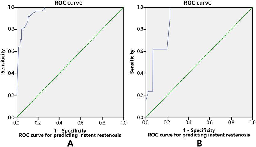
www.nature.com/scientificreports/
Figure 1. Severe stenosis at the lower segment of the basilar artery. (A) Magnetic resonance imaging (DWI)
showed disperse lesions of new infarction in the right hemisphere of the cerebellum. (B) Computed tomography
demonstrated severe stenosis at the lower segment of the basilar artery without calcification. (C) Digital
subtraction angiography revealed severe stenosis at the lower segment of the basilar artery. (D) An Enterprise
stent was deployed at the stenosis leading to patent flow. (E) Follow-up angiography at 6 months after stenting
demonstrated good patency of the basilar artery at the lesion.
segment), calcification (with 1/2 circular calcification as the borderline), balloon inflation pressure, intraproce-
dural arterial dissection, residual stenosis and cerebral flow TICI grade were the dominant risk factors (P < 0.05)
(Table 3).
Based on multivariate analysis in the modeling group, the in-stent restenosis probability prediction model was
established as follows: P = 1/[1 + e−( - 6.070–1.391 location+2.745 calcification+4.117 balloon inflation pressure+2.195 intraprocedural dissection+1.163
residual stenosis+1.174 flow TC grade)
]. In this equation, e is the natural constant. In the modeling group with 359 lesions in
the first angiographic follow-up, the ROC curve was established using the equation to calculate the probability of
in-stent restenosis in each case for comparison with the angiographic follow-up outcome (Fig. 5A). The AUC in
the receiver operating characteristic analysis was 0.963 (95% CI: 0.945–0.982), and the sensitivity and specificity
of this equation were 78.69% and 95.64%, respectively, when the prediction cutoff value was 0.50. The logistic
regression model was validated in the validation group (n = 89), and in-stent restenosis was predicted with this
equation for all patients with follow-up. The AUC was calculated as 0.902 (95% CI = 0.836 − 0.969) (Fig. 5B),
and the Youden index was 0.7890. When the same cutoff value of 0.50 was adopted, the sensitivity was 76.92%,
the specificity was 80.26%, and the total coincidence rate of the model was 79.78% (71/89) (Table 4), indicating
the good clinical application value of this model.
Discussion
In this study, it was found that lesion location, calcification degree, balloon expansion pressure, residual stenosis,
intraprocedural arterial dissection, and cerebral blood flow TICI grade were the factors significantly affecting in-
stent restenosis after Enterprise stent angioplasty for intracranial atherosclerotic stenoses. The in-stent restenosis
prediction model established using these factors was validated to be able to effectively predict in-stent restenosis
with good sensitivity and specificity.
Although the Enterprise stent has improved properties with regard to treating intracranial vascular diseases20,
in-stent stenosis still occurs within the stent. Feng et al4 found > 50% in-stent restenosis in 6.81% (three) of
patients 22 months after deployment of the Enterprise stent even though the initial symptomatic intracranial
Scientific Reports | (2021) 11:10479 | https://doi.org/10.1038/s41598-021-89670-x 3
Vol.:(0123456789)www.nature.com/scientificreports/
Figure 2. Right internal carotid artery severe stenosis. (A) Magnetic resonance imaging (MRI) showed an
ischemic infarct at three days after onset. (B) MRI at 14 days after onset revealed increased area of the infarction.
(C) Computed tomography revealed calcification (arrows) around the posterior communicating artery segment
of the right internal carotid artery. (D) Cerebral digital subtraction angiography demonstrated severe stenosis
at the posterior communicating artery segment of the right internal carotid artery. (E) Stent angioplasty was
performed with an Enterprise stent and the severe stenosis disappeared. (F) Nine months after stent angioplasty,
the stent remained patent.
arterial stenosis was decreased from 79.3 ± 8.1% to 14.9 ± 12.3% immediately following stenting. In the study with
189 patients harboring 209 intracranial stenoses treated with the Enterprise stent investigated by Vajda et al13, in-
stent restenosis was observed in 43 (24.7%) cases out of 174 stenoses with angiographic follow-up at a mean time
of 10 months even though the poststenting stenosis rate was reduced to 25.1 ± 1% from 65.4 ± 1% before stenting.
Salik et al. also detected in-stent restenosis in two (3.3%) of 60 patients with available angiographic follow-up
22 months after deployment of the Enterprise stent for symptomatic high-grade intracranial stenosis, which was
decreased from presenting to poststenting (92 ± 6% vs. 12 ± 10%)12. Our study identified some significant factors
affecting the in-stent restenosis of the Enterprise stent in treating intracranial atherosclerotic diseases, and the
logistic model constructed with these significant risk factors can be used to effectively predict in-stent restenosis,
which is a major drawback of intracranial stent angioplasty for atherosclerotic diseases.
Hypertension and diabetes mellitus can chronically affect the atherosclerotic process but do not acutely affect
in-stent stenosis. During stent-assisted coiling of wide-necked intracranial aneurysms, a certain rate (10.2%-
32.12%) of in-stent stenosis can also h appen4,23,24, with some studies displaying in-stent restenosis as a significant
dynamic and spontaneously resolvable process23,24. These studies may indicate that in-stent stenosis or restenosis
in stents assisting coiling embolization of cerebral aneurysms is not the progression of atherosclerosis but rather
the result of intimal hyperplasia. In stent angioplasty for intracranial atherosclerotic stenosis, balloon inflation
and stent deployment can tear plaques and damage the intima of the stented artery, leading to an inflammatory
reaction of endothelial and smooth muscle cells23,26. The intimal reaction to the stent as a foreign body can also
cause thrombosis within the stent, which, together with the inflammatory reaction, can bring about in-stent
restenosis. Balloon inflation will damage not only the intimal and medial membrane but also the adventitial
nourishing blood vessels, and injury of the adventitial nourishing vessels plays an important role in in-stent
restenosis. Therefore, balloon inflation pressure should not be too high, and the diameter of the balloon should
not be too large to avoid damage to the artery wall. The diameter of a balloon selected for inflating the stenotic
Scientific Reports | (2021) 11:10479 | https://doi.org/10.1038/s41598-021-89670-x 4
Vol:.(1234567890)www.nature.com/scientificreports/
Figure 3. Severe stenosis in the lower trunk of the left middle cerebral artery (MCA). (A) Magnetic resonance
imaging (MRI) showed bead-like high intensity lesion near the ventricular body. (B) MRI angiography revealed
severe stenosis in the lower trunk of the left MCA (small arrow) and an aneurysm (large arrow) at the anterior
communicating artery. (C) Nine days after admission, the symptoms were aggravated and the second MRI scan
demonstrated increased infarction area. (D) Perfusion imaging showed that greater time is needed for the blood
flow to reach the left parietal occipital lobe. E&F. Digital subtraction angiography demonstrated severe stenosis
in the lower trunk of the left MCA (arrow, E), and stent angioplasty was performed with an Enterprise stent to
relieve the stenosis (F). G. Endovascular coil embolization was performed to completely occlude the anterior
communicating aneurysm. (H). Follow-up angiography at six months following stenting revealed patent flow in
the lower trunk of the left MCA. (I). Perfusion imaging showed the time is significantly improved for the blood
flow to reach the left parietal occipital lobe.
Scientific Reports | (2021) 11:10479 | https://doi.org/10.1038/s41598-021-89670-x 5
Vol.:(0123456789)www.nature.com/scientificreports/
Figure 4. Endovascular recanalization of occluded left middle cerebral artery (MCA) and angiographic
follow-up. (A) DWI of magnetic resonance imaging (MRI) immediately after admission revealed bead-like high
signal in the left hemisphere. (B) Computed tomography angiography showed occluded M1 segment of the left
MCA. (C) Two days after admission, DWI of MRI demonstrated increased area of high signal lesions in the
left hemisphere with fusion of the original bead-like lesions. (D) PWI of MRI revealed greater time needed for
the blood flow to reach the left hemisphere. (E) Digital subtraction angiography showed occluded M1 segment
of the left MCA with no display of the distal arterial branches. (F) A microguidewire was used to explored the
occluded M1 segment, and a microcatheter was navigated along the microguidewire through the occluded M1
segment into the M2 segment of the left MCA, and angiography through this microcatheter indicated patency
of distal arterial branches. (G) An Enterprise 4.5 mm*22 mm stent was deployed at the occluded segment
after balloon angioplasty, with recanalized arterial branches of the left MCA. (H) PWI of MRI demonstrated
improved blood flow to the left hemisphere, much better than before recanalization. (I) Digital subtraction
angiography six months later revealed patent left MCA with no instent stenosis.
segment of the artery should be 80% of the normal artery diameter near the stenotic segment, and optimal expan-
sion of the stenotic artery should not be pursued to prevent damage to the adventitial nourishing blood v essels13.
The lesion nature rather than the type was a significant risk factor for in-stent restenosis in our study. Calci-
fication in the lesion can significantly affect in-stent restenosis. For patients with apparent plaque calcification,
the Enterprise stent may not be suitable for stent angioplasty because of its low radial force. In atherosclerosis
progression, plaque calcification develops via inflammation-dependent mechanisms involved in the progression
Scientific Reports | (2021) 11:10479 | https://doi.org/10.1038/s41598-021-89670-x 6
Vol:.(1234567890)www.nature.com/scientificreports/
Follow-up angiography
Group Patent Restenosis X2 P value
BA&MCA 154(91.7%) 14(8.3%)
Location 316.8 < 0.0001
VA&ICA 144(75.4%) 47(24.6%)
> 1/2 circle 41(52.6%) 37(47.4%)
Calcification 65.5 < 0.0001
≤ 1/2 circle 257(91.5%) 24(8.5%)
> 4 ATM 46(47.4%) 51(52.6%)
Balloon inflation pressure 119.3 < 0.0001
≤ 4 ATM 252(96.2%) 10(3.8%)
Yes 52(56.5%) 40(43.5%)
Intraprocedural dissection 61.5 < 0.0001
No 246(92.1%) 21(7.9%)
> 30% 40(43.0%) 53(57.0%)
Residual stenosis 142.4 < 0.0001
≤ 30% 258(97.0%) 8(3.0%)
> 2b 51(61.4%) 32(38.6%)
TICI 35.6 < 0.0001
≤ 2b 247(89.5%) 29(10.5%)
> 50y 173(82.0%) 38(18.0%)
Age 0.376 0.5398
≤ 50y 125(84.5%) 23(15.5%)
Yes 140(80.9%) 33(19.1%)
Smoking history 1.028 0.3107
No 158(84.9%) 28(15.1%)
Yes 104(84.6%) 19(15.4%)
Alcoholic abuse 0.316 0.5738
No 194(82.2%) 42(17.8%)
Yes 249(82.5%) 53(17.5%)
Hypertension 0.420 0.5170
No 49(86.0%) 8(14.0%)
Yes 197(81.7%) 44(18.3%)
Diabetes mellitus 0.833 0.3615
No 101(85.6%) 17(14.4%)
Yes 266(84.1%) 50(15.8%)
Dyslipidemia 2.556 0.1099
No 32(74.4%) 11(25.6%)
Yes 140(80.9%) 33(19.1%)
Coronary heart disease 1.028 0.3107
No 158(84.9%) 28(15.1%)
Yes 104(84.6%) 19(15.4%)
Hyperhomocysteinemia 0.316 0.5738
No 194(82.2%) 42(17.8%)
> 30 s 173(82.0%) 38(18.0%)
Pre-expansion time 0.376 0.5398
≤ 30 s 125(84.5%) 23(15.5%)
Table 2. Single factor analysis of risk factors for in-stent restenosis. BA, basilar artery; MCA, middle cerebral
artery; VA, vertebral artery; ICA, internal carotid artery; TICI, thrombolysis in cerebral infarction grade.
Indexes Regression coefficient estimated value (B) P value Dominance ratio 95%CI
Constant − 6.070 0.000 0.002
Location − 1.391 0.005 0.249 0.095–0.652
Calcification 2.745 0.000 15.564 5.61–43.21
Balloon inflation pressure 4.117 0.000 61.367 17.757–212.080
Intraprocedural dissection 2.195 0.000 8.982 3.132–25.763
Residual stenosis 1.163 0.013 3.199 1.284–7.973
TICI 1.174 0.012 3.234 1.291–8.101
Table 3. Regression model of instent restenosis based on imaging and clinical features of lesions.
and regression of atherosclerosis27. Intracranial atherosclerotic calcification is not isolated and frequently coexists
with arterial angulation or tortuosity28, which results in a poor reaction to balloon angioplasty. In plaque calcifi-
cation, the compliance of the arteries is very poor and may need a greater balloon inflation pressure, which may
cause a greater rate of arterial dissection and rupture compared with noncalcified plaques. For severe calcification,
the artery may not be expanded sufficiently, and in such insufficiently expanded arteries, the Enterprise stent will
not be easily or totally expanded, leading to poor adherence to the wall or irregular deformation of the s tent4.
Intraprocedural arterial dissection is one risk factor for in-stent restenosis. Based on our experience with
carotid stenting, the use of a small balloon for predilation and suboptimal balloon angioplasty before stenting are
better for reducing arterial dissection. Follow-up studies revealed that intraprocedural arterial dissection could
significantly increase the in-stent restenosis rate. Therefore, the balloon used for dilatation should be selected
Scientific Reports | (2021) 11:10479 | https://doi.org/10.1038/s41598-021-89670-x 7
Vol.:(0123456789)www.nature.com/scientificreports/
Figure 5. Receiver operating characteristic (ROC) curve for predicting instent restenosis based on imaging and
clinical features in the modelling (A) and validation (B) group.
Follow-up angiographic
outcome
Restenosis predicted by restenosis equation Restenosis Non-restenosis Total
No 3 61 64 (71.9%)
Yes 10 15 25 (28.1%)
Total 13 (14.6%) 76 (85.4%) 89
Table 4. Prediction of instent restenosis based on imaging and clinical features of lesions in the validation
group (n = 89).
with the balloon diameter being 80% that of the adjacent normal artery to prevent dissecting the atherosclerotic
plaque. Residual stenosis may also increase the in-stent restenosis rate, and a higher rate of residual stenosis was
permitted during the procedure to increase the safety of intracranial stent angioplasty. The cerebral blood flow
TICI grade is used to evaluate the recanalization degree of occluded arteries with four grades: zero, minimal,
partial, and complete recanalization, which significantly affects residual stenosis after stent deployment and
subsequently in-stent restenosis at follow-up.
Regarding lesion location in affecting restenosis, the in-stent restenosis rate was significantly higher in the
vertebral and internal carotid arteries than in the basilar and middle cerebral arteries (78.4% vs 21.6%, P = 0).
The Enterprise stent is a closed-cell intracranial stent with low adherent capability in tortuous a rteries29, which
may lead to greater intimal hyperplasia and consequently greater in-stent restenosis in the vertebral and internal
carotid arteries. Ebrahimi et al.29 found that the Enterprise stent with closed cells has less flexibility to conform
to a curved or irregular vessel because the unsegmented closed-cell design does not permit the stent to extend
at the outer curvature or to shorten at the inner curvature, resulting in maladaptation to the vascular curvature
and flattening or even kinking of the stent in acute curves. This maladaptation will cause intimal hyperplasia in
addition to long-term material fatigue damage.
Some limitations existed in this study: we included only participants of Chinese ethnicity in our popula-
tion, and this was a single-center, retrospective study. Future studies will have to resolve these issues for better
outcomes.
In conclusion, in-stent restenosis after Enterprise stenting for atherosclerotic arterial stenosis is affected by
stenosis location, calcification, balloon inflation pressure, intraprocedural arterial dissection, residual stenosis
and cerebral flow grade, and establishment of a logistic model with these factors can effectively predict in-stent
restenosis.
Scientific Reports | (2021) 11:10479 | https://doi.org/10.1038/s41598-021-89670-x 8
Vol:.(1234567890)www.nature.com/scientificreports/
Materials and methods
This study was approved by the ethics committee of Henan Provincial People’s Hospital. Informed consent was
obtained from all patients to participate in the study. All methods were performed in accordance with the relevant
guidelines and regulations. Between January 2017 and March 2019, consecutive patients with angiography-
confirmed intracranial atherosclerotic diseases (≥ 50% stenosis) who were treated with the Enterprise stent were
enrolled. The exclusion criteria were patients with identified or suspected vasculitis or vascular dissection. A
total of 400 patients were enrolled in the modeling group to establish a prediction model for in-stent restenosis,
and 89 patients with matched baseline data were enrolled for validation of the prediction model (Table 1). The
endovascular procedure (Figs. 1, 2, 3, and 4) was performed under general anesthesia with oral administration of
clopidogrel (75 mg/d) and aspirin (300 mg/d) at least three days before stenting. All procedures were performed
by surgeons with over five years of experience with endovascular techniques. After a 6F guiding catheter was
introduced into the vessel, and angiographic evaluation was performed for the atherosclerotic lesion, including
location, length and vascular diameter. Then, a Transcend microwire (Transcend, 0.014, 205 cm, Stryker) was
navigated together with a microcatheter through the lesion into the distal vessel. An exchange microwire was
sent across the lesion, and a Gateway balloon catheter was introduced to the stenosis or occlusion location and
gradually inflated to full expansion for 30 s using nominal pressure. The Gateway balloon was selected with the
balloon diameter reaching approximately 80% of the diameter of the normal vessel adjacent to the lesion to be
treated, and underdilation was recommended to avoid arterial dissection, vessel rupture, and plaque displace-
ment. After inflation, the Gateway balloon catheter was withdrawn, and an Enterprise stent with a diameter of
4.5 mm (common specification of the stent) was sent via a microcatheter to the lesion and deployed to cover
the target lesion. The length of the Enterprise stent was selected so that the stent covered the whole lesion and
extended approximately 1–2 mm beyond each end of the lesion. After stent deployment, angiographic evaluation
was performed for residual stenosis and blood flow through the stent. Technical success was defined as residual
stenosis < 50%. If the residual stenosis was > 50%, poststenting dilatation was performed with a balloon catheter.
For recanalization of occluded intracranial arteries, a microguidewire was first used to explore the occluded
segment, and after the wire was sent through the occluded segment into distal branches, a microcatheter was
navigated along the microguidewire through the occluded segment into the distal arterial branches. Angiography
was performed through the microcatheter to show whether the microcatheter was in the distal arterial branch.
Later, an exchange microwire was sent across the lesion for introduction of a balloon catheter for angioplasty
and stenting. After the endovascular procedure, computed tomography was conducted to rule out intracranial
hemorrhage or thrombosis. Antiplatelet treatment was conducted with aspirin 100 mg/d and clopidogrel 75 mg/d.
Six to 12 months after the procedure, clopidogrel was stopped while aspirin was continued14,17.
After stent angioplasty, the following data were recorded and analyzed: the stenosis location, length and
degree; normal vascular diameter distal and proximal to the lesion; lesion type; balloon size; inflation pressure
and duration; stent type; residual stenosis; perioperative complications; morbidity; and mortality within 30 days.
All patients were followed up within 30 days after stenting for possible stroke, death, and transient ischemic attack
(TIA). At 1, 3 and 6 months after discharge, clinical evaluation of the patients’ neurological system was performed
by means of telephone contact and outpatient services. Angiographic follow-up was performed once between
3–12 months after stenting, and telephone follow-up was performed once yearly afterwards. Head CT angiog-
raphy (CTA), magnetic resonance imaging (MRI), or digital subtraction angiography (DSA) were performed
for those with suspected stroke once a year. For stable patients, the follow-up interval might be longer than two
years. Recanalization of the lesion was evaluated before and after the procedure with the thrombolysis in cerebral
infarction grade (TICI) grading system18, which classifies the recanalization of the occluded artery into 0—no
recanalization, 1—minimal, 2—partial, and 3—complete recanalization. The definition of intracranial arterial
stenosis provided by Samuels et al19 was adopted, and the stenosis was measured using the equation percent
stenosis = [(1 − (Dstenosis/Dnormal))] × 100, where Dstenosis indicates the arterial diameter at the location of
the most severe stenosis and Dnormal indicates the diameter of the proximal normal artery. According to the
study by Levy et al8, in-stent restenosis refers to a lesion demonstrating stenosis greater than 50% adjacent to or
within the stent and absolute luminal loss greater than 20% on follow-up imaging.
The following variables were analyzed. Lesion calcification was evaluated in terms of location and degree, and
the section of the narrowest lesion on CTA was used to evaluate the arterial calcification degree. Calcification
location: 0, basilar artery or middle cerebral artery; 1, internal carotid artery siphon segment or vertebral artery
V4 segment. Calcification degree: 0, calcification arc ≤ 180°; 1, calcification arc > 180°. Lesion length: 0, ≤ 15 mm;
1, > 15 mm. Balloon inflation pressure: 0, ≤ 4 ATM; 1, > 4 ATM. Intraprocedural arterial dissection following bal-
loon expansion: 0, no dissection; 1, with dissection. Residual stenosis: 0, with residual stenosis ≤ 30%; 1, residual
stenosis > 30%. Cerebral blood flow grade: 0, TICI ≤ 2b; 1, TICI > 2b.
Statistical analysis
The statistical analysis was performed with SPSS 19.0 software (IBM, Chicago, IL, USA). All continuous data are
expressed as the mean ± standard deviation and were tested with paired t test and single-factor analysis. Categori-
cal data are expressed as frequencies and were tested with the chi-square test or Fisher’s exact test. The prediction
model was identified with multivariate binary logistic regression analysis. A forward-conditional method was
used to identify variables significant (P < 0.05) for predicting intracranial atherosclerotic lesions. Based on the
selected imaging and clinical variables, a simple diagnosis equation was established: P (in-stent restenosis prob-
ability) = 1/[1 + e−z] for calculation of the in-stent restenosis probability compared with the follow-up data of CTA
and digital subtraction angiography. The specificity and sensitivity of the prediction model were evaluated, and
the receiver operating characteristic (ROC) curve and the area under the curve (AUC) were calculated in both
the modeling and validation groups. A P value less than 0.05 was considered statistically significant.
Scientific Reports | (2021) 11:10479 | https://doi.org/10.1038/s41598-021-89670-x 9
Vol.:(0123456789)www.nature.com/scientificreports/
Received: 23 June 2020; Accepted: 19 April 2021
References
1. Feldmann, E. et al. Chinese-white differences in the distribution of occlusive cerebrovascular disease. Neurology 40, 1541–1545.
https://doi.org/10.1212/wnl.40.10.1540 (1990).
2. Qureshi, A. I. et al. Intracranial atherosclerotic disease: an update. Ann. Neurol. 66, 730–738. https://doi.org/10.1002/ana.21768
(2009).
3. Chimowitz, M. I. et al. Stenting versus aggressive medical therapy for intracranial arterial stenosis. N. Engl. J. Med. 365, 993–1003.
https://doi.org/10.1056/NEJMoa1105335 (2011).
4. Feng, Z. et al. Enterprise stent for the treatment of symptomatic intracranial atherosclerotic stenosis: an initial experience of 44
patients. BMC Neurol 15, 187. https://doi.org/10.1186/s12883-015-0443-9 (2015).
5. Park, S. C. et al. Long-term outcome of angioplasty using a wingspan stent, post-stent balloon dilation and aggressive restenosis
management for intracranial arterial stenosis. Clin. Neuroradiol. https://doi.org/10.1007/s00062-019-00793-1 (2019).
6. Henkes, H. et al. Treatment of intracranial atherosclerotic stenoses with balloon dilatation and self-expanding stent deployment
(WingSpan). Neuroradiology 47, 222–228. https://doi.org/10.1007/s00234-005-1351-2 (2005).
7. Derdeyn, C. P. et al. Aggressive medical treatment with or without stenting in high-risk patients with intracranial artery stenosis
(SAMMPRIS): the final results of a randomised trial. Lancet 383, 333–341. https://d oi.o
rg/1 0.1 016/S 0140-6 736(13)6 2038-3 (2014).
8. Levy, E. I. et al. Wingspan in-stent restenosis and thrombosis: incidence, clinical presentation, and management. Neurosurgery
61, 644–650. https://doi.org/10.1227/01.NEU.0000290914.24976.83 (2007) (discussion 650-641).
9. Turk, A. S. et al. Influence of patient age and stenosis location on wingspan in-stent restenosis. AJNR Am. J. Neuroradiol. 29, 23–27.
https://doi.org/10.3174/ajnr.A0869 (2008).
10. Bose, A. et al. A novel, self-expanding, nitinol stent in medically refractory intracranial atherosclerotic stenoses: the Wingspan
study. Stroke 38, 1531–1537. https://doi.org/10.1161/STROKEAHA.106.477711 (2007).
11. Natarajan, S. K. et al. Primary stenting for acute ischemic stroke using the enterprise intracranial stent: 2-year results of a phase-I
trial. J. Vasc. Int. Neurol. 8, 62–67 (2015).
12. Salik, A. E. et al. Medium-term results of undersized angioplasty and stenting for symptomatic high-grade intracranial athero-
sclerotic stenosis with Enterprise. Interv Neuroradiol, 1591019919832244, doi:https://doi.org/10.1177/1591019919832244 (2019).
13. Vajda, Z. et al. The modified Bose method for the endovascular treatment of intracranial atherosclerotic arterial stenoses using
the Enterprise stent. Neurosurgery 70, 91–101. https://doi.org/10.1227/NEU.0b013e31822dff0f (2012) (discussion 101).
14. Wang, X. et al. Application of the enterprise stent in atherosclerotic intracranial arterial stenosis: a series of 60 cases. Turk. Neu-
rosurg. 26, 69–76. https://doi.org/10.5137/1019-5149.JTN.13350-14.1 (2016).
15. Higashida, R. T., Halbach, V. V., Dowd, C. F., Juravsky, L. & Meagher, S. Initial clinical experience with a new self-expanding nitinol
stent for the treatment of intracranial cerebral aneurysms: the Cordis Enterprise stent. AJNR Am. J. Neuroradiol. 26, 1751–1756
(2005).
16. Krischek, O., Miloslavski, E., Fischer, S., Shrivastava, S. & Henkes, H. A comparison of functional and physical properties of self-
expanding intracranial stents [Neuroform3, Wingspan, Solitaire, Leo+, Enterprise]. Minim Invasive Neurosurg. 54, 21–28. https://
doi.org/10.1055/s-0031-1271681 (2011).
17. Zsolt Vajda, E. S. et al. The modified Bose method for the endovascular treatment of intracranial atherosclerotic arterial stenoses
using the Enterprise stent. Neurosurgery 70, 91–101 (2012).
18. Almekhlafi, M. A. et al. Not all “successful” angiographic reperfusion patients are an equal validation of a modified TICI scoring
system. Int. Neuroradiol. 20, 21–27. https://doi.org/10.15274/INR-2014-10004 (2014).
19. Samuels, O. B., Joseph, G. J., Lynn, M. J., Smith, H. A. & Chimowitz, M. I. A standardized method for measuring intracranial
arterial stenosis. AJNR Am. J. Neuroradiol. 21, 643–646 (2000).
20. (!!! INVALID CITATION !!! 13, 14).
21. Groschel, K., Schnaudigel, S., Pilgram, S. M., Wasser, K. & Kastrup, A. A systematic review on outcome after stenting for intracranial
atherosclerosis. Stroke 40, e340-347. https://doi.org/10.1161/STROKEAHA.108.532713 (2009).
22. Albuquerque, F. C. et al. Angiographic patterns of Wingspan in-stent restenosis. Neurosurgery 63, 23–27. https://doi.org/10.1227/
01.NEU.0000335067.53190.A2 (2008) (discussion 27-28).
23. Gao, B., Safain, M. G. & Malek, A. M. Enterprise stenting for intracranial aneurysm treatment induces dynamic and reversible
age-dependent stenosis in cerebral arteries. J. Neurointerv. Surg. 7, 297–302. https://doi.org/10.1136/neurintsurg-2013-011074
(2015).
24. Kim, Y. S., Lee, S. W., Yeom, J. A., Yoon, C. H. & Baik, S. K. Angiographic findings of in-stent intimal hyperplasia after stent-assisted
coil embolization: are they permanent findings?. J. Neurosurg. 124, 328–333. https://doi.org/10.3171/2015.2.JNS142557 (2016).
25. Feng, X. et al. Comparison of recanalization and in-stent stenosis between the low-profile visualized intraluminal support stent
and enterprise stent-assisted coiling for 254 intracranial aneurysms. World Neurosurg. 109, e99–e104. https://doi.org/10.1016/j.
wneu.2017.09.112 (2018).
26. Shin, Y. S. et al. Wingspan stenting for intracranial atherosclerotic stenosis: clinical outcomes and risk factors for in-stent restenosis.
Neurosurgery 72, 596–604. https://doi.org/10.1227/NEU.0b013e3182846e09 (2013) (discussion 604).
27. Shioi, A. & Ikari, Y. Plaque calcification during atherosclerosis progression and regression. J. Atheroscler. Thromb. 25, 294–303.
https://doi.org/10.5551/jat.RV17020 (2018).
28. Pikija, S., Magdic, J. & Hojs-Fabjan, T. Calcifications of vertebrobasilar arteries on CT: detailed distribution and relation to risk
factors in 245 ischemic stroke patients. Biomed Res. Int. 2013, 918970. https://doi.org/10.1155/2013/918970 (2013).
29. Ebrahimi, N., Claus, B., Lee, C. Y., Biondi, A. & Benndorf, G. Stent conformity in curved vascular models with simulated aneurysm
necks using flat-panel CT: an in vitro study. AJNR Am. J. Neuroradiol. 28, 823–829 (2007).
Author contributions
Study design: K.Z., T.-X.L.Data collection: K.Z., Z.-L.W., B.-L.G., J.-J.G., H.-L.G., Y.-F.W. Data analysis: K.Z.*,
J.-C.X., T.-X.L.Supervision: Y.-F.W. Writing of the original version: K.Z. Revision of the original version: B.-L.G.
Approval of the article: all authors.
Competing interests
The authors declare no competing interests.
Additional information
Correspondence and requests for materials should be addressed to Z.-L.W.
Reprints and permissions information is available at www.nature.com/reprints.
Scientific Reports | (2021) 11:10479 | https://doi.org/10.1038/s41598-021-89670-x 10
Vol:.(1234567890)www.nature.com/scientificreports/
Publisher’s note Springer Nature remains neutral with regard to jurisdictional claims in published maps and
institutional affiliations.
Open Access This article is licensed under a Creative Commons Attribution 4.0 International
License, which permits use, sharing, adaptation, distribution and reproduction in any medium or
format, as long as you give appropriate credit to the original author(s) and the source, provide a link to the
Creative Commons licence, and indicate if changes were made. The images or other third party material in this
article are included in the article’s Creative Commons licence, unless indicated otherwise in a credit line to the
material. If material is not included in the article’s Creative Commons licence and your intended use is not
permitted by statutory regulation or exceeds the permitted use, you will need to obtain permission directly from
the copyright holder. To view a copy of this licence, visit http://creativecommons.org/licenses/by/4.0/.
© The Author(s) 2021
Scientific Reports | (2021) 11:10479 | https://doi.org/10.1038/s41598-021-89670-x 11
Vol.:(0123456789)You can also read