Fluorescent light energy modulates healing in skin grafted mouse model
←
→
Page content transcription
If your browser does not render page correctly, please read the page content below
Open Medicine 2021; 16: 1240–1255
Research Article
Jie Ding, Maiken Mellergaard, Zhensen Zhu, Peter Kwan, Deirdre Edge, Zengshuan Ma,
Lise Hebert, Saad Alrobaiea, Takashi Iwasaki, Michael Canova Engelbrecht Nielsen*,
Edward E. Tredget*
Fluorescent light energy modulates healing in
skin grafted mouse model
https://doi.org/10.1515/med-2021-0329 collagen deposition, myofibroblast and mast cell accumu-
received March 26, 2021; accepted July 7, 2021 lation, and connective tissue growth factor expression.
Abstract: Skin grafting is often the only treatment for skin While there was no visible difference in gross morphology,
trauma when large areas of tissue are affected. This sur- we found that FLE treatment promoted a balanced col-
gical intervention damages the deeper dermal layers of lagen remodeling. Collectively, these findings suggest
the skin with implications for wound healing and a risk of that FLE has a conceivable effect at balancing healing after
scar development. Photobiomodulation (PBM) therapy skin grafting, which reduces the risk of infections, chronic
modulates biological processes in different tissues, with wound development, and fibrotic scarring.
a positive effect on many cell types and pathways essen- Keywords: skin grafting, dermal fibrotic mouse model, fluor-
tial for wound healing. This study investigated the effect escent light energy, photobiomodulation, wound healing
of fluorescent light energy (FLE) therapy, a novel type of
PBM, on healing after skin grafting in a dermal fibrotic
mouse model. Split-thickness human skin grafts were
transplanted onto full-thickness excisional wounds on 1 Introduction
nude mice. Treated wounds were monitored, and excised
xenografts were examined to assess healing and patho- Therapy involving skin grafting has significantly advanced
physiological processes essential for developing chronic over the last decade. Skin grafts are characterized by (1)
wounds or scarring. Results demonstrated that FLE treat- donor and recipient of tissue and include: autografts
ment initially accelerated re-epithelialization and rete (same individual), allografts (same species), and xeno-
ridge formation, while later reduced neovascularization, grafts (different species) and (2) thickness of graft tissue,
that is classified as split-thickness human skin grafts
(STSGs) or full-thickness skin grafts (FTSGs) consisting
* Corresponding author: Michael Canova Engelbrecht Nielsen, of epidermis and part of dermis or epidermis and full
Department of Research and Development, Klox R&D Center, thickness dermis, respectively [1]. FTSGs are mostly used
Guangdong Klox Biomedical Group Co., Ltd, Room 603, 6/F, for smaller areas and on more exposed body sites, since
Building 8, No. 6, Nanjiang Second Road, Zhujiang Street, Nansha
they need proper vascularity to survive but contract less
District, Guangzhou, China, e-mail: men@kloxasia.com
* Corresponding author: Edward E. Tredget, Divisions of Plastic and
than STSGs, thus reducing the risk of hypertrophic scar
Reconstructive Surgery and Critical Care, 2D2.28 Walter C (HTS) development [1].
MacKenzie Health Sciences Centre & Wound Healing Research Skin repair, including rapid wound closure, tissue
Group, 161 HMRC, Department of Surgery, University of Alberta, healing, and reduced scarring after grafting, is essential
Edmonton, Alberta, Canada, e-mail: etredget@ualberta.ca to generate a fully functional tissue [2]. However, wound
Jie Ding, Zhensen Zhu, Peter Kwan, Zengshuan Ma, Saad Alrobaiea,
healing is a profoundly complex process relying on close
Takashi Iwasaki: Wound Healing Research Group, Department of
Surgery, Faculty of Medicine and Dentistry, University of Alberta, collaboration between skin cells, infiltrating immune cells,
161 HMRC, Edmonton, Canada and the extracellular matrix (ECM) to warrant a balanced
Maiken Mellergaard: Department of Veterinary and Animal healing process [3]. Skin grafting therapies often result in
Sciences, Faculty of Health and Medical Sciences, University of some degree of fibrosis and scarring, thus the development
Copenhagen, Denmark
of new effective therapeutic strategies for deep injury to
Deirdre Edge, Maiken Mellergaard: Department of Research and
Development, Klox Technologies Europe Ltd, Dublin, Ireland
the dermis e.g., in relation to skin grafting are required.
Lise Hebert: Department of Research and Development, Klox Photobiomodulation (PBM) describes the therapeutic
Technologies Inc., Laval, Canada application of light to stimulate regeneration [4–7].
Open Access. © 2021 Jie Ding et al., published by De Gruyter. This work is licensed under the Creative Commons Attribution 4.0 International
License.
FLE balances healing after skin grafting 1241
Indeed, studies have demonstrated that PBM induces cellular infiltration, and connective tissue growth factor
anti-inflammatory activity and tissue repair by modu- (CTGF) were assessed in situ.
lating neovascularization and the early formation of col-
lagen fibers [8–12]. Fluorescent light energy (FLE) is a
novel form of PBM consisting of a topical component
containing specific chromophores that need activation 2 Materials and methods
by a LED lamp, whereby FLE is generated. The chromo-
phores absorb photons from the LED lamp (440–460 nm)
2.1 Animals and study design
and through the phenomenon of Stokes shift, emit lower-
energy fluorescent light (500–610 nm) that penetrates
The experiment followed Canadian rules for animal treat-
intact or wounded tissues [12–14]. In vitro studies have
ment and welfare, and the study was approved by Animal
shown that FLE is superior at inducing collagen produc-
Care and Use Committees (ACUCs) of the University
tion in human dermal fibroblast cell cultures when com-
of Alberta. A 4-week-old male BALB/c-nu/nu nude mice
pared with the blue LED light alone or a lamp mimicking
(n = 69) weighing an average of 20 g were purchased from
the spectral output of FLE [12]. FLE also significantly
Charles River Laboratories International, Inc. (Wilmington,
downregulates several pro-inflammatory cytokines, including
MA). The mice were first conditioned for 1–2 weeks in the
interleukin 6 and tumor necrosis factor α, as well as facili-
university animal facility and then grafted with split-thick-
tating early modulation of angiogenesis [12–15]. Furthermore,
ness human grafts from discarded skin flaps of patients
FLE has been shown to significantly impact healthy [16],
undergoing abdominoplasty.
inflamed, and disease-affected skin tissues. The latter being
Mice were divided into four groups according to the treat-
well-documented in clinical trials and case studies, for acne
ments: (1) untreated (Control), (2) LED light alone (Light),
[17,18], rosacea [19,20], acne conglobate and hidradenitis
(3) solid FLE formulation plus LED light (sFLE), and (4) gel
suppurativa [21], acneiforme eruption [22], senile lenti-
FLE formulation plus LED light (gFLE) (16–18 mice/group
gines [23,24], and wound healing [13,25–27]. Finally, a
and 5–6 mice/group/time point, Table 1). Treatment was
series of case studies investigating the effect of FLE treat-
performed twice per week from day 7 after grafting and
ment on healing of acute second-degree burns showed
for six consecutive weeks. Wound healing was monitored
accelerated wound healing as well as overall improvement
weekly using digital photography. After 28, 56, or 84 days,
of tissue structure in two cases of severe HTS after burn
the mice were euthanized, using a CO2 chamber, and xeno-
injuries, suggesting that FLE balances wound healing at
grafts were collected for histology and biochemistry analysis
different stages of the wound healing and remodeling pro-
(Figure 1a).
cess [28].
Based on these previously reported potent anti-inflam-
matory and tissue regenerating properties of FLE, we
speculated that FLE treatment has the potential to be an 2.2 Preparation of human STSGs
effective and preventative therapeutic strategy ensuring
rapid and balanced wound healing in patients after skin Abdominal human skin tissues were collected from five
grafting. Thus, the aim of this study was to investigate the white female patients (19, 42, 50, 55, and 56 years old) who
effects of FLE on skin repair after grafting. We used a pre- had cosmetic abdominoplasty. STSGs were harvested ex vivo
viously described in vivo dermal scar model [29]. This using a Padgett electric dermatome (Padgett Instruments,
model provides a useful tool for investigating the direct INC., Kansas City, MO) set at 0.03 cm. They were cut into
effect of FLE on healing after skin grafting including essen-
tial cellular factors associated with dermal fibrotic disor-
ders [30]. Nude mice with human STSGs were treated with Table 1: Groups and skin donors
either FLE or control LED light and the results were com-
pared with an untreated control (and for some assays Groups 28 days 56 days 84 days Total
normal human skin or human HTS tissue). Wound closure Control n =5 n =5 n = 6 n = 16
was assessed by monitoring morphological changes and LED n =6 n =6 n = 6 n = 18
measuring the wound area. Furthermore, histological sFLE n =6 n =6 n = 6 n = 18
analysis was used for investigating the healing process gFLE n =5 n =6 n = 6 n = 17
Total n = 22 n = 23 n = 24 n = 69
focusing on thickness of the dermal layers, vascularity,
Skin donor n =1 n =2 n = 2 n =5
and re-epithelialization. Finally, collagen deposition,
1242 Jie Ding et al. Figure 1: Study procedure and generation of STSG mouse model. (a) Human split-thickness skin grafts (STSGs) were collected from patients who had cosmetic abdominoplasty (1) and transplanted onto full-thickness excisional wounds on the backs of mice (2). The wounds were treated either with sFLE or gFLE plus LED light or LED light alone from Day 7 post-grafting, twice a week for 6 consecutive weeks, or left untreated (3). The wounds were monitored weekly by digital photography (4). The mice were euthanized at day 28, 56, and 84 post- treatment and xenografts were collected for histological analysis. Collected tissue from each mouse was divided into three parts, one was embedded in paraffin (red square), one was prepared and stored for collagen quantification (black square), and one was prepared and stored as OCT blocks (blue square, not used in this study) (5). (b) Representative pictures showing generation of the STSG mouse model used in the study. Human skin from cosmetic abdominoplasty (6) was prepared for STSG grafting on nude mice (7). Excision wound was prepared on recipient mouse (8) prior to STSG grafting (9). Xenograft developed on recipient mouse (10) before initiation of treatment. pieces of 2.0 cm × 1.5 cm (Figure 1b) and kept in sterile protocol. All human subjects who provided tissues gave normal saline for grafting. The Health Research Ethics Board written informed consent, which was documented in the of the University of Alberta Hospital approved the patient patient’s health record before participation in the study.
FLE balances healing after skin grafting 1243
2.3 Establishment of human dermal fibrotic 2.6 Quantification of wound closure
mouse model
Wound closure was assessed and quantified at day 28, 56,
A full-thickness excisional wound (2.0 cm × 1.5 cm) was made or 84 post-treatment by measuring the wound area and
on the back of each mouse under isoflurane anesthesia. A normalizing measurements according to a scale embedded
human STSG, randomized to the four groups, was trans- in each picture using ImageJ (National Institutes of Health,
planted onto the wound and secured with sutures (Figure 1b). Bethesda, MD). Wound closure was presented as percen-
The surgical site was then covered with a non-adherent tage of wound area.
petrolatum (Xeroform™, Covidien, Mansfield, MA) and gauze
in a tie-over bolster dressing for 7 days after grafting to ensure
adherence of the human skin graft to the wound bed of the 2.7 Evaluation of re-epithelialization in
mouse tissue, as previously described [31]. xenografts
Xenografts were collected after the mice were euthanized
using a CO2 chamber at day 28, 56, or 84 post-treatment
2.4 FLE treatment
and prepared for histological analysis. Sections were
stained with hematoxylin and eosin (H&E), imaged using
Treatment was initiated on day 7 after grafting when the
a light microscope (Nikon Canada Inc., Mississauga, ON,
dressing was removed. Mice were treated under anesthesia
Canada), and analyzed according to a scale in each
via nasal halothane and treated twice per week for 6 conse-
image. Epithelialization (integrity of epidermal layer)
cutive weeks. Two different FLE systems (Klox Technologies
and re-epithelialization (upward migration of epithelial
Inc., Québec, Canada) were used. The gFLE was prepared by
cells) were assessed in the H&E images and examined
mixing a chromophore gel with a hydro-carrier gel immedi-
against sections from a skin sample (non-grafted) from
ately before use and the mixed gFLE was applied as a thick
one donor at each time point to compare grafted tissue
layer of 2 mm on the wound. The sFLE (solid membrane form
with normal human skin tissue (NS samples).
of the FLE gel containing chromophores) was cut into pieces
of 1.5 cm2 × 2 cm2 and placed on the wound. Both the FLE
systems emit similar FLE emission spectra between 500 and
2.8 Quantification of epidermal and dermal
610 nm, as previously described [13,15]. Illumination was per-
formed for 5 min at a distance of 5 cm from the wound. The
thicknesses and vascularity in
LED lamp (Klox Technologies Inc., Laval, Canada) used to xenografts
illuminate the chromophores in the gel or membrane delivers
a non-coherent blue light with a single peak wavelength and ImageJ was used to measure epidermal and dermal thick-
a maximum emission between 440 and 460 nm. The power nesses using high-power magnification in H&E images.
density of the Klox lamp (Klox Technologies Inc., Laval, Epidermal thickness was defined as the distance from
Canada) was measured using a multifunctional spectroradio- skin surface down to the bottom of epidermal papillae and
meter system (SP-100, Orb Optronix inc., Kirkland, WA, USA) dermal thickness as the distance between the epidermal–
with a spectral range between 380 and 780 nm. The wave- dermal junction and the dermal–adipose layer junction,
length binning resolution of the optical system was 1 nm. as previously described for dermal thickness [32]. Three
The optical densities of the lamp were determined at a measurements were taken per sample, two adjacent to
5 cm distance. The power output of the Klox lamp is certified the wound site at 50 μm on each side and one in the
to be within 120–125 mW/m3 and the fluency between 33 and middle of each section. The vascularity was assessed by
45 J/cm2 at 5 min treatment [12–15]. counting the number of blood vessels in 20× magnifica-
tion views taken diagonally from top left to bottom right
in the dermis, in five random high-power fields (HPFs) for
each animal.
2.5 Digital photography
Wounds were monitored morphologically by digital photo- 2.9 Quantification of collagen in xenografts
graphy every week after grafting to document wound
healing and scar formation. A scale was embedded in Collagen content was quantified in the xenografts by
each photo to allow for direct measurements. 4-hydroxyproline analysis using liquid chromatography/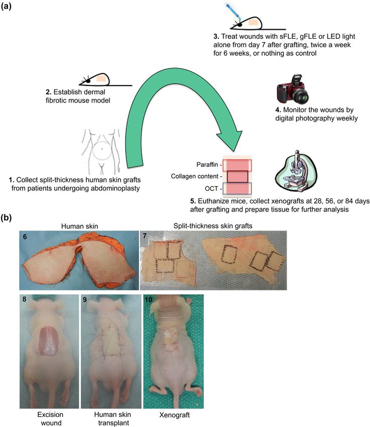
1244 Jie Ding et al.
mass spectrometry (LC/MS), as previously modified and smooth muscle actin (αSMA) and CTGF after paraffin-
described [33]. 5–10 mg of xenograft tissues (wet weight) embedded sections were deparaffinized and hydrated.
were freeze-dried overnight. The dried tissues were then Antigen retrieval was performed on paraffin sections
hydrolyzed in 6 N hydrochloric acid (HCL) solution at using 0.05% of trypsin in PBS for 15 min. The sections
110°C, resulting in collagen being cleaved into its compo- were blocked with serum for 5 min at room temperature
nent amino acids and 4-hydroxyproline being released (RT), rinsed, and incubated with primary antibodies of mono-
from the collagen protein. Then, a known amount of clonal rabbit anti-αSMA (EMD Millipore, Billerica, MA) and
N-methyl-proline was added as an internal standard along polyclonal goat anti-CTGF (Santa Cruz Biotechnology, Dallas,
with the n-butyl-ester reagent for derivatization. After the TX) for 1 h at RT. Secondary antibodies used were biotiny-
mixture was dried under vacuum, the determination of lated anti-rabbit IgG (Vector Laboratories, Burlingame,
4-hydroxyproline was performed using an HP Hewlett CA) for αSMA staining or biotinylated anti-goat IgG
Packard 1100 LC linked to 6130 MS (Agilent Technologies, (Dako, Glostrup, Denmark) for CTGF staining for 1 h at
Santa Clara, CA) selective detector monitoring the ions 188 RT. Detection was done using VECTASTAIN Elite ABC
(N-butyl-ester of 4-hydroxyproline) and 186 (N-butyl-ester Reagent and Vector DAB substrate (Vector Laboratories,
of N-methyl-proline). Each sample was run in triplicate, Burlingame, CA) and counterstained in hematoxylin. Finally,
and data are displayed as ng of 4-hydroxyproline per mg the sections were mounted after dehydration and imaged
dry tissue obtained by reference to a standard curve. using an optical microscope.
Myofibroblasts and endothelial cells around blood
vessels were all stained by anti-αSMA antibody and
2.10 Collagen assessment were distinguished based on location and morphology.
Myofibroblasts were counted in five HPFs/animal. CTGF
Paraffin-embedded sections were deparaffinized and hydrated was qualified in the sections by brown staining using
after being washed twice in acidified water, dehydrated ImageJ software. Color devolution was done by choosing
and mounted before staining in picrosirius red (Abcam, H&E DAB plugin on a CTGF stained image, subtracting
Toronto, ON, Canada) for 1 h. Picrosirius red stain provides background by adjusting threshold, and measuring the
a simple, specific, and sensitive method to localize col- CTGF stained area, this was expressed as a percentage of
lagen in the tissues by reacting with sulfonic acid groups the image area.
present in the collagen molecule [34]. Collagen bundles
appear as green, red, or yellow on a black background
allowing quantitative morphometric analysis under polar-
ized light. Collagen orientation was evaluated by Fourier 2.12 Mast cell assessment
analysis using ImageJ software (ImageJ v.1.51 u NIH, USA),
represented by the collagen orientation index (COI). Xenograft sections were stained in toluidine blue (Fisher
Briefly, the original single collagen bundle in each picro- Chemical, Geel, Belgium) working solution for 2–3 min
sirius red image taken under polarized microscope was after deparaffinization and hydration. After they were
converted to a representation in the frequency domain by washed with distilled water, the sections were then dehy-
ImageJ software. The threshold was adjusted to make it clear. drated quickly through 95 and 100% of ethyl alcohol
The width and length of the representation was measured (Commercial Alcohols, Brampton, ON, Canada) and cleared
and the COI was calculated using the following equation: 1 − in xylene. Finally, the stained sections were mounted for
(short axis/long axis), as previously described [35], and pre- observation by light microscopy. Toluidine blue is a cellular
sented as arbitrary units (a.u.). The collagen networks in dye with high affinity for acidic tissue components such as
xenografts were compared with NS and HTS from patients heparin- and histamine-rich metachromatic granules in the
recovered from burn injury. Random collagen bundle tissue cytoplasm of mast cells, staining mast cells red-purple and
like NS has an index of 0 whereas the parallel organization the background blue [30]. The red-purple mast cells were
seen in tissue like HTS leads to an index closer to 1. counted in five HPFs/animal.
2.11 Myofibroblast and CTGF assessment 2.13 Statistical analysis
To detect involvement of myofibroblasts and fibrotic fac- Data are presented as mean value ± SE. Group data (5–6
tors in scar formation, xenografts were stained for alpha mice/group, Table 1) at each time point was analyzedFLE balances healing after skin grafting 1245
using GraphPad Prism version 9.0.0 (GraphPad Soft- measured wound area at day 56 or 84 (Figure 2b and
ware), and each treatment group was statistically com- Figure S1).
pared with the control group using two-way ANOVA with
Dunnett´s multiple comparison test, with the exception of
the NS versus HTS comparison in Figure 5c, which was
done using an unpaired, two-tailed t-test. A p-value ≤ 3.2 FLE treatment induced re-epithelization
0.05 was considered statistically significant. and reduced epidermal thickness after
skin grafting
Re-epithelialization is an essential measure of wound
3 Results healing that is often impaired in chronic wounds, and is
therefore used as a defining parameter for successful
wound closure [36]. Since FLE treatment seemingly accel-
3.1 FLE treatment modulated wound closure erated early wound closure, we next examined re-epithe-
after human STSG transplantation lialization. Light microscopy of xenograft sections stained
on mice with H&E revealed that after 28 days, FLE treatment (sFLE
and gFLE) induced a flat epidermal layer with complete re-
Wound closure was monitored after human STSGs were epithelialization (Figure 3a, red arrows) compared with
transplanted onto full-thickness excisional wounds on NS or control groups (Control or Light) where re-epithelia-
the back of the mice. Standard morphology assessment lization was delayed and only clearly appearing after
showed that FLE treatment (sFLE and gFLE) accelerated 56 days (Figure 3a, red arrows). Interestingly, over time
wound closure 28 days after treatment when compared the epithelium expanded and rete ridges were formed
with control or light treatment alone (Figure 2a, upper in the groups treated with FLE (Figure 3a, green arrows),
row). Quantification of wound closure by measuring but not in the two control groups (Figure 3a). Next the
the wound area revealed a reduction in the wound size thickness of the epidermis (Figure 3a, purple arrows)
of the sFLE treated group (sFLE: 49.9 ± 10.7%) compared and the dermis (Figure 3a, yellow arrows) were measured
with the control group (Control: 62.4 ± 8.2%) at 28 days and quantified. All three PBM-treated groups showed
post-treatment (Figure 2b), although this reduction reduced epidermal thickness of xenografts compared
was not significant (Figure S1). Furthermore, complete with the control group at 84 days post-treatment,
healing of the graft edges were observed, scabs were although this reduction was only statistically significant
almost completely gone, and smooth epithelium covered in the light-treated group (Control: 0.15 ± 0.02 mm vs
the entire wound on FLE-treated mice (Figure 2a, upper Light: 0.07 ± 0.01 mm; p = 0.0463) (Figure 3b and Figure
row). Meanwhile, in both control groups (Control and S2a), while PBM treatments already significantly dimin-
Light), large scabs remained at 28 days after treatment ished the dermal thickness at 28 days post-treatment
(indicated by red arrows in Figure 2a, upper row), sug- (Light: 2.07 ± 0.08 mm; p = 0.0106, sFLE: 1.35 ± 0.07 mm;
gesting incomplete healing of the wounds and delayed p = 0.0246, and gFLE: 1.35 ± 0.08 mm; p = 0.0328) com-
wound healing compared with FLE-treated mice at this pared with the control group (Control: 1.69 ± 0.13 mm)
time point. Over time, all wounds healed and started to (Figure 3c and Figure S2b). Besides, sFLE significantly
contract (Figure 2a, middle row). Moreover, parts of the increased dermal thickness at 56 days (sFLE: 1.48 ± 0.14 mm
developing scars became thicker, shiny, and raised in all vs Control: 1.16 ± 0.00 mm; p = 0.0363), and no signifi-
of the groups 56 days after treatments (Figure 2a, middle cant difference in dermal thickness between groups was
row). At day 84 after treatment, the grafts had expanded observed at day 56 or 84 post-treatment (Figure 3c and
and developed HTS-like tissues, clearly distinguishable Figure S2b).
from the surrounding mouse skin. Although FLE acceler- As angiogenesis is vital for wound healing [37,38], we
ated wound healing at 28 days after treatment, there evaluated the number of blood vessels in the collected
was no obvious superficial difference in scar morphology xenografts (Figure 3a, blue arrows). Given that STSGs
at day 56 or 84 after treatment (Figure 2a), or in the does not contain a dermis when transplanted, all blood1246 Jie Ding et al. Figure 2: sFLE accelerated wound closure after skin grafting. The wounds were treated with LED light alone (Light) or two different FLE treatments plus LED light (sFLE or gFLE) twice a week for six consecutive weeks, or left untreated (Control). (a) Representative pictures showing wounds monitored by digital photography every week after grafting. Pictures show appearance of healing at day 28 (upper row), 56 (middle row), and 84 (lower row) after last treatment. Scale bar, 0.5 cm. (b) Wound closure was quantified as percentage of measured wound area and presented as bar graphs showing mean value ± SE. vessels in the xenograft are newly formed. Compared 6.17 number/HPFs; p = 0.0097) after treatment (Figure 3d with the control group, we found a significant reduction and Figure S2c). in blood vessel numbers after treatment with gFLE during Collectively, these data suggest that FLE treatment the first 28 (gFLE: 15.33 ± 3.38 number/HPFs vs Control: enhances wound closure and re-epithelialization while 30.67 ± 4.33 number/HPFs; p = 0.0466) and 56 days reducing scar thickness and neovascularization within (gFLE: 8.00 ± 2.08 number/HPFs vs Control: 26.33 ± 28 days after injury.
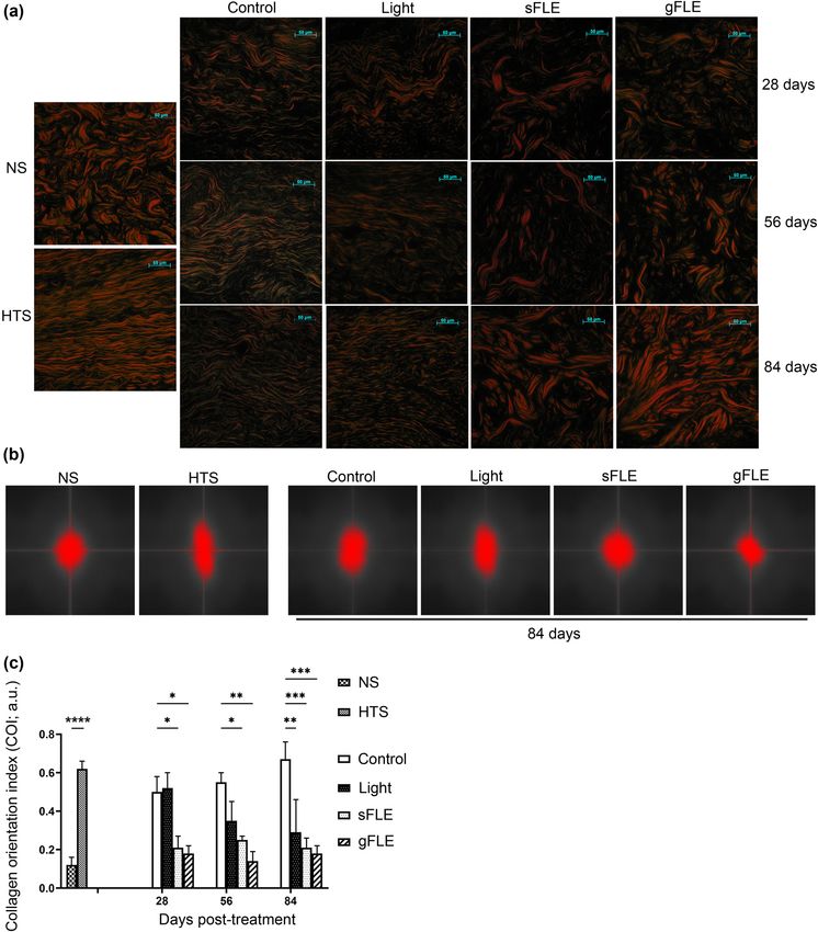
FLE balances healing after skin grafting 1247 Figure 3: FLE promoted reepithelization, and reduced epidermal thickness and vascularity. (a) Representative images showing H&E staining of human normal skin (NS) and xenografts harvested from mice treated with LED light alone (Light) or two different FLE treatments plus LED light (sFLE or gFLE) at day 28, 56, and 84 post-treatment, or untreated (Control). Assessment of epidermis thickness (purple arrows), dermis thickness (yellow arrows), blood vessels (blue arrows), epithelialization (red arrows), and rete ridges (green arrows) were observed, measured, and counted. Scale bar, 20 µm. (b–d) Quantified data are presented as bar graphs showing the mean value ± SE of (b) epidermis thickness, (c) dermis thickness, and (d) blood vessel numbers/HPFs. *p ≤ 0.05; **p ≤ 0.01.
1248 Jie Ding et al.
3.3 FLE treatment modulated collagen resembled that of human HTS (Figure 5a, Control and left
deposition and remodeling after skin column). Conversely, in xenografts from mice treated with
grafting FLE, collagen structure resembled the arrangement seen in
human NS (Figure 5a, sFLE, gFLE, and left column).
Collagen is a key component of the ECM and a balanced Finally, collagen organization was quantified using
decomposition is vital for ensuring tissue healing without the COI by Fourier analysis [41,42]. The short and long
causing fibrosis [35,39]. Quantification of collagen in the axes of the collagen bundles were measured for each scar
xenografts demonstrates that collagen deposition was (Figure 5b and Figure S4a) and the COIs were calculated
significantly induced by sFLE (sFLE: 57.21 ± 2.46 ng/mg; (Figure 5c and Figure S4b). First, the COI of NS (NS: 0.12 ±
p = 0.0036) treatment at day 28 compared with the con- 0.04 a.u.) was significantly lower compared with HTS
trol group (Control: 45.88 ± 2.46 ng/mg), and reduced in (HTS: 0.62 ± 0.04 a.u.; p < 0.0001). Furthermore, data
all three groups of mice treated with PBM (Light: 40.76 ± demonstrated a significant decrease in COI in FLE-treated
1.80 ng/mg; p < 0.0001, sFLE: 46.98 ± 1.31 ng/mg; p = mice (sFLE: 0.21 ± 0.06 a.u.; p = 0.0423, and gFLE: 0.18 ±
0.0007, and gFLE: 46.15 ± 1.00 ng/mg; p = 0.0003) com- 0.04 a.u.; p = 0.0300) compared to that in control group
pared with the control group (Control: 60.18 ± 3.15 ng/mg) (Control: 0.50 ± 0.08 a.u.) at day 28, day 56 (Control: 0.55
at day 56 post-treatment, while no difference was observed ± 0.05 a.u. vs sFLE: 0.25 ± 0.02 a.u.; p = 0.0341, or gFLE:
at day 84 (Figure 4 and Figure S3). 0.14 ± 0.05 a.u.; p = 0.0025), and for all PBM-treated
Next the structure of the collagen network was assessed groups on day 84 (Control: 0.67 ± 0.09 a.u. vs Light:
using picrosirius red staining. The orientation of collagen 0.29 ± 0.17 a.u.; p = 0.0034, sFLE: 0.21 ± 0.05 a.u.; p =
molecules is an important determinant of their functionality 0.0004, or gFLE: 0.18 ± 0.04 a.u.; p = 0.0001) (Figure 5c
in connective tissues [40]. It is furthermore known that in and Figure S4b). Furthermore, COI of xenografts from
scar tissue, the collagen network differs from the collagen FLE-treated mice resembled COI of NS tissue (Figure
structure in normal skin, in which collagen forms a “basket- 5c), suggesting an improvement in collagen orientation
weave” structure with perpendicular collagen fibers [39]. and remodeling mediated by FLE treatments.
First, we saw that the structure of collagen bundles in
human HTS tissue consisted of thin collagen fibers and
smaller bundles that aligned parallel with the epidermis 3.4 FLE treatment reduced levels of
compared with human NS tissue (Figure 5a, left column).
myofibroblasts, mast cells, and CTGF
In xenografts of control mice, the collagen bundle structure
in xenografts
Myofibroblasts play a crucial role in wound healing via
ECM synthesis and wound contraction. However, exces-
sive proliferation of differentiated αSMA-expressing
myofibroblasts is associated with increased fibrosis [43].
Hence, the impact of FLE on myofibroblasts was investi-
gated by quantifying these cells in the dermis of xeno-
grafts by determining αSMA staining (exemplified in
Figure S5a). Our results demonstrate that FLE treatment
decreased the number of myofibroblasts in xenograft tis-
sues (Figure 6a). Although these findings were not sig-
nificant on day 28 or 56 (Figure 6a and Figure S5b), day
84 findings showed that both sFLE and gFLE significantly
reduced myofibroblast numbers in the tissue post-treat-
Figure 4: FLE modulated collagen production. Collagen production in ment (Control: 18.0 ± 3.46 cells/HPFs vs sFLE: 7.0 ± 2.04
xenografts harvested from mice treated with LED light alone (Light) cells/HPFs; p = 0.0114, or gFLE: 1.33 ± 0.88 cells/HPFs;
or two different FLE treatments plus LED light (sFLE or gFLE) at day p < 0.0001), while treatment with LED light alone signif-
28, 56, and 84 post-treatment, or untreated (Control) was quantified
icantly promoted myofibroblasts within the first 56 days
by 4-hydroxyproline assessment. Bar graphs present the mean
value ± SE of 4-hydroxyproline production ng/mg of dry tissue
(Control: 12.0 ± 4.36 cells/HPFs vs Light: 25.3 ± 1.45 cells/
referring to a standard curve. Experiments were performed in tri- HPFs; p = 0.0029) after treatment when compared with
plicate for each sample. **p ≤ 0.01; ***p ≤ 0.001; ****p ≤ 0.0001. the control group (Figure 6a and Figure S5b).FLE balances healing after skin grafting 1249 Figure 5: FLE improved collagen orientation and remodeling. (a) Representative images showing picrosirius red staining of human hyper- trophic scar (HTS), site-matched human normal skin (NS), and xenografts harvested from mice treated with LED light alone (Light) or two different FLE treatments plus LED light (sFLE or gFLE) at day 28, 56, and 84 post-treatment, or untreated (Control) was performed to appraise collagen networks. Under polarized light, collagen bundles appeared green, red, or yellow. Scale bar, 50 µm. (b) Representative images of collagen orientation evaluated by Fournier analysis in the xenografts harvested from mice at day 84 post-treatment. (c) Bar graphs present the mean value ± SE of the COI calculated using the equation: 1 − (short axis/long axis) as arbitrary units (a.u.). *p ≤ 05; **p ≤ 0.01; ***p ≤ 0.001; ****p ≤ 0.0001.
1250 Jie Ding et al.
gFLE: 8.67 ± 1.90 cells/HPFs; p < 0.0001) post-treatment com-
pared with the control group (Figure 6b and Figure S6b).
Interestingly, the light-mediated down-modulation observed
at day 56 had partly increased at 84 days after treatment
(Figure 6b and Figure S6b).
Finally, expression of CTGF, which is an important
growth factor known to be overexpressed and involved in
fibrosis and scar formation [45], was assessed in the graft
tissue (Figure 7a). CTGF expression was not significantly
modulated in any of the groups at 28 and 56 days post-
treatment (Figure 7b and Figure S7). However, CTGF
expression was significantly reduced by Light and sFLE
(Light: 2.93 ± 0.43% of area; p = 0.0220 or sFLE: 2.11 ±
0.10% of area; p = 0.0011) compared with control group
(Control: 5.11 ± 1.08% of area) at day 84 (Figure 7b and
Figure S7).
Together these data suggest that FLE-treatment mod-
ulates wound closure by accelerating re-epithelialization,
while normalizing the collagen organization, blood vessel
formation, and reducing the risk of scarring through reduced
numbers of myofibroblasts, mast cells, and CTGF levels.
4 Discussion
Figure 6: FLE reduced myofibroblast formation and mast cell accu- Ensuring swift and balanced wound healing following
mulation. (a) Bar graphs present the mean value ± SE of myofibro- skin grafting is essential to reduce the risk of infection
blasts in 5 HPFs in the xenografts harvested from mice treated with which can lead to prolonged inflammation, increasing
LED light alone (Light) or two different FLE treatments plus LED light the risk of chronic wounds, fibrosis, and severe scarring
(sFLE or gFLE) at day 28, 56, and 84 post-treatment, or untreated
[1]. This study sought to investigate the effect of FLE
(Control). *p ≤ 0.05; **p ≤ 0.01; ****p ≤ 0.0001. (b) Graph presents
the mean value ± SE of mast cells in 5 HPFs in the xenografts har-
therapy in wound healing after grafting, based on the
vested from mice treated with LED light alone (Light) or two different hypothesis that FLE represents a novel approach to ensure
FLE treatments plus LED light (sFLE or gFLE) at day 28, 56, and 84 balanced healing with reduced risk of scarring for graft
post-treatment, or untreated (Control). patients.
We used our previously described modified mouse
scar model [29] that offers the advantages of low cost,
Recent studies have highlighted the importance of easy manipulation, and short time frame for scar forma-
mast cells in the release of various mediators that affect tion and remodeling, compared with several other dermal
cell proliferation and collagen remodeling during wound wound models, including the in vivo human scratch
healing, with high numbers of activated mast cells asso- wound model [46], rabbit ear [47], or red Duroc pig ding
ciating with scarring [44]. The number of mast cells were [48]. Our model [29], in which STSGs instead of FTSGs are
therefore analyzed in xenografts after FLE treatment. Tolui- transplanted on nude mice, results in development of red,
dine blue staining was done to assess mast cell numbers raised, and thickened scars that have intrinsic properties
in grafted tissue (exemplified in Figure S6a). PBM treatment closely resembling HTS formation in humans [30].
significantly reduced mast cell numbers in the xenografts at Although this model is prone to develop scarring, it is
day 56 (Control: 27.3 ± 5.00 cells/HPFs vs Light: 12.0 ± 2.60 highly valuable for investigating several aspects of the
cells/HPFs; p = 0.0118, sFLE: 8.70 ± 1.70 cells/HPFs; p = wound healing process because of its significant increase
0.0018, or gFLE: 8.70 ± 3.50 cells/HPFs; p = 0.0018) and day in the number of macrophages, mast cells, and fibrocytes
84 (Control: 40.0 ± 2.90 cells/HPFs vs Light: 21.3 ± 3.70 cells/ [30], along with an increase in biglycan and a decrease in
HPFs; p = 0.0010, sFLE: 6.00 ± 3.00 cells/HPFs; p < 0.0001, or decorin expression in the grafted skin [49]. We found thatFLE balances healing after skin grafting 1251 Figure 7: FLE decreased fibrotic factor production. (a) Representative images showing connective tissue growth factor (CTGF) staining (dark brown) in xenografts harvested from mice treated with LED light alone (Light) or two different FLE treatments plus LED light (sFLE or gFLE) at day 28, 56, and 84 post-treatment, or untreated (Control). Scale bar, 100 µm. (b) Bar graphs present the mean value ± SE of CTGF stained area (% of area). *p ≤ 0.05; **p ≤ 0.01. FLE treatment accelerated re-epithelialization and rete diabetic foot ulcers, and pressure ulcers as well as acute ridges formation in the early stages of healing, which are second-degree burns [13,27,28], while highlighting a poten- key parameters in scar pathophysiology [50]. Although tial superior effect of FLE in the earlier stages of healing. scar morphology was not significantly different between Further studies are needed to clarify the effect of FLE on FLE-treated and control groups in this dermal fibrotic reducing the risk of development of scarring. Introducing mouse model, accelerated wound healing is advantageous FLE earlier (before 7 days) after skin grafting might be ben- in itself since is reduces the risk of infections that often eficial and is an interesting aim of a subsequent investiga- complicate the healing process and increases the risk of tion. In addition, investigating FLE in a model less prone for developing chronic wounds or fibrosis, including HTS. default development of HTS would also be insightful. These results complement recent studies investigating Fibroblastic proliferation and excess collagen deposi- the effect of FLE on different phases of wound healing tion are associated with imbalanced healing and scarring showing accelerated healing of chronic venous leg ulcers, [3,51,52]. Interestingly, studies have shown that light
1252 Jie Ding et al.
within the red part of the visual spectrum suppresses col- may reflect the different time points when vascularization
lagen production in human skin fibroblast cultures [51] was assessed after stimulation in each study and that FLE
and inhibits type I collagen expression as well as TGF-β- thereby modulates angiogenesis differently in different
induced fibroblast to myofibroblast differentiation [53]. phases during wound healing.
Previous studies focusing on FLE have investigated col- Although a number of parameters measured in this
lagen production early after treatment showing that FLE study were improved by LED light stimulation alone, FLE
can modulate collagen production both in vitro and in vivo yielded additional effects demonstrating that the FLE
[12,16]. In the current study, we found that FLE increased spectrum specifically was critical to the therapeutic ben-
collagen 28 days after treatment, whereas it was down- efits observed. Specifically, FLE reduced myofibroblast
modulated after 56 days. Different types of collagen accumulation whereas Light treatment significantly
come into play at different time points during healing; increased the number of myofibroblasts in the tissue after
collagen III is produced in the proliferative phase and 56 days. This indicates that blue light directly promotes
replaced by collagen I in the remodeling phase [39]. Since myofibroblast differentiation which increases the risk of
the 4-hydroxyproline-assay used does not distinguish fibrosis whereas the full FLE spectrum reduced myofibro-
between collagen types and thus kinetics of these, more blast numbers. Furthermore, mast cell recruitment and
detailed investigations of collagen types are needed to CTGF expression were decreased by FLE in the later phase
fully understand how FLE controls collagen production of repair and remodeling. Combined, these effects resulted
in different steps of the wound healing process. However, in increased wound maturity, which is vital for minimizing
collagen production alone is inadequate to assess its role the risk of fibrosis in patients [39]. The FLE biophotonic
in healing, since deposition and remodeling is central for platform utilized in this study has the advantage of gen-
constructing new and fully functional tissues [39,40]. We erating a broad spectrum of fluorescence within the visible
found that the COI was significantly decreased already at range (440–460 and 500–700 nm) [12,13]. This enables the
28 days and remained low at 56 and 84 days after treat- light to penetrate various layers of the skin and interact
ment, suggesting that FLE normalizes collagen deposition with endogenous chromophores, such as flavins/ribo-
and remodeling in the tissue in the long-term. Considering flavins, cytochrome c oxidase subunit IV, and nicotina-
that modulation of collagen production is an essential com- mide adenine dinucleotide hydrogen [56], in different
ponent of any effective tissue repair and anti-fibrotic therapy layers in a single treatment. Moreover, previous clinical
[54], our data provide support for a new therapeutic mod- studies investigating FLE therapy has reported the treat-
ality for managing development of skin fibrosis. ment as applicable, safe, and with few adverse effects
Angiogenesis is vital to restore circulation in the [17,18,25–28], underlining few clinical implications for the
grafted and damaged tissue, and is induced by secreted use of FLE. For treatment of acute second-degree burns or
growth factors, including vascular endothelial growth chronic ulcers, patients did initially experience some pain
factor (VEGF) produced by keratinocytes, fibroblasts, and in relation to the FLE treatment, which was, however, not
macrophages [37]. While the importance of angiogenesis considered extraordinary compared with the standard of
in the early/proliferative phase of tissue healing is well- care for these conditions [27,28].
documented, less is known about regulation later in the PBM was previously shown to (1) accelerate the
repair process due to the difficulties in monitoring this healing of formocresol-induced oral ulcers and diabetic
process in vivo. A study has shown that long term over- wounds in rats due to certain wavelengths stimulating
expression of VEGF-A associates with pathological condi- fibroblast proliferation and collagen production [57,58];
tions such as muscle fibrosis [55], underlining the impor- (2) induce comparable levels of cell migration and wound
tance of resolving this process later in the wound healing closure in a scratch wound model [59]; (3) promote donor
process. We have previously shown that FLE-treatment site wound healing of the free gingival graft, potentially
facilitates angiogenesis, by measuring branching and via reducing reactive oxygen species production [60];
tube formation in cultures of human aortic endothelial (4) modulate angiogenesis [61]; (5) promote provisional
cells 18 h after stimulation in vitro [12]. In this study, we matrix and wound reorganization [60]; and (6) enhance
found a significant reduction in the number of blood ves- the healing process of third-degree burns in rats [62]
sels in the grafted tissues 28 and 56 days after FLE stimu- and modulate mitochondrial physiology [13] and gene
lation. Earlier, we described that the number of newly expression [15]. Further experiments are needed to deter-
formed blood vessels in grafted skin peaked on day 10 mine which molecular pathways are specifically activated
and declined again by day 28 after grafting [38]. These by FLE treatment underpinning its beneficial effects. Poten-
observed differences in the effect of FLE on angiogenesis tial mechanisms of action likely include photon absorptionFLE balances healing after skin grafting 1253
by endogenous chromophores, photonic energy utilization, visualization: M.M. and J.D.; writing – original draft: J.D.;
and modulation of mitochondrial activity including ATP writing – review & editing: M.M., J.D., D.E, L.H., M.C.E.N.,
production, which regulate cellular activation, migration, E.E.T., Z.Z., P.K., Z.M., S.A., and T.I.
and protein synthesis that are essential in wound healing
[56]. Conflict of interest: M.M., L.H., and M.C.E.N. are employees
In summary, the aim of this study was to investigate and D.E. is a former employee of Klox Technologies. This
the impact of FLE on tissue repair after skin grafting and does not alter our adherence to Open Medicine policies on
examine its therapeutic potential for improving healing sharing data and materials. The other authors have no
and reducing the risk of complication. We found that FLE conflicts of interest in this manuscript.
treatment stimulated healing by increasing re-epithelia-
lization, significantly increasing epidermal thickness and Data availability statement: Supplemental information
reducing dermal thickness after 28 days, while decreasing is available online and further data is available upon
number of blood vessels after 56 days. Moreover, collagen request to corresponding authors.
production was enhanced at day 28 and significantly
reduced at day 56 with COI significantly reduced at all
three time points. Finally, mast cells infiltration, myofibro-
blast formation, and angiogenesis were lowered later in References
the healing process (84 days after treatment). However,
FLE did not directly alter the morphology of the default- [1] Sun BK, Siprashvili Z, Khavari PA. Advances in skin grafting
and treatment of cutaneous wounds. Science.
developed HTSs in this mouse model.
2014;346(6212):941–5.
To conclude, these findings suggest that FLE helps
[2] Brown JE, Holloway SL. An evidence-based review of split-
balance the wound healing process at different stages thickness skin graft donor site dressings. Int Wound J.
following skin grafting, although a thorough clinical 2018;15(6):1000–9.
assessment is necessary. This study supports FLE therapy [3] Larouche J, Sheoran S, Maruyama K, Martino MM. Immune
as a possible safe treatment for skin graft patients to help regulation of skin wound healing: mechanisms and novel
therapeutic targets. Adv Wound Care (N Rochelle).
ensure a balanced healing process and a lowered risk of
2018;7(7):209–31.
developing chronic wounds. [4] Avci P, Gupta A, Sadasivam M, Vecchio D, Pam Z, Pam N, et al.
Low-level laser (light) therapy (LLLT) in skin: stimulating,
Acknowledgments: The authors are grateful to Firefighters healing, restoring. Semin Cutan Med Surg. 2013;32(1):41–52.
Burn Trust Fund from Edmonton Firefighter Association. [5] Anders JJ, Lanzafame RJ, Arany PR. Low-level light/laser
therapy versus photobiomodulation therapy. Photomed Laser
Surg. 2015;33(4):183–4.
Funding information: The Innovation Fund Denmark has
[6] Hamblin MR. Shining light on the head: photobiomodulation
supported with a Postdoc stipend to MM (#8054-00028B). for brain disorders. BBA Clin. 2016;6:113–24.
Klox Technologies has supplied FLE systems used in the [7] de Freitas LF, Hamblin MR. Proposed mechanisms of photo-
study and has provided support and guidance in the biomodulation or low-level light therapy. IEEE J Sel Top
application of these. In addition, Klox Technologies has Quantum Electron. 2016;22:3.
[8] Dancáková L, Vasilenko T, Kováč I, Jakubčová K, Hollý M,
provided support in the form of salaries for authors M.M.,
Revajová V, et al. Low-level laser therapy with 810 nm wave-
L.H., D.E., and M.C.E.N. M.M., L.H., and D.E. have been length improves skin wound healing in rats with streptozo-
involved in the final revision of the manuscript and sup- tocin-induced diabetes. Photomed Laser Surg.
port publication, but did not have any additional role in 2014;32(4):198–204.
the study design, data collection and analysis, or pre- [9] Zhang H, Liu S, Yang X, Chen N, Pang F, Chen Z, et al. LED
phototherapy with gelatin sponge promotes wound healing in
paration of the original manuscript. The specific roles of
mice. Photochem Photobiol. 2018;94(1):179–85.
these authors are articulated in the author contributions
[10] Hamblin MR. Mechanisms and applications of the anti-
section. inflammatory effects of photobiomodulation. AIMS Biophys.
2017;4(3):337–61.
Author contributions: Conceptualization: J.D. and E.E.T.; [11] Hamblin MRFC, Huang YY, Freitas de Freitas L, Carroll J. Low-
data curation: J.D., Z.Z., P.K., Z.M., S.A., T.I., and E.E.T.; level light therapy: photobiomodulation. Bellingham, WA USA:
SPIE PRESS BOOK; 2018, p. 388.
formal analysis: J.D., M.M., M.C.E.N., and E.E.T.; investi-
[12] Edge D, Mellergaard M, Dam-Hansen C, Corell DD, Jaworska J,
gation: J.D., Z.Z., P.K., Z.M., S.A., T.I., and E.E.T.; meth- Scapagnini G, et al. Fluorescent light energy: the future for
odology: J.D. and E.E.T.; project administration: J.D. and treating inflammatory skin conditions? J Clin Aesthet Dermatol.
E.E.T.; supervision: E.E.T.; validation: J.D. and E.E.T.; 2019;12(5):E61–8.1254 Jie Ding et al.
[13] Scapagnini G, Marchegiani A, Rossi G, Zago M, Jowarska J, [26] Nikolis AGD, Pesant Y, Scapagnini G, Vezina D. A prospective
Wael M, et al. Management of all three phases of wound case series evaluating the safety and efficacy of the Klox bio-
healing through the induction of fluorescence biomodulation photonic system in venous leg ulcers. Chronic Wound Care
using fluorescence light energy. Proc. SPIE 10863, Photonic Manag Res. 2016;3:101–11.
Diagnosis and Treatment of Infections and Inflammatory [27] Romanelli M, Piaggesi A, Scapagnini G, Dini V, Janowska A,
Diseases II, 108630W (7 March 2019). SPIE; 2019. https:// Iacopi E, et al. Evaluation of fluorescence biomodulation in the
spie.org/Publications/Proceedings/Paper/10.1117/ real-life management of chronic wounds: the EUREKA trial.
12.2508066. J Wound Care. 2018;27(11):744–53.
[14] Zago M, Dehghani M, Jaworska J, Mellergaard M, Edge D, [28] Mellergaard M, Fauverghe S, Scarpa C, Pozner VL, Skov S,
Corell DD, et al. Fluorescent light energy in wound healing: Hebert L, et al. Evaluation of fluorescent light energy for the
when is a photon something more? Proceedings Volume 11221, treatment of acute second-degree burns. Mil Med.
Mechanisms of Photobiomodulation Therapy XV; 112210A 2021;186(Supplement_1):416–23.
(2020). San Francisco, California, United States: SPIE [29] Yang DY, Li SR, Wu JL, Chen YQ, Li G, Bi S, et al. Establishment
BiOS; 2020. of a hypertrophic scar model by transplanting full-thickness
[15] Ferroni L, Zago M, Patergnani S, Campbell SE, Hébert L, human skin grafts onto the backs of nude mice. Plast Reconstr
Nielsen M, et al. Fluorescent light energy (FLE) acts on mito- Surg. 2007;119(1):104–9. discussion 10-1.
chondrial physiology improving wound healing. J Clin Med. [30] Wang J, Ding J, Jiao H, Honardoust D, Momtazi M,
2020;9(2):559. Shankowsky HA, et al. Human hypertrophic scar-like nude
[16] Nikolis A, Bernstein S, Kinney B, Scuderi N, Rastogi S, mouse model: characterization of the molecular and cellular
Sampalis JS. A randomized, placebo-controlled, single- biology of the scar process. Wound Repair Regen.
blinded, split-faced clinical trial evaluating the efficacy and 2011;19(2):274–85.
safety of KLOX-001 gel formulation with KLOX light-emitting [31] Ding J, Tredget EE. Transplanting human skin grafts onto
diode light on facial rejuvenation. Clin Cosmet Investig nude mice to model skin scars. Methods Mol Biol.
Dermatol. 2016;9:115–25. 2017;1627:65–80.
[17] Antoniou C, Dessinioti C, Sotiriadis D, Kalokasidis K, [32] Wang J, Jiao H, Stewart TL, Shankowsky HA, Scott PG,
Kontochristopoulos G, Petridis A, et al. A multicenter, rando- Tredget EE. Increased severity of bleomycin-induced skin
mized, split-face clinical trial evaluating the efficacy and fibrosis in mice with leukocyte-specific protein 1 deficiency.
safety of chromophore gel-assisted blue light phototherapy J Invest Dermatol. 2008;128(12):2767–76.
for the treatment of acne. Int J Dermatol. 2016;55(12):1321–8. [33] Tredget EE, Falk N, Scott PG, Hogg AM, Burke JF. Determination
[18] Nikolis A, Fauverghe S, Scapagnini G, Sotiriadis D, of 4-hydroxyproline in collagen by gas chromatography/mass
Kontochristopoulos G, Petridis A, et al. An extension of a spectrometry. Anal Biochem. 1990;190(2):259–65.
multicenter, randomized, split-face clinical trial evaluating the [34] Junqueira LC, Bignolas G, Brentani RR. Picrosirius staining
efficacy and safety of chromophore gel-assisted blue light plus polarization microscopy, a specific method for
phototherapy for the treatment of acne. Int J Dermatol. collagen detection in tissue sections. Histochem J.
2018;57(1):94–103. 1979;11(4):447–55.
[19] Braun SA, Gerber PA. A photoconverter gel-assisted blue light [35] Osman OS, Selway JL, Harikumar PE, Stocker CJ, Wargent ET,
therapy for the treatment of rosacea. Int J Dermatol. Cawthorne MA, et al. A novel method to assess collagen
2017;56(12):1489–90. architecture in skin. BMC Bioinforma. 2013;14(1):260.
[20] Sannino M, Lodi G, Dethlefsen MW, Nistico SP, Cannarozzo G, [36] Martin P, Nunan R. Cellular and molecular mechanisms of
Nielsen MCE. Fluorescent light energy: treating rosacea sub- repair in acute and chronic wound healing. Br J Dermatol.
types 1, 2, and 3. Clin Case Rep. 2018;6(12):2385–90. 2015;173(2):370–8.
[21] Koceva I, Rümmelein B, Gerber PA, Edge D, Nielsen MCE. [37] Guerra A, Belinha J, Jorge RN. Modelling skin wound healing
Fluorescent light energy: a new therapeutic approach to angiogenesis: a review. J Theor Biol. 2018;459:1–17.
effectively treating acne conglobata and hidradenitis suppur- [38] Shaterian A, Borboa A, Sawada R, Costantini T, Potenza B,
ativa. Clin Case Rep. 2019;7(9):1769–72. Coimbra R, et al. Real-time analysis of the kinetics of angio-
[22] Mahendran A, Wong XL, Kao S, Sebaratnam DF. Treatment of genesis and vascular permeability in an animal model of
erlotinib-induced acneiform eruption with chromophore gel- wound healing. Burns. 2009;35(6):811–7.
assisted phototherapy. Photodermatol Photoimmunol [39] Reinke JM, Sorg H. Wound repair and regeneration. Eur Surg
Photomed. 2019;35(3):190–2. Res. 2012;49(1):35–43.
[23] Gerber PA, Scarcella G, Edge D, Nielsen MCE. Biophotonic [40] Bi X, Li G, Doty SB, Camacho NP. A novel method for deter-
pretreatment enhances the targeting of senile lentigines with mination of collagen orientation in cartilage by Fourier trans-
a 694 nm QS-ruby laser. Photodermatol Photoimmunol form infrared imaging spectroscopy (FT-IRIS). Osteoarthr
Photomed. 2020;36(2):159–60. Cartil. 2005;13(12):1050–8.
[24] Scarcella GGP, Edge D, Nielsen MCE. Effective removal of solar [41] Verhaegen PD, Schouten HJ, Tigchelaar-Gutter W, van Marle J,
lentigines by combination of pre- and post- fluorescent light van Noorden CJ, Middelkoop E, et al. Adaptation of the dermal
energy treatment with picosecond laser treatment. Clin Case collagen structure of human skin and scar tissue in response
Rep. 2020;8(8):1429–32. to stretch: an experimental study. Wound Repair Regen.
[25] Nikolis AFS, Vezina D, Scapagnini G. Evaluation of biophotonic 2012;20(5):658–66.
therapy in a non-healing diabetic foot ulcer: a case report. [42] van Zuijlen PP, de Vries HJ, Lamme EN, Coppens JE, van Marle J,
Diabet Foot Can. 2016;2016(4):25–30. Kreis RW, et al. Morphometry of dermal collagen orientation byFLE balances healing after skin grafting 1255
Fourier analysis is superior to multi-observer assessment. fibroblast-myofibroblast transition reducing TRPC1 channel
J Pathol. 2002;198(3):284–91. expression/activity: new perspectives for tissue fibrosis
[43] Hinz B. Formation and function of the myofibroblast during treatment. Lasers Surg Med. 2016;48(3):318–32.
tissue repair. J Invest Dermatol. 2007;127(3):526–37. [54] McDougall S, Dallon J, Sherratt J, Maini P. Fibroblast migration
[44] Wilgus TA, Wulff BC. The importance of mast cells in dermal and collagen deposition during dermal wound healing: math-
scarring. Adv Wound Care (N Rochelle). 2014;3(4):356–65. ematical modelling and clinical implications. Philos Trans A
[45] Lian N, Li T. Growth factor pathways in hypertrophic scars: Math Phys Eng Sci. 2006;364(1843):1385–405.
Molecular pathogenesis and therapeutic implications. Biomed [55] Karvinen H, Pasanen E, Rissanen TT, Korpisalo P,
Pharmacother. 2016;84:42–50. Vähäkangas E, Jazwa A, et al. Long-term VEGF-A expression
[46] Morris DE, Wu L, Zhao LL, Bolton L, Roth SI, Ladin DA, et al. promotes aberrant angiogenesis and fibrosis in skeletal
Acute and chronic animal models for excessive dermal scar- muscle. Gene Ther. 2011;18(12):1166–72.
ring: quantitative studies. Plast Reconstr Surg. [56] Lubart R, Lavi R, Friedmann H, Rochkind S. Photochemistry and
1997;100(3):674–81. photobiology of light absorption by living cells. Photomed
[47] Dunkin CSJ, Pleat JM, Gillespie PH, Tyler MPH, Roberts AHN, Laser Surg. 2006;24(2):179–85.
McGrouther DA. Scarring occurs at a critical depth of skin [57] Lau PS, Bidin N, Krishnan G, Nassir Z, Bahktiar H. Biophotonic
injury: Precise measurement in a graduated dermal scratch in effect of diode laser irradiance on tensile strength of diabetic
human volunteers. Plastic Reconstr Surg. rats. J Cosmetic Laser Ther. 2015;17(2):86–9.
2007;119(6):1722–32. [58] de Carvalho FB, Andrade AS, Rasquin LC, de Castro IV,
[48] Zhu KQ, Engrav LH, Gibran NS, Cole JK, Matsumura H, Cangussu MC, Pinheiro AL, et al. Effect of laser (lambda 660
Piepkorn M, et al. The female, red Duroc pig as an animal nm) and LED (lambda 630 nm) photobiomodulation on for-
model of hypertrophic scarring and the potential role of the mocresol-induced oral ulcers: a clinical and histological study
cones of skin. Burns. 2003;29(7):649–64. on rodents. Lasers Med Sci. 2015;30(1):389–96.
[49] Momtazi M, Kwan P, Ding J, Anderson CC, Honardoust D, [59] Spitler R, Berns MW. Comparison of laser and diode sources
Goekjian S, et al. A nude mouse model of hypertrophic scar for acceleration of in vitro wound healing by low-level light
shows morphologic and histologic characteristics of human therapy. J Biomed Opt. 2014;19(3):38001.
hypertrophic scar. Wound Repair Regen. 2013;21(1):77–87. [60] Wang CY, Tsai SC, Yu MC, Lin YF, Chen CC, Chang PC. Light-
[50] Arno AI, Gauglitz GG, Barret JP, Jeschke MG. Up-to-date emitting diode irradiation promotes donor site wound healing
approach to manage keloids and hypertrophic scars: a useful of the free gingival graft. J Periodontol. 2015;86(5):674–81.
guide. Burns. 2014;40(7):1255–66. [61] de Sousa AP, Paraguassú GM, Silveira NT, de Souza J,
[51] Mamalis A, Siegel D, Jagdeo J. Visible red light emitting diode Cangussú MC, dos Santos JN, et al. Laser and LED photo-
photobiomodulation for skin fibrosis: key molecular pathways. therapies on angiogenesis. Lasers Med Sci. 2013;28(3):981–7.
Curr Dermatology Rep. 2016;5:121–8. [62] de Vasconcelos Catão MH, Nonaka CF, de Albuquerque RL Jr,
[52] Biernacka A, Dobaczewski M, Frangogiannis NG. TGF-beta Bento PM, de Oliveira, Costa R. Effects of red laser, infrared,
signaling in fibrosis. Growth Factors. 2011;29(5):196–202. photodynamic therapy, and green LED on the healing process
[53] Sassoli C, Chellini F, Squecco R, Tani A, Idrizaj E, Nosi D, et al. of third-degree burns: clinical and histological study in rats.
Low intensity 635 nm diode laser irradiation inhibits Lasers Med Sci. 2015;30(1):421–8.You can also read