Folding of cohesin's coiled coil is important for Scc2/4-induced association with chromosomes
←
→
Page content transcription
If your browser does not render page correctly, please read the page content below
RESEARCH ARTICLE

                                    Folding of cohesin’s coiled coil is
                                    important for Scc2/4-induced association
                                    with chromosomes
                                    Naomi J Petela1†, Andres Gonzalez Llamazares2†, Sarah Dixon1, Bin Hu3,
                                    Byung-Gil Lee2, Jean Metson1, Heekyo Seo4, Antonio Ferrer-Harding1,
                                    Menelaos Voulgaris1, Thomas Gligoris1, James Collier1, Byung-Ha Oh4,
                                    Jan Löwe2*, Kim A Nasmyth1*
                                    1
                                     Department of Biochemistry, University of Oxford, Oxford, United Kingdom; 2MRC
                                    Laboratory of Molecular Biology, Cambridge, United Kingdom; 3Institute of Medical
                                    Sciences, University of Aberdeen, Aberdeen, United Kingdom; 4Department of
                                    Biological Sciences, KAIST Institute for the Biocentury, Cancer Metastasis Control
                                    Center, Korea Advanced Institute of Science and Technology, Daejeon, Republic of
                                    Korea

                                    Abstract Cohesin’s association with and translocation along chromosomal DNAs depend on an
                                    ATP hydrolysis cycle driving the association and subsequent release of DNA. This involves DNA
                                    being ‘clamped’ by Scc2 and ATP-dependent engagement of cohesin’s Smc1 and Smc3 head
                                    domains. Scc2’s replacement by Pds5 abrogates cohesin’s ATPase and has an important role in
                                    halting DNA loop extrusion. The ATPase domains of all SMC proteins are separated from their
                                    hinge dimerisation domains by 50-nm-long coiled coils, which have been observed to zip up along
*For correspondence:                their entire length and fold around an elbow, thereby greatly shortening the distance between
jyl@mrc-lmb.cam.ac.uk (JL);         hinges and ATPase heads. Whether folding exists in vivo or has any physiological importance is not
ashley.nasmyth@bioch.ox.ac.uk       known. We present here a cryo-EM structure of the apo form of cohesin that reveals the structure
(KAN)                               of folded and zipped-up coils in unprecedented detail and shows that Scc2 can associate with
†
 These authors contributed          Smc1’s ATPase head even when it is fully disengaged from that of Smc3. Using cysteine-specific
equally to this work                crosslinking, we show that cohesin’s coiled coils are frequently folded in vivo, including when
                                    cohesin holds sister chromatids together. Moreover, we describe a mutation (SMC1D588Y) within
Competing interests: The
                                    Smc1’s hinge that alters how Scc2 and Pds5 interact with Smc1’s hinge and that enables Scc2 to
authors declare that no
competing interests exist.          support loading in the absence of its normal partner Scc4. The mutant phenotype of loading
                                    without Scc4 is only explicable if loading depends on an association between Scc2/4 and cohesin’s
Funding: See page 28
                                    hinge, which in turn requires coiled coil folding.
Received: 05 February 2021
Accepted: 21 June 2021
Published: 14 July 2021

Reviewing editor: Adèle L
                                    Introduction
Marston, University of
                                    SMC complexes are highly conserved from prokaryotes to eukaryotes. Best characterised among
Edinburgh, United Kingdom           this family are cohesin and condensin, both of which are DNA translocases (Ganji et al., 2018;
                                    Davidson et al., 2019; Kim et al., 2019; Golfier et al., 2020). Cohesin and condensin are thought
   Copyright Petela et al. This
                                    to organise chromosomes in eukaryotes during interphase and mitosis respectively by producing
article is distributed under the
                                    long loops of DNA (Nasmyth, 1982), a process called loop extrusion (LE). Cohesin has an additional
terms of the Creative Commons
Attribution License, which          property, namely the ability to hold sister DNAs together from their genesis during S phase till their
permits unrestricted use and        eventual disjunction to opposite poles of the cell during anaphase.
redistribution provided that the       Cohesin is composed of two rod-shaped SMC proteins, Smc1 and Smc3, with a dimerisation
original author and source are      interface at one end that is connected to an ABC-like ATPase domain via a 50-nm-long coiled coil
credited.                           (Figure 1A). Interaction via their dimerisation domains produces a V-shaped Smc1/3 heterodimer

Petela, Gonzalez Llamazares, et al. eLife 2021;10:e67268. DOI: https://doi.org/10.7554/eLife.67268                                 1 of 32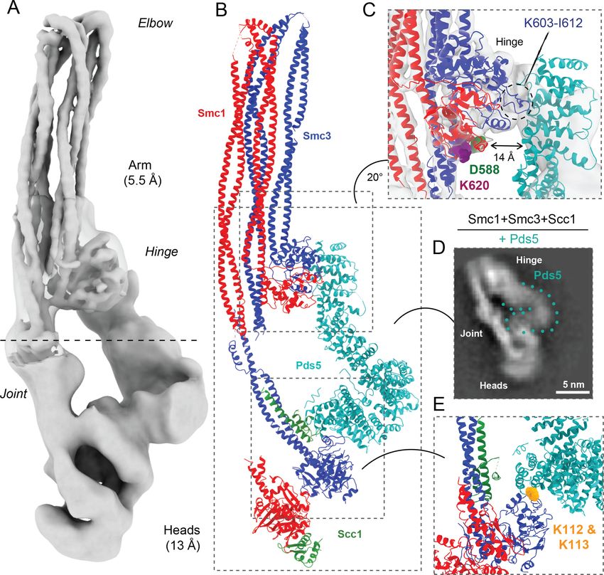
Research article                                       Chromosomes and Gene Expression Structural Biology and Molecular Biophysics

Figure 1. A mutation in the hinge domain of Smc1 restores viability in the absence of Scc4. (A) Schematic representation of Saccharomycescerevisiae
cohesin complex and its folding cycle. (B) Comparison of growth of wild-type (WT), scc4-4, and scc4-4 smc1D588Y strains at 35.5˚C (K699, K8326,
K19813). (C) Tetrad dissection of diploid strains containing SCC4/scc4D SMC1/smc1D588Y grown at 30˚C. Spores expressing smc1D588Y are circled in
red, and spores that lack Scc4 are indicated with blue hexagons. (D) Structure of the mouse Smc3-Smc1D574Y hinge domain (PDB: 7DG5). (E) Multiple
Figure 1 continued on next page

Petela, Gonzalez Llamazares, et al. eLife 2021;10:e67268. DOI: https://doi.org/10.7554/eLife.67268                                           2 of 32
Research article                                       Chromosomes and Gene Expression Structural Biology and Molecular Biophysics

Figure 1 continued
sequence alignment indicating conservation of Smc1D588. (F) Structural superposition of the WT hinge and the D574Y mutant hinge. Tyr574 swings out
relative to the position of D574 with a concomitant local conformational change of the mutated loop.
The online version of this article includes the following figure supplement(s) for figure 1:
Figure supplement 1. A mutation in the hinge domain of Smc1 restores viability in the absence of Scc4.

                                    whose two arms are connected by a central ‘hinge’ domain. The two ATPase ‘head’ domains at the
                                    apices of this dimer are meanwhile inter-connected by a kleisin subunit, Scc1. Scc1’s N- and C-termi-
                                    nal domains bind respectively to the coiled coil emerging from Smc3’s head (its neck) and the base
                                    of Smc1’s ATPase, thereby creating a tripartite SMC-kleisin (S-K) ring (Figure 1A). Cohesin’s associa-
                                    tion with DNA as well as its abilities to hold sisters together and extrude DNA loops are facilitated
                                    by three large hook-shaped HAWK (HEAT repeat proteins associated with kleisins) proteins; Scc2,
                                    Scc3, and Pds5 (Figure 1A). Scc3 is thought to be permanently bound to the complex, whereas Scc2
                                    and Pds5, which are mutually exclusive, are more dynamic. Of these, Scc2 has a crucial role in activ-
                                    ating cohesin’s ATPase at least in vitro, whether in the presence or absence of DNA (Petela et al.,
                                    2018).
                                        The discovery that anaphase is initiated through the opening of S-K rings due to cleavage of their
                                    kleisin moiety by the protease separase (Uhlmann, 2001) led to the suggestion that cohesion is
                                    mediated by the co-entrapment of sister DNAs within individual S-K rings (Haering et al., 2002).
                                    This hypothesis, known as the ring model, made the key prediction that site-specific chemical cross-
                                    linking of all three of the ring’s subunit interfaces would create a covalent topological linkage resis-
                                    tant to protein denaturation between small circular sister DNAs. Such catenated dimers (CDs) are
                                    indeed found in cells (Haering et al., 2008; Gligoris et al., 2014) and only under conditions in which
                                    cells form sister chromatid cohesion (Srinivasan et al., 2018).
                                        The ring model envisages that once established during DNA replication, maintenance of sister
                                    chromatid cohesion during G2 and M phases would not require continued ATP hydrolysis. This
                                    notion, namely that cohesion is a passive process, explains why Scc2, though essential for loading
                                    and for maintaining cohesin’s association with unreplicated DNA in vivo, has no role in maintaining
                                    cohesion during G2/M phases (Ciosk et al., 2000; Srinivasan et al., 2019). Cohesin’s ATPase is
                                    strictly dependent on Scc2 in vitro and is presumably inactive in vivo upon Scc2’s departure. LE in
                                    contrast requires continuous ATP hydrolysis dependent on Scc2, at least in vitro (Davidson et al.,
                                    2019).
                                        Yet another difference is that cohesion depends on passage of DNAs inside S-K rings while LE
                                    does not (Srinivasan et al., 2018; Davidson et al., 2019). Given that cohesion and LE involve at
                                    least some different mechanisms, it is perhaps not surprising that there is increasing evidence that
                                    the two processes are mutually exclusive in vivo (Srinivasan et al., 2018; Davidson et al., 2019).
                                    Complexes engaged in cohesion do not extrude loops and vice versa.
                                        Though maintenance of cohesion may have little in common with LE, the process by which cohe-
                                    sion is created in the first place may utilise mechanisms common to LE. This is supported by the fact
                                    that Scc2 is required for entrapping DNA within S-K rings as well as for the DNA-dependent ATPase
                                    activity necessary for LE. DNA entrapment assays combined with cryo-EM structures suggest that a
                                    key intermediate common to both processes is the passage of DNA between disengaged ATPase
                                    heads followed by its ‘clamping’ by Scc2 on a surface on top of them created by ATP-dependent
                                    head engagement (Collier et al., 2020; Shi et al., 2020; Higashi et al., 2020). It is envisaged that
                                    DNA translocation during LE involves recurrent rounds of DNA clamping followed by its release
                                    upon ATP hydrolysis. If so, each round presumably involves clamping of DNA successively further
                                    along the chromatin fibre. Clamping in this manner may be an important feature of cohesin’s associ-
                                    ation with chromatin, at least during G1 when LE is possibly its main activity. Crucially, clamping in
                                    vitro does not require Scc3, which is necessary for cohesin’s stable association with chromatin in vivo
                                    and ensures, at least in vitro, that clamping is followed or accompanied by transient opening of the
                                    S-K ring and thereby entrapment of DNAs within (Collier et al., 2020). The key point is that clamp-
                                    ing may be a feature not only of LE but also of the entrapment of DNAs within S-K rings necessary
                                    for cohesion.

Petela, Gonzalez Llamazares, et al. eLife 2021;10:e67268. DOI: https://doi.org/10.7554/eLife.67268                                         3 of 32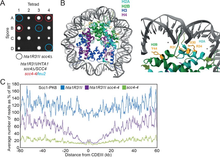
Research article                                       Chromosomes and Gene Expression Structural Biology and Molecular Biophysics

                                       Which interface of the S-K ring is opened through the action of Scc3 is uncertain as is the mecha-
                                    nism, either when individual DNAs are entrapped during G1 (or G2) or when sister DNAs are
                                    entrapped during the passage of replication forks. Complexes containing co-translational fusions,
                                    either between the C-terminus of Smc3 and the NTD of Scc1, or between Scc1’s C-terminus and the
                                    NTD of Smc1 are functional and capable of entrapping individual or sister DNAs within S-K rings. In
                                    contrast, the artificial connection of the Smc1 and Smc3 hinge domains using rapamycin blocks the
                                    establishment but not maintenance of sister chromatid cohesion (Gruber et al., 2006), leading to
                                    the suggestion that DNAs enter the S-K ring via a gate created by transient dissociation of the
                                    hinge. Whether this is really the case awaits more rigorous types of experiments.
                                       Cohesin complexes defective in ATP hydrolysis, due to Smc1E1158Q and Smc3E1155Q (EQEQ)
                                    mutations, accumulate in the clamped state in vitro (Collier et al., 2020; Shi et al., 2020;
                                    Higashi et al., 2020). Along with Scc2, they also accumulate at Saccharomyces cerevisiae CEN
                                    sequences (Hu et al., 2011), which are sites at which cohesin loads onto chromosomes with espe-
                                    cially high efficiency, due to an interaction between the kinetochore protein Ctf19 and Scc4 bound
                                    to Scc2’s largely unstructured N-terminal domain (Hinshaw et al., 2017). This suggests that in addi-
                                    tion to being a recurrent feature of LE, formation of the clamped state may be an early step in cohe-
                                    sin’s de novo association with chromosomal DNA. Scc4 facilitates Scc2-mediated loading
                                    throughout chromosome arms as well as at CEN sequences, though how it does so is poorly
                                    understood.
                                       When cohesin’s ATPase heads are disengaged, the coiled coils of Smc1 and Smc3 associate with
                                    each other along much of their length (Chapard et al., 2019). When this ‘zipping up’ includes the
                                    sections of coiled coils close to the ATPase heads, it forces them to adopt a configuration in which
                                    they are juxtaposed in a ‘J’ state that is distinct from, and incompatible with ATP-driven head
                                    engagement known as the ‘E’ state. Crucially, the zipping up of coiled coils in this manner is incom-
                                    patible with the clamping of DNA by Scc2 on top of engaged heads and the latter is therefore
                                    accompanied by extensive unzipping, at least up to the elbow (Collier et al., 2020). Coiled coil zip-
                                    ping up is a feature of cohesin engaged in holding sister chromatids together, with sister DNAs
                                    entrapped within J-K compartments, namely between juxtaposed (J) heads and the kleisin associ-
                                    ated with them (Chapard et al., 2019). Extensive zipping up may have an important role in prevent-
                                    ing unregulated ATP hydrolysis or precocious head engagement.
                                       Along with coiled coil zipping up, the generation of cohesive structures during S phase is accom-
                                    panied by acetylation of Smc3’s K112 and K113 residues (Guacci et al., 2015; Beckouët et al.,
                                    2016). The double acetylation stabilises cohesin’s association with chromosomes and increases the
                                    residence time of Pds5, which unlike Scc2 is necessary for maintaining cohesion as well as preventing
                                    de-acetylation of K112 and K113 (Chan et al., 2012; Chan et al., 2013). Complexes occupied by
                                    Pds5 cannot hydrolyse ATP, and in addition to maintaining cohesive structures in post-replicative
                                    cells, replacement of Scc2 or its human orthologue Nipbl by Pds5 appears to block the DNA translo-
                                    cation necessary for LE throughout interphase (Petela et al., 2018; Wutz et al., 2017;
                                    Dauban et al., 2020).
                                       An important property of complexes occupied by Pds5, but not those by Scc2, is their ability to
                                    dissociate from chromosomes (Chan et al., 2013). This releasing activity is blocked by acetylation of
                                    Smc3 K112 and K113 during S phase by Eco1, substitution of both residues by glutamine, fusion of
                                    Scc1’s NTD to Smc3’s C-terminus (Chan et al., 2012), or mutations that affect the interface between
                                    Smc1 and Smc3 ATPase heads when engaged in the presence of ATP (Elbatsh et al., 2016). These
                                    findings have led to the suggestion that dissociation of Scc1’s NTD from Smc3’s neck during head
                                    engagement in the presence of Pds5 has a key role in triggering release (Beckouët et al., 2016).
                                    This process normally requires binding of Wapl to Pds5 and Scc3, along with head engagement
                                    (Kueng et al., 2006; Muir et al., 2020). Crucially, neither Wapl nor Pds5 are intrinsic to the release
                                    process as neither protein is necessary when Scc2 is inactivated in G1 cells, suggesting that the dis-
                                    sociation of Scc1 from Smc3 necessary for release takes place when heads engage in the absence of
                                    Scc2 and that Pds5 and Wapl facilitate the process at least partly by occluding Scc2
                                    (Srinivasan et al., 2019). How Smc3’s K112 and K113 residues contribute to release when unmodi-
                                    fied is not understood. If release involved an intermediate similar to the clamped state, albeit with
                                    Pds5 replacing Scc2, then these residues could contribute to the binding of DNA to engaged heads.
                                       As well as their tendency to zip up, a striking feature of cohesin’s SMC coiled coils is their folding
                                    around an elbow (Figure 1A) situated two thirds of the way between the heads and hinge

Petela, Gonzalez Llamazares, et al. eLife 2021;10:e67268. DOI: https://doi.org/10.7554/eLife.67268                                    4 of 32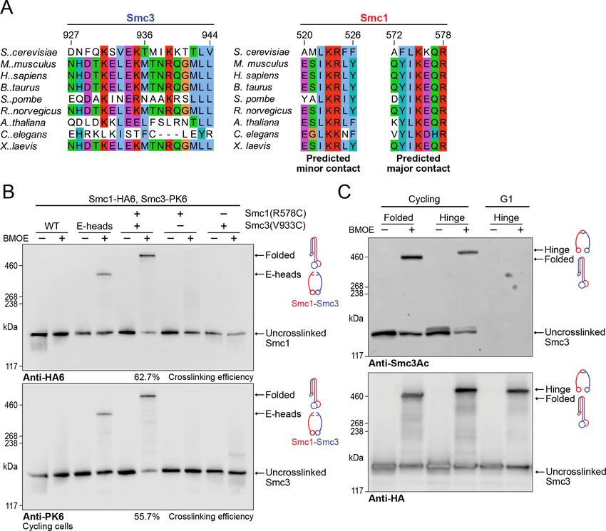
Research article                                       Chromosomes and Gene Expression Structural Biology and Molecular Biophysics

                                    (Bürmann et al., 2019). Folding around this discontinuity results in association of the hinge with a
                                    section of the coiled coil close to the so-called joint region, a break in the coiled-coils above the
                                    ATPase heads. Folding is a widely conserved feature of SMCs when observed using EM in vitro,
                                    both when heads are engaged (Collier et al., 2020) or disengaged (Bürmann et al., 2019), but
                                    whether folding occurs in vivo and has an important physiological function is not known. It has been
                                    postulated that the elbow could be involved in LE by coupling cycles of folding and unfolding with
                                    DNA translocation (Bürmann et al., 2019; Hassler et al., 2018). Further, it has been noted that a
                                    potential simultaneous interaction of a HAWK with the hinge and kleisin would require some sort of
                                    folding (Murayama and Uhlmann, 2015, Huis in ’t Veld et al., 2014, Bürmann et al., 2019).
                                        Despite the discovery that Scc2 facilitates binding of DNA to engaged ATPase heads in vitro
                                    (Shi et al., 2020; Higashi et al., 2020) and does so in the absence of Scc3 without entry inside the
                                    S-K ring (Collier et al., 2020), the mechanism by which Scc2 promotes cohesin’s association with
                                    and translocation along chromosomes in vivo remains poorly understood. Scc2’s unstructured NTD
                                    is bound by a superhelical array of 13 tetratricopeptide repeats (TPRs) belonging to its partner Scc4
                                    (Hinshaw et al., 2015). Cells lacking either Scc4 or Scc2’s NTD are not viable and have greatly
                                    reduced levels of chromosomal cohesin. Nevertheless, a version of Scc2 lacking the NTD is fully
                                    capable of activating cohesin’s ATPase (Petela et al., 2018) and clamping DNA on top of engaged
                                    ATPase heads in vitro (Shi et al., 2020; Collier et al., 2020). To gain insight into the role of Scc4, we
                                    recently undertook a genetic screen, isolating mutations that suppress lethality caused by loss of
                                    Scc4 activity (Petela et al., 2018). This identified two different scc2 point mutations, E822K and
                                    L937F. scc2E822K lies in the interface between Scc2 and Smc3’s K112 and K113, and as such, all
                                    three residues are in the vicinity of DNA clamped by Scc2 on top of engaged ATPase heads.
                                    Because acetylation of Smc3 K112 K113 greatly reduces cohesin loading as well as release,
                                    Scc2E822K might bypass Scc4 by increasing the avidity with which DNA is clamped (Collier et al.,
                                    2020).
                                        Here, we describe two other types of mutations that suppress scc4 lethality. One type includes
                                    mutations in histone H2A that loosen the association between nucleosomes and DNA, which con-
                                    ceivably act like Scc2E822K, by facilitating cohesin’s interaction with naked DNA. The other is an
                                    aspartic acid on the surface of Smc1’s hinge domain that is replaced by an aromatic residue:
                                    smc1D588Y. UV-induced crosslinking in cells whose Smc1 hinge contains p-benzoyl
                                    L-phenylalanine (BPA) at defined positions revealed that it contacts Scc2, Scc3, and Pds5. Inactiva-
                                    tion of Scc4 reduced crosslinking with Scc2, but increased that with Pds5, while smc1D588Y had the
                                    opposite effect. These findings suggest that Smc1’s hinge contacts Scc2 and Pds5 directly, Scc4
                                    facilitates association with Scc2 and hinders that with Pds5, and smc1D588Y does likewise and com-
                                    pensates for a lack of Scc4. To explain how Scc2 contacts Smc1’s hinge while also bound to Smc1’s
                                    ATPase, we suppose that cohesin’s coiled coil is folded around its elbow, thereby bringing the hinge
                                    into contact with HAWK regulatory subunits associated with cohesin’s ATPase heads, as recently
                                    observed in a cryo-EM structure of the ATP-bound clamped state (Collier et al., 2020).
                                        Using cryo-EM, we have now determined the structures of the folded cohesin complex associated
                                    with either Scc2 or Pds5, both in the absence of ATP. The structures demonstrate that both Scc2
                                    and Pds5, while attached to the ATPase domains of Smc1 and Smc3, respectively, reach up to the
                                    hinge, thus providing a clue regarding the effects of Smc1D588Y and an explanation for previously
                                    observed K620BPA crosslinks (Bürmann et al., 2019). The resolution of the folded coiled coils and
                                    hinge (5–6 Å) not only permitted the identification of the contacts involved in folding but also
                                    allowed       identification   of     candidates       for    cysteine      substitution       for    potential
                                    bismaleimidoethane (BMOE) crosslinking to assay folding. One such residue pair, Smc1R578C-
                                    Smc3V933C, gave rise to efficient BMOE-induced crosslinking in vivo even when Smc3 was acety-
                                    lated. Therefore, folding takes place not only when Scc2 is bound but also when cohesin is engaged
                                    in holding sister chromatids together in post-replicative cells. Our findings demonstrate that folding
                                    of cohesin’s coiled coils is not an in vitro artefact. Folding occurs in living cells, it is a feature of cohe-
                                    sin engaged in holding sister chromatids together, and it is of physiological importance during Scc2-
                                    mediated cohesin loading.

Petela, Gonzalez Llamazares, et al. eLife 2021;10:e67268. DOI: https://doi.org/10.7554/eLife.67268                                          5 of 32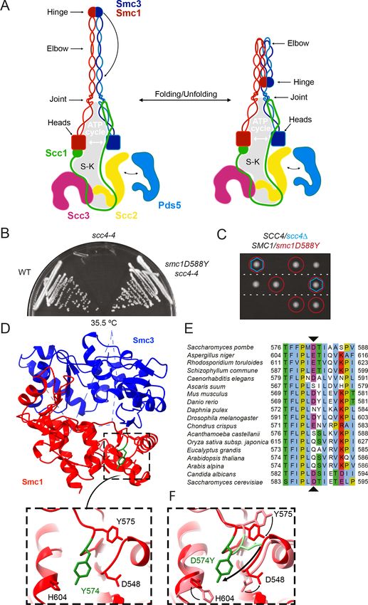
Research article                                       Chromosomes and Gene Expression Structural Biology and Molecular Biophysics

                                    Results
                                    To understand better how Scc4 helps Scc2 to load cohesin onto chromosomes, we isolated muta-
                                    tions that enable temperature-sensitive scc4-4 cells to grow at the restrictive temperature (35.5˚C).
                                    This yielded both intragenic and extragenic mutations. The scc4-4 allele was created by error-prone
                                    PCR (Ciosk et al., 2000) and contains several different mutations, including Y40N. Sequencing of
                                    intragenic revertants revealed that wild-type (WT) growth was restored either by restoring tyrosine
                                    at position 40 or by substituting it with histidine, implying that the mutation responsible for scc4-4’s
                                    thermosensitive proliferation is Y40N. When integrated at the LEU2 locus, this mutation alone con-
                                    ferred temperature sensitive (ts) growth (Figure 1—figure supplement 1A). Y40 is a highly con-
                                    served residue that is buried in Scc4’s superhelical array of TPR motifs (Figure 1—figure
                                    supplement 1B). It is unlikely that it contacts the Scc2 polypeptide directly, but despite this, Y40N
                                    disrupts co-immunoprecipitation of Scc4 and Scc2 (Figure 1—figure supplement 1C).

                                    A mutation in the hinge domain of Smc1 restores viability in the
                                    absence of Scc4
                                    All of the extragenic scc4-4 suppressors (Figure 1B) contained a mutation tightly linked to SMC1.
                                    Indeed, all 12 independently isolated mutations contained the same single base change causing sub-
                                    stitution of aspartic acid by tyrosine at position 588 in Smc1 (Figure 1E). Tetrad dissection of SCC4/
                                    scc4D SMC1/smc1D588Y diploids revealed that smc1D588Y enabled cells to proliferate in the com-
                                    plete absence of Scc4 (Figure 1C). Smc1D588 is located in the hinge domain, at the C-terminal end
                                    of a b strand that interacts in an antiparallel fashion with a strand in Smc3 (Figure 1D). Despite its
                                    proximity to the Smc1-Smc3 interface, D588 does not appear to contact Smc3 residues. To address
                                    whether suppression arises due to the loss of a relatively conserved acidic residue (Figure 1E) or
                                    due to the substitution of a bulky aromatic, we tested the ability of a variety of other amino acid sub-
                                    stitutions to rescue viability in the absence of Scc4. Mutant or WT alleles of SMC1 were introduced
                                    into the TRP1 locus of a SMC1/smc1D SCC4/scc4D diploid. Subsequent dissection revealed that
                                    mutation to phenylalanine or tryptophan was able to restore growth to a similar degree as tyrosine
                                    in the absence of Scc4 (Figure 1—figure supplement 1D) but not histidine, arginine, alanine, gluta-
                                    mic acid, or asparagine (Supplementary file 1). Suppression of scc4D lethality also occurred in the
                                    presence of WT SMC1 but was much less effective (Figure 1—figure supplement 1D). Thus, we
                                    conclude that suppression is due to the introduction of a bulky aromatic amino acid at this crucial
                                    position and not through loss of the conserved aspartic acid. It is notable that the DNA base change
                                    observed in all 12 suppressors is the only one capable of creating such a transition via a single
                                    change and that the equivalent position is never a bulky aromatic in SMC2, SMC3, and SMC4 as well
                                    as SMC1. smc1D588Y was able to rescue the proliferation defect of the temperature-sensitive scc2-4
                                    allele at 30˚C, but not scc2D (Figure 1—figure supplement 1E, F), implying that it acts by enhancing
                                    the activity of Scc2, not by replacing it.
                                        To determine whether the smc1D588Y mutation alters the hinge structure, we introduced the
                                    equivalent mutation (D574Y) into an isolated mouse hinge. X-ray crystallography revealed that a
                                    clash between the tyrosine residue and neighbouring loop causes Y574 to instead swing out relative
                                    to the position of D574, causing a local conformational change of the mutated loop (Figure 1D, F,
                                    Figure 1—figure supplement 1G, Supplementary file 2). Importantly, the change had little or no
                                    impact on the overall structure of the hinge. Despite this, both D588Y and D588W reduced the
                                    amount of Smc1 hinge that co-precipitated with Smc3 hinges (Figure 1—figure supplement 1H).
                                    To address whether Smc1D588Y affects dissociation of pre-assembled Smc1/3 hinge complexes, we
                                    co-expressed either WT Smc1 or Smc1D588Y hinge domains with Smc3 hinges, purified Smc1/3
                                    complexes, and compared their persistence in the presence of a fivefold excess of SNAP-tagged
                                    Smc1 hinge domains. This revealed that the amount of Smc1D588Y associated with Smc3 hinges
                                    declined more rapidly in the presence of a WT competitor than WT Smc1, indicating that
                                    Smc1D588Y at least increases the off rate (Figure 1—figure supplement 1I).
                                        Our finding that Smc1D588Y increases dissociation of Smc1 from Smc3 hinge domains in vitro
                                    raises the possibility that suppression depends on, or indeed is caused by, the greater ease with
                                    which hinges can dissociate. We therefore tested whether other non-lethal mutations within the
                                    Smc1/Smc3 hinge interface are also capable of bypassing the need for Scc4. A highly conserved
                                    lysine residue within Smc3 (Smc3K652) that opposes Smc1D588 was mutated to tyrosine, alanine, or

Petela, Gonzalez Llamazares, et al. eLife 2021;10:e67268. DOI: https://doi.org/10.7554/eLife.67268                                    6 of 32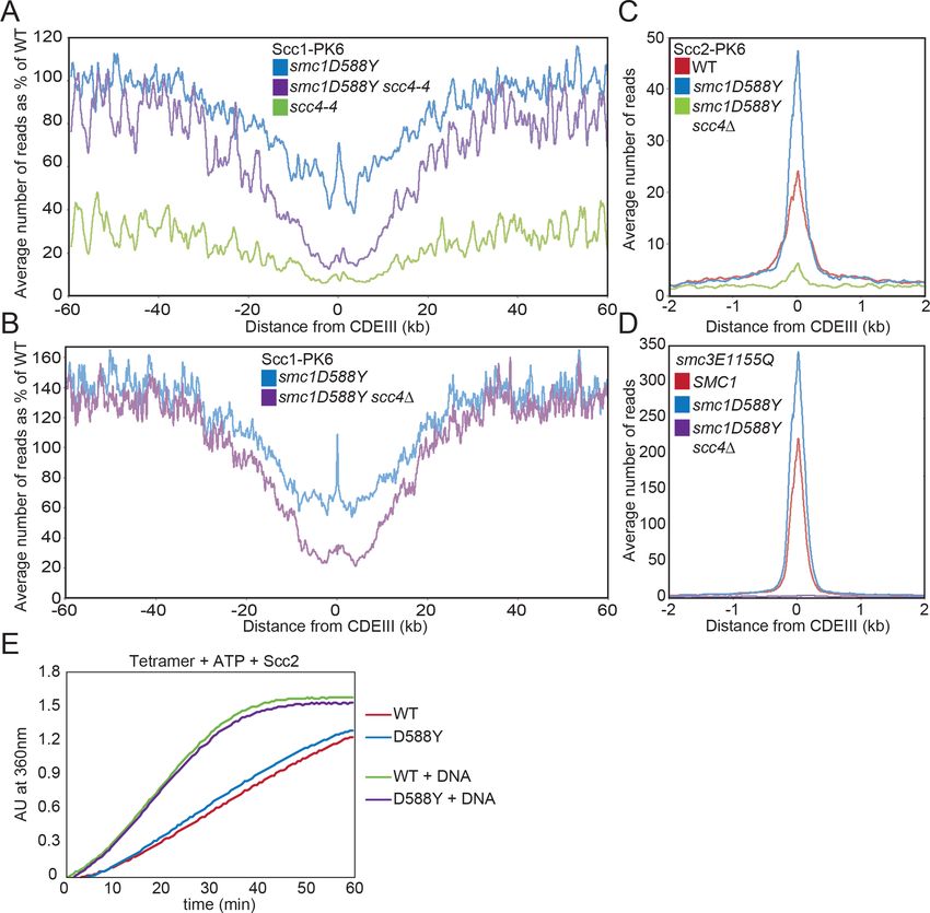
Research article                                       Chromosomes and Gene Expression Structural Biology and Molecular Biophysics

                                    valine, with no effect. Similarly, previously published mutations in both hinges, designed to weaken
                                    their interaction, smc1L635K K639E; smc1I590K; smc1L564K; smc3E570K; smc3L672R (Mishra et al.,
                                    2010), were also unable to support growth in the absence of Scc4. The failure of these other muta-
                                    tions to suppress scc4D lethality, together with our finding that smc1D588Y was identified in 12 out
                                    of 12 spontaneous extragenic suppressors, suggests that decreased affinity of Smc1/Smc3 hinges is
                                    not the mechanism by which Smc1D588Y enables Scc2 to load cohesin without Scc4.

                                    smc1D588Y restores cohesin occupancy on chromosome arms in the
                                    absence of Scc4
                                    Calibrated ChIP-seq revealed that scc4-4 causes a substantial reduction in the level of chromosomal
                                    cohesin when G1 cells undergo S phase and enter G2/M at the restrictive temperature (37˚C)
                                    (Figure 2A, Figure 2—figure supplement 1A). The reduction is more marked within pericentric
                                    sequences, where there is a 10-fold reduction, than along arms where is there merely a fourfold
                                    reduction. Average chromosome profiles centred around the centromeric CDEIII, plotted as a per-
                                    centage of the reads obtained for WT, revealed that smc1D588Y restores cohesin occupancy to
                                    approximately WT levels on chromosome arms (>30 kb from the centromere), but not around cen-
                                    tromeres (Figure 2A). The failure to restore loading around centromeres is perhaps not surprising as
                                    most pericentric cohesin is loaded at CENs in a process that involves binding of Scc4 to the kineto-
                                    chore protein Ctf19, a requirement that is apparently not bypassed by smc1D588Y. Interestingly,
                                    smc1D588Y caused a substantial reduction of cohesin occupancy around centromeres even in the
                                    presence of WT SCC4 (Figure 2A), an effect that will also have contributed to the lack of suppres-
                                    sion in this region of the chromosome.
                                        To investigate the effect of smc1D588Y at a more physiological temperature, we used calibrated
                                    ChIP-seq to compare cohesin’s occupancy of the genome in SCC4, SCC4 smc1D588Y, and scc4D
                                    smc1D588Y cells following their release from a pheromone-induced G1 arrest and subsequent arrest
                                    in G2/M phase at 25˚C. Average chromosome profiles around centromeres plotted as a percentage
                                    of WT (SCC4) revealed that smc1D588Y increased cohesin occupancy on chromosome arms to 120–
                                    150% of WT levels, both in the presence and absence of SCC4 (Figure 2B). In other words,
                                    smc1D588Y enhances cohesin’s loading on chromosome arms via a mechanism that is completely
                                    independent of Scc4. In contrast, smc1D588Y reduced association around centromeres to approxi-
                                    mately 60% of WT levels, which was further reduced to 20% by scc4D. In scc4D smc1D588Y cells,
                                    cohesin occupancy within pericentric chromatin resembles that along chromosome arms as if a single
                                    Smc1D588Y-driven mechanism is responsible for loading at both locations in these cells (Figure 2—
                                    figure supplement 1B). Importantly, smc1D588Y does not increase occupancy on chromosome
                                    arms merely because defective loading at CENs increases the amount of cohesin available to load
                                    onto chromosome arms because the scc4m35 mutation, which disrupts Scc4’s association with Ctf19
                                    and also reduces loading at CENs, has no such effect (Petela et al., 2018; Hinshaw et al., 2015).
                                        It is striking that in SCC4 cells smc1D588Y had far less effect on cohesin’s association at CEN
                                    loading sites themselves. For example, it was 110% of WT in cells growing at 25˚C and 75% of WT at
                                    37˚C. Because association was greatly reduced in scc4-4 and scc4D cells (Figure 2A, B), it presum-
                                    ably arises as a consequence of Scc4’s association with Ctf19. If so, these complexes should be asso-
                                    ciated with Scc2, which was confirmed by calibrated ChIP-seq showing that Scc2’s association with
                                    CENs far from being reduced was in fact substantially increased by smc1D588Y and fully dependent
                                    on Scc4 (Figure 2C). Cohesin occupied by Scc2 at CENs could either be in the process of loading
                                    (Petela et al., 2018; Hu et al., 2011) or engaged in LE (Dauban et al., 2020; Paldi et al., 2020). In
                                    both cases, the complexes are likely to adopt at least transiently the clamped state, which is stabi-
                                    lised by smc1E1158Q and smc3E1155Q, at least in vitro (Collier et al., 2020). In cells, cohesin com-
                                    plexes containing these mutations accumulate to especially high levels at CENs, albeit with a short
                                    residence time (Hu et al., 2011), suggesting that they initiate an early step in the loading process,
                                    namely the clamped state, but in the absence of ATP hydrolysis fail to undergo a later step required
                                    for stable association and translocation into neighbouring pericentric sequences. Interestingly,
                                    smc1D588Y not only increased Scc2’s association with CENs but also caused a similar increase in
                                    Smc3E1155Q’s association (Figure 2D). This implies that the reduced loading around centromeres
                                    arises not from defective formation of the clamped state at CENs by Scc2/4 complexes associated
                                    with Ctf19 but from a defect in a subsequent step in the loading/translocation reaction that requires
                                    ATP hydrolysis. Because accumulation of Smc1D588Y complexes at CENs resembles that of

Petela, Gonzalez Llamazares, et al. eLife 2021;10:e67268. DOI: https://doi.org/10.7554/eLife.67268                                    7 of 32
Research article Chromosomes and Gene Expression Structural Biology and Molecular Biophysics Figure 2. smc1D588Y restores cohesin occupancy on chromosome arms in the absence of Scc4. (A) Average calibrated ChIP-seq profiles of Scc1-PK6 in smc1D588Y, scc4-4, and smc1D588Y scc4-4 cells 60 kb either side of CDEIII plotted as a percentage of the average number of reads obtained for wild- type (WT) cells. Cells were pheromone arrested in G1 at 25˚C before release at 37˚C into medium containing nocodazole. Samples were taken 75 min after release (K22005, K22009, K21999, K22001). (B) Average calibrated ChIP-seq profiles of Scc1-PK6 in smc1D588Y, and smc1D588Y scc4D cells 60 kb either side of CDEIII plotted as a percentage of the average number of reads obtained for WT cells. Cells were pheromone arrested in G1 at 25˚C before release at 25˚C into medium containing nocodazole. Samples were taken 60 min after release (K22005, K22009, K19624). (C) Average calibrated ChIP-seq profiles of Scc2-PK6 2 kb either side of CDEIII in cycling WT, smc1D588Y, and smc1D588Y scc4D cells at 25˚C (K21388, K24680, K24678). (D) Average calibrated ChIP-seq profiles of ectopically expressed Smc3E1155Q-PK6 2 kb either side of CDEIII in cycling WT, smc1D588Y, and smc1D588Y scc4D cells at 25˚C (K24562, K24689, K24564). (E) ATPase activity of WT or mutant tetramers on addition of ATP and Scc2 in the presence and absence of DNA. Figure 2 continued on next page Petela, Gonzalez Llamazares, et al. eLife 2021;10:e67268. DOI: https://doi.org/10.7554/eLife.67268 8 of 32
Research article                                           Chromosomes and Gene Expression Structural Biology and Molecular Biophysics

Figure 2 continued
The online version of this article includes the following figure supplement(s) for figure 2:
Figure supplement 1. smc1D588Y restores cohesin occupancy on chromosome arms in the absence of Scc4.

                                      complexes containing Smc3E1155Q, we tested the effect of Smc1D588Y on cohesin’s ATPase activ-
                                      ity but found little or no effect either in the presence or absence of DNA (Figure 2E, Figure 2—fig-
                                      ure supplement 1D).

                                      Mutations in SCC2 and histone genes also suppress scc4D lethality
                                      To address whether it is possible to identify extragenic scc4D suppressor mutations besides
                                      smc1D588Y, we isolated a second set in a smc1D588E yeast strain that cannot mutate residue 588
                                      to an aromatic residue through a single base pair mutation (Petela et al., 2018). We identified using
                                      genetic crosses and genomic sequencing 12 mutations within SCC2 (described in Petela et al.,
                                      2018), 49 within HTA1 (one of two histone H2As), and a single mutation within HTB1 (one of two
                                      H2Bs). All permitted proliferation of scc4D cells, albeit to a greater or lesser extent (Figure 3A).

                                      Scc4 helps overcome inhibition of loading by nucleosomes
                                      The H2A mutations affected three residues, namely G30, R31, and R34. These mutations (G30D,
                                      R31I/T/S/G, and R34I) are all located on a defined patch on the surface of the nucleosome that inter-
                                      acts with DNA and the single H2B mutation (Y44D) is located nearby (Figure 3B). Because substitu-
                                      tion of two positively charged residues causes suppression, we surmise that the mutations act by
                                      weakening the association between histones and DNA. hta1R31I was made de novo and shown to
                                      suppress the lethality of scc4-4 cells (Figure 3A). Its effect on cohesin loading in SCC4 and scc4-4
                                      cells was measured using calibrated ChIP-seq to measure Scc1’s association with the genome after
                                      cells had undergone DNA replication at 35.5˚C following a pheromone-induced G1 arrest at 25˚C. A
                                      lower restrictive temperature (35.5˚C) was used in this instance because hta1R31I is itself lethal at 37˚
                                      C. Consistent with its poor suppression of scc4D lethality (Figure 3A), hta1R31I increased loading
                                      along chromosome arms more modestly than smc1D588Y, raising loading in scc4-4 cells from 20%
                                      to 70% of WT (HTA1 SCC4) (Figure 3C). As in the case of both smc1D588Y and scc2E822K L937F
                                      (Petela et al., 2018), hta1R31I failed to suppress the loading defect of scc4-4 mutants in the vicinity
                                      of centromeres (Figure 3C). Interestingly, in the presence of WT SCC4, hta1R31I actually increased
                                      loading along chromosome arms over WT by 20%. This implies that the association between histo-
                                      nes and DNA within the nucleosome restricts cohesin loading, at least along chromosome arms, not
                                      only in scc4 mutants but also in WT cells. Like scc2E822K L937F (Petela et al., 2018) but unlike
                                      smc1D588Y, hta1R31I does not per se reduce loading of cohesin around centromeres (Figure 3C),
                                      suggesting that hta1R31I and smc1D588Y affect different aspects of the loading process.
                                          It has been suggested that the chromatin structure remodelling complex (RSC) has a key role in
                                      loading cohesin onto yeast chromosomes (Huang et al., 2004) and that an important function of
                                      Scc2/4 along chromosome arms is to facilitate nucleosome remodelling catalysed by RSC (Lopez-
                                      Serra et al., 2014). This raised the possibility that mutations like hta1R31I suppress the loading
                                      defects of scc4 mutants because they bypass the need for RSC and smc1D588Y might act likewise.
                                      To address this, we used calibrated ChIP-seq to reinvestigate the loading defects of sth1-3 cells,
                                      which contain a temperature sensitive mutation within RSC’s ATPase subunit. WT and sth1-3 cells
                                      were arrested in G1 by a-factor at 25˚C and then released from the block at 37˚C. sth1-3 delayed
                                      budding, DNA replication, and the onset of Smc3 acetylation (Figure 3—figure supplement 1A–C),
                                      complicating the comparison with WT. We therefore compared the calibrated ChIP-seq profiles
                                      sth1-3 cells 105 min after release, when most but not all cells had both budded and undergone DNA
                                      replication, with WT cells at 75 min, a time point at which their cell cycle progression was most simi-
                                      lar. Western blotting confirmed that the levels of Scc1 in this pair of samples were also similar (Fig-
                                      ure 3—figure supplement 1C). Surprisingly, their calibrated ChIP-seq profiles were also very similar
                                      not only in the vicinity of centromeres (Figure 1—figure supplement 1D, F) but also throughout an
                                      interval 60 kb either side of centromeres (Figure 3—figure supplement 1F). Crucially, sth1-3 caused
                                      only a modest reduction in the occupancy ratio (OR), which denotes the overall level of association
                                      throughout the genome (Figure 3—figure supplement 1D). These findings contradict the previous

Petela, Gonzalez Llamazares, et al. eLife 2021;10:e67268. DOI: https://doi.org/10.7554/eLife.67268                                         9 of 32
Research article Chromosomes and Gene Expression Structural Biology and Molecular Biophysics
Figure 3. Mutations in SCC2 and histone genes also suppress scc4D lethality. (A) Tetrad dissection of diploid strains containing SCC4/scc4D leu2/scc4-4
HTA1/hta1R31I. Spores in which scc4D is rescued by hta1R31I are circled in blue. (B) Structure of the yeast nucleosome (PDB: 1ID3; White et al., 2001).
H2A is shown in blue and H2B in green. Suppressor mutations are shown in yellow. (C) Average calibrated ChIP-seq profiles of Scc1-PK6 in hta1R31I,
scc4-4, and hta1R31I scc4-4 cells 60 kb either side of CDEIII plotted as a percentage of the average number of reads obtained for wild-type (W)T cells.
Cells were pheromone arrested in G1 at 25˚C before release at 35.5˚C into medium containing nocodazole. Samples were taken 60 min after release
(K22005, K24574, K24568, K22001).
The online version of this article includes the following figure supplement(s) for figure 3:
Figure supplement 1. Scc4 helps overcome inhibition of loading by nucleosomes.
claim that RSC has a crucial role in cohesin loading, based on qPCR measurements at individual loci
of the very same sth1-3 strain (Lopez-Serra et al., 2014). Because of this discrepancy, we compared
the calibrated ChIP-seq profiles of WT (75 min) and sth1-3 (105 min) in the vicinity of three loci
whose association was previously reported to be 30% of WT. There was little or no effect of the
mutation at CEN3, a modest reduction at POA1, and more surprisingly an increase at MET10 (Fig-
ure 3—figure supplement 1E).
Given the pleiotropic consequences of sth1-3 on cell cycle progression, it is difficult to exclude
the possibility that RSC has a modest effect on cohesin loading. However, if Scc4 promoted loading
by helping chromatin remodelling by RSC, then smc1D588Y should suppress any apparent loading
defect caused by RSC. The fact that the Scc1 calibrated ChIP-seq profile of smc1D588Y sth1-3 dou-
ble mutants is indistinguishable to that of sth1-3 single mutants (Figure 3—figure supplement 1G)
shows that insofar that there is any defect, it is clearly unaffected by smc1D588Y. In other words, a
Petela, Gonzalez Llamazares, et al. eLife 2021;10:e67268. DOI: https://doi.org/10.7554/eLife.67268 10 of 32Research article Chromosomes and Gene Expression Structural Biology and Molecular Biophysics
version of cohesin that no longer requires Scc4 does not alter sth1-3’s albeit modest defect. It may
therefore be a pleiotropic consequence of the mutant’s retarded cell cycle progression and not due
to an Scc4-dependent RSC activity that creates nucleosome-free regions necessary for cohesin
loading.
Recent work has revealed that Scc2 has a key role in clamping DNA onto engaged heads and
that Scc2E822K, which also suppresses scc4D, might function by enhancing DNA binding within the
clamped state (Collier et al., 2020; Shi et al., 2020; Higashi et al., 2020). We therefore suggest
that the reason why histone mutations suppress the lethality of scc4 mutants is because they increase
the accessibility of DNA and thereby facilitate formation of the clamped state.
Scc4 regulates an interaction between the hinge domain and HAWKs
How might replacement of a specific surface residue on the Smc1 hinge by a bulky aromatic one
help Scc2 function without Scc4? One possibility is that it strengthens a hydrophobic interaction with
another cohesin subunit. We have previously described the UV-dependent crosslinking in living yeast
cells between Pds5 and a version of Smc1 containing BPA at position K620, which is located in an
alpha helix adjacent to the loop containing D588 (Figure 4A; Bürmann et al., 2019). Pds5 is not
required for cohesin loading, and therefore strengthening its interaction with Smc1’s hinge cannot
be responsible for suppression. We therefore tested whether UV induces crosslinking of
Smc1K620BPA to other regulatory subunits. To do this, cells expressing FLAG-tagged versions of
Scc2, Scc3, Scc4, or Pds5 in cells whose sole source of Smc1 was Myc-tagged Smc1K620BPA were
exposed to UV, and subsequent western blotting was used to detect FLAG-tagged proteins in
immunoprecipitates (IPs) of Scc1-containing complexes (Figure 4B).
Western blotting for the Myc epitope confirmed that all samples contained a high molecular
weight version of Smc1, consisting of proteins crosslinked to K620 (Figure 4B). As expected for a
subunit that is stably associated with cohesin Smc-kleisin trimers, high levels of Scc3 were detected
in IPs from Scc3-FLAG cells, most of which had an electrophoretic mobility expected of uncros-
slinked protein, but a small fraction co-migrated with the high molecular weight version of Smc1,
suggesting that UV also induces crosslinking of Smc1K620BPA to Scc3. Pds5 is less stably associ-
ated, explaining why only modest amounts of uncrosslinked Pds5 are detected in the IPs. Despite
this, we observed much more Smc1-Pds5 than Smc1-Scc3 crosslinked protein, confirming that
Smc1K620BPA crosslinks to Pds5 with high efficiency (Bürmann et al., 2019). Because co-precipita-
tion of unstably associated proteins will be greatly enhanced by crosslinking, it is not possible to
assess the actual fraction of crosslinked protein. Scc2’s residence time on chromosomal cohesin of
approximately 2–4 s (Hu et al., 2011) is even less than that of Pds5 and the former is therefore diffi-
cult to detect in Smc1 IPs. Nevertheless, the level of Smc1-Scc2 crosslinked protein was comparable
to that of Scc3, despite being overall threefold less abundant (Tóth et al., 1999). In contrast, we
detected no Smc1-Scc4 crosslinked proteins in Scc4FLAG cells. Cryo-EM has revealed that the N-ter-
minal HEAT repeats of Scc2 as well as those of its human ortholog Nipbl are found in close proximity
to Smc1’s hinge within complexes that have clamped DNA on top of their engaged ATPase domains
(Shi et al., 2020; Higashi et al., 2020; Collier et al., 2020) and the crosslinking between
Smc1K620BPA and Scc2 may reflect this state. However, they could also reflect an alternative one in
which Scc2 is bound to cohesin whose heads are disengaged as described in the next section.
As association of cohesin with Scc2 and Pds5 is mutually exclusive and the latter incapable of
activating cohesin’s ATPase or association with chromatin (Petela et al., 2018), Scc4 and
Smc1D588Y could facilitate loading either by enhancing association of the hinge with Scc2 or
decreasing it with Pds5. To test this, we measured the effect of scc4-4 in the presence or absence of
smc1D588Y on Smc1K620BPA-Pds5 and Smc1K620BPA-Scc2 crosslinking. This revealed that Scc4
inactivation (scc4-4) increased Pds5 crosslinking threefold while smc1D588Y had the opposite effect.
Crucially, smc1D588Y was largely epistatic to scc4-4. In other words, the elevated crosslinking
observed when Scc4 was inactivated dropped in the presence of smc1D588Y to the depressed level
of SCC4 smc1D588Y cells (Figure 4C, D). The mutations had the opposite, albeit less dramatic,
effects on Smc1K620BPA-Scc2 crosslinking. Scc4 inactivation halved it while smc1D588Y restored it
80% of WT levels (Figure 4E, F). These results are consistent with the notion that a key function of
Scc4 is to facilitate interaction between Scc2 and the Smc1 hinge, either directly or indirectly by
impeding the latter’s interaction with Scc2’s competitor Pds5 or conceivably via both mechanisms.
Petela, Gonzalez Llamazares, et al. eLife 2021;10:e67268. DOI: https://doi.org/10.7554/eLife.67268 11 of 32Research article Chromosomes and Gene Expression Structural Biology and Molecular Biophysics Figure 4. Scc4 regulates an interaction between the hinge domain and HAWKs. (A) Modelled structure of the yeast cohesin hinge domain based on bacterial SMC hinge from Thermotoga maritima (PDB: 1GXL; Haering et al., 2002). (B) Identification of proteins that crosslink to Smc1 hinge. Strains expressing various cohesin regulators tagged with either FLAG6 or HA6 in combination with Smc1K620BPA-myc were treated with UV prior to immunoprecipitation with PK-tagged Scc1 and the products analysed by western blotting (B1969, B1976, B1983, B2020, B2072, B2079). (C) Effect of Figure 4 continued on next page Petela, Gonzalez Llamazares, et al. eLife 2021;10:e67268. DOI: https://doi.org/10.7554/eLife.67268 12 of 32
Research article                                          Chromosomes and Gene Expression Structural Biology and Molecular Biophysics

Figure 4 continued
Scc4 and Smc1D588Y on crosslinking between Pds5 and Smc1 hinge. Cells expressing Smc1K620BPA in the presence or absence of scc4-4 and
Smc1D588Y were exponentially grown at 25˚C and shifted to 35.5˚C for 1 hr. Cells were irradiated with UV, and the cohesin complex was isolated by
immunoprecipitation of PK-tagged Scc1. The Myc-tagged Smc1K620BPA was examined by western blot (B2072, B2212, B2214, B2215). (D)
Quantification of the crosslinks in (C) as a percentage of the wild-type (WT) Smc1 crosslinking efficiency. (E) Effect of Scc4 and Smc1D588Y on
crosslinking between Scc2 and Smc1 hinge. Strains were treated as described in (C) (B1969, B2213, B2216, B2217). (F) Quantification of the crosslinks in
(E) as a percentage of the WT Smc1 crosslinking efficiency. The experiments shown in (C–F) were performed twice with the same result. (G) In vivo
cysteine crosslinking of Smc1 hinge with Scc2 protein. Yeast cells expressing Smc1K620C and Scc2N200C were incubated with
bismaleimidoethane (BMOE) (B3082, B3107, B3114, and B3116). The crosslinked Smc1/Scc2 was isolated by immunoprecipitation of PK-tagged Scc1
and examined by western blot. * Unspecific crosslink band.
The online version of this article includes the following figure supplement(s) for figure 4:
Figure supplement 1. Scc4 regulates an interaction between the hinge domain and HAWKs.

                                         If the essential role of Scc4 were merely to hinder an interaction between the Smc1 hinge and
                                     Pds5, then Scc4 should be unnecessary for cohesin’s association with chromosome arms in cells lack-
                                     ing Pds5. We therefore used calibrated ChIP-seq to measure the effect of depleting Pds5 (using the
                                     auxin-dependent AID degron) on cohesin’s occupancy of chromosome arms after scc4-4 cells
                                     undergo S phase at 37˚C, which revealed that Pds5 depletion had no effect (Figure 4—figure sup-
                                     plement 1A). In other words, Pds5 is not necessary for depressing cohesin’s association with chro-
                                     mosomes in scc4-4 mutants. Likewise, if by reducing Pds5’s interaction with the Smc1 hinge
                                     smc1D588Y reduced Pds5’s occupancy of chromosomal cohesin, then it should depress the fraction
                                     of chromosomal cohesin associated with Pds5. The fact that smc1D588Y has no such effect (Fig-
                                     ure 1—figure supplement 1B, C) implies that though the mutation alters how Pds5 interacts with
                                     the Smc1 hinge, this does not in fact alter chromosomal cohesin’s occupancy by Pds5.
                                         Our finding that Scc4 does not act solely by hindering Pds5 suggests that Scc4 and Smc1D588Y
                                     facilitate Scc2 activity by promoting its interaction with the hinge. To elucidate where the hinge con-
                                     tacts Scc2, we inserted TEV protease cleavage sites at various positions within Scc2 to determine
                                     whether Smc1K620BPA crosslinked to the N- or C-terminal fragments created by TEV cleavage (Fig-
                                     ure 1—figure supplement 1D, E). Analysis of those TEV insertions that were functional in vivo
                                     revealed that crosslinking occurred within Scc2’s N-terminal sequences, between residues 150 and
                                     215. This interval is between the N-terminal domain that binds Scc4 (Hinshaw et al., 2015) and the
                                     hook-shaped structure composed of HEAT repeats. This part of Scc2 is not sufficiently ordered to
                                     have been visualised in the cryo-EM structure of a complex containing DNA clamped between Scc2
                                     and engaged Smc1/3 ATPases (Collier et al., 2020). To confirm the location, we measured BMOE-
                                     induced crosslinking in vivo between Smc1K620C and a variety of Scc2 cysteine substitutions
                                     between residues 153 and 212. Although Smc1K620C alone gave rise to a Smc1-Scc2 crosslinked
                                     species, the crosslinking was more efficient on the introduction of Scc2N200C, suggesting that Smc1
                                     is likely also crosslinking to a natural cysteine in Scc2 (most likely Scc2C224, which sits on a small
                                     helix just below N200) (Figure 4G). Importantly, the region of Scc2 whose association with the Smc1
                                     hinge is reduced by scc4-4 and restored by smc1D588Y is close to where Scc4 binds to Scc2
                                     (Hinshaw et al., 2015). In other words, Scc4 would be close enough to directly influence Scc2’s
                                     interaction with the hinge.

                                     Cryo-EM structures of cohesin trimers associated with Scc2 or Pds5
                                     reveal folded coiled coils
                                     The notion that smc1D588Y suppresses scc4D by altering the interaction between Smc1’s hinge
                                     domain and cohesin’s HAWK subunits Scc2 and Pds5 raises a conundrum: how can HAWK proteins,
                                     which are known to associate with cohesin’s kleisin subunit and its ATPase domains, interact with a
                                     hinge domain that is separated from the ATPase domains by a 50-nm-long coiled coil? One possibil-
                                     ity is that the HAWK proteins interact with cohesin’s hinge and ATPase domains at different points
                                     in time. Alternatively, if in fact they interact with hinge and heads simultaneously, then the coiled coil
                                     cannot be fully extended. For example, folding at an elbow in the middle of the coiled coil
                                     (Bürmann et al., 2019) may bring the hinge into proximity of HAWKs associated with the ATPases.
                                     Folding has recently been observed at low resolution in a complex between DNA, Scc2, and hydroly-
                                     sis-impaired EQEQ ATPases engaged in the presence of ATP (Collier et al., 2020; Shi et al., 2020;

Petela, Gonzalez Llamazares, et al. eLife 2021;10:e67268. DOI: https://doi.org/10.7554/eLife.67268                                              13 of 32
Research article Chromosomes and Gene Expression Structural Biology and Molecular Biophysics
Higashi et al., 2020). To investigate this further, we used cryo-EM to determine the structures of
the S. cerevisiae cohesin trimer (Smc1, Smc3, and Scc1 containing cysteines specifically crosslinking
the three intermolecular interfaces; Smc1-Scc1, Smc3-Scc1, and the Smc1-Smc3 hinge [Collier et al.,
2020] at an efficiency of 20% [data not shown]) bound to either Scc2 (Figure 5A) (EMD-12880) or
Pds5 (Figure 6A) (EMD-12888) in the absence of nucleotide and DNA. The former revealed a coiled
Figure 5. Folded cohesin allows interaction of hinge with Scc2 N-terminus. (A) Views of cryo-EM reconstruction of Scc2-bound cohesin coloured by
subunit. (B) Full pseudo-atomic model of folded cohesin trimer bound to Scc2. (C) Close-up of breaks in the coiled coils of Smc3 and Smc1 that
constitute the elbow region of cohesin (PDB: 7OGT; EMD-12887). (D) Close-up of the interaction between the hinge and Smc3 that stabilises the folded
state. (E) Close-up of Scc2 N-terminus in proximity of hinge residues K620 and D588Y. (F, G) Comparison of cryo-EM densities between Scc2-bound
and ATP-free cohesin seen in (F) (EMD-12880) and ATP-bound cohesin seen in (G) (EMD-12889), demonstrating that head engagement is not sufficient
for coiled coil unzipping.
The online version of this article includes the following figure supplement(s) for figure 5:
Figure supplement 1. Folded cohesin allows interaction of hinge with Scc2 N-terminus.
Figure supplement 2. Data processing and reconstruction schematics of all cryo-EM maps.
Petela, Gonzalez Llamazares, et al. eLife 2021;10:e67268. DOI: https://doi.org/10.7554/eLife.67268 14 of 32Research article Chromosomes and Gene Expression Structural Biology and Molecular Biophysics
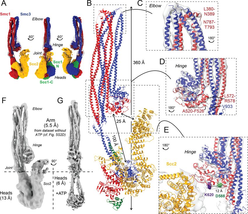
Figure 6. Pds5 binds to Smc3 head while contacting the hinge. (A) Composite map of cryo-EM reconstructions of Pds5-bound cohesin (EMD-12888). (B)
Full pseudo-atomic model of folded cohesin trimer bound to Pds5 coloured by subunit. (C) Close-up of interaction between hinge and Pds5 showing
proximity of N-terminus of the HAWK to hinge residues D588 and K620. (D) 2D classes of Pds5-bound ATPase heads. (E) Close-up of Pds5 binding to
K112- and K113-proximal region of the Smc3 head.
The online version of this article includes the following figure supplement(s) for figure 6:
Figure supplement 1. Detailed view of fitted atomic structures in cryo-EM maps.
coil folded at its elbow (Figure 5B, C), causing the hinge to interact with sections of the coiled coil
that are approximately 10 nm away from the point at which they emerge from the ATPase domains
(Figure 5B). The cryo-EM reconstruction not only revealed the path of the coiled coils around the
hinge-coiled coil interface (where the map is at 5–6 Å resolution; EMD-12887), but also enabled the
Petela, Gonzalez Llamazares, et al. eLife 2021;10:e67268. DOI: https://doi.org/10.7554/eLife.67268 15 of 32Research article Chromosomes and Gene Expression Structural Biology and Molecular Biophysics
production of a pseudo-atomic model of the folded form (PDB: 7OGT). Folding brings a pair of heli-
ces within Smc1’s hinge, namely the end of the coiled coil around A520-F526 and another short helix
around L564-R578, into close proximity of a short stretch of Smc3’s coiled coil (Figure 5D). Very sim-
ilar folding was observed when Pds5 was bound instead of Scc2 (Figure 6A, B). Though folding per-
mits an association between the hinge and the N-terminal Scc2 sequences, which could in principle
stabilise the folded conformation, we also observed similar, if not identical, folding in samples lack-
ing all HAWK proteins (Figure 5—figure supplement 1E).
Coiled coil folding enables interaction of Scc2 or Pds5 with the Smc1
hinge
Initial 2D classes of Scc2-bound cohesin revealed floppiness not only within the HAWK, especially
within its C-terminus, but also between the joint and the ATPase heads (Figure 5—figure supple-
ment 1B). We therefore split the complex computationally into two regions, with a boundary at the
joint, and processed their densities separately (Figure 5F). This yielded an overall resolution of 13 Å
for the HAWK-bound part, which enabled fitting of a homologous Scc2 crystal structure (PDB:
5ME3; Chao et al., 2015) together with both head crystal structures (PDB: 1W1W; Haering et al.,
2004; PDB: 4UX3; Gligoris et al., 2014) to produce a pseudo-atomic model. Analysis revealed that
Scc2 binds rigidly to the Smc1 ATPase head in a manner resembling but distinct from its interaction
in the clamped state (Figure 5—figure supplement 1B; Collier et al., 2020). Scc2’s C-terminal
HEAT repeats 18–24 (residues 1127–1493) dock onto Smc1’s F-loop (residues 1095–1118) as well as
the emerging coiled coils, a mode of interaction analogous to that between condensin’s HAWK Ycs4
and Smc4 (Lee et al., 2020). This mode of interaction therefore takes place whether or not heads
are engaged. Unlike the engaged and clamped head state, Scc2 makes no contact with Smc3 in the
non-engaged, nucleotide-free structure. Contrary to its C-terminal part, Scc2’s N-terminal region
adopts a range of conformations. Bending around the mid-region of Scc2 enables its N-terminus to
contact the joint region of Smc3’s coiled coil in the clamped state. However, when heads are disen-
gaged in our nucleotide-free structure, Scc2 is straightened and its N-terminal half adopts the con-
formation observed in crystals of Scc2 alone (Chao et al., 2015). Because cohesin’s elbow is further
away from its hinge than is the case for condensin, folding of its coiled coils brings the hinge to
within 12 nm of the ATPase heads. As a consequence, the N-terminal part of Scc2 molecules bound
to Smc1 ATPase heads is in proximity to the hinge, thereby explaining not only its crosslinking to
Smc1K620BPA in vivo but also how Smc1D588Y could circumvent the need for Scc4 (Figure 5E). We
suggest that the addition of a bulky amino acid into Smc1 through D588Y may be sufficient to help
bind an otherwise floppy Scc2 N-terminal domain, whose interaction with the hinge is normally stabi-
lised by Scc4.
We processed data collected on Pds5-bound complexes in a similar manner, producing a 13 Å
resolution structure, which revealed that Pds5 binds to Smc3 and not, like Scc2, to Smc1’s ATPase
head domain (placed PDB: 5F0O; Lee et al., 2016; Figure 6A, B). The contact takes place between
the most C-terminal HEAT repeats of Pds5 and the top region of the N-terminal lobe of Smc3’s
ATPase. Strikingly, this part of Smc3 contains the pair of highly conserved lysine residues K112 and
K113 (Figure 6E), whose acetylation by Eco1 not only prevents releasing activity (Unal et al., 2008;
Rolef Ben-Shahar et al., 2008) but also stabilises Pds5’s interaction with chromosomal cohesin com-
plexes (Chan et al., 2012). Unlike Scc2, Pds5 does not rely on negatively charged amino acids for its
interaction with the K112/K113 region and may therefore be better suited than Scc2 for binding the
acetylated and less positively charged version of Smc3. Furthermore, binding in this manner shields
both lysine residues when acetylated, hence explaining how Pds5 hinders de-acetylation during G2/
M phases (Chan et al., 2013). Like Scc2, Pds5’s N-terminal HEAT repeats approach Smc1’s hinge
domain, which explains the crosslinking to Smc1K620BPA in vivo (Figure 6C). The low resolution
and flexibility apparent in our map mean that we cannot be sure whether Pds5’s C-terminal domain
reaches beyond the hinge and contacts the coiled coils. Importantly, the modes of interaction of
Scc2 and Pds5 with Smc subunits appear to be incompatible with each other, as has been postulated
previously through in vivo and in vitro work (Petela et al., 2018).
Petela, Gonzalez Llamazares, et al. eLife 2021;10:e67268. DOI: https://doi.org/10.7554/eLife.67268 16 of 32You can also read