Foliage response to heavy metal contamination in Sycamore Maple (Acer pseudoplatanus L.)
←
→
Page content transcription
If your browser does not render page correctly, please read the page content below
For. Snow Landsc. Res. 80, 3: 275–288 (2006)                                                    275

Foliage response to heavy metal contamination in Sycamore
Maple (Acer pseudoplatanus L.)
Olivier André1, Pierre Vollenweider2* and Madeleine S. Günthardt-Goerg2

1 Present address INRA-CNRS Laboratoire des Interactions Plante-Microorganismes
  (UMR 441-2594 ), BP 52627, Chemin de Borde Rouge – Auzeville, 31326 Castanet Tolosan, France.
  Olivier.Andre@toulouse.inra.fr
2 Swiss Federal Institute for Forest, Snow and Landscape Research (WSL), Zürcherstrasse 111, 8903

  Birmensdorf, Switzerland. pierre.vollenweider@wsl.ch, madeleine.goerg@wsl.ch
* Corresponding author

Abstract
Sycamore Maple (Acer pseudoplatanus L.) belongs to the typical pioneer trees found in contami-
nated ecosystems where it can contribute to the phytostabilisation of heavy metals (HM) in soils.
In this study, foliage reactions caused by the experimental exposure to copper / zinc / cadmium /
lead = 640 / 3000 / 10 / 90 mg/kg were investigated in trees following development of distinct
visible symptoms of stress in crown foliage during the third year of exposure. Low amounts of
HM were translocated to the foliage and no reduction of foliage biomass was recorded. Only zinc
reached concentrations near the toxicity level, especially in older foliage, and its involvement in
stress reactions was confirmed by the morphology of visible symptoms, cytochemical micro-
localisation and structural changes in the leaves. Low quantities of zinc reached sensitive sites in
the leaf blade and triggered oxidative stress. Phytoremediation potential of Sycamore Maple
includes both a phytostabilisation and bioindication component. The latter can be useful for mon-
itoring and surveillance purposes.

Keywords: phytoremediation, phytostabilisation, bioindication, zinc, cytochemistry, condensed
tannins, symptom validation

1       Introduction

Different tree species growing spontaneously or after planting in sites with heavy metal
(HM) pollution seem to have a considerable potential for the phytoremediation of contami-
nated soils (PULFORD and WATSON 2003). Pioneer species are not demanding for substrate
structure, texture and nutrient availability and have other advantages like a deep root
system and fast growth rate (PUNSHON et al. 1996). Species with either low or high HM
uptake are best suited for phytostabilisation or phytoextraction respectively (PULFORD and
WATSON 2003). As a pioneer and anemochore species, Sycamore Maple (Acer pseudopla-
tanus L.) often grows in contaminated ecosystems from central Europe and has until now
mainly shown a phytostabilisation potential (PULFORD and WATSON 2003; MERTENS et al.
2004). A good tolerance for HM toxicity based on species-specific growth, stress avoidance
and detoxification strategies can increase the phytoremediation capacity of trees (PULFORD
and WATSON 2003; VOLLENWEIDER et al. 2006). Partitioning and rate of HM accumulation
in the different organs widely varies between HM, tree species and even individuals within a
single clone (LEPP 1996; PULFORD and WATSON 2003). In trees however, the micro-localis-
ation of HM to date has rarely been characterised down to the tissue and cell level, because
sizable amounts of HM are required to detect a microscopical signal with most methods.
Translocation of HM to leaf threatens physiological activities in the tree crown as several
276                                                                           Olivier André et al.

sites in the leaf blade and vein are sensitive to oxidative stress (DIETZ et al. 1999; PRASAD
and STRZALKA 1999; VOLLENWEIDER et al. 2006). According to where HM have accumulated
at tissue and cell level, they can trigger either little (COSIO et al. 2006; VOLLENWEIDER et al.
2006) or cascades of oxidative stress reactions leading to hypersensitive-like responses (HR-
like, VOLLENWEIDER et al. 2003; MARTIN et al. 2006). Intolerant cell and tissue reactions
result in visible leaf symptoms which constitute useful bioindications to (1) diagnose the
stress factor, (2) rapidly assess contaminated spots in heterogeneously contaminated
environments and (3) analyse the response variability between different populations and
species of trees (VOLLENWEIDER and GUNTHARDT-GOERG 2005). Visible symptoms can be
validated with microscopical analyses: by micro-localising the deposits of HM at tissue and
cell level and identifying the physiological reactions on the basis of their structural signature
(VOLLENWEIDER et al. 2003, 2006; MARTIN et al. 2006), the link between the accumulation of
a given HM and the occurrence of visible symptoms in foliage can be demonstrated. This
combination of methods also provides insights about direct and indirect HM-effects useful
to estimate the specificity of the different visible symptom features.
    This study was realised within the framework project “From Cell to Tree” (GUNTHARDT-
GOERG 2005) after the observation of distinct visible symptoms in the foliage during the
third year of exposure to a mixed HM contamination in the soil. It is focused on responses at
the leaf, tissue and cell level in the foliage of Sycamore Maple. Investigations were per-
formed to (1) elucidate the underlying structural changes and corresponding physiological
reactions (VOLLENWEIDER et al. 2006), (2) distinguish between direct and indirect HM
effects, (3) determine which HM caused the symptoms at cell, tissue and leaf level, (4)
validate the symptoms and evaluate their bioindication potential and (5) analyse possible
contributions of Sycamore Maple to phytoremediation. HM effects on foliage were
characterised by measuring changes in foliage biomass and foliar HM concentration, micro-
localising zinc (Zn) at tissue and cell level, observing microscopical symptoms of stress and
analysing the leaf content for amounts of total phenols and condensed tannins. These latter
compounds can complex different heavy metals (MCDONALD et al. 1996; MORAN et al.
1997), accumulate in the same apoplastic and symplastic cell compartment where HM have
been translocated (VOLLENWEIDER et al. 2006) and contribute with antioxidant functions to
detoxify oxidative stress (RICE-EVANS et al. 1997; SAINT-CRICQ DE GAULEJAC et al. 1999)
caused by HM (DIETZ et al. 1999). They therefore constitute important markers of the
defence and detoxification reactions induced by HM stress.

2      Materials and methods

2.1    Experimental design and plant material

The experiment was carried out at WSL in Birmensdorf (450 m above sea level),
Switzerland, using twenty circular field plots with four treatments and five repetitions each.
Each field plot (diameter: 2 m) consisted of a two-layered soil substrate isolated by concrete
walls from the surrounding ground: 45 cm of an acidic forest subsoil (loamy sand [Alisol],
initial pH in 0.01 M CaCl2 = 4.2, from Eiken in the Rhine valley, Aargau, Switzerland) was
packed inside each field plot and topped by 15 cm of an agricultural topsoil (loamy soil,
initial pH in 0.01 M CaCl2 = 6.4, Corg = 15.1 gkg–1, from Birr, Aargau, Switzerland). This top-
soil was selected to ensure sufficient nutrient availability in experiments within the frame-
work “From Cell to Tree” and no fertiliser was added prior to or during the experimentation
(see HERMLE et al. 2006a for physical and chemical soil properties). Ten plots received
For. Snow Landsc. Res. 80, 3 (2006) 277 topsoil with and 10 without artificial HM contamination. HM dust from a non ferrous smelter filter was applied manually resulting in copper (Cu) / Zn / cadmium (Cd) / lead (Pb) = 640 / 3000 / 10 / 90 mgkg–1 of which 40 % (Cu), 70 % (Zn), 85 % (Cd), and 10 % (Pb) was either mobile or easily mobilizable (NOWACK et al. 2006). This level of soil contamination and HM bio-availability clearly exceeded Swiss VBBo limits1 but remained partly below levels found at different polluted sites throughout Europe (DICKINSON 2000; HORVARTH and GRUIZ 1996; ERNST 1972). It thus reproduced, in a realistic way, soil pollution levels existing at “brown field” sites. The control treatment (CO) without HM contamination con- tained: Cu / Zn / Cd / Pb = 28 / 97 / 0.1 / 37 mgkg–1. The plots were irrigated by means of six sprinklers during periods with insufficient natural precipitation. The irrigation water ionic composition was taken as the 30 years mean of the ambient rain at the location. Its pH was adjusted by HCl to either 5.5 or 3.5. During winter (November to April) no irrigation was given. Four treatments were replicated 5 times each and included (1) control: uncontaminated topsoil irrigated with rainwater pH 5.5 (CO), (2) acid rain: uncontaminated topsoil plus rainwater pH 3.5 (AR), (3) HM: HM-contaminated topsoil plus rainwater pH 5.5 (HM) and (4) combination: HM-contaminated topsoil plus rainwater pH 3.5 (HMAR). Each plot was planted with different native tree and understorey plants to simulate the ecosystem con- ditions in a young forest plantation. The tree layer included seven different tree species at 14 equidistant places, including one Sycamore Maple (Acer pseudoplatanus L.) per plot (planted as six-months-old rooted cuttings before bud break in spring 2000). 2.2 Leaf biomass and HM content in foliage Foliage of each tree was harvested in September 2002 and 2003, prior to the onset of autumnal coloration. The mean single leaf area and mass, specific leaf area (leaf area per leaf mass) and specific leaf mass (leaf mass per area) was determined in 10 leaves, taken randomly over each tree, after drying at 65 °C. In order to determine the leaf content of HM, an aliquot of the entire foliage dry mass per tree, was milled (ultra centrifuge mill wolfram carbide coated), digested with a high pressure microwave system (UltraClav by Milestone: 240 °C, 120 bar) and analysed in duplicates (spread
278                                                                        Olivier André et al.

2.4    Cytochemical and structural analyses

In late summer (20.09.2002), disks 1 cm in diameter were excised at the leaf basis from
symptomatic and asymptomatic leaves at three different leaf position along the top shoot of
two CO and two HM-treated trees. They were fixed either in methanol or by infiltration
with buffered (pH 7.0) 2.5 % glutaraldehyde. Zinc was cytochemically revealed
(VOLLENWEIDER et al. 2005) for the purpose of micro-localisation. Changes in the cell and
tissue structure and the physiological responses to HM stress were analysed in light and
fluorescence microscopy according to VOLLENWEIDER et al. (2003 and 2006) and
GUNTHARDT-GOERG et al. (1997).

2.5    Biochemical analysis of total phenols and condensed tannins

Amounts of total phenols and condensed tannins (CT) were analysed in five to seven leaves
across each tree crown in 2002 harvested at the same time as disks for microscopy. Leaf
samples were immediately frozen in liquid nitrogen, freeze-dried and stored at –20 °C until
analysis. An aliquot of 0.5 g of dry foliar material was frozen in liquid nitrogen prior to
homogenization (1 min) with steel balls in a Mikro-Dismembrator II of B. Braun,
Melsungen, Germany (60 × 15 mm oscillations per second). The still frozen powder was
transferred to a separating funnel and extracted 4 times during 3 min under magnet stirring
at room temperature with 4.5 ml acetone 70 % containing 0.1 % ascorbic acid. The clear
filtrates were then combined and partially purified (BROADHURST and JONES 1978). Total
phenols (TP) were analysed using the Folin-Ciocalteu method (ESTERBAUER et al. 1975;
WATERMAN and MOLE 1994). CT were quantified with the proanthocyanidin (mainly the
CT polymers; PORTER et al. 1986; WATERMAN and MOLE 1994) and acid-vanillin assay
(mainly the CT oligomers; BROADHURST and JONES 1978; WATERMAN and MOLE 1994).
Absorbances were read on an UV-160 spectrophotometer from Shimadzu, Kyoto, Japan.
The treatment effect was tested with a non parametric test (Wilcoxon).

3      Results

3.1    Leaf biomass and HM content in foliage

Treatments had only minor effects on foliage biomass: HM or AR caused no significant
change in mean single leaf area or mass and total foliage mass per tree in 2002 (mean single
leaf mass: CO / AR / HM / HMAR ± SE = 630.3 ± 88.87 / 584.6 ± 86.04 / 639 ± 38.15 / 532.6 ±
67.71 mg) or 2003 (CO / AR / HM / HMAR ± SE = 446.6 ± 55.41 / 614.6 ± 48.21 / 546.8 ±
14.50 / 434.6 ± 62.52 mg). The only significant effect was provided by the HM x AR interac-
tion in 2003 for the leaf area (P = 0.022) and leaf mass (P = 0.028). On a single leaf basis,
leaves in 2003 were smaller (P = 0.012) and tended to have a reduced mass (P = 0.076), but
mean total foliage mass per tree was almost doubled (P = 0.002) as crown volume was larger
in 2003 compared to 2002. Specific leaf area and specific leaf mass remained unchanged.
   HM concentration in foliage of Sycamore Maple remained very low whatever the applied
treatment and year (Table 1); only Zn and Cu reached levels above the detection limit. Zinc
concentration showed a slight but significant increase due to exposure to soil contamination
with HM whereas that of Cu was enhanced only by the AR treatment in 2003. The
interaction between the HM and AR treatment was in both years and for both elements not
For. Snow Landsc. Res. 80, 3 (2006) 279
significant. The above treatments had similar effects in 2002 and 2003 with a tendency (P =
0.052) to more Cu in 2003 particularly in the AR treatment. According to KABATA-PENDIAS
and PENDIAS (2001), Cu concentrations were close to the deficiency limit (about 5 mgkg–1
dry mass) indicated for leaves of crop species whereas those of Zn were rather normal and
hardly reached toxicity level (100–150 mgkg–1 dry mass) in HM-treated trees.
Table 1. Heavy metal concentration ± SE (mgkg–1 dry mass) in the foliage of Sycamore maple at the end
of the 2002 and 2003 vegetation season and significance of the AR and HM treatment with regard to
Co. Pb and Cd concentration remained below the detection limit (3 and 0.6 mgkg–1 dry mass, respective-
ly). N = 5 trees.
Year Co AR HM HMAR
2002 Zn 67.0 ± 7.00 61.0 ± 5.42 119 ± 10.2 120 ± 9.5
Significance (ANOVA) n.s. 0.0001
Cu 4.05 ± 0.415 4.60 ± 0.283 4.05 ± 0.275 4.42 ± 0.202
Significance (ANOVA) n.s. n.s.
2003 Zn 75.2 ± 8.05 76.2 ± 8.63 124 ± 7.3 123 ± 8.7
Significance (ANOVA) n.s. 0.0001
Cu 4.73 ± 0.852 6.0 ± 1.006 3.82 ± 0.136 5.64 ± 0.460
Significance (ANOVA) 0.0419 n.s.
3.2 Development of visible symptoms
For the first time in July 2002, stippling and chlorosis gradients starting from the annual
shoot basis and decreasing towards the shoot tip were recorded in leaves of HM-exposed
trees. Necrotic dots and flecks appeared later in August next to veins on the oldest leaves
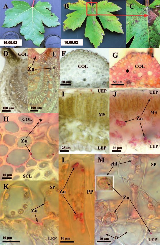
and also formed gradients increasing towards the leaf basis (Fig. 1B and C vs. A). These
symptoms occurred again in 2003 (and 2004, not shown) starting 50 days after bud break
(early June, Fig. 2). The proportion of symptomatic leaves increased until late July and was
significantly different (P < 0.0001) from that in trees without HM exposure (over all dates).
3.3 Cytochemical detection of zinc
In HM-treated trees, traces of Zn were observed in leaves sampled at the basis of top shoot
only. No HM was detected in leaves at higher shoot positions or control trees. Zinc was
micro-localised in different vein tissues (Fig. 1E, G, H vs. D, F; Table 2) noteworthy the upper
collenchyma (Fig. 1G, H vs. F) which showed the largest apparent concentrations among all
leaf tissues. In leaf blades (Fig. 1 J–M vs. I; Table 2) a few Zn-enriched spots scattered
throughout the leaf blade showed apparently unimpaired Zn accumulation throughout most
tissues (Fig. 1J vs. I). At cell level, Zn was micro-localised in different compartments (Table
2). Accumulation in cell walls was generally homogeneous except in sclerenchyma (more
signal in middle lamella, not shown), leaf blade upper (more signal in outer cell wall) and
lower epidermis (more signal in middle lamella of inside-facing cell wall, Fig. 1K). In
symplast, Zn was never observed in vacuoles or nucleus (Fig. 1M) but in the cytosol
(Fig. 1H, L and M) and mesophyll chloroplasts (Fig. 1K, M). Inside this latter organelle,
several Zn granules could generally be micro-localised (detail in Fig. 1M).280 Olivier André et al.
For. Snow Landsc. Res. 80, 3 (2006) 281
1.0
0.0766 0.0528
0.9 P= 0.0057 0.0495
0.5
0.4
0.3
0.2
0.1
0.0
0 2 40 60 80 100 120 Days
30.4.3 6.6.3 20.6.3 8.7.3 22.7.3 19.8.3 2.9.3 Date
Fig. 2. Symptom development in Sycamore Maple (means ± SE, N = 5 trees) as indicated by the propor-
tion of leaves showing visible symptoms of HM stress on the top shoot at each sampling date during the
fourth year of exposure (2003). Over all data, the difference between trees in treatments with (closed
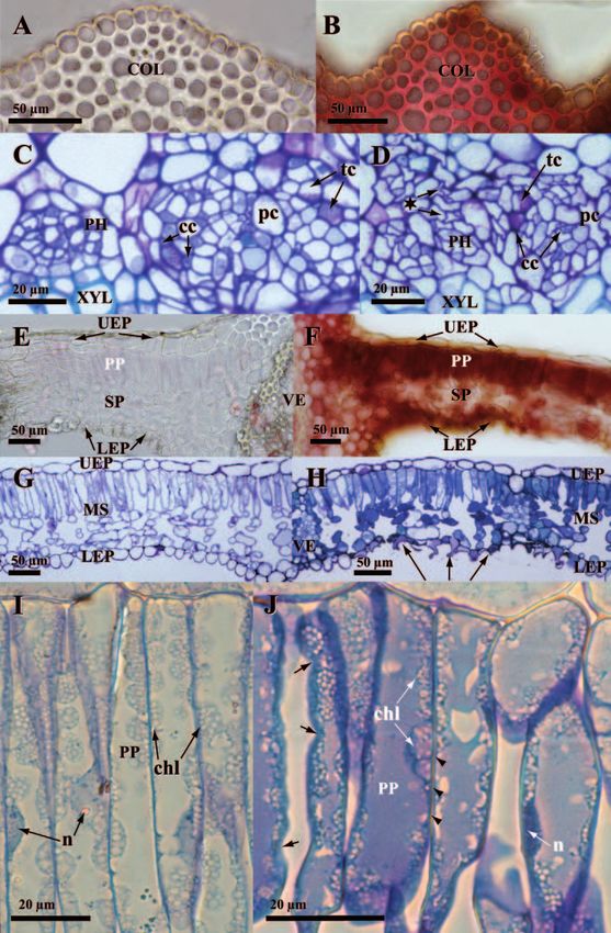
symbols) or without (open symbols) HM was significant (P282 Olivier André et al. 3.4 Structural changes and defence reactions The HM treatment caused different stress and defence reactions in the vein and leaf blade resulting in several structural changes. In veins of leaves at lower shoot position, phloem degenerated as indicated by cell wall folding (Fig. 3D vs. C) and condensation of cell content in non conducting cells. Cell walls in adaxial collenchyma were thickened noteworthy with CT (Fig. 3B vs. A). Other apoplastic CT were found in the epidermis and xylem whereas those in the symplast occurred in the adaxial collenchyma, epidermis, phloem and medullar rays (not shown). Structural changes in the leaf blade were often linked to those in veins as shown by gradients of symptom intensity decreasing with distance from veins (Fig. 3F, H vs. E, G). The most characteristic changes included gradients of necrosis in lower epidermis (Fig. 3H vs. G) as shown by cell collapse, cell wall thickening and accumulation of secondary compounds (Fig. 3F, H). Palisadic and spongy cells in mesophyll showed different degener- ation symptoms including cell wall thickening, folding and partial collapse (Fig. 3J vs. I), accumulation of secondary compounds (Fig. 3F, J vs. E, I), condensation of the cell nucleus, reduction of the chloroplast size and condensation of its stroma (Fig. 3J vs. I). In reference to VOLLENWEIDER et al. (2006), these changes were indicative of accelerated cell senescence processes (ACS). CT formed one class of the secondary compounds which were accumulated inside mesophyll of HM-exposed samples. They were strongly increased in comparison to control samples (Fig. 3F vs. 3E) and preferentially accumulated in the lower spongy parenchyma layers. The micro-localisation of CT thus closely matched that of Zn in the vein (Fig. 3B vs. 1G) as well as leaf blade (Fig. 3F vs. 1J). Structural injuries induced by HM decreased at higher shoot positions: necroses were no more detected and symptoms most frequently recurring included phloem degeneration, increase of secondary compounds in the vacuole and ACS gradients apart from veins (not shown). 3.5 Quantification of total phenols and condensed tannins in foliage Spectrophotometrical measurements of TP and CT showed to which extent the phenolic compounds cytochemically detected in HM-treated leaves from the basis of top shoot were also quantitatively increased throughout the whole crown foliage. The acid vanillin reagent with which the oligomeric fraction of CT was assessed (Fig. 4) was the same as that used to reveal CT cytochemically (Fig. 3B, F). TP reached 6.2 % of the foliage dry mass on average in control samples of which CT represented a small fraction (less than 5 %) only (Fig. 4). The Fig. 3. Stress and defence reactions to HM in leaves of Sycamore Maple sampled from the basis of the top shoot by the end of the third year of treatment (2002). A to D changes in vein. B vs. A collenchyma (COL) cell walls and to a lesser extent lumen contained increased amounts of CT (coloured in red) matching Zn micro-localization. D vs. C phloem (PH) showed stress reactions in the form of folded cell walls (★) and slightly condensed cell content especially in non conducting cells. E to J changes in leaf blade. F vs. E gradient of vacuolar CT close to main vein (VE). CT especially accumulated in palisade parenchyma (PP) and in the lower layer of spongy parenchyma (SP) forming a gradient culminating at the junction with the main vein. A few lower epidermis (LEP) cells contained CT, however phenolics were otherwise too oxidized to still react with vanillin, as indicated by brown tones. H vs. G stress reac- tions in the leaf blade next to a secondary vein. Mesophyll (MS) cells accumulated secondary com- pounds in their vacuole (coloured in blue). Lower epidermis became largely necrotic as shown by cell collapse (arrows). J vs. I changes in palisade cells. Cells underwent ACS as shown by thickening (arrow- heads) and folding (arrows) of cell walls, vacuoles full of secondary compounds, smaller and denser nuclei (n) and condensed chloroplasts (chl) with more lipids (dark grey shades). UEP: upper epidermis.
For. Snow Landsc. Res. 80, 3 (2006) 283
284                                                                                  Olivier André et al.

HM treatment did not change the TP content in the foliage whereas the polymeric and
oligomeric CT fractions tended to increase (Fig. 4): amounts of the oligomeric fraction were
raised from 1.2 % (CO) to 2.1 % (HM) and those of the polymeric form from 2.7 % (CO) to
5 % (HM) of the TP on average. These moderate increases thus confirmed that the exposure
to HM tended to specifically enhance the leaf content of CT and that this did not only occur
in older leaf material but also in the rest of the crown.

                                                     Total phenols         P = 0.4849
                              70                     CT (mainly polymers) P = 0.0605
                                                     CT (mainly oligomers) P = 0.089

                              60
             mgg-1 dry mass

                              50
                               5
                              4
                              3
                              2
                              1
                              0
                                   CO + AR                     HM + HMAR

Fig. 4. Phenolic content (means ± SE) in foliage of Sycamore Maple at the end of the third vegetation
season (2002). Samples from treatments without (CO, AR) and with heavy metals (HM, HMAR) were
pooled. Level of statistical significance (Wilcoxon test) between both groups is indicated in the legend.
N = 10 trees.

4       Discussion

Since the foliage biomass and crown development was not affected by the HM and AR
treatment, the phytoremediation capacity of the investigated Sycamore Maples remained
unchanged, at least during the whole period of observation despite the stress reactions
observed in older foliage. It could be a consequence of the low amounts of HM which were
translocated to the tree crown. Among the HM enriched in foliage, the uptake level of Zn
was superior to that of Cu as shown by bio-accumulation factors (BAF, calculated as the
ratio between the leaf and soil concentration) around 0.04 vs. 0.006 respectively. Lower BAF
for Cu than Zn is generally observed in trees (PULFORD and WATSON 2003) as well as in
other vascular plants (SIEDLECKA 1995). Our BAF were at best similar to and generally
smaller than those derived from data in other studies using Sycamore Maples growing on
dredged sediments (0.21 for Zn and 0.11 for Cu, MERTENS et al. 2004), or in two woodland
sites with different level of HM contamination (0.04 and 0.78 for Zn, 1.45 and 0.04 for Cu,
WATMOUGH and DICKINSON 1996). This could relate to the experimental soil contamination
with HM as mineral oxides in HM dust within the framework “From Cell to Tree” (NOWACK
For. Snow Landsc. Res. 80, 3 (2006) 285 et al. 2006) whereas HM might be in a more readily available form for plants with other sources of contamination. Since, in our case, unfavourable physical and chemical soil charac- teristics, as frequently occurring in contaminated sites (PULFORD and WATSON 2003), did not further impair tree establishment and HM uptake, the little HM uptake in treated Sycamore Maple may be attributed to HM avoidance and/or blocking in the rhizosphere. In reference to the higher HM uptake observed, noteworthy, in Populus tremula and Salix viminalis (HERMLE et al. 2006b) within the same “From Cell to Tree” framework, the results obtained here confirm the high phytostabilisation and low phytoextraction potential of Sycamore maple regarding the investigated HM and suggest it to be a species-related characteristic. Stress reactions provided useful bioindications in the form of visible symptoms in older foliage without changing tree growth. Gradients of stress reactions at foliage and leaf level showing a connection to the leaf vein were typical for an abiotic soil-borne stress factor (MARSCHNER 1995; VOLLENWEIDER and GUNTHARDT-GOERG 2005). Symptom devel- opment resulted from accumulative effects suggesting the involvement of Zn and Cd since both elements accumulate in foliage during the vegetation season (LAUREYSENS et al. 2004). Zinc was the only HM however, whose concentration reached near toxic levels in the foliage of the HM-exposed Sycamore Maples. Gradients of visible leaf symptoms in foliage and Zn cytochemical signal as a function of leaf position on the shoot suggest that Zn reached higher concentrations in older foliage compared to the rest of the crown. At the leaf level, symptom distribution along gradients and its connection to veins were closely matching the accumulation and distribution of Zn (as observed after cytochemical detection) as well as the distribution of microscopical stress reactions. These distribution characteristics were also found in other species e.g. Phaseolus vulgaris (MARTIN et al. 2006) and Populus tremula (VOLLENWEIDER and GUNTHARDT-GOERG 2005) under the same mixed HM-exposure suggesting that the visible symptoms, detected here, formed typical and specific bioindications of Zn intoxication in Sycamore maple. The micro-localisation results for Zn helped to understand how and why so moderate Zn concentrations in older foliage nevertheless resulted in necroses visible with the naked eye. Only a part of the Zn contaminants were deposited in structures like cell walls in adaxial collenchyma where it did not threaten sensitive and vitally important processes of the leaf physiology (VOLLENWEIDER et al. 2006). Another part accumulated apparently unimpaired in different leaf blade sites, generally close to veins, where it was in the position to sig- nificantly increase oxidative stress as Zn is an abiotic elicitor of reactive oxygen species (ROS; DIETZ et al. 1999). Zinc accumulation in cytosol and chloroplasts of mesophyll cells seems particularly critical as the photosynthetic electron transport system is the major source of ROS in plants (YAMASAKI et al. 1997). These cell compartments contain con- tinuously running detoxification systems (ELSTNER 1996) but supplementary sources of oxidative stress can however definitely unbalance the buffering capacities (VOLLENWEIDER et al. 2003). Zinc was thus in the position to directly interfere with the photosynthetic and gas exchange activities in mesophyll. No detoxification processes like Zn excretion through secretory hairs (CHOI et al. 2001; MARTIN et al. 2006), storage in vacuoles (VAZQUEZ et al. 1994; CLEMENS 2001), or massive inlays in cell walls (LICHTENBERGER and NEUMANN 1997; VOLLENWEIDER et al. 2006) was detected, suggesting that lesions to tissues resulted from insufficiently controlled Zn storage. Structural changes caused by stress reactions were found in sites where Zn was micro- localised and through which Zn was at least translocated (phloem), but where Zn con- centration remained below the detection limit. Indeed, cycling of mineral elements in plants, probably through the medullar rays, contributes to HM allocation to phloem (MARSCHNER 1995; VOLLENWEIDER et al. 2006). In reference to the latter publication, the microscopical
286                                                                          Olivier André et al.

changes observed here in phloem probably resulted from ACS processes. In the leaf blade,
advanced ACS processes were observed in connection to Zn-induced injury gradients starting
from veins. In contrast to Raphanus sativus (MARTIN et al. 2006) or Populus tremula
(GUNTHARDT-GOERG and VOLLENWEIDER 2003), no HR-like reactions were observed in
mesopyll in relation to Zn deposition here, most likely because of the lower Zn concentration
inside the leaves of Sycamore Maple. Changes at tissue and cell levels and the microscopical
gradients were otherwise similar to those observed in Populus tremula. The micro-localisation
of CT matched remarkably well with that of Zn as observed in the case of Salix viminalis
under Cd stress (VOLLENWEIDER et al. 2006). CT were found in both apoplastic and
symplastic sites and in places with or without structural injuries. They were specifically
induced by the HM treatment among other phenolic compounds and formed one of the few
cell defence reactions induced by Zn and recorded in HM-contaminated leaves. Their
eventual Zn-chelating or oxidative stress-quenching effects cannot be estimated on the basis
of our investigations. The results obtained here on Sycamore Maple show however that they
form interesting stress markers for several HM and tree species.

5      Conclusion

In Sycamore Maples growing on soils with mixed HM contamination, low amounts of Zn
triggered oxidative stress in older leaf foliage because Zn could reach, unimpaired, sensitive
sites inside leaf blade. They caused ACS and necrosis resulting in unspecific leaf chlorosis
and necrotic spots and flecks next to veins. The latter were arranged along gradients at leaf
and shoot level and were characteristic for Zn. Visible symptoms provided a reliable bio-
indication of Zn contamination without changing the phytostabilisation potential of the
treated Sycamore Maple, since Zn average concentration inside crown foliage hardly
reached the toxicity limit. Most stress reactions occurred in older foliage where they could
little influence the growth of this pioneer tree species with unlimited apical growth (at least
as a young tree). The phytoremediation potential of Sycamore Maple was found to include a
bioindication component which can be useful for surveillance purposes in sites where
phytostabilisation is achieved.

Acknowledgments
Technical assistance by Michael Lautenschläger (tending the plants, sampling and sample pre-
paration), WSL central Laboratory (element analysis) and Terry Menard (microscopical analysis)
are gratefully acknowledged.

6        References

BROADHURST, R.B.; JONES, W.T., 1978: Analysis of condensed tannins using acidified vanillin. J.
  Sci. Food Agric. 29: 788–794.
CHOI, Y.E.; HARADA, E.; WADA, M.; TSUBOI, H.; MORITA, Y.; KUSANO, T.; SANO, H., 2001:
  Detoxification of cadmium in tobacco plants: formation and active excretion of crystals
  containing cadmium and calcium through trichomes. Planta 213: 45–50.
CLEMENS, S., 2001: Molecular mechanisms of plant metal tolerance and homeostasis. Planta 212:
  475–486.
For. Snow Landsc. Res. 80, 3 (2006)                                                          287

COSIO, C.; VOLLENWEIDER, P.; KELLER, C., 2006: Localization and effects of cadmium in leaves
   of tolerant willows (Salix viminalis L.). Part I. Macrolocalization and phytotoxic effects of
   cadmium. Environ. Exp. Bot. 58: 64–74.
DICKINSON, N.M., 2000: Strategies for sustainable woodland on contaminated soils. Chemosphere
   41: 259–263.
DIETZ, K.-J.; BAIER, M.; KRAMER, U., 1999: Free radicals and reactive oxygen species as mediators
   of heavy metal toxicity in plants. In: PRASAD, M.N.V.; HAGEMEYER, J. (eds) Heavy metal stress
   in plants. Berlin, Springer. 73–97.
ELSTNER, E.F., 1996: Die Sauerstoffaktivierung als Basis pflanzlicher Stressreaktionen. In:
   BRUNOLD, C.; RUEGSEGGER, A.; BRANDLE, R. (eds) Stress bei Pflanzen. Ökologie, Physio-
   logie, Biochemie, Molekularbiologie. Bern, Stuttgart, Paul Haupt. 347–362.
ERNST, W., 1972: Zink- und Cadmium-Immissionen auf Böden und Pflanzen in der Umgebung
   einer Zinkhütte. Ber. Dtsch. bot. Ges. 85: 295–300.
ESTERBAUER, H.; GRILL, D.; BECK, G., 1975: Phenols in needles of Picea-Abies. Phyton-Annales
   Rei Botanicae 17: 87–99.
GUNTHARDT-GOERG, M.S., 2005: From Cell to Tree. In: Swiss Federal Institute for Forest, Snow,
   and Landscape Research WSL (ed) Metal fluxes and stresses in terrestrial ecosystems.
   Abstracts. Birmensdorf, Swiss Federal Institute for Forest, Snow and Landscape Research
   WSL. 38.
GUNTHARDT-GOERG, M.S.; MCQUATTIE, C.J.; SCHEIDEGGER, C.; RHINER, C.; MATYSSEK, R.,
   1997: Ozone-induced cytochemical and ultrastructural changes in leaf mesophyll cell walls.
   Can. J. For. Res. 27: 453–463.
GUNTHARDT-GOERG, M.S.; VOLLENWEIDER, P., 2003: Cellular injury, heavy metal uptake and
   growth of poplar, willow and spruce influenced by heavy metal and soil acidity. In: MENCH,
   M.J.; MOCQUOT, B. (eds) Risk assessment and sustainable land management using plants in
   trace element-contaminated soils. COST Action 837, 4th WG2 workshop, Bordeaux 2002.
   INRA, Centre Bordeaux-Aquitaine, Villenave d’Ornon cedex. Bordeaux, France. 165–171.
HERMLE, H.; GUNTHARDT-GOERG, M.S.; SCHULIN, R., 2006a: Effects of metal contaminated soil
   on the performance of young trees growing in model ecosystems under field conditions.
   Environ. Pollut. 144, 2: 703–714.
HERMLE, S.; VOLLENWEIDER, P.; GUNTHARDT-GOERG, M.S.; MCQUATTIE, C.J.; MATYSSEK, R.,
   2006b: Responsiveness of leaf gas exchange and structure in field-grown, juvenile Populus
   tremula and Salix viminalis trees to heavy metal contamination and soil and rainwater acidity.
   Tree Physiol. accepted.
HORVARTH, B.; GRUIZ, K., 1996: Impact of metalliferous ore mining activity on the environment
   in Gyongyosoroszi, Hungary. Sci. Total Environ. 184: 215–227.
KABATA-PENDIAS, A.; PENDIAS, H., 2001: Trace elements in soils, 3rd Edition. London, New York,
   CRC Press, Boca Raton.
LAUREYSENS, I.; BLUST, R.; DE TEMMERMAN, L.; LEMMENS, C.; CEULEMANS, R., 2004: Clonal
   variation in heavy metal accumulation and biomass production in a poplar coppice culture: I.
   Seasonal variation in leaf, wood and bark concentrations. Environ. Pollut. 131: 485–494.
LEPP, N.W., 1996: Uptake, mobility and loci of concentration of heavy metals in trees. In:
   GLIMMERVEEN, I. (ed) Heavy metals and trees. Proceedings of a discussion meeting, Glasgow.
   Institute of chartered foresters, Edinburgh. 68–82.
LICHTENBERGER, O.; NEUMANN, D., 1997: Analytical electron microscopy as a powerful tool in
   plant cell biology: examples using electron energy loss spectroscopy and X-ray microanalysis.
   Eur. J. Cell Biol. 73: 378–386.
MARSCHNER, H., 1995: Mineral nutrition of higher plants. London, Academic press.
MARTIN, D.; VOLLENWEIDER, P.; GUNTHARDT-GOERG, M.S., 2006: Bioindication of heavy metal
   contamination in vegetable gardens. For. Snow Landsc. Res. 80, 2: 169–180.
MCDONALD, M.; MILA, I.; SCALBERT, A., 1996: Precipitation of metal ions by plant polyphenols:
   Optimal conditions and origin of precipitation. J. Agric. Food Chem. 44: 599–606.
MERTENS, J.; VERVAEKE, P.; DE SCHRIJVER, A.; LUYSSAERT, S., 2004: Metal uptake by young
   trees from dredged brackish sediment: limitations and possibilities for phytoextraction and
   phytostabilisation. Sci. Total Environ. 326: 209–215.
288                                                                             Olivier André et al.

MORAN, J.F.; KLUCAS, R.V.; GRAYER, R.J.; ABIAN, J.; BECANA, M., 1997: Complexes of iron with
   phenolic compounds from soybean nodules and other legume tissues: Prooxidant and anti-
   oxidant properties. Free Radical Biology and Medicine 22: 861–870.
NOWACK, B.; RAIS, D.; FREY, B.; MENON, M.; SCHULIN, R.; GUNTHARDT-GOERG, M.S.; LUSTER, J.,
   2006: Influence of heavy metal contamination on soil parameters in a lysimeter experiment
   designed to evaluate phytostabilization by afforestation. For. Snow Landsc. Res. 80, 2: 201–211.
PORTER, L.J.; HRSTICH, L.N.; CHAN, B.G., 1986: The conversion of procyanidins and prodel-
   phinidins to cyanidin and delphinidin. Phytochemistry 25: 223–230.
PRASAD, M.N.V.; STRZALKA, K., 1999: Impacts of heavy metals on photosynthesis. In: PRASAD,
   M.N.V.; HAGEMEYER, J. (eds) Heavy metal stress in plants. Berlin, Springer. 117–138.
PULFORD, I.D.; WATSON, C., 2003: Phytoremediation of heavy metal-contaminated land by trees –
   a review. Environ. Int. 29: 529–540.
PUNSHON, T.; DICKINSON, N.M.; LEPP, N.W., 1996: The potential of Salix clones for bioremediating
   metal polluted soil. In: GLIMMERVEEN, I. (ed) Heavy metals and trees. Proceedings of a dis-
   cussion meeting, Glasgow. Institute of chartered foresters, Edinburgh. 93–104.
RICE-EVANS, C.A.; MILLER, N.J.; PAGANGA, G., 1997: Antioxidant properties of phenolic com-
   pounds. Trends Plant Sci. 2: 152–159.
SAINT-CRICQ DE GAULEJAC, N.S.; PROVOST, C.; VIVAS, N., 1999: Comparative study of polyphenol
   scavenging activities assessed by different methods. J. Agric. Food Chem. 47: 425–431.
SIEDLECKA, A., 1995: Some aspects of interactions between heavy metals and plant mineral
   nutrients. Acta Soc. Bot. Pol. 64: 265–272.
VAZQUEZ, M.D.; POSCHENRIEDER, C.; BARCELO, J.; BAKER, A.J.M.; HATTON, P.; COPE, G.H.,
   1994: Compartmentation of zink in roots and leaves of the zinc hyperaccumulator Thlaspi
   caerulescens J & C Presl. Bot. Acta 107: 243–250.
VOLLENWEIDER, P.; GUNTHARDT-GOERG, M.S., 2005: Diagnosis of abiotic and biotic stress
   factors using visible symptoms in foliage. Environ. Pollut. 137: 455–46.
VOLLENWEIDER, P.; OTTIGER, M.; GUNTHARDT-GOERG, M.S., 2003: Validation of leaf ozone
   symptoms in natural vegetation using microscopical methods. Environ. Pollut. 124: 101–118.
VOLLENWEIDER, P.; MENARD, T.; GUNTHARDT-GOERG, M.S., 2005: Microlocalisation and
   mapping of zink in poplar with cytochemical methods. In: Swiss Federal Institute for Forest,
   Snow, and Landscape Research WSL (eds) Metal fluxes and stresses in terrestrial ecosystems.
   Abstracts. Birmensdorf, Swiss Federal Institute for Forest, Snow and Landscape Research
   WSL. 105.
VOLLENWEIDER, P.; COSIO, C.; GUNTHARDT-GOERG, M.S.; KELLER, C., 2006: Localization and
   effects of cadmium in leaves of a cadmium-tolerant willow (Salix viminalis L.). Part II
   Microlocalization and cellular effects of cadmium. Environ. Exp. Bot. 58: 25–40.
WATERMAN, P.G.; MOLE, S., 1994: Analysis of phenolic plant metabolites. In: LAWTON, J.H.,
   LIKENS, G.E. (eds) Methods in ecology. Oxford, London, Edinburgh, Boston, Melbourne,
   Paris, Berlin, Vienna, Blackwell scientific publications.
WATMOUGH, S.A.; DICKINSON, N.M., 1996: Variability of metal resistance in Acer pseudoplatanus
   L. (Sycamore) callus tissue of different origins. Environ. Exp. Bot. 36: 293–302.
YAMASAKI, H.; SAKIHAMA, Y.; IKEHARA, N., 1997: Flavonoid-peroxidase reaction as a detoxi-
   fication mechanism of plant cells against H2O2. Plant Physiol. 115: 1405–1412.

                                                       Revised version accepted October 30, 2006
You can also read