GAF is essential for zygotic genome activation and chromatin accessibility in the early Drosophila embryo
←
→
Page content transcription
If your browser does not render page correctly, please read the page content below
RESEARCH ARTICLE

                                    GAF is essential for zygotic genome
                                    activation and chromatin accessibility in
                                    the early Drosophila embryo
                                    Marissa M Gaskill†, Tyler J Gibson†, Elizabeth D Larson, Melissa M Harrison*

                                    Department of Biomolecular Chemistry, University of Wisconsin School of Medicine
                                    and Public Health, Madison, United States

                                    Abstract Following fertilization, the genomes of the germ cells are reprogrammed to form the
                                    totipotent embryo. Pioneer transcription factors are essential for remodeling the chromatin and
                                    driving the initial wave of zygotic gene expression. In Drosophila melanogaster, the pioneer factor
                                    Zelda is essential for development through this dramatic period of reprogramming, known as the
                                    maternal-to-zygotic transition (MZT). However, it was unknown whether additional pioneer factors
                                    were required for this transition. We identified an additional maternally encoded factor required for
                                    development through the MZT, GAGA Factor (GAF). GAF is necessary to activate widespread
                                    zygotic transcription and to remodel the chromatin accessibility landscape. We demonstrated that
                                    Zelda preferentially controls expression of the earliest transcribed genes, while genes expressed
                                    during widespread activation are predominantly dependent on GAF. Thus, progression through the
                                    MZT requires coordination of multiple pioneer-like factors, and we propose that as development
                                    proceeds control is gradually transferred from Zelda to GAF.

*For correspondence:                Introduction
mharrison3@wisc.edu                 Pronounced changes in cellular identity are driven by pioneer transcription factors that act at the top
†
 These authors contributed          of gene regulatory networks. While nucleosomes present a barrier to the DNA binding of many tran-
equally to this work                scription factors, pioneer factors can bind DNA in the context of nucleosomes. Pioneer-factor bind-
                                    ing establishes accessible chromatin domains, which serve to recruit additional transcription factors
Competing interests: The
                                    that drive gene expression (Zaret and Mango, 2016; Iwafuchi-Doi and Zaret, 2014; Zaret and Car-
authors declare that no
                                    roll, 2011). These unique characteristics of pioneer factors enable them to facilitate widespread
competing interests exist.
                                    changes in cell identity. Nonetheless, cell-fate transitions often require a combination of pioneering
Funding: See page 24                transcription factors to act in concert to drive the necessary transcriptional programs. Indeed, the
Received: 19 January 2021           reprogramming of a specified cell type to an induced pluripotent stem cell requires a cocktail of
Accepted: 14 March 2021             transcription factors, of which Oct4, Sox2, and Klf4 function as pioneer factors (Takahashi and
Published: 15 March 2021            Yamanaka, 2006; Takahashi et al., 2007; Chronis et al., 2017; Soufi et al., 2015; Soufi et al.,
                                    2012). Despite the many examples of multiple pioneer factors functioning together to drive reprog-
Reviewing editor: Yukiko M
Yamashita, Whitehead Institute/
                                    ramming, how these factors coordinate gene expression changes within the context of organismal
MIT, United States                  development remains unclear.
                                       Pioneer factors are also essential for the reprogramming that occurs in the early embryo. Follow-
   Copyright Gaskill et al. This
                                    ing fertilization, specified germ cells must be rapidly and efficiently reprogrammed to generate a
article is distributed under the
                                    totipotent embryo capable of differentiating into all the cell types of the adult organism. This
terms of the Creative Commons
Attribution License, which          reprogramming is initially driven by mRNAs and proteins that are maternally deposited into the
permits unrestricted use and        oocyte. During this time, the zygotic genome remains transcriptionally quiescent. Only after cells
redistribution provided that the    have been reprogrammed is the zygotic genome gradually activated. This maternal-to-zygotic transi-
original author and source are      tion (MZT) is broadly conserved among metazoans and essential for future development (Schulz and
credited.                           Harrison, 2019; Vastenhouw et al., 2019). Activators of the zygotic genome have been identified

Gaskill, Gibson, et al. eLife 2021;10:e66668. DOI: https://doi.org/10.7554/eLife.66668                                              1 of 30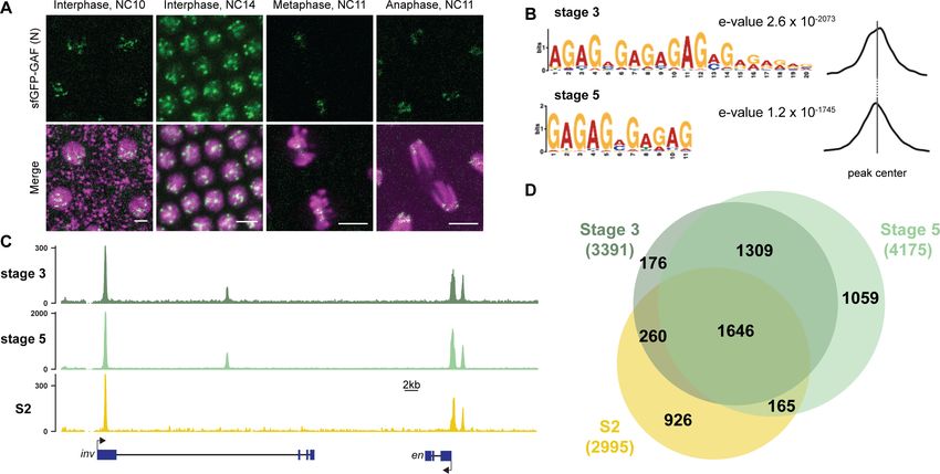
Research article                                                                                 Chromosomes and Gene Expression

                                    eLife digest Most cells in an organism share the exact same genetic information, yet they still
                                    adopt distinct identities. This diversity emerges because only a selection of genes is switched on at
                                    any given time in a cell. Proteins that latch onto DNA control this specificity by activating certain
                                    genes at the right time. However, to perform this role they first need to physically access DNA: this
                                    can be difficult as the genetic information is tightly compacted so it can fit in a cell. A group of
                                    proteins can help to unpack the genome to uncover the genes that can then be accessed and
                                    activated. While these ‘pioneer factors’ can therefore shape the identity of a cell, much remains
                                    unknown about how they can work together to do so. For instance, the pioneer factor Zelda is
                                    essential in early fruit fly development, as it enables the genetic information of the egg and sperm
                                    to undergo dramatic reprogramming and generate a new organism. Yet, it was unclear whether
                                    additional helpers were required for this transition.
                                       Using this animal system, Gaskill, Gibson et al. identified GAGA Factor as a protein which works
                                    with Zelda to open up and reprogram hundreds of different sections along the genome of fruit fly
                                    embryos. This tag-team effort started with Zelda being important initially to activate genes;
                                    regulation was then handed over for GAGA Factor to continue the process. Without either protein,
                                    the embryo died.
                                       Getting a glimpse into early genetic events during fly development provides insights that are
                                    often applicable to other animals such as fish and mammals. Ultimately, this research may help
                                    scientists to understand how things can go wrong in human embryos.

                                    in a number of species (zebrafish – Pou5f3, Sox19b, Nanog; mice – Dux, Nfy; humans – DUX4,
                                    OCT4; fruit flies – Zelda), and all share essential features of pioneer factors (Schulz and Harrison,
                                    2019; Vastenhouw et al., 2019).
                                        Early Drosophila development is characterized by 13, rapid, synchronous nuclear divisions.
                                    Zygotic transcription gradually becomes activated starting about the eighth nuclear division, and
                                    widespread transcription occurs at the 14th nuclear division when the division cycle slows and the
                                    nuclei are cellularized (Schulz and Harrison, 2019; Vastenhouw et al., 2019). The transcription fac-
                                    tor Zelda (Zld) is required for transcription of hundreds of genes throughout zygotic genome activa-
                                    tion (ZGA) and was the first identified global genomic activator (Liang et al., 2008; Harrison et al.,
                                    2011; Nien et al., 2011). Embryos lacking maternally encoded Zld die before completing the MZT
                                    (Liang et al., 2008; Harrison et al., 2011; Nien et al., 2011; Fu et al., 2014; Staudt et al., 2006).
                                    Zld has the defining features of a pioneer transcription factor: it binds to nucleosomal DNA
                                    (McDaniel et al., 2019), facilitates chromatin accessibility (Schulz et al., 2015; Sun et al., 2015) and
                                    this leads to subsequent binding of additional transcription factors (Yáñez-Cuna et al., 2012;
                                    Xu et al., 2014; Foo et al., 2014).
                                        By contrast to the essential role for Zld in flies, no single global activator of zygotic transcription
                                    has been identified in other species. Instead, multiple transcription factors function together to acti-
                                    vate zygotic transcription (Schulz and Harrison, 2019; Vastenhouw et al., 2019). Work from our
                                    lab and others has implicated additional factors in regulating reprogramming in the Drosophila
                                    embryo. Specifically, the enrichment of GA-dinucleotides in regions of the genome that remain
                                    accessible in the absence of Zld and at loci that gain accessibility late in ZGA, suggest that a protein
                                    that binds to these loci functions with Zld to define cis-regulatory regions during the initial stages of
                                    development (Schulz et al., 2015; Sun et al., 2015; Blythe and Wieschaus, 2016). Three proteins,
                                    GAGA factor (GAF), chromatin-linked adaptor for MSL proteins (CLAMP), and Pipsqueak (Psq) are
                                    known to bind to GA-dinucleotide repeats and are expressed in the early embryo, implicating one
                                    or all these proteins in reprogramming the embryonic transcriptome (Rieder et al., 2017;
                                    Soruco et al., 2013; Kuzu et al., 2016; Biggin and Tjian, 1988; Bhat et al., 1996; Soeller et al.,
                                    1993; Lehmann et al., 1998).
                                        CLAMP was first identified based on its role in targeting the dosage compensation machinery,
                                    and it preferentially localizes to the X chromosome (Larschan et al., 2012; Soruco et al., 2013). Psq
                                    is essential for oogenesis (Horowitz and Berg, 1996), and it has been suggested to have a role as a
                                    repressor and in chromatin looping (Gutierrez-Perez et al., 2019; Huang et al., 2002). GAF,

Gaskill, Gibson, et al. eLife 2021;10:e66668. DOI: https://doi.org/10.7554/eLife.66668                                                  2 of 30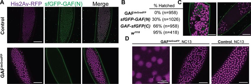
Research article                                                                               Chromosomes and Gene Expression

                                    encoded by the Trithorax-like (Trl) gene, has broad roles in transcriptional regulation, including func-
                                    tioning as a transcriptional activator (Farkas et al., 1994; Bhat et al., 1996), repressor
                                    (Mishra et al., 2001; Busturia et al., 2001; Bernués et al., 2007; Horard et al., 2000), and insulator
                                    (Ohtsuki and Levine, 1998; Wolle et al., 2015; Kaye et al., 2017). Through interactions with chro-
                                    matin remodelers, GAF is instrumental in driving regions of accessible chromatin both at promoters
                                    and distal cis-regulatory regions (Okada and Hirose, 1998; Tsukiyama et al., 1994; Xiao et al.,
                                    2001; Tsukiyama and Wu, 1995; Fuda et al., 2015; Judd et al., 2021). Analysis of hypomorphic
                                    alleles has suggested an important function for GAF in the early embryo in both driving expression
                                    of Ultrabiothorax (Ubx), Abdominal B (Abd-B), engrailed (en), and fushi tarazu (ftz) and in maintain-
                                    ing normal embryonic development (Bhat et al., 1996; Farkas et al., 1994). Given these diverse
                                    functions for GAF, and that it shares many properties of a pioneer transcription factor, we sought to
                                    investigate whether it has a global role in reprogramming the zygotic genome for transcriptional
                                    activation.
                                        Investigation of the role of GAF in the early embryo necessitated the development of a system to
                                    robustly eliminate GAF, which had not been possible since GAF is essential for maintenance of the
                                    maternal germline and is resistant to RNAi knockdown in the embryo (Bhat et al., 1996;
                                    Bejarano and Busturia, 2004; Rieder et al., 2017). For this purpose, we generated endogenously
                                    GFP-tagged GAF, which provided the essential functions of the untagged protein, and used the
                                    deGradFP system to deplete GFP-tagged GAF in early embryos expressing only the tagged con-
                                    struct (Caussinus et al., 2012). Using this system, we identified an essential function for GAF in driv-
                                    ing chromatin accessibility and gene expression during the MZT. Thus, at least two pioneer-like
                                    transcription factors, Zld and GAF, must cooperate to reprogram the zygotic genome of Drosophila
                                    following fertilization.

                                    Results
                                    GAF binds the same loci throughout ZGA
                                    To investigate the role of GAF during the MZT, we used Cas9-mediated genome engineering to tag
                                    the endogenous protein with super folder Green Fluorescent Protein (sfGFP) at either the N- or
                                    C-termini, sfGFP-GAF(N) and GAF-sfGFP(C), respectively (Pédelacq et al., 2006). There are two pro-
                                    tein isoforms of GAF (Benyajati et al., 1997). Because the N-terminus of GAF is shared by both iso-
                                    forms, the sfGFP tag on the N-terminus labels all GAF protein (Figure 1—figure supplement 1A).
                                    By contrast, the two reported isoforms differ in their C-termini, and thus the C-terminal sfGFP labels
                                    only the short isoform (Figure 1—figure supplement 1B). Whereas null mutants in Trithorax-like
                                    (Trl), the gene encoding GAF, are lethal (Farkas et al., 1994), both sfGFP-tagged lines are homozy-
                                    gous viable and fertile. Additionally, in embryos from both lines sfGFP-labeled GAF is localized to
                                    discrete nuclear puncta and is retained on the mitotic chromosomes in a pattern that recapitulates
                                    what has been previously described for GAF based on antibody staining in fixed embryos
                                    (Figure 1A, Figure 1—figure supplement 1C; Raff et al., 1994). Together, these data demonstrate
                                    that the sfGFP tag does not interfere with essential GAF function and localization.
                                        To begin to elucidate the role of GAF during early embryogenesis, we determined the genomic
                                    regions occupied by GAF during the MZT. We hand sorted homozygous GAF-sfGFP(C) stage 3
                                    (nuclear cycle (NC) 9) and stage 5 (NC14) embryos and performed chromatin immunoprecipitation
                                    coupled with high-throughput sequencing (ChIP-seq) using an anti-GFP antibody. While alternative
                                    splicing generates two GAF protein isoforms that differ in their C-terminal polyQ domain, in the
                                    early embryo only the short isoform is detectable (Benyajati et al., 1997). We confirmed the expres-
                                    sion of the short isoform in the early embryo by blotting extract from 0 to 4 hr AEL (after egg laying)
                                    N- and C-terminally tagged GAF embryos with an anti-GFP antibody (Figure 1—figure supplement
                                    1D). The long isoform was undetectable in extract from embryos of both sfGFP-tagged lines har-
                                    vested 0–4 hr AEL, but was detectable at low levels in extract from sfGFP-GAF(N) embryos har-
                                    vested 13–16 hr AEL. Thus, we conclude that during the MZT the C-terminally sfGFP-labeled short
                                    isoform comprises the overwhelming majority of GAF present in the GAF-sfGFP(C) embryos. We
                                    identified 3391 GAF peaks at stage 3 and 4175 GAF peaks at stage 5 (Supplementary file 1). To
                                    control for possible cross-reactivity with the anti-GFP antibody, we performed ChIP-seq on w1118
                                    stage 3 and stage 5 embryos in parallel. Since there are no GFP-tagged proteins in the w1118

Gaskill, Gibson, et al. eLife 2021;10:e66668. DOI: https://doi.org/10.7554/eLife.66668                                               3 of 30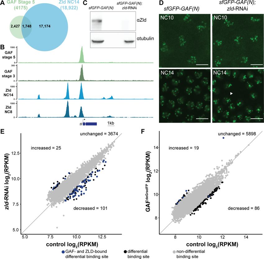
Research article                                                                                     Chromosomes and Gene Expression

Figure 1. GAF binds thousands of loci throughout the MZT. (A) Images of His2Av-RFP; sfGFP-GAF(N) embryos at the nuclear cycles (NC) indicated
above. sfGFP-GAF(N) localizes to puncta during interphase and is retained on chromosome during mitosis. His2AvRFP is shown in magenta. sfGFP-GAF
(N) is shown in green. Scale bars, 5 mm. (B) Binding motif enrichment of GA-dinucleotide repeats at GAF peaks identified at stage 3 and stage 5
determined by MEME-suite (left). Distribution of the GA-repeat motif within peaks (right). Gray line indicates peak center. (C) Representative genome
browser tracks of ChIP-seq peaks for GAF-sfGFP(C) from stage 3 and stage 5 embryos and GAF ChIP-seq from S2 cells (Fuda et al., 2015). (D) Venn
diagram of the peak overlap for GAF as determined by ChIP-seq for GAF-sfGFP(C) from sorted stage 3 embryos and stage 5 embryos and by ChIP-seq
for GAF from S2 cells (Fuda et al., 2015). Total number of peaks identified at each stage is indicated in parentheses. See also Figure 1—figure
supplements 1–3.
The online version of this article includes the following figure supplement(s) for figure 1:
Figure supplement 1. N- and C- terminal sfGFP-tags label distinct GAF isoforms.
Figure supplement 2. GAF binds thousands of regions in the embyo and S2 cells.
Figure supplement 3. GAF has tissue-specific binding.

                                    embryos, any peaks identified with the anti-GFP antibody would be the result of non-specific interac-
                                    tions and excluded from further analysis. No peaks were called in the w1118 dataset for either stage,
                                    confirming the specificity of the peaks identified in the GAF-sfGFP(C) embryos (Figure 1—figure
                                    supplement 2A,B). Further supporting the specificity of the ChIP data, the canonical GA-rich GAF-
                                    binding motif was the most highly enriched sequence identified in ChIP peaks from both stages, and
                                    these motifs were centrally located in the peaks (Figure 1B). Peaks were identified in the regulatory
                                    regions of several previously identified GAF-target genes, including the heat shock promoter
                                    (hsp70), even-skipped (eve), Krüppel (Kr), Ubx, and en (Figure 1C; Biggin and Tjian, 1988;
                                    Gilmour et al., 1989; Soeller et al., 1988; Lee et al., 1992; Read et al., 1990; Kerrigan et al.,
                                    1991). Peaks were enriched at promoters, which fits with the previously defined role of GAF in
                                    establishing paused RNA polymerase (Figure 1—figure supplement 2C; Lee et al., 2008;
                                    Fuda et al., 2015; Judd et al., 2021).
                                       There was a substantial degree of overlap between GAF peaks identified at both stage 3 and
                                    stage 5. A total of 2955 peaks were shared between the two time points, representing 87% of total
                                    stage 3 peaks and 71% of total stage 5 peaks (Figure 1C,D, Figure 1—figure supplement 2D). This
                                    demonstrates that GAF binding is established prior to widespread ZGA and remains relatively
                                    unchanged during early development, similar to what has been shown for Zld, the major activator of
                                    the zygotic genome (Harrison et al., 2011). We compared GAF-binding sites we identified in the

Gaskill, Gibson, et al. eLife 2021;10:e66668. DOI: https://doi.org/10.7554/eLife.66668                                                        4 of 30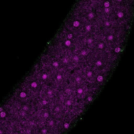
Research article                                                                              Chromosomes and Gene Expression

                                    early embryo to previously identified GAF-bound regions in S2 cells, which are derived from 20 to
                                    24 hr old embryos (Fuda et al., 2015). Despite the difference in cell-type and antibody used, 56%
                                    (1906) of stage 3 peaks and 43% (1811) of stage 5 peaks overlapped peaks identified in S2 cells
                                    (Figure 1C,D, Figure 1—figure supplement 2D). It was previously noted that GAF binding in 8–16
                                    hr embryos was highly similar to GAF occupancy in the wing imaginal disc harvested from the larva
                                    (Slattery et al., 2014), and our data indicate that this binding is established early in development
                                    prior to activation of the zygotic genome. Nonetheless, peaks unique to each tissue likely represent
                                    GAF-binding events as they are centrally enriched for GA-rich GAF-binding motifs (Figure 1—figure
                                    supplement 3). Thus, while a majority of GAF-binding sites are maintained in both the embryo and
                                    cell culture, a subset of GAF-binding sites is likely tissue specific.

                                    Maternal GAF is required for embryogenesis and progression through
                                    the MZT
                                    Early embryonic GAF is maternally deposited in the oocyte, and this maternally deposited mRNA
                                    can sustain development until the third instar larval stage (Farkas et al., 1994). To investigate the
                                    role of GAF during the MZT therefore necessitated a system to eliminate this maternally encoded
                                    GAF. RNAi failed to successfully knockdown GAF in the embryo (Rieder et al., 2017), and female
                                    germline clones cannot be generated as GAF is required for egg production (Bhat et al., 1996;
                                    Bejarano and Busturia, 2004). To overcome these challenges, we leveraged our N-terminal sfGFP-
                                    tagged allele and the previously developed deGradFP system to target knockdown at the protein
                                    level (Caussinus et al., 2012). The deGradFP system uses a genomically encoded F-box protein
                                    fused to a nanobody recognizing GFP, which recruits GFP-tagged proteins to a ubiquitin ligase com-
                                    plex. Ubiquitination of the GFP-tagged protein subsequently leads to efficient degradation by the
                                    proteasome. To adapt this system for efficient use in the early embryo, we generated transgenic flies
                                    in which the deGradFP nanobody fusion was driven by the nanos (nos) promoter for strong expres-
                                    sion in the embryo 0–2 hr after fertilization (Wang and Lehmann, 1991). In embryos laid by nos-
                                    deGradFP; sfGFP-GAF(N) females all maternally encoded GAF protein is tagged with GFP and thus
                                    subject to degradation by the deGradFP nanobody fusion. These embryos are hereafter referred to
                                    as GAFdeGradFP.
                                        We verified the efficiency of the deGradFP system by imaging living embryos in which nuclei
                                    were marked by His2Av-RFP. GAFdeGradFP embryos lack the punctate, nuclear GFP signal identified
                                    in control embryos that do not carry the deGradFP nanobody fusion, indicating efficient depletion of
                                    sfGFP-GAF(N) (Figure 2A). This knockdown was robust, as we failed to identify any NC10 - 14
                                    embryos with nuclear GFP signal, and none of the embryos carrying both the deGradFP nanobody
                                    and the sfGFP-tagged GAF hatched (Figure 2B). Based on live embryo imaging, the majority of
                                    embryos died prior to NC14, indicating that maternal GAF is essential for progression through the
                                    MZT. We identified a small number of GFP-expressing, gastrulating escapers. It is unclear if these
                                    embryos had an incomplete knockdown of maternally encoded sfGFP-GAF(N), or if a small percent-
                                    age of embryos survived until gastrulation in the absence of GAF and that the GFP signal was the
                                    result of zygotic gene expression. Nonetheless, none of these embryos survived until hatching.
                                    Despite being able to maintain a strain homozygous for the N-terminal, sfGFP-tagged GAF, quanti-
                                    tative analysis revealed an effect on viability. Embryos homozygous for sfGFP-GAF(N) had only a
                                    30% hatching rate (Figure 2B). By contrast, embryos homozygous for GAF-sfGFP(C) hatched at a
                                    rate of 66%. To determine whether the differences in hatching rates were due to differences in pro-
                                    tein expression levels, we performed in vivo fluorescent quantification of the GFP signal in nuclei of
                                    sfGFP-GAF(N) and GAF-sfGFP(C) homozygous stage 5 embryos (2–2.5 hr AEL). There was no statisti-
                                    cally significant difference in GFP signal between the genotypes (Figure 2—figure supplement 1),
                                    suggesting that the lower hatching rate for sfGFP-GAF(N) embryos is unlikely to be caused by
                                    reduced protein levels. Nonetheless, all future experiments controlled for the effect of the N-termi-
                                    nal tag by using sfGFP-GAF(N) homozygous embryos as paired controls with GAFdeGradFP embryos.
                                        Having identified a dramatic effect of eliminating maternally expressed GAF on early embryonic
                                    development, we used live imaging to investigate the developmental defects in our GAFdeGradFP
                                    embryos. GAFdeGradFP embryos in which nuclei were marked by a fluorescently labeled histone
                                    (His2Av-RFP) were imaged through several rounds of mitosis. We observed defects such as asyn-
                                    chronous mitosis, anaphase bridges, disordered nuclei, and nuclear dropout (Figure 2C,D; Videos 1
                                    and 2). Nevertheless, embryos were able to complete several rounds of mitosis without GAF before

Gaskill, Gibson, et al. eLife 2021;10:e66668. DOI: https://doi.org/10.7554/eLife.66668                                             5 of 30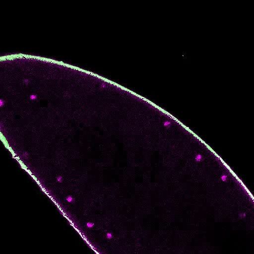
Research article                                                                                       Chromosomes and Gene Expression

Figure 2. Embryos lacking maternal GAF die during early embryogenesis with nuclear and mitotic defects. (A) Images of control (maternal genotype:
His2Av-RFP; sfGFP-GAF(N)) and GAFdeGradFP (maternal genotype: His2Av-RFP/nos-deGradFP; sfGFP-GAF(N)) embryos at NC14, demonstrating loss of
nuclear GFP signal specifically in GAFdeGradFP embryos. His2Av-RFP marks the nuclei. (B) Hatching rates after > 24 hr. (C) Confocal images of His2Av-
RFP in arrested/dying GAFdeGradFP embryos with blocky nuclei, mitotic arrest, and nuclear fallout. (D) Confocal images of His2Av-RFP in NC13 control
and GAFdeGradFP embryos, showing disordered nuclei and anaphase bridges in GAFdeGradFP embryos. Scale bars, 50 mm except where
indicated. See also Figure 2—figure supplement 1.
The online version of this article includes the following figure supplement(s) for figure 2:
Figure supplement 1. sfGFP-GAF(N) and GAF-sfGF(C) are expressed at similar levels in the early embryo.

                                    arresting. Nuclear defects became more pronounced as GAFdeGradFP embryos developed and
                                    approached NC14. The arrested/dead embryos were often arrested in mitosis or had large, irregular
                                    nuclei (Figure 2C), similar to the nuclear ‘supernovas’ in GAF-deficient nuclei reported in Bhat et al.,
                                    1996. Our live imaging allowed us to detect an additional nuclear defect: that in the absence of
                                    maternal GAF nuclei become highly mobile and, in some cases, adopt a ‘swirling’ pattern (Video 2).
                                                                                         While it is unclear what causes in this phenotype,
                                                                                         GAF is suggested to have a role in maintaining
                                                                                         genome stability. Thus, loss of GAF may lead to
                                                                                         activation of the DNA-damage response path-
                                                                                         way, which can result in similar nuclear pheno-
                                                                                         types (Bhat et al., 1996; Sibon et al., 2000;
                                                                                         Takada et al., 2003). As a control, we imaged
                                                                                         sfGFP-GAF(N) homozygous embryos through

                                    Video 1. Video of a GAFdeGradFP embryo going               Video 2. Video of a severely disordered GAFdeGradFP
                                    through several rounds of mitosis prior to gastrulation.   embryo going through several rounds of mitosis prior
                                    Nuclei are marked by His2Av-RFP.                           to gastrulation. Nuclei are marked by His2Av-RFP.
                                    https://elifesciences.org/articles/66668#video1            https://elifesciences.org/articles/66668#video2

Gaskill, Gibson, et al. eLife 2021;10:e66668. DOI: https://doi.org/10.7554/eLife.66668                                                           6 of 30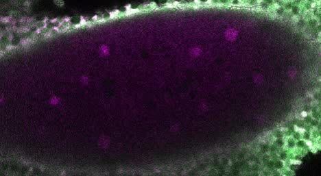
Research article                                                                               Chromosomes and Gene Expression

                                    several rounds of mitosis. These embryos proceeded normally through NC10-14, demonstrating that
                                    the mitotic defects in the GAFdeGradFP embryos were caused by the absence of GAF and not the
                                    sfGFP tag (Figure 2D; Video 3). The defects in GAFdeGradFP embryos are reminiscent of the nuclear-
                                    division defects previously reported for maternal depletion of zld (Liang et al., 2008; Staudt et al.,
                                    2006) and identify a fundamental role for GAF during the MZT.

                                    GAF is required for the activation of hundreds of zygotic genes
                                    During the MZT, there is a dramatic change in the embryonic transcriptome as developmental con-
                                    trol shifts from mother to offspring. Having demonstrated that maternal GAF was essential for devel-
                                    opment during the MZT, we investigated the role of GAF in regulating these transcriptional
                                    changes. We performed total-RNA seq on bulk collections of GAFdeGradFP and control (sfGFP-GAF
                                    (N)) embryos harvested 2–2.5 hr AEL, during the beginning of NC14 when the widespread genome
                                    activation has initiated. Our replicates were reproducible (Figure 3—figure supplement 1), allowing
                                    us to identify 1452 genes that were misexpressed in GAFdeGradFP embryos as compared to controls.
                                    Importantly, by using sfGFP-GAF(N) homozygous embryos as a control we have excluded from our
                                    analysis any genes misexpressed as a result of the sfGFP tag on GAF. Of the misexpressed genes
                                    884 were down-regulated and 568 were up-regulated in the absence of GAF (Figure 3A, Figure 3—
                                    figure supplement 2A). The gene encoding GAF, Trithorax-like, was named because it was required
                                    for expression of the homeotic genes Ubx and Abd-B (Farkas et al., 1994). Our RNA-seq analysis
                                    identified 7 of the 8 Drosophila homeotic genes (Ubx, Abd-B, adb-A, pb, Dfd, Scr, and Antp) down-
                                    regulated in GAFdeGrad embryos. Additionally, many of the gap genes, essential regulators of ante-
                                    rior-posterior patterning, are down-regulated: giant (gt), knirps (kni), huckebein (hkb), Krüppel (Kr),
                                    and tailless (tll) (Supplementary file 2). Gene ontology (GO)-term analysis showed down-regulated
                                    genes were enriched for functions in system development and developmental processes as would
                                    be expected for essential genes activated during ZGA (Figure 3—figure supplement 2B). GO-term
                                    analysis of the up-regulated genes showed weak enrichment for response to stimulus and metabolic
                                    processes (Figure 3—figure supplement 2C).
                                        To determine whether GAF is functioning predominantly in transcriptional activation or repres-
                                    sion, we used our stage 5 ChIP-seq data to determine the likely direct targets of GAF by assigning
                                    GAF-ChIP peaks to the nearest gene. We found that 45% (397) of the down-regulated genes were
                                    proximal to a GAF peak. By contrast, only 17% (99) of up-regulated genes are near GAF peaks, simi-
                                    lar to the 15% of genes with unchanged expression levels that were proximal to GAF sites
                                    (Figure 3A). The significant enrichment for GAF-binding sites proximal to down-regulated genes as
                                    compared to up-regulated supports a role for
                                    GAF specifically in transcriptional activation
                                    (p
Research article Chromosomes and Gene Expression
contributed (pResearch article Chromosomes and Gene Expression Figure 3. GAF is required for zygotic genome activation. (A) Volcano plot of transcripts mis-expressed in GAFdeGradFP embryos as compared to sfGFP- GAF(N) controls. Stage 5 GAF-sfGFP(C) ChIP-seq was used to identify GAF-bound target genes. (B) The percentage of up-regulated and down- regulated transcripts in GAFdeGradFP embryos classified as maternal, zygotic, or maternal-zygotic based on Lott et al., 2011. (C) Overlap of down- regulated embryonic transcripts in the absence of GAF or Zld activity (p
Research article                                                                                  Chromosomes and Gene Expression

Figure 4. At the majority of loci, GAF and Zld bind chromatin independently. (A) Overlap of Zld- and GAF-binding sites determined by GAF-sfGFP(C)
stage 5 ChIP-seq and Zld NC14 ChIP-seq (Harrison et al., 2011). (B) Representative genome browser tracks of Zld and GAF ChIP-seq peaks at the
tailless locus. (C) Immunoblot for Zld on embryo extracts from zld-RNAi; sfGFP-GAF(N) and sfGFP-GAF(N) control embryos harvested 2–3 hr AEL.
Tubulin was used as a loading control. (D) Images of zld-RNAi; sfGFP-GAF(N) embryos and sfGFP-GAF(N) control embryos at NC10 and NC14 as
marked. Arrowhead shows nuclear dropout, a phenotype indicative of zld loss-of-function. Scale bar, 10 mm. (E) Correlation between log2(RPKM) of
ChIP peaks for GAF from GAF-sfGFP(N) stage 5 embryos (control) and zld-RNAi; sfGFP-GAF(N) embryos (zld-RNAi). Color highlights significantly
changed peaks (adjusted p-value 2) and those that are bound by both GAF and Zld as indicated below. (F) Correlation between
log2(RPKM) of ChIP peaks for Zld from sfGFP-GAF(N) embryos (control) and GAFdeGradFP embryos fixed 2–2.5 hr AEL. Color highlights significantly
changed peaks (adjusted p-value 2) and those that are bound by both GAF and Zld as indicated below. See also Figure 4—figure
supplements 1–2.
The online version of this article includes the following figure supplement(s) for figure 4:
Figure supplement 1. GAF and Zld bind to shared and unique regions of the genome.
Figure supplement 2. Independent chromatin binding by GAF and Zld.

Gaskill, Gibson, et al. eLife 2021;10:e66668. DOI: https://doi.org/10.7554/eLife.66668                                                   10 of 30
Research article                                                                              Chromosomes and Gene Expression

                                    Yamada et al., 2019). Indeed, 19% of Dorsal sites depend on Zld for occupancy (Sun et al., 2015).
                                    We therefore examined GAF localization upon depletion of maternal zld using RNAi driven in the
                                    maternal germline by mata-GAL4-VP16 in a background containing sfGFP-GAF(N) (Sun et al.,
                                    2015). Immunoblot confirmed a nearly complete knockdown of Zld (Figure 4C), and we verified that
                                    RNAi-treated embryos failed to hatch. To assess the depletion of Zld on the subnuclear localization
                                    of GAF, we imaged sfGFP-GAF(N); zld-RNAi and control (sfGFP-GAF(N)) embryos using identical
                                    acquisition settings. We observed no difference in puncta formation of GAF at the beginning of
                                    ZGA (NC10) or late ZGA (NC14) (Figure 4D). We conclude that Zld is not required for GAF to form
                                    subnuclear puncta during the MZT.
                                        To more specifically determine the impact of loss of Zld on GAF chromatin occupancy, we per-
                                    formed ChIP-seq with an anti-GFP antibody on embryos expressing zld-RNAi and sfGFP-GAF(N)
                                    along with paired sfGFP-GAF(N) controls at 2–2.5 hr AEL (stage 5). Mouse H3.3-GFP chromatin was
                                    used as a spike-in to normalize for immunoprecipitation efficiency between samples. In control
                                    sfGFP-GAF(N) embryos, we identified 6373 peaks, and these largely overlapped with the peaks
                                    identified in the GAF-sfGFP(C) embryos; 91% of the 4175 peaks identified in the GAF-sfGFP(C)
                                    embryos overlap with the peaks identified for the N-terminally tagged GAF (Figure 4—figure sup-
                                    plement 1B). This high degree of overlap indicates that our ChIP-seq experiments identified a
                                    robust set of high-confidence GAF-bound regions. To determine whether GAF requires Zld for bind-
                                    ing, we analyzed GAF occupancy upon zld knockdown at this set of 3800 high-confidence peaks.
                                    The majority (3674) of these high-confidence GAF peaks were maintained in the zld-RNAi back-
                                    ground and were bound at roughly equivalent levels when normalized to the spike-in control
                                    (Figure 4E and Figure 4—figure supplement 2A). Using DESeq2, we identified 126 GAF-bound
                                    regions that were significantly different between the sfGFP-GAF(N) controls and the zld-RNAi
                                    embryos (Figure 4E): 101 sites that were decreased and 25 sites that were increased.
                                        To assess the impact of the loss of GAF on Zld binding, we completed the reciprocal experiment
                                    and performed ChIP-seq for Zld on GAFdeGradFP and control homozygous (sfGFP-GAF(N)) embryos
                                    at 2–2.5 hr AEL (stage 5). We identified a set of high-confidence Zld peaks, by overlapping the Zld
                                    peaks from our control (sfGFP-GAF(N)) embryos with previously published data for Zld at NC14
                                    (Harrison et al., 2011). Similar to GAF binding when Zld is depleted, the majority of the 6003 high-
                                    confidence Zld peaks were maintained when GAF was depleted (Figure 4F). However, in the GAFde-
                                    GradFP
                                            ChIP data we observed a global reduction in Zld peak heights as compared to the control
                                    (Figure 4—figure supplement 2B). Therefore, it is possible that GAF knockdown causes a global
                                    reduction in Zld binding. However, technical, rather than biological differences, may account for this
                                    overall decrease (see Materials and methods). Among the high-confidence Zld peaks, DESeq2 identi-
                                    fied 105 Zld-binding sites that differed in occupancy between GAFdeGradFP embryos and controls: 86
                                    sites decreased and 19 sites increased.
                                        Because of the limited number of significantly different peaks identified upon removal of either
                                    Zld (3.3% of GAF peaks changed) or GAF (1.7% of Zld peaks changed), we sought to determine
                                    whether there were global changes in the distribution of these factors. For this purpose, we ranked
                                    peaks in each dataset based on the number of reads per kilobase per million mapped reads (RPKM)
                                    and determined if the peak ranks were correlated between the mutant and control. We identified a
                                    high degree of correlation in peak rank for both comparisons (Pearson correlation, r = 0.93 for GAF
                                    ChIP in sfGFP-GAF(N) and sfGFP-GAF(N); zld-RNAi embryos and r = 0.88 for Zld ChIP in sfGFP-GAF
                                    (N) and GAFdeGradFP embryos; Figure 4—figure supplement 2C,D). Thus, the overall distribution of
                                    binding sites is maintained for each factor in the absence of the other.
                                        At some loci, pioneer factors have been shown to function together to stabilize binding
                                    (Donaghey et al., 2018; Chronis et al., 2017). If either GAF or Zld stabilized genomic occupancy of
                                    the other factor, we would expect to identify a loss of binding specifically at loci that are co-bound
                                    by both factors. We identified a set of GAF and Zld co-occupied sites by overlapping our high-confi-
                                    dence GAF peaks and high-confidence Zld peaks. Peaks with at least 100 bp overlap were consid-
                                    ered to be shared and used for our set of co-bound regions. To test likely direct effects of GAF on
                                    Zld occupancy, we determined the number regions that showed significant changes in Zld binding in
                                    GAFdeGradFP embryos that were bound by GAF. One out of the 86 regions at which Zld binding
                                    decreased in the GAF knockdown and 5 out of 19 that increased overlapped with regions bound by
                                    GAF (Figure 4F). Contrary to our expectation if GAF was directly promoting Zld occupancy, there
                                    was no enrichment for co-bound regions and those that lost Zld binding. We investigated the

Gaskill, Gibson, et al. eLife 2021;10:e66668. DOI: https://doi.org/10.7554/eLife.66668                                            11 of 30
Research article Chromosomes and Gene Expression
reciprocal effect of Zld on GAF occupancy by determining the number regions that showed signifi-
cant changes in GAF binding in zld-RNAi embryos that were also bound by Zld. Fifty-one of the 101
regions that had decreased GAF binding were also bound by Zld, suggesting a potential direct
effect of Zld on GAF occupancy at these regions. By contrast, only 6 of the 25 regions that had
increased GAF binding were also bound by Zld (Figure 4E). GAF-binding regions that were
decreased in the absence of Zld were significantly enriched for co-bound sites as compared to those
GAF-binding sites that were maintained upon Zld depletion (p=4.810 5, log2(odds ratio)=1.19,
two-tailed Fisher’s exact test). Based on the enrichment for Zld binding at these GAF-bound
decreased sites, we hypothesized that Zld might be pioneering chromatin accessibility at these
regions to promote GAF binding. Indeed, these regions were enriched for loci that depend on Zld
for chromatin accessibility (p=2.210 16, log2(odds ratio)=4.07, two-tailed Fisher’s exact test): 38 of
the 51 GAF and Zld co-bound sites at which GAF requires Zld for occupancy also require Zld for
accessibility (Hannon et al., 2017). Thus, at this limited set of regions the pioneer factor Zld may
function to establish accessible chromatin that is necessary to promote robust GAF binding. None-
theless, the majority of Zld- and GAF-binding sites are occupied in the absence of the other factor.
GAF is essential for accessibility at hundreds of loci during ZGA
GAF interacts with chromatin remodelers and is required to maintain chromatin accessibility in tissue
culture (Okada and Hirose, 1998; Tsukiyama et al., 1994; Tsukiyama and Wu, 1995; Xiao et al.,
2001; Judd et al., 2021; Fuda et al., 2015). Furthermore, GAF-binding motifs are enriched at
regions of open chromatin that are established at NC12 and NC13 and that are bound by Zld but
do not depend on Zld for accessibility (Schulz et al., 2015; Sun et al., 2015; Blythe and Wieschaus,
2016). To directly test the function of GAF in determining accessible chromatin domains during the
MZT, we performed the assay for transposase-accessible chromatin (ATAC)-seq on six replicates of
single GAFdeGradFP and control (sfGFP-GAF(N)) embryos harvested 2–2.5 hr AEL. His2AV-RFP signal
for each embryo was visually inspected prior to performing ATAC-seq to ensure that embryos did
not have grossly distorted nuclear morphology. In contrast to our control, replicates from the GAFde-
GradFP
embryos showed higher variability (Figure 5—figure supplement 1A), suggesting that devel-
opmental defects caused by the lack of GAF might have lowered our ability to detect subtle
changes in accessibility. Nonetheless, we identified 1523 regions with significant changes in accessi-
bility in GAFdeGradFP embryos as compared to controls; 607 regions lost accessibility and 916 regions
gained accessibility (Figure 5A, Figure 5—figure supplement 1B,C). Sites that lost accessibility
were among the regions with the highest ATAC-signal (Figure 5—figure supplement 1C). 32%
(197) of the regions that lost accessibility were significantly enriched for GAF binding, as determined
by stage 5 GAF-sfGFP(C) ChIP-seq, when compared to open regions with no change in accessibility
(p=2.210 16, log2(odds ratio)=2.4, two-tailed Fisher’s exact test, Figure 5—figure supplement
1D). The enrichment for GAF-binding sites in those regions that depend on GAF for accessibility
suggests that GAF may directly drive accessibility at these regions. Therefore, we focused our analy-
sis on the subset of regions that depend on GAF for accessibility, which we define as those that lost
accessibility in the absence of GAF. Consistent with the enrichment of GAF-binding sites in pro-
moters, 45% of all regions that depend on GAF for accessibility were in promoters. Enrichment for
promoters was even larger in those regions that were bound by GAF and dependent on GAF for
accessibility (Figure 5B). By contrast, regions that gain accessibility in the absence of GAF are not
enriched for promoters, supporting that this is likely an indirect effect of GAF knockdown
(Figure 5B). Ubx, a previously identified embryonic GAF-target gene, is an example of a gene that
requires GAF for chromatin accessibility at the promoter (Figure 5C; Farkas et al., 1994). Ubx simi-
larly requires GAF occupancy for gene expression as the transcript was significantly down-regulated
in GAFdeGradFP embryos (Figure 5C). Thus, GAF binding at the Ubx promoter is required for both
chromatin accessibility and gene expression. Together, our data demonstrate that GAF is required
for chromatin accessibility at hundreds of loci during the MZT, and that this activity occurs preferen-
tially at promoters.
Gaskill, Gibson, et al. eLife 2021;10:e66668. DOI: https://doi.org/10.7554/eLife.66668 12 of 30Research article Chromosomes and Gene Expression
Figure 5. GAF is required for chromatin accessibility. (A) Volcano plot of regions that change in accessibility in GAFdeGradFP embryos as compared to
sfGFP-GAF(N) controls, stage 5 GAF-sfGFP(C) ChIP-seq was used to identify GAF-bound target regions. (B) Genomic distribution of all regions that lose
accessibility (Lose accessibility (all)), regions that lose accessibility and are GAF-bound (Lose accessibility (GAF-bound), regions that did not change
significantly in accessibility (Non-significant), and regions that gain accessibility (Gain accessibility)). (C) Genome browser tracks of GAF-sfGFP(C) ChIP-
seq at stage 3 and stage 5, ATAC-seq on wild-type embryos at NC12, NC13, and stage 5 along with control (sfGFP-GAF(N)) and GAFdeGradFP embryos,
and RNA-seq from control (sfGFP-GAF(N)) and GAFdeGradFP embryos. NC12 and NC13 ATAC-seq data is from Blythe and Wieschaus, 2016. ATAC-seq
data for stage 5 embryos is from Nevil et al., 2020. Region highlighted in green indicates the GAF-dependent, GAF-bound Ubx promoter. See also
Figure 5—figure supplement 1.
The online version of this article includes the following figure supplement(s) for figure 5:
Figure supplement 1. GAF is required for chromatin accessibility.
GAF and Zld are individually required for chromatin accessibility at
distinct regions
Previous work from our lab and others suggested that during the MZT, GAF may be responsible for
maintaining chromatin accessibility at Zld-bound regions in the absence of Zld (Schulz et al., 2015;
Moshe and Kaplan, 2017; Sun et al., 2015). To more broadly investigate how GAF and Zld shape
chromatin accessibility during early development, we focused on regions co-occupied by both fac-
tors as identified by ChIP-seq (this work and Harrison et al., 2011). We compared our single embryo
ATAC-seq data to ATAC-seq data for wild-type NC14 embryos and NC14 embryos lacking maternal
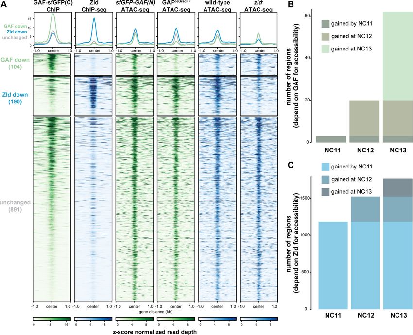
zld (zld-) (Figure 6A; Hannon et al., 2017). Only seven of the 1192 regions co-bound by both Zld
and GAF decreased in accessibility upon removal of either factor. By contrast, 104 regions require
GAF for accessibility and 190 require Zld. Regions that require GAF for accessibility had a higher
average GAF ChIP-seq peak height than regions that require Zld. Similarly, the Zld ChIP-seq signal
was higher in regions where Zld is necessary for accessibility. Thus, both GAF and Zld are individually
required for accessibility at distinct genomic regions co-occupied by both factors, and this require-
ment is correlated with occupancy as reflected in ChIP-seq peak height (Figure 6A). GAF is also
required for accessibility at 83 additional regions at which GAF is bound without Zld (Figure 6—fig-
ure supplement 1). The majority of sites bound by both Zld and GAF (891) did not change in chro-
matin accessibility when either factor was removed, suggesting that at these locations GAF and Zld
may function redundantly to facilitate chromatin accessibility or that other factors were sufficient to
maintain accessibility at these sites. Thus, during the MZT GAF and Zld are individually required for
Gaskill, Gibson, et al. eLife 2021;10:e66668. DOI: https://doi.org/10.7554/eLife.66668 13 of 30Research article Chromosomes and Gene Expression
Figure 6. GAF and Zld independently shape chromatin accessibility over the MZT. (A) Heatmaps of ChIP-seq and ATAC-seq data, as indicated above,
for regions bound by both GAF and Zld and subdivided based on the change of accessibility in the absence of either factor. (B) Number of regions that
depend on GAF for accessibility and are bound by GAF that are accessible at NC11, NC12, and NC13. (C) Number of regions that depend on Zld for
accessibility and are bound by Zld that are accessible at NC11, NC12, and NC13. NC11, NC12, and NC13 data are from Blythe and Wieschaus, 2016.
Zld ChIP-seq data are from Harrison et al., 2011. ATAC-seq data from zld germline clones (zld-) are from Hannon et al., 2017. Total number of
regions accessible at NC11 = 3084, NC12 = 6487, and NC13 = 9824. See also Figure 6—figure supplements 1–2.
The online version of this article includes the following figure supplement(s) for figure 6:
Figure supplement 1. A subset of regions bound by GAF, and not Zld, depend on GAF for accessibility.
Figure supplement 2. Regions that gain accessibility late during the MZT are accessible in GAFdeGradFP embryos used for ATAC-seq.
chromatin accessibility at distinct co-bound regions. At very few co-bound regions are both factors
individually required.
Previous work demonstrated that the Zld-binding motif is enriched at accessible regions that are
established by NC11. By contrast, GAF-binding motifs are enriched at regions that dynamically gain
chromatin accessibility later during the MZT at NC12 or NC13 (Blythe and Wieschaus, 2016). If Zld
preferentially drives chromatin accessibility early in the MZT and GAF is preferentially required later,
we would expect that regions that require GAF for accessibility would be enriched for regions that
Gaskill, Gibson, et al. eLife 2021;10:e66668. DOI: https://doi.org/10.7554/eLife.66668 14 of 30Research article Chromosomes and Gene Expression
gain accessibility at NC12 and NC13. To test this prediction, we compared our ATAC-seq data to
ATAC-seq on embryos precisely staged by nuclear cycle (Blythe and Wieschaus, 2016). Of the
GAF-bound, GAF dependent accessible regions that were identified in the staged ATAC-seq data,
three were open by NC11, 17 were newly opened at NC12, and 42 were newly opened at NC13
(Figure 6B, Figure 6—figure supplement 2A), demonstrating that GAF-bound, GAF-dependent
regions are enriched for regions that dynamically open at NC12 or NC13 as compared to regions
that are already accessible at NC11 (p=5.710 7, log2(odds ratio)=3.2, two-tailed Fisher’s exact
test). Indeed, the Ubx promoter dynamically gained accessibility at NC13 (Figure 5C). Confirming
that the loss of accessibility observed at late-opening regions was not due to differences in staging
between the control (sfGFP-GAF(N)) and GAFdeGradFP embryos, the vast majority of sites that gained
accessibility at NC13 (2452) were unchanged in accessibility in the GAFdeGradFP embryos (Figure 6—
figure supplement 2). In contrast to GAF, Zld-bound, Zld-dependent accessible regions were
enriched for regions already accessible at NC11: 1182 were open by NC11, 343 were newly opened
at NC12, and 244 were newly opened at NC13 (pResearch article Chromosomes and Gene Expression
Figure 7. Zld and GAF independently regulate embryonic reprogramming. (A) During the minor wave of ZGA (NC10-13), Zld is the predominant factor
required for driving expression of genes bound by Zld alone and genes bound by both Zld and GAF. (B) As the genome is more broadly activated
during NC14, GAF becomes the major factor in driving zygotic transcription. (C) Early in the MZT, Zld is required for chromatin accessibility at many
regions co-bound by GAF and Zld. When zld is depleted, accessibility is lost at a subset of regions, but GAF remains bound at the majority of sites. (D)
Late during the MZT, GAF is required for chromatin accessibility at many Zld-bound regions. In GAFdeGradFP embryos accessibility is lost at a subset of
sites, but Zld remains bound.
misregulation of previously identified embryonic GAF-targets in the GAFdeGradFP embryos demon-
strate an essential function for GAF in the early embryo.
Live imaging of GFP-tagged, endogenously encoded GAF demonstrated that GAF is mitotically
retained in small foci. This is similar to what was reported for antibody staining on fixed embryos,
which showed GAF localized at pericentric heterochromatin regions of GA-rich satellite repeats
(Raff et al., 1994; Platero et al., 1998). Because this prior imaging necessitated fixing embryos, it
was unclear if mitotic retention of GAF was required for mitosis. Our system enabled us to deter-
mine that in the absence of GAF nuclei can undergo several rounds of mitosis albeit with noticeable
defects (Videos 1 and 2). We conclude that GAF is not strictly required for progression through
mitosis. However, the nuclear defects observed in GAFdeGradFP embryos support the model that
GAF is broadly required for nuclear division and chromosome stability, in addition to its role in tran-
scriptional activation during ZGA (Bhat et al., 1996). Our imaging also identified high nuclear mobil-
ity in GAFdeGradFP embryos as compared to control embryos; nuclei of a subset of embryos showed
a dramatic ‘swirling’ pattern of movement (Video 2). This defect is potentially due a DNA damage
response that inactivates centrosomes to promote nuclear fallout (Sibon et al., 2000; Takada et al.,
2003), or general disorder in the cytoskeletal network that is responsible for nuclear migration and
division in the syncytial embryo (Sullivan and Theurkauf, 1995). Altogether, our phenotypic analysis
of GAFdeGradFP embryos shows for the first time that maternal GAF is required for progression
Gaskill, Gibson, et al. eLife 2021;10:e66668. DOI: https://doi.org/10.7554/eLife.66668 16 of 30Research article Chromosomes and Gene Expression
through the MZT, and suggests GAF has an early, global role in nuclear division and chromosome
stability.
In addition to GAF and Zld, the transcription factor CLAMP is expressed in the early embryo and
functions in chromatin accessibility and transcriptional activation (Rieder et al., 2017; Soruco et al.,
2013; Urban et al., 2017a; Urban et al., 2017b; Rieder et al., 2019). While CLAMP, like GAF,
binds GA-dinucleotide repeats, the two proteins preferentially bind to slightly different GA-repeats
(Kaye et al., 2018). GAF and CLAMP can compete for binding sites in vitro, and, in cell culture,
when one factor is knocked down the occupancy of the other increases, suggesting that CLAMP and
GAF compete for a subset of binding sites and may have partial functional redundancy (Kaye et al.,
2018). We demonstrate that GAF, like CLAMP, is essential in the early embryo, indicating that these
two GA-dinucleotide-binding proteins cannot completely compensate for each other in vivo during
early development (Rieder et al., 2017). Furthermore, our sequencing analysis identified that a
majority of genes that require GAF for expression are distinct from those that are regulated by
CLAMP. Thus, while GAF and CLAMP may have some overlapping functions, they are independently
required to regulate embryonic development during the MZT.
GAF is necessary for widespread zygotic genome activation and
chromatin accessibility
GAF is a multi-purpose transcription factor with known roles in transcriptional regulation at pro-
moters and enhancers as well as additional suggested roles in high-order chromatin structure. Our
analysis showed that during the MZT GAF acts largely as an activator, directly binding and activating
hundreds of zygotic transcripts during ZGA (Figure 3). We identified thousands of regions bound by
GAF throughout the MZT, and these regions were preferentially associated with genes whose tran-
scription decreased when maternally encoded GAF was degraded. This function may be driven, in
part, through GAF-mediated chromatin accessibility as we identified hundreds of regions that
depend on GAF for accessibility (Figure 5). This activity, in both transcriptional activation and medi-
ating open chromatin, is similar to Zld, the only previously identified essential activator of the zygotic
genome in Drosophila, and is shared with genome activators in other species, such as Pou5f3 and
Nanog (zebrafish) and Dux (mammals) (Schulz and Harrison, 2019; Vastenhouw et al., 2019). Our
data support a pioneer-factor like role for GAF in this process as we demonstrated that GAF is
already bound early in the MZT to regions that require GAF for accessibility later. Thus, GAF occu-
pancy precedes GAF-mediated accessibility at a subset of sites.
In addition to a direct role in mediating chromatin accessibility through the recruitment of chro-
matin remodelers (Okada and Hirose, 1998; Tsukiyama et al., 1994; Tsukiyama and Wu, 1995;
Xiao et al., 2001; Judd et al., 2021; Fuda et al., 2015), GAF may also indirectly affect chromatin
accessibility through a role in shaping three-dimensional chromatin structure. Sixty-seven percent of
the regions that lost chromatin accessibility in the absence of GAF did not overlap a GAF-binding
site as identified by ChIP-seq. Thus, at these regions GAF may function indirectly through the ability
to facilitate enhancer-promoter loops (Mahmoudi et al., 2002; Melnikova et al., 2004;
Petrascheck et al., 2005). In addition, GAF-binding motifs are enriched at TAD boundaries which
form during the MZT (Hug et al., 2017), and GAF binding is enriched at Polycomb group dependent
repressive loops that form following NC14 (Ogiyama et al., 2018). Further investigation is necessary
to determine the role of GAF in establishing three-dimensional chromatin architecture in the early
embryo.
GAF and Zld are uniquely required to reprogram the zygotic genome
Reprogramming in culture requires a cocktail of transcription factors that possess pioneer factor
activity (Takahashi and Yamanaka, 2006; Soufi et al., 2012; Soufi et al., 2015). Similarly, in zebra-
fish and mice multiple pioneering factors are required for the rapid and efficient reprogramming
that occurs during the MZT (Schulz and Harrison, 2019; Vastenhouw et al., 2019). Here we have
shown that, in addition to the essential pioneer factor Zld, GAF is a pioneer-like factor required for
both gene expression and chromatin accessibility in the early Drosophila embryo. By analyzing the
individual contributions of these two factors, we have begun to elucidate the different mechanisms
by which multiple pioneer factors can drive dramatic changes in cell identity.
Gaskill, Gibson, et al. eLife 2021;10:e66668. DOI: https://doi.org/10.7554/eLife.66668 17 of 30Research article Chromosomes and Gene Expression
While some pioneer factors work together to stabilize their interaction on chromatin
(Chronis et al., 2017; Donaghey et al., 2018; Liu and Kraus, 2017; Swinstead et al., 2016) our
data support primarily independent genome occupancy by Zld and GAF. Our reciprocal ChIP-seq
data demonstrated that at regions bound by GAF and Zld, binding of each factor was largely
retained in the absence of the other (Figure 4). Analysis of chromatin accessibility further supports
these independent roles. We would predict that if Zld binding were lost in the absence of GAF or
vice versa that at regions occupied by both factors either Zld or GAF individually would be required
for accessibility. However, we identified only seven regions that lost accessibility in the absence of
either Zld or GAF. By contrast, we identified more than one hundred regions that were individually
dependent on each factor alone, and 891 regions that did not change in accessibility upon loss of
either Zld or GAF. These data support independent roles for Zld and GAF in establishing or main-
taining accessibility. Furthermore, we propose that because each factor can retain genome occu-
pancy in the absence of the other that this may explain the 891 regions that remain accessible in the
absence of either Zld or GAF: in the absence of Zld, GAF may be able to maintain accessibility and
vice versa. However, until we investigate the chromatin landscape upon the removal of both Zld and
GAF we cannot rule out that at these regions other factors may be instrumental in maintaining acces-
sibility. Indeed, Duan et al. identify an essential role for the GA-dinucleotide-binding protein CLAMP
in directing Zld binding and promoting chromatin accessibility (Duan et al., 2020).
At most loci, our data support independent binding for GAF and Zld. Nonetheless, we cannot
eliminate a possible global role for GAF on Zld occupancy as the Zld ChIP-seq signal is considerably
lower in the GAFdeGradFP embryos as compared to controls. We would predict that if GAF is directly
functioning to stabilize Zld binding then this effect would be more evident at regions where both
GAF and Zld bind, but this is not what we observed. Nonetheless, because of the proposed role of
GAF in regulating three-dimensional chromatin architecture, GAF may be globally required for
robust Zld occupancy. Furthermore, at a small subset of loci our data support a pioneering role for
Zld in promoting GAF binding. In this experiment, the endogenously tagged GAF allowed us to use
antibodies directed against the GFP epitope for ChIP. The use of the epitope tag enabled us to
robustly control for immunoprecipitation efficiency by using another GFP-tagged protein as a spike-
in control for the immunoprecipitation of GAF. In this manner, we identified 101 GAF-bound regions
that decreased upon Zld knockdown. Of these, 51 were Zld-bound regions and 38 of these depend
on Zld for chromatin accessibility. Thus, at a small subset of sites the pioneering function of Zld may
be required to stabilize GAF binding.
We propose that there is a handoff between Zld and GAF pioneer-like activity as the embryo pro-
gresses through the MZT: Zld functions primarily at the initiation of zygotic genome activation and
GAF functions primarily later during the major wave of zygotic transcription. We identified that
genes regulated by Zld are enriched for those activated during NC10-13, while genes that depend
on GAF are enriched for those that initiate expression during NC14. Similarly, we determined that
the majority of regions that require Zld for accessibility are already accessible at NC11 while those
that require GAF are enriched for regions that gain accessibility at NC12 and NC13. This is sup-
ported by prior analysis that showed that the Zld-binding motif is enriched at regions that are
already accessible at NC11 and the GAF-binding motif is enriched at regions that dynamically gain
accessibility later during the MZT (Blythe and Wieschaus, 2016). Based on this evidence, we pro-
pose that there is a gradual handoff in the control of transcriptional activation and chromatin remod-
eling from Zld to GAF as the MZT progresses (Figure 7). During the initial stages of the MZT, the
nuclear division cycle is an incredibly rapid series of synthesis and mitotic phases. At NC14, this cycle
slows, and these different dynamics likely influence the mechanisms by which accessibility can be
established. While it is unclear how Zld establishes accessibility, GAF interacts with chromatin
remodelers. It is possible that the activities of these complexes may have a more substantial impact
once the division cycle slows.
Together our data support the requirement for at least two pioneer-like transcription factors, Zld
and GAF, to sequentially reprogram the zygotic genome following fertilization and allow for future
embryonic development. It is likely that there are additional factors that function along with Zld and
GAF to define accessible cis-regulatory regions and drive genome activation. Future studies will
enable more detailed mechanistic insights into how multiple pioneering factors work together to
reshape the transcriptional landscape and transform cell fate in the early embryo.
Gaskill, Gibson, et al. eLife 2021;10:e66668. DOI: https://doi.org/10.7554/eLife.66668 18 of 30You can also read