Gaining Back What Is Lost: Recovering the Sense of Smell in Mild to Moderate Patients After COVID-19
←
→
Page content transcription
If your browser does not render page correctly, please read the page content below

Chemical Senses, 2020, Vol XX, 1–7
                                                                                                doi:10.1093/chemse/bjaa066
                                                                                                             Original Article
                                                                                   Advance Access publication 9 October 2020

Original Article

Gaining Back What Is Lost: Recovering the

                                                                                                                                    Downloaded from https://academic.oup.com/chemse/advance-article/doi/10.1093/chemse/bjaa066/5919785 by guest on 04 November 2020
Sense of Smell in Mild to Moderate Patients
After COVID-19
Lucia Iannuzzi1,*, Anna Eugenia Salzo1,*, Gioacchino Angarano2,
Vincenzo Ostilio Palmieri3, Piero Portincasa3, Annalisa Saracino2,
Matteo Gelardi4, Michele Dibattista5 and Nicola Quaranta1
1
 ENT Clinic, Department of Biomedical Sciences, Neurosciences and Sense Organs, University of Bari, Bari, Italy,
2
 Clinic of Infectious Diseases, Department of Biomedical sciences and Human Oncology, University of Bari, Bari,
Italy, 3Clinica Medica “A. Murri,” Department of Biomedical sciences and Human Oncology, University of Bari, Bari,
Italy, 4ENT Clinic, University of Foggia, Foggia, Italy and 5Department of Basic Medical Sciences, Neuroscience and
Sense Organs, University of Bari A. Moro, Bari, Italy

Correspondence to be sent to: Michele Dibattista, Department of Basic Medical Sciences, Neuroscience and Sense
Organs, University of Bari A. Moro, Piazza Giulio Cesare n.11, 70125 Bari, Italy. e-mail: michele.dibattista@uniba.it

*These authors have equally contributed to this work and should be regarded as joint first authors.

Editorial Decision 2 October 2020.

Abstract
The purpose of our cohort study was to quantify olfactory deficits in Coronavirus disease 2019
(COVID-19) patients using Sniffin’ Sticks and a pre-post design to evaluate olfactory recovery. Thirty
adult patients with laboratory-confirmed mild to moderate forms of COVID-19 underwent a quan-
titative olfactory test performed with the Sniffin’ Sticks test (SST; Burghardt, Wedel, Germany),
considering olfactory threshold (T), odor discrimination (D), and odor identification (I). Results
were presented as a composite TDI score (range 1–48) that used to define functional anosmia (TDI
≤ 16.5), hyposmia (16.5 < TDI < 30.5), or functionally normal ability to smell (TDI ≥ 30.5). Patients
also self-evaluated their olfactory function by rating their ability to smell on a visual analogue scale
(Visual Analog Scale rating) and answering a validated Italian questionnaire (Hyposmia Rating
Scale). Patients were tested during hospitalization and about 2 months after symptoms onset.
During the hospitalization, the overall TDI score indicated that our cohort had impairments in their
olfactory ability (10% was diagnosed with anosmia and more than 50% were hyposmic). Almost all
patients showed a significant improvement at around 1 month following the first test and for all
the parts of the SST except for odor identification. None of the subjects at 1 month was still diag-
nosed with anosmia. We also quantified the improvement in the TDI score based on initial diag-
nosis. Anosmic subjects showed a greater improvement than hyposmic and normosmic subjects.
In conclusion, within a month time window and 2 months after symptoms’ onset, in our cohort of
patients we observed a substantial improvement in the olfactory abilities.

Key words COVID-19, olfactory deficits, olfactory test, Sniffin’ Sticks test—SST

© The Author(s) 2020. Published by Oxford University Press. All rights reserved.
                                                                                                                                1
For permissions, please e-mail: journals.permissions@oup.com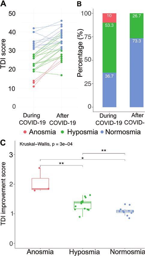
2                                                                                                  Chemical Senses, 2020, Vol. XX, No. XX

Introduction                                                             Table 1. Demographic and anamnestic characteristics of patients

The novel Coronavirus disease 2019 (COVID-19) has grown to                                                                            Number of
be a global public-health emergency since patients were first de-                                                                     patients (%)
tected in Wuhan, China, in December 2019. The disease burden
                                                                         Sample size                                                       30
of COVID-19 caused by Severe Acute Respiratory Syndrome
                                                                         Age (mean ± SD)                                              47.47 ± 13
Coronavirus-2 (SARS-CoV-2) has been increasing continuously
                                                                         Gender                                                       16 F (53.3)
with more than 2 676 300 confirmed cases as of 6 September                                                                            14 M (46.7)
2020 (World Health Organization n.d.). SARS-CoV-2 typically              Current smoker                                                  2 (6.7)

                                                                                                                                                       Downloaded from https://academic.oup.com/chemse/advance-article/doi/10.1093/chemse/bjaa066/5919785 by guest on 04 November 2020
causes common cold symptoms with a wide range of clinical                Ex-smoker                                                      7 (23.3)
manifestations, but can also cause severe pneumonia, respiratory         Never a smoker                                                 21 (70)
failure, and death (Gandhi et al. 2020). Presently, many reports         Hypertension                                                   7 (23.3)
have summarized the clinical features of patients infected with          Thyroid related                                                 3 (10)
SARS-CoV-2, revealing that most patients developed smell and             Diabetes                                                        2 (6.7)
taste alterations, namely ageusia and anosmia (Hopkins et al.            Neoplastic diseases                                             3 (10)
                                                                         Previous pulmonary embolism                                     1 (3.3)
2020; Paderno et al. 2020). Total or partial loss of olfactory per-
                                                                         Fibromyalgia                                                    1 (3.3)
ception has been proposed as an early marker of SARS-CoV-2
                                                                         Polycystic ovary                                                1 (3.3)
infection (Gerkin et al. 2020; Moein, Hashemian, Mansourafshar,          Allergy                                                         9 (30)
et al. 2020).
    Interestingly, chemosensory dysfunction is not associated with
nasal symptoms such as rhinorrhea and nasal obstruction (Mercante        Table 2. Otolaryngology, flu-like, and neurological symptoms
et al. 2020; Parma et al. 2020) and may be caused by different and
                                                                                                                                      Number of
yet unidentified factors. Mechanisms behind olfactory loss could in-
                                                                                                                                      patients (%)
clude the “cytokine storm” or the direct damage of the olfactory epi-
thelium expressing host receptors required for efficient SARS-CoV-2      Flu-like symptoms                                            30 (100)
infection in humans (Butowt and Bilinska 2020; Cooper et al. 2020).      Nasal obstruction                                             7 (23.3)
    Recent investigations about smell and taste dysfunction in           Epistaxis                                                     4 (13.3)
COVID-19 patients, even if conducted on large cohorts of subjects,       Nasal discharge                                               5 (16.7)
are mostly based on interviews and surveys (Giacomelli et al. 2020;      Neurological symptoms                                         9 (30)
Mercante et al. 2020; Menni et al. 2020; Parma et al. 2020). Using         Headache                                                    8 (26.7)a
                                                                           Nausea                                                      2 (6.7)a
objective evaluation methods, fewer studies reported a recovery of
                                                                           Dizziness                                                   1 (3.3)a
olfactory abilities after recovery from SARS-CoV-2 infection. Some
authors reported, using surveys, an early smell recovery. For example,
                                                                           a
                                                                            Symptoms are not mutually exclusive.
Lechien, Chiesa-Estomba, et al. (2020), in a study on 417 COVID-19
patients, 25% reported smell recovery 2 weeks after disease reso-
                                                                         of the study as well patients needing invasive or noninvasive venti-
lution, Lee et al. (2020) described complete recovery within 3 weeks
                                                                         lation were excluded.
with a median time of improvement of 7 days.
                                                                             During hospitalization, an average of 25 days after COVID-19 diag-
    The purpose of our study was to quantify olfactory deficits in
                                                                         nosis, we collected an accurate medical history and performed quan-
COVID-19 patients using Sniffin’ Sticks and to study the recovery of
                                                                         titative olfactory testing using the Sniffin’ Sticks test (SST) (Burghardt,
olfactory impairments.
                                                                         Wedel, Germany) (Hummel et al. 2007; Oleszkiewicz et al. 2019).
                                                                             Patient follow-ups (after COVID-19), using the same test, were
                                                                         carried out a month after the first evaluation, about 2 months after
Subjects and Methods                                                     symptom onset. The study was approved by the Ethics Committee
Participants                                                             of “Azienda Ospedaliero Universitaria Policlinico of Bari,” Italy
The study included 34 adult patients admitted to the COVID-              (n.623/2020).
Hospital of the Policlinico Hospital of Bari, Italy, between 22 April
and 6 May 2020. Four patients dropped-out after the first step of        Psychophysical olfactory function
examination: 3 of them did not shown up to the second control and        We assessed olfactory function using the SST including olfactory
1 needed intensive care. All patients were positive to nasopharyn-       threshold (T), odor discrimination (D), and odor identification (I).
geal swabs for SARS-CoV-2 molecular tests and suffered of mild to        The maximum score for each of the 3 subsections of the SST is 16.
moderate symptoms (Tables 1 and 2) (Gandhi et al. 2020; Lechien,         Results from the 3 tests were presented as a composite TDI score
Chiesa-Estomba, et al. 2020). Patients were enrolled on a volun-         (range 1–48) (Rumeau et al. 2016) and then used to define func-
tary basis and signed the informed consent form. All tests were per-     tional anosmia (TDI ≤ 16.5), functional hyposmia (16.5 < TDI <
formed with the highest regard for examiner safety with appropriate      30.5), or functionally normal ability to smell (TDI ≥ 30.5) (Hummel
personal protective equipment. Exclusion criteria included patients      et al. 2007; Oleszkiewicz et al. 2019). These values were in the 10th
with olfactory dysfunctions before the epidemic, patients affected by    percentile for the reference group (Hummel et al. 2007 updated in
rhino-sinusal pathologies, previous rhino-sinusal surgery, current or    Oleszkiewicz et al. 2019).
previous use of recreational drugs by inhalation, recent use of nasal        SSTs were administered first during hospitalization, when pa-
topical vasoconstrictors, head injuries, previous head–neck radio-       tients had been tested positive for SARS-CoV 2 (during COVID-
therapy, neurodegenerative pathology, and psychiatric pathology. In      19), and after all but one tested negative using viral swab (after
addition, noncompliant patients unable to fully understand the aims      COVID-19).
Chemical Senses, 2020, Vol. XX, No. XX                                                                                                                3

Subjective evaluation of smell                                                   during and after COVID-19. First, during the hospitalization, the
Rating olfactory abilities on a Visual Analogue Scale                            overall TDI score (mean = 27.07, SD = 7.88 and 95% confidence
Participants were asked to quantify their olfactory abilities by rating          interval [CI] [24.8, 30.6]) indicated that our cohort had impairments
on a Visual Analogue Scale. The examiner asked patients to quantify              in their olfactory ability (Table 3 and Figure 1, during COVID-19).
their olfactory function by putting a mark on a millimetric hori-                Ten percentage of our patients was diagnosed with anosmia based
zontal line ranging from 0, indicating the perception of a totally im-           on their TDI score being lower than 16 (score set by Hummel et al.
paired olfactory function, to 100, indicative excellent sense of smell.          2007) and updated in 2019 in their normative data (Oleszkiewicz
This evaluation was referred to the time of tests (during and after              et al. 2019) and more than 50% were hyposmic (Figure 2B). In par-
COVID-19) and retrospectively to the onset of symptoms (onset).                  ticular, the olfactory threshold (mean = 5.47, SD = 2.50, and 95%

                                                                                                                                                            Downloaded from https://academic.oup.com/chemse/advance-article/doi/10.1093/chemse/bjaa066/5919785 by guest on 04 November 2020
Results were then calculated by measuring the distance from 0 in                 CI [4.5, 6.4]) and Identification (mean = 9.7, SD = 3.97, and 95%
millimeter.                                                                      CI [8.2, 11.2]) were indicative of this group of patients presenting
                                                                                 severe hyposmia.
Questionnaire                                                                        Interestingly, olfactory threshold, odor discrimination, and
The Hyposmia Rating Scale (HRS) was developed for assessing ol-                  total score (TDI) were significantly different between during and
factory dysfunction in patients with Parkinson’s disease. It is a ques-          after COVID-19, whereas odor identification was not (repeated-
tionnaire based on 6 questions related to the ability to smell different         measures ANOVA F (7,203) = 291.5, P < 0.05 followed by post hoc
odors (flowers, gas, garbage, perfume, sweat [body odor], cooked                 Bonferroni). Overall, the TDI score After COVID-19 (mean = 33.67,
food) (Millar Vernetti et al. 2012). Each item is measured by a Likert           SD = 6.52, and 95% CI [31.2, 36.1]) indicated a significant improve-
scale from 1 to 5, where the value 1 corresponds to “no difficulty or            ment of the olfactory abilities (Figure 1D). The olfactory threshold
absence of disturbance” and the value 5 corresponds to “maximum                  after COVID-19 (mean = 8.07, SD = 2.6, and 95% CI [7.1, 9.1])
difficulty or maximum intensity disturbance” (HRS range 6–30). The               improved almost 2-fold while less of an improvement was ob-
questionnaire was administered as described above.                               served in odor discrimination (around 1.2-fold, now mean = 14.20,
                                                                                 SD = 2.27, and 95% CI [13.8, 15.0]) and odor identification (after
                                                                                 COVID-19 mean = 11.4, SD = 3.6, and 95% CI [10.2, 12.6]). Among
Statistical analysis
                                                                                 our patients all but three had an improvement in their TDI score,
Data analysis was performed with R via Rstudio (R Core Team
                                                                                 and interestingly, one of these subjects reporting the lowest score
2018). Jamovi (The Jamovi Project 2020) was used for repeated-
                                                                                 after 1 month, was still positive for the virus. None of the subjects
measures ANOVA. Post hoc comparisons were performed as stated
                                                                                 at 1 month was still diagnosed with anosmia. Also, we observed
in the figure legends. In our analysis, we do not report the effect of
                                                                                 a decrease in patients considered to be hyposmic (Figure 2B, from
age on our results as by correcting for the effect of age we obtained
                                                                                 53.3% to 26.7% of the total number of patients) and an increased
the same significance level as reported in Results. In our sample, we
                                                                                 in normosmic patients (Figure 2B, from 36.7% to 73.3%). We also
did not consider sex as between-subject factor because of our rela-
                                                                                 quantified the improvement in the TDI score based on the initial
tively small number of participants (see Table 1).
                                                                                 diagnosis. Anosmic subjects showed a greater improvement than
                                                                                 hyposmic and normosmic subjects (Figure 2C). Also, the improve-
Results                                                                          ment in the group initially considered hyposmic was significantly
                                                                                 larger than that of the normosmic (which was not significantly dif-
Sniffin’ Sticks                                                                  ferent from 1, P = 0.05).
We were able to directly asses the olfactory abilities of patients, by
performing SST, at 2 time points of the disease, in particular during
hospitalization, when patients had tested positive for SARS-CoV-2
                                                                                 Visual Analog Scale and HRS rating
(during COVID-19), and after they had tested negative for the viral              Although we could directly test our participants by using the SSTs,
swab (after COVID-19). Figure 1 shows several parameters that we                 we also asked them to self-rate their olfactory abilities using a Visual
evaluated and they revealed significant differences between the score            Analog Scale (VAS rating) and to self-report the abilities to smell
                                                                                 common odors as described by the HRS questionnaire (see Subjects
                                                                                 and Methods). By using a VAS rating, we could also obtain informa-
                                                                                 tion about how that subjects self-reported their olfactory abilities at
                                                                                 the onset of the disease. Therefore, we had 3 time points at which to
                                                                                 evaluate self-reported rating: onset, during, and after. We could ob-
                                                                                 serve a progressive increase in ratings (Figure 3A), being significantly
                                                                                 lower at the onset (median = 30 with interquartile rage [iqr] = 67.5;
                                                                                 respectively, P = 0.00002 and P = 0.00001 after Bonferroni cor-
                                                                                 rection) compared with the other 2 time points. Also, a complete
                                                                                 subjective recovery was reported at 1 month (after COVID-19:
                                                                                 median = 100, iqr = 17.5) and being significantly improved com-
                                                                                 pared with the rating during COVID-19 (median = 80, iqr = 30,
                                                                                 and P = 0.0004 after Bonferroni correction). Also, HRS ratings
Figure 1. Plots representing ratings for threshold (A), discrimination (B),      (during: median = 10, iqr = 8.75 and after: median = 6, iqr = 3 and
identification (C), and combined TDI score (D) during (pink) and after (blue)    P = 0.0009) showed a significant decrease in the score indicative
COVID-19. Within each subplot (from left to right), boxplots show the first to
                                                                                 of an improvement of the self-rated olfactory abilities (Figure 3B,
third quartiles, the horizontal line denotes the median, and whiskers denote
1.5 times the interquartile range. The density distribution of the data shows
                                                                                 during: median = 10, iqr = 8.75 and after: median = 6, iqr = 3).
the proportions of given ratings. Black dots and lines in each subplot repre-        Are these methods reporting a real improvement? We correlated
sent the mean and the 95% confidence interval.                                   the VAS and HRS questionnaires with the TDI score at different
4 Chemical Senses, 2020, Vol. XX, No. XX
Table 3. Sniffin’ Sticks score means, standard deviation (SD), standard error (SE), and 95% confidence interval (CI)
Threshold Discrimination Identification TDI
During After During After During After During After
COVID-19 COVID-19 COVID-19 COVID-19 COVID-19 COVID-19 COVID-19 COVID-19
N 30 30 30 30 30 30 30 30
Mean 5.47 8.07 11.90 14.20 9.70 11.40 27.07 33.67
SD 2.50 2.70 3.49 2.27 3.97 3.16 7.88 6.52
Downloaded from https://academic.oup.com/chemse/advance-article/doi/10.1093/chemse/bjaa066/5919785 by guest on 04 November 2020
SE 0.46 0.49 0.64 0.41 0.72 0.58 1.44 1.19
CI [4.5, 6.4] [7.1, 9.1] [10.6, 13.2] [13.8, 15.0] [8.2, 11.2] [10.2, 12.6] [24.8, 30.6] [31.2, 36.1]
P 0.003 0.015 0.252 0.001
P values are from post hoc Bonferroni comparisons after repeated-measures ANOVA F (7,203) = 291.5, P < 0.05.
time points (Figure 3C). We found a significant anticorrelation be-
tween VAS and HRS ratings during COVID-19 (−0.66, P < 0.05) and
between VAS and HRS after COVID-19 (−0.78, P < 0.05). Also, a
lower but significant correlation between TDI score and VAS during
COVID-19 (0.56, P < 0.05) emerged. TDI score and VAS rating after
COVID-19 were correlated (0.59, P < 0.05).
There was a significant anticorrelation between TDI score and
HRS during COVID-19 (−0.50, P < 0.05) and between TDI score
and HRS after COVID-19 (−0.48, P < 0.05).
Discussion
During the developing COVID-19 pandemic, it quickly emerged
from early anecdotal reports to large-scale studies that the sense of
smell is severely impaired in affected subjects. Several reports ad-
dressed the degree of the impairments by using self-reported surveys
that may be unable to precisely characterize the degree of loss in the
absence of objective olfactory testing (Welge-Lüssen 2005; Hoffman
et al. 2016; Lötsch and Hummel 2019). It is worth noticing, though,
that due to complete lockdown procedures and isolation of patients
these could be the only methods to address and quantify the degree
of the olfactory loss during that time.
We were able to overcome the problem of the isolation and used
a more objective testing method: the SST. In our clinic, we had ini-
tially 34 mild to moderate hospitalized patients; among which, 30
were tested around 20 days after hospitalization and with positive
SARS-CoV-2 viral swabs.
We administered the complete battery of tests and found that
10% of our subjects could be classified as anosmic based on their
TDI score and more than half of our participants were hyposmic
during COVID-19. During the COVID-19 pandemic, it has been
reported that the SST is more sensitive in detecting anosmia and
hyposmia in comparison to self-reporting or taking a medical his-
tory (Hornuss et al. 2020), making the SST an appropriate test to
use. In our case, we could identify 63% of participants with reduced
olfactory ability.
Lechien, Cabaraux, et al. (2020), also using the SST on 86 pa-
tients with COVID-19, found a very similar percentage of partici-
pants with olfactory deficits (48% anosmic, 14% hyposmic, 62% Figure 2. Plots representing TDI score ratings (A) for each subject during (left)
total) compared with our study. Other studies used different ol- and after (right) COVID-19. (B) Bar plots represent the percentage of individ-
factory tests and found different percentages, in particular highly uals classified as anosmic, hyposmic, and normosmic during (left) and after
variable were the proportion of anosmic and hyposmic participants (right) COVID-19. (C) Boxplots show the first to third quartiles, the horizontal
(Bocksberger et al. 2020; Tsivgoulis et al. 2020; Vaira, Hopkins, line denotes the median, and whiskers denote 1.5 times the interquartile
range. Dots represent single subjects in each group. For the differences be-
et al. 2020).
tween the groups: Krustal–Wallis test followed by Wilcox–Mann U test with
By using University of Pennsylvania Identification Tests (UPSIT), Bonferroni correction. P values for the pairwise comparisons are P = 0.0085
Moein, Hashemian, Tabarsi, et al. (2020) reported some degree for anosmia versus hyposmia, P = 0.0126 and P = 0.0015 for hyposmia versus
of smell loss in 96% of their tested COVID-19 participants. The normosmia.Chemical Senses, 2020, Vol. XX, No. XX 5
differences between studies using objective methods could stem
from the relative smaller sample size (as in our case) and the dif-
ferent timing of testing during the disease progression. Indeed, in
our work, we tested at around 20 days since disease onset com-
pared with shorter onset in other studies (Bocksberger et al. 2020;
Moein, Hashemian, Tabarsi, et al. 2020; Tsivgoulis et al. 2020; Vaira,
Hopkins, et al. 2020). Despite the delayed timing of our tests, our an-
osmic participants were around 10%, similar to the 8% reported by
Le Bon et al. (2020) who tested their participants 5 weeks after the
Downloaded from https://academic.oup.com/chemse/advance-article/doi/10.1093/chemse/bjaa066/5919785 by guest on 04 November 2020
onset of olfactory loss and more or less 15 days after their confirmed
COVID-19 diagnosis obtained either by RT-PCR on nasopharyngeal
swab or by serology testing.
Finally, a recent meta-analysis deposited as pre-print reported
that studies that used objective methods (i.e., Sniffing Sticks) (Vaira,
Deiana, et al. 2020; Vaira, Hopkins, et al. 2020; Vaira, Salzano, et al.
2020; Iravani et al. 2020; Moein, Hashemian, Tabarsi, et al. 2020)
to asses olfactory deficits were, in general, more sensitive than those
that used subjective methods (i.e., questionnaires) and on average
77% of COVID-19 patients had been found with olfactory deficits
(with a 95% CI of 61.4–89.2) (Hannum et al. 2020). Again, our data
are in line with these results.
Olfactory thresholds during COVID-19 had, as observed also for
TDI, a score lower than the cutoff value for hyposmia (Oleszkiewicz
et al. 2019). Interestingly, olfactory threshold score was also found
to affect the lower overall TDI of a cohort of 72 patients that tested
positive (either via viral swab or via serological tests) to COVID-19
(Le Bon et al. 2020).
The olfactory threshold part of the test is mainly dependent on
the peripheral olfactory system (i.e., olfactory epithelium in the
nasal cavity, which is most easily reached by the virus), and, in our
case, it diminished to a larger degree than odor identification and
discrimination which are more strongly related to higher cogni-
tive processes (Hummel et al. 2016; Oleszkiewicz et al. 2019). This
might point to a preferential peripheral damage to olfactory percep-
tion. This would be consistent with the findings on animal models
showing that SARS-CoV-2 through its Spike glycoprotein can bind
ACE2 receptors abundantly expressed in the sustentacular cells of
the olfactory epithelium and most likely start inflammatory pro-
cesses, a so-called “cytokines storm” that could exacerbate the im-
mune response. In the olfactory epithelium, a variety of cytokines
are secreted by infiltrating leukocytes and those can affect olfac-
tory receptor neurons and the stem cell niche, this impairing both
the odorant responses and the regeneration capability (Sultan et al.
2011; Chen et al. 2019; Cooper et al. 2020).
Although sustentacular cells are not directly involved in the
conversion of a chemical odor signal into an electrical nerve signal
by olfactory receptor neurons that is then sent to the brain, they
regulate several aspects of the tissue homeostasis, which in turn as-
sures a normal function of olfactory receptor neurons (Dibattista
et al. 2020). All these events could drive a partial or complete loss
Figure 3. Plots representing ratings for VAS (A) at the onset of (left), during
of smell. It has been reported recently that a French woman affected (middle), and after (right) COVID-19. HRS (B) rating during (left) and after
by COVID-19, who was tested for olfactory sensitivity and found (right) COVID-19. Within each subplot, boxplots show the first to third quar-
to be anosmic, presented obstructive bilateral inflammation in the tiles, the horizontal line denotes the median, and whiskers denote 1.5 times
olfactory cleft. Although local tissue inflammation was present, it is the interquartile range. The density distribution of the data shows the pro-
portions of given ratings. The pairwise comparisons test used are annotated
not clear whether it affected the integrity of the olfactory epithelium
under the graphs in (A) and (B). (C) Correlation matrices for the different
(Eliezer et al. 2020).
methods we used to quantify olfactory abilities of the patients before and
It has been suggested that the combined assessment of odor de- after COVID-19. Crosses indicate nonsignificant correlations.
tection threshold and odor identification would be the most appro-
priate test to detect olfactory impairments in COVID-19 patients identification is the other score worth mentioning because of its
(Le Bon et al. 2020). Although in our participants odor discrim- lower values during COVID-19. It is dependent on semantic memory
ination is higher than 10th percentile (cutoff value for hyposmia), and involves more difficult cognitive tasks than olfactory threshold6 Chemical Senses, 2020, Vol. XX, No. XX
and therefore requires intact cognitive skills. So, does this mean that collect large dataset that are important to create training and testing
also cognitive skills are impaired in COVID-19 patients? Not ne- set of data for machine learning algorithms that could be imple-
cessarily so, as we could consider that these patients, affected by a mented as diagnostic tools (Gerkin et al. 2020; Menni et al. 2020).
peripheral olfactory loss, require active relearning of odor identifi-
cation, which is potentially a more complex and therefore slower Strengths and limitations
process than odor discrimination (Hummel et al. 2016).
Our work has both strength and limitations. The use of the SST as an
Another hypothesis could be that the deficit in odor identifica-
objective test to quantify olfactory impairments is a major strength.
tion and in particular the lack of a significant improvement could be
Few studies have applied objective measurements (to the best of
indicative of more extensive nonfunctional pathways due to the po-
Downloaded from https://academic.oup.com/chemse/advance-article/doi/10.1093/chemse/bjaa066/5919785 by guest on 04 November 2020
our knowledge 6 vs. 35 and growing studies that used subjective
tential neuroinvasiveness of the SARS-CoV-2. At our understanding,
methods). Also, our group was quite homogeneous and balanced for
although neurological symptoms have been reported, this area is
sex (in our case, we could not see an effect of sex in our analysis
subject to an extensive debate (Mao et al. 2020).
similar to Le Bon et al. (2020) and we had the opportunity to have a
We also used a self-reported questionnaire to gather more infor-
pre-post longitudinal design).
mation about how participants self-evaluated their sense of smell
Limitations arise mainly from our sample size, smaller than
and also to obtain retrospective data (i.e., VAS at the onset).
other studies where objective methods were used (Bocksberger et al.
Interestingly, we found correlations for VAS ratings at different
2020; Iravani et al. 2020; Le Bon et al. 2020; Lechien, Cabaraux,
time points and between VAS and HRS (anticorrelation since the
et al. 2020; Moein, Hashemian, Tabarsi, et al. 2020; Tsivgoulis et al.
HRS scoring system is reversed, i.e., lower score better olfaction).
2020; Vaira, Deiana, et al. 2020; Vaira, Hopkins, et al. 2020; Vaira,
Still significant, although milder, was the correlations between the
Salzano, et al. 2020).
TDI scores and the questionnaire ratings, indicating that VAS and
This could limit the generalization of our conclusions and other
HRS with their scoring system based on self-evaluation could be a
type of analysis that could reveal more about the relation between
good proxy to test olfactory abilities.
COVID-19 symptoms and olfactory deficits. For example, Iravani
et al. (2020) found that intensity ratings during COVID-19 is de-
Improvement of the sense of smell pendent on the number of symptoms (i.e., the more symptoms the
Questions that up to now do not have clear answers are whether an- less intense was the rating of household odors).
osmic symptom persist and for how long after a person had a nega-
tive viral swab. At around 1 month from the first test and more or less
2 months from the onset of COVID-19, we performed a follow-up Conclusions
study with our patients. All but one had been cleared from the viral By using a psychophysical test to directly asses the olfactory abilities
load and all but three showed an improvement of the TDI score. of the patients, we found a decreased sense of smell of COVID-19-
The higher scores in the follow-up study was overall significantly affected patients. The choice of performing the SST allowed us to not
improved from that compared with during COVID-19 except for rely exclusively on self-reports and to compare our results with nor-
odor identification. Therefore, although we could not definitely con- mative data sets available for the general population. In addition, by
clude that after 2 months from the onset of the disease, there is a full following-up with our subjects we are beginning to answer the ques-
recovery of the sense of smell, certainly we could state that in this tion about gaining back the sense of smell. In our cohort, we could
time window there is a substantial improvement in mild to moderate show a clear improvement in the olfactory abilities with a negative
COVID-19 patients. Our results still showed a portion of participants viral swab within 1 month time window.
that remained hyposmic (27%) and similar to the 29% found by Le
Bon et al. (2020). A higher percentage of hyposmic was found by
Otte et al. (2020) after 7 weeks after COVID-19 onset. Discrepancies References
could be due to several factors such as different study populations, World Health Organization. n.d. Coronavirus disease 2019 (COVID-19).
sample size, duration of initial anosmia (Le Bon et al. 2020), and days Situation Report. Available from: https://www.who.int/docs/default-
since the onset. Indeed, by applying a machine learning algorithm, it source/coronaviruse/situation-reports/20200907-weekly-epi-update-4.
has been shown that recovery has the number of days from the onset pdf?sfvrsn=f5f607ee_2.
of the disease as best predictor (Gerkin et al. 2020). We observed a Bocksberger S, Wagner W, Hummel T, Guggemos W, Seilmaier M, Hoelscher M,
Wendtner CM. 2020. Temporary hyposmia in COVID-19 patients. HNO.
larger improvement in those subjects initially diagnosed as anosmic,
68(6):440–443.
which was larger compared with hyposmic and normosmic subjects.
Butowt R, Bilinska K. 2020. SARS-CoV-2: olfaction, brain infection, and the
Indeed, in the follow-up no one was anosmic and all but 3 (10%) urgent need for clinical samples allowing earlier virus detection. ACS
of our patients improved their scores. Interestingly, the only subject Chem Neurosci. 11(9):1200–1203.
still positive for a viral test was the one with the lowest score. The Chen M, Reed RR, Lane AP. 2019. Chronic inflammation directs an olfactory
other two that did not improve belonged to the normosmic group stem cell functional switch from neuroregeneration to immune defense.
that showed less or nonsignificant improvement compared with the Cell Stem Cell. 25(4):501–513.e5.
anosmic and hyposmic group. The overall improvement was also Cooper KW, Brann DH, Farruggia MC, Bhutani S, Pellegrino R, Tsukahara T,
detected by the VAS and HRS questionnaires. Due the opportunity Weinreb C, Joseph PV, Larson ED, Parma V, et al. 2020. COVID-19
of asking the patients to rate their sense of smell at the onset, we and the chemical senses: supporting players take center stage. Neuron.
107(2):219–233.
could observe a progressive increase of the ratings during and after
Dibattista M, Pifferi S, Menini A, Reisert J. 2020. Alzheimer’s disease: what can
COVID-19. Although participants can both underestimate or over-
we learn from the peripheral olfactory system? Front Neurosci. 14:440.
estimate olfactory acuity, the use of VAS to self-rate olfactory abilities Eliezer M, Hautefort C, Hamel AL, Verillaud B, Herman P, Houdart E, Eloit C.
during COVID-19 pandemic has been crucial to detect impairments 2020. Sudden and complete olfactory loss function as a possible symptom
(e.g., Parma et al. 2020; Giacomelli et al. 2020; and summarized by of COVID-19. JAMA Otolaryngol Head Neck Surg. 146(7):674–675.
Pellegrino et al. 2020). Also, subjective methods could be used to doi:10.1001/jamaoto.2020.0832.Chemical Senses, 2020, Vol. XX, No. XX 7
Gandhi RT, Lynch JB, Del Rio C. 2020. Mild or moderate Covid-19. N Engl Real-time tracking of self-reported symptoms to predict potential COVID-
J Med. doi:10.1056/NEJMcp2009249. Epub ahead of print. PMID: 19. Nat Med. 26(7):1037–1040.
32329974. Mercante G, Ferreli F, De Virgilio A, Gaino F, Di Bari M, Colombo G, Russo E,
Gerkin RC, Ohla K, Veldhuizen MG, Joseph PV, Kelly CE, Bakke AJ, Steele KE, Costantino A, Pirola F, Cugini G, et al. 2020. Prevalence of taste and smell
Farruggia MC, Pellegrino R, Pepino MY, et al. 2020. The best COVID-19 dysfunction in Coronavirus disease 2019. JAMA Otolaryngol Head Neck
predictor is recent smell loss: a cross-sectional study. MedRxiv. doi:10.11 Surg. 146(8):1–6.
01/2020.07.22.20157263. Millar Vernetti P, Perez Lloret S, Rossi M, Cerquetti D, Merello M. 2012.
Giacomelli A, Pezzati L, Conti F, Bernacchia D, Siano M, Oreni L, Rusconi S, Validation of a new scale to assess olfactory dysfunction in patients with
Gervasoni C, Ridolfo AL, Rizzardini G, et al. 2020. Self-reported olfactory Parkinson’s disease. Parkinsonism Relat Disord. 18(4):358–361.
and taste disorders in patients with severe acute respiratory Coronavirus 2 Moein ST, Hashemian SM, Mansourafshar B, Khorram-Tousi A, Tabarsi P,
Downloaded from https://academic.oup.com/chemse/advance-article/doi/10.1093/chemse/bjaa066/5919785 by guest on 04 November 2020
infection: a cross-sectional study. Clin Infect Dis. 71(15):889–890. Doty RL. 2020. Smell dysfunction: a biomarker for COVID-19. Int Forum
Hannum ME, Ramirez VA, Lipson SJ, Herriman RD, Toskala AK, Lin C, Allergy Rhinol. 10(8):944–950.
Joseph PV, Reed DR. 2020. Objective sensory testing methods reveal a Moein ST, Hashemian SM, Tabarsi P, Doty RL. 2020. Prevalence and revers-
higher prevalence of olfactory loss in COVID-19 positive patients com- ibility of smell dysfunction measured psychophysically in a cohort of
pared to subjective methods: a systematic review and meta-analysis. COVID-19 patients. Int Forum Allergy Rhinol. doi:10.1002/alr.22680.
medRxiv, 6 July 2020, doi:10.1101/2020.07.04.20145870, preprint. Oleszkiewicz A, Schriever VA, Croy I, Hähner A, Hummel T. 2019. Updated
Hoffman HJ, Rawal S, Li CM, Duffy VB. 2016. New chemosensory com- Sniffin’ Sticks normative data based on an extended sample of 9139
ponent in the U.S. National Health and Nutrition Examination Survey subjects. Eur Arch Otorhinolaryngol. 276(3):719–728.
(NHANES): first-year results for measured olfactory dysfunction. Rev Otte MS, Eckel HNC, Poluschkin L, Klussmann JP, Luers JC. 2020.
Endocr Metab Disord. 17(2):221–240. Olfactory dysfunction in patients after recovering from COVID-19. Acta
Hopkins C, Surda P, Whitehead E, Kumar BN. 2020. Early recovery following Otolaryngol. 27:1–4. doi:10.1080/00016489.2020.1811999.
new onset anosmia during the COVID-19 pandemic – an observational Paderno A, Schreiber A, Grammatica A, Raffetti E, Tomasoni M, Gualtieri T,
cohort study. J Otolaryngol Head Neck Surg. 49(1):26. Taboni S, Zorzi S, Lombardi D, Deganello A, et al. 2020. Smell and taste
Hornuss D, Lange B, Schröter N, Rieg S, Kern WV, Wagner D. 2020. alterations in COVID-19: a cross-sectional analysis of different cohorts.
Anosmia in COVID-19 patients. Clin Microbiol Infect. 26:1426–1427. Int Forum Allergy Rhinol. 10(8):955–962.
doi:10.1016/j.cmi.2020.05.017. Parma V, Ohla K, Veldhuizen MG, Niv MY, Kelly CE, Bakke AJ, Cooper KW,
Hummel T, Kobal G, Gudziol H, Mackay-Sim A. 2007. Normative data for the Bouysset C, Pirastu N, Dibattista M, et al. 2020. More than smell –
“Sniffin’ Sticks” including tests of odor identification, odor discrimination, COVID-19 is associated with severe impairment of smell, taste, and
and olfactory thresholds: an upgrade based on a group of more than 3,000 chemesthesis. Chem Senses. 45:609–622. doi:10.1093/chemse/bjaa041.
subjects. Eur Arch Otorhinolaryngol. 264(3):237–243. Pellegrino R, Cooper KW, Di Pizio A, Joseph PV, Bhutani S, Parma V. 2020.
Hummel T, Whitcroft KL, Andrews P, Altundag A, Cinghi C, Costanzo RM, Corona viruses and the chemical senses: past, present, and future. Chem
Damm M, Frasnelli J, Gudziol H, Gupta N, et al. 2016. Position paper on Senses. 45:415–422. doi:10.1093/chemse/bjaa031.
olfactory dysfunction. Rhinology. 56(1):1–30. R Core Team. 2018. R: a language and environment for statistical computing.
Iravani B, Arshamian A, Ravia A, Mishor E, Snitz K, Shushan S, Roth Y, Perl O, Vienna (Austria): R Foundation for Statistical Computing.
Honigstein D, Weissgross R, et al. 2020. Relationship between odor inten- The Jamovi Project. 2020. Jamovi (version 1.2) [computer software]. Available
sity estimates and COVID-19 prevalence prediction in a Swedish popula- from https://www.jamovi.org.
tion. Chem Senses. 45:449–456. doi:10.1093/chemse/bjaa034. Rumeau C, Nguyen DT, Jankowski R. 2016. How to assess olfactory perform-
Le Bon SD, Pisarski N, Verbeke J, Prunier L, Cavelier G, Thill MP, Rodriguez A, ance with the Sniffin’ Sticks test®. Eur Ann Otorhinolaryngol Head Neck
Dequanter D, Lechien JR, Le Bon O, et al. 2020. Psychophysical evaluation Dis. 133(3):203–206.
of chemosensory functions 5 weeks after olfactory loss due to COVID-19: a Sultan B, May LA, Lane AP. 2011. The role of TNF-α in inflammatory olfac-
prospective cohort study on 72 patients. Eur Arch Otorhinolaryngol. 4:1–8. tory loss. Laryngoscope. 121(11):2481–2486.
doi:10.1007/s00405-020-06267-2. Tsivgoulis G, Fragkou PC, Delides A, Karofylakis E, Dimopoulou D,
Lechien JR, Cabaraux P, Chiesa-Estomba CM, Khalife M, Hans S, Calvo- Sfikakis PP, Tsiodras S. 2020. Quantitative evaluation of olfactory dys-
Henriquez C, Martiny D, Journe F, Sowerby L, Saussez S. 2020. Objective function in hospitalized patients with Coronavirus [2] (COVID-19). J
olfactory evaluation of self-reported loss of smell in a case series of 86 Neurol. 267(8):2193–2195.
COVID-19 patients. Head Neck. 42(7):1583–1590. Vaira LA, Deiana G, Fois AG, Pirina P, Madeddu G, De Vito A, Babudieri S,
Lechien JR, Chiesa-Estomba CM, De Siati DR, Horoi M, Le Bon SD, Petrocelli M, Serra A, Bussu F, et al. 2020. Objective evaluation of anosmia
Rodriguez A, Dequanter D, Blecic S, El Afia F, Distinguin L, et al. 2020. and ageusia in COVID-19 patients: single-center experience on 72 cases.
Olfactory and gustatory dysfunctions as a clinical presentation of mild- Head Neck. 42(6):1252–1258.
to-moderate forms of the coronavirus disease (COVID-19): a multicenter Vaira LA, Hopkins C, Salzano G, Petrocelli M, Melis A, Cucurullo M,
European study. Eur Arch Otorhinolaryngol. 277(8):2251–2261. Ferrari M, Gagliardini L, Pipolo C, Deiana G, et al. 2020. Olfactory and
Lee Y, Min P, Lee S, Kim SW. 2020. Prevalence and duration of acute loss of gustatory function impairment in COVID-19 patients: Italian objective
smell or taste in COVID-19 patients. J Korean Med Sci. 35(18):e174. multicenter-study. Head Neck. 42(7):1560–1569.
Lötsch J, Hummel T. 2019. Machine-learned analysis of side-differences in Vaira LA, Salzano G, Petrocelli M, Deiana G, Salzano FA, De Riu G. 2020.
odor identification performance. Neuroscience. 422:44–53. Validation of a self-administered olfactory and gustatory test for the re-
Mao L, Jin H, Wang M, Hu Y, Chen S, He Q, Chang J, Hong C, Zhou Y, motely evaluation of COVID-19 patients in home quarantine. Head Neck.
Wang D, et al. 2020. Neurologic manifestations of hospitalized pa- 42(7):1570–1576.
tients with Coronavirus disease 2019 in Wuhan, China. JAMA Neurol. Welge-Lüssen A. 2005. Gestörte Riech- und Schmeckfunktion.
77(6):683–690. Therapieoptionen bei Riech- und Schmeckstörungen [Impaired
Menni C, Valdes AM, Freidin MB, Sudre CH, Nguyen LH, Drew DA, sense of smell and taste. Therapy options in anosmia and dysgeusia].
Ganesh S, Varsavsky T, Cardoso MJ, El-Sayed Moustafa JS, et al. 2020. Laryngorhinootologie. 84(Suppl 1):S92–S100.You can also read