GBA, Gaucher Disease, and Parkinson's Disease: From Genetic to Clinic to New Therapeutic Approaches - MDPI
←
→
Page content transcription
If your browser does not render page correctly, please read the page content below
cells
Review
GBA, Gaucher Disease, and Parkinson’s Disease:
From Genetic to Clinic to New
Therapeutic Approaches
Giulietta M. Riboldi 1,2, * and Alessio B. Di Fonzo 2,3
 1    The Marlene and Paolo Fresco Institute for Parkinson’s and Movement Disorders, NYU Langone Health,
      New York, NY 10017, USA
 2    Dino Ferrari Center, Neuroscience Section, Department of Pathophysiology and Transplantation, University
      of Milan, 20122 Milan, Italy; alessio.difonzo@policlinico.mi.it
 3    Foundation IRCCS Ca’ Granda Ospedale Maggiore Policlinico, Neurology Unit, 20122 Milan, Italy
 *    Correspondence: giulietta.riboldi@nyulangone.org or giulietta.riboldi@unimi.it; Tel.: +1-929-455-5652

 Received: 21 March 2019; Accepted: 16 April 2019; Published: 19 April 2019

 Abstract: Parkinson’s disease (PD) is the second most common degenerative disorder. Although
 the disease was described more than 200 years ago, its pathogenetic mechanisms have not yet been
 fully described. In recent years, the discovery of the association between mutations of the GBA gene
 (encoding for the lysosomal enzyme glucocerebrosidase) and PD facilitated a better understating
 of this disorder. GBA mutations are the most common genetic risk factor of the disease. However,
 mutations of this gene can be found in different phenotypes, such as Gaucher’s disease (GD),
 PD, dementia with Lewy bodies (DLB) and rapid eye movements (REM) sleep behavior disorders
 (RBDs). Understanding the pathogenic role of this mutation and its different manifestations is
 crucial for geneticists and scientists to guide their research and to select proper cohorts of patients.
 Moreover, knowing the implications of the GBA mutation in the context of PD and the other associated
 phenotypes is also important for clinicians to properly counsel their patients and to implement their
 care. With the present review we aim to describe the genetic, clinical, and therapeutic features related
 to the mutation of the GBA gene.

 Keywords: glucocerebrosidase; Parkinson’s disease; Gaucher’s disease; Lewy Body Dementia; REM
 sleep behavior disorders

1. Introduction
     GBA is a gene located on chromosome 1 (1q21) encoding for the glucocerebrosidase (GCase),
a lysosomal enzyme involved in the metabolism of glucosylceramide. The mutation of this gene
has been classically associated with Gaucher’s disease, a systemic disorder with a variable degree
of involvement of the central nervous system. Surprisingly, about 14 years ago it was observed that
mutations in this same gene were associated with an increased incidence of Parkinson’s disease (PD),
in both Gaucher’s patients as well as asymptomatic carriers [1–4]. PD is the second most common
neurodegenerative disorder, affecting 2–3% of the world population over the age of 65 [5]. It is caused
by the progressive loss of dopaminergic neurons in the substantia nigra. Classically it presents with
a combination of bradykinesia, rigidity, resting tremor, and postural instability. However, a list of
non-motor features, such as hyposmia, constipation, urinary symptoms, orthostatic hypotension,
anxiety, depression, impaired sleep, and cognitive impairment can present as well in various degrees [5].
Since the first observations of GBA and PD, their association has been extensively explored. Different
hypotheses have been formulated to explain the causative role of this mutation in PD [6]. First of all,
GCase is part of the endolysosomal pathway, which seems to be particularly crucial in the pathogenesis

Cells 2019, 8, 364; doi:10.3390/cells8040364                                        www.mdpi.com/journal/cells
Cells 2019, 8, 364 2 of 16
of PD. Indeed, many different monogenic familial forms of PD are caused by genes involved in this
pathway [7]. Moreover, mutated GCase is not able to fold properly and thus can accumulate in
different cellular compartments of the dopaminergic neurons, causing a cell stress response that can
be deleterious of the cells. In addition, impaired GCase activity seems to cause an accumulation of
alpha-synuclein (for a comprehensive review see [8]).
Today we know that GBA mutations are the major genetic risk factor for PD. Impaired GCase
activity has been identified also in idiopathic cases of PD patients who did not carry a mutation in the
gene, suggesting a central role of this enzyme in the pathogenesis of the disease [9,10].
In the present review, we aim to summarize the genetic changes and the characteristic features
associated with the mutations of this gene, spanning from Gaucher’s disease to PD and the other
described phenotypes. This will aid in a better understanding of the pathogenic role of this mutation.
The identification of these phenotypes will allow for clinicians to offer more appropriate counseling to
the patients and their families.
2. Pathogenetic Mutations of the GBA Gene
2.1. GBA Mutation and Gaucher’s Disease (GD)
Gaucher’s disease (GD) is a systemic disorder that can present with a various degree of systemic
and neurological manifestations. According to the severity of the disease and the neurological
involvement, three different types of GD have been identified. GD type 1 has been classically considered
only a systemic disorder, with no neurological involvement whatsoever. Anemia, leukopenia,
thrombocytopenia with frequent bleeding, osteopenia with bone pain, easy fractures, Erlenmeyer
flask deformity, as well as hepatosplenomegaly, failure to grow, and puberty delay can be presenting
features of this disease [11–14]. Monoclonal gammopathy has been reported as well [15]. The disease
can manifest early in childhood but it may remain undiagnosed until adulthood when the phenotype
is mild. The pathological hallmarks of the disease are the so-called Gaucher cells, macrophages
engorged with aberrant lysosomes as a consequence of the GCase-impaired activity. Symptoms are
caused by the infiltration of these cells in the reticuloendothelial system of the affected organs [16].
In recent years, the natural history of GD type 1 has dramatically changed since the introduction
of target treatments, such as enzyme replacement therapy (ERT) (human recombinant enzyme to
be administered intravenously every other week) and oral substrate reduction therapy (SRT) [17].
Treatments with these two approaches are able to address the majority of the systemic symptoms
associated with GD type 1 and those in GD type 3. So far, SRT has been approved only for subjects over
the age of 18 years. However, in the adult population it represents an important alternative first line
treatment. Unfortunately, these therapies are not able to cross the blood-brain barrier and therefore
they are not suitable for the treatment of the neurological complications associated with GD type 2 and
3. The two latest forms are also referred to as the acute (type 2) and chronic (type 3) neuronopathic form.
Patients affected with GD type 2 start manifesting severe symptoms very early, usually within the first
six months of life. They usually present a combination of severe neurological manifestations, with
brainstem involvement (i.e., eye movement abnormalities, spasticity, hypotonia) and seizure, as well
as life-threatening systemic symptoms, such as respiratory distress and aspiration pneumonia [18,19].
Skin manifestations, like ichthyosis or collodion abnormalities, as well as hydrops fetalis, can be
present. Prognosis is very poor and death usually occurs before the age of four [20]. GD type 3 (chronic
neuronopathic form) has been further classified as GD type 3a,b,c. GD type 3a presents a milder
visceral phenotype, but can be associated with severe and life-threatening myoclonic seizures. GD
type 3b, instead, is characterized by a more prominent visceral involvement [21]. Interestingly, one of
the features that have been used to try to discriminate between patients with GD type 1 and the milder
neuropathic form GD type 3 is the assessment of the eye movements. Indeed, patients with GD type 3,
especially type b, present with characteristic eye movement abnormalities. In particular they show loss
of horizontal before vertical gaze palsy and slowing of the saccades, suggesting involvement of theCells 2019, 8, 364 3 of 16
brainstem. GD type 3c, instead, is the only subtype of the disease presenting with cardiac mitral and
aortic calcification and poor prognosis [21]. A particular cluster of patients with GD type 3 has been
identified among the Swedish population. This is also referred as Norrbottnian form, because of its
geographical distribution. It is associated with the c.1448T > G mutation and it presents with an early
and severe splenomegaly and a combination in the first or second decade of ataxia, spastic paresis,
horizontal supranuclear gaze palsy, kyphoscoliosis and other orthopedic abnormalities, cognitive
impairment, and seizures [22].
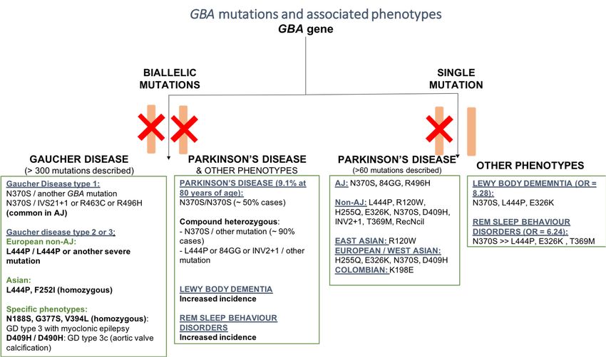
Those different phenotypes are associated with discrete genetic mutations, as detailed below.
Different Pathogenic Mutations of GBA Associated with Gaucher’s Disease (GD) Subtypes
More than 300 variants of the GBA gene have been associated with Gaucher’s disease [23]. GD is
an autosomal recessive disorder. In order for the disease to manifest, patients need to carry a pathogenic
mutation on both alleles of the GBA gene, either in a homozygous or compound heterozygous fashion.
Point mutations, insertion, deletion, missense mutations, splice junctions, and concomitant multiple
mutations have been reported [24]. The different variants can be more represented in particular ethnic
groups as well as in particular phenotypes. The c.1226A < G (N370S; or N409S according to the new
nomenclature) mutation is the most common one among Ashkenazi Jew (AJ) patients, followed by the
c.84dupG (84GG) mutation, which is more rare. The c.115 + 1G > A (IVS2 + 1), c.1504C > T (R463C),
and c.1604G > A (R496H) are commonly found in AJ patients with GD type 1 [24]. On the contrary,
the N370S mutation is rarely found among Chinese and Japanese patients [24] (Hruska et al., 2008).
Among Asian ethnic groups, the c.1448T > C (L444P, or L483P according to the new nomenclature) and
the c.754T > A (F252I), usually associated with GD type 2 and 3, are more prevalent, also explaining
why among these populations the neuropathic forms of GD are more frequent [20]. c.1448T > C
(L444P) is also the most frequent mutation among Caucasians with a non-Ashkenazi Jew ancestry [25]
(Figure 1).8, x FOR PEER REVIEW
Cells 2019, 4 of 17
Figure1.1.Schematic
Figure Schematicrepresentation
representation ofof the
the most common
common pathogenic
pathogenicmutations
mutationsofofthe
theGBA
GBAgenes
genesand
and
associatedphenotypes.
associated phenotypes. Phenotypes
Phenotypes werewere grouped
grouped basedbased on homozygous
on homozygous and heterozygous
and heterozygous mutations,
mutations,
ancestry, andancestry, and specificfeatures.
specific associated associated features.
2.2.Different
GBA Mutation and Parkinson’s
mutations can lead todisease (PD)phenotypes of GD. The c.1226A > G (N370S) mutation
different
is associated only with Gaucher’s disease type 1 and it seems to be protective for the development
2.2.1. Pathogenic Mutations of GBA Associated with PD
More than a decade ago, the association between an increased risk of developing PD and the
presence of GBA mutations was initially noticed in large Gaucher’s disease clinics. The incidence of
PD among GD patients and their relatives, which were supposedly carriers for the mutation, seemed
to be higher than the general population. Initially, only single case reports were suggesting thisCells 2019, 8, 364 4 of 16
of the neurological involvement characteristic of GD type 2 and 3. Indeed, patients who present the
c.1226A > G (N370S) mutation on at least one allele of the GBA gene will manifest only GD type 1 [24].
Interestingly, subjects who are homozygous for the N370S variant can also remain asymptomatic for
the disease. On the other hand, the c.1448T > C (L444P) mutation is usually associated with GD
type 2 or 3, even when presenting in a compound heterozygous state [19]. Homozygous c.1448T >
C (L444P) mutation [c.1448T > C]1[c.1448T > C] (L444P/L444P) with no recombinant alleles can be
associated with very severe but also milder phenotypes [26]. The c.1342G > C (D409H) variant is
responsible for GD type 3c which presents with characteristic cardiac valve calcifications [27]. c.680A
> G (N188S), c.1246G > A (G377S), and c.1297G > T (V394L) are more likely to be associated with
myoclonic epilepsy [28–30]. Despite previously reported observations, it is commonly found that
members of the same family report variability in the manifestation of symptoms even with an identical
genotype, suggesting that a genotype/phenotype correlation is tentative still. Other reported mutations
are uniquely rare and oftentimes private among specific families. [12]. Hence, it is difficult to make
generalizations about these mutations specific phenotypical profiles.
Another interesting mutation is the c.1093G > A (E326K), which caused a lot of debate in the
literature [31]. Indeed, it is not clear whether this mutation is really pathogenic for GD, since it was
found also in a significant number of asymptomatic carriers in homozygosity [32,33]. However, when
associated with other GBA mutations on the same allele, it can cause severe impairment of the GCase
activity [34,35]. Interestingly enough, the same mutation seems to be significantly associated with an
increased risk of PD [33].
2.2. GBA Mutation and Parkinson’s Disease (PD)
2.2.1. Pathogenic Mutations of GBA Associated with PD
More than a decade ago, the association between an increased risk of developing PD and the
presence of GBA mutations was initially noticed in large Gaucher’s disease clinics. The incidence of PD
among GD patients and their relatives, which were supposedly carriers for the mutation, seemed to be
higher than the general population. Initially, only single case reports were suggesting this association.
Interestingly, PD was noticed in patients with GD type 1, which has always been considered the
non-neuropathic form of the disease [36–41]. It was only when larger populations of PD patients were
screened for mutations of this gene that the important role of GBA in the pathogenesis of PD was
assessed worldwide.
So far, more than 50 population studies have screened the GBA gene among PD patients, covering
a large number of ancestries (reviewed in [42,43]). Overall, these studies demonstrated that the
incidence of GBA mutations is significantly higher among PD patients, compared to non-affected
subjects. Compared to GD, a smaller number of GBA mutations have been reported in patients with
PD (about 130 GBA mutations) [42]. However, in many of these studies, only the mutations that are
most commonly associated with PD were screened. Therefore, less frequent variants still associated
with the disease could go undetected. Among all, the c.1226A > G (N370S) and the c.1448T > C
(L444P) mutations are the two most common mutations worldwide. Indeed, in some populations they
account for the 70–80% of the total number of variants of GBA associated with PD [44]. Among subjects
from eastern Europe with an AJ ancestry, the c.1226A > G (N370S) mutation is definitely the most
frequent one among PD patients, as already reported for GD (Figure 1). Among the non-AJ European
descendants, the c.1448T > C (L444P) mutation is more common. Interestingly, it has been reported
that some mutations are able to increase the risk of PD only in the context of specific ancestry [42]. This
is the case of the c.84dupG (84GG) and c.1604G > A (R496H) for AJ subjects, the c. 475 C > T (R120W)
for East Asian populations, and the c.882T > G (H255Q), c.1093G > A (E326K), c.1342G > C (D409H),
and c.1226A < G (N370S), which are only found in subjects of European or West Asian ancestry [42]
(Figure 1). A recent study identified an increased incidence of the K198E variant (previously described
in GD1 and GD2 patients) in a population of PD patients from Columbia compared to controls [43].Cells 2019, 8, 364 5 of 16
It seems that severe GBA mutations (as classified according to the subtype of GD that they are associated
with), such as c.84dupG (84GG), c.115 + 1G >A (IVS2 + 1), c.1297G > T (V394L), c.1342G > C (D409H),
c.1448T > C (L444P), and c.1263del + RecTL, are associated with a higher risk of causing PD compared
to milder mutations, such as the N370S and c.84dupG (84GG) [45]. Moreover, severe mutations are
associated with an earlier age of onset, as well as a more rapid progression and increased involvement
of cognitive functions [45–47]. In one study, the motor and some of the non-motor symptoms (such as
depression, REM sleep behavior disorders, and hyposmia) were significantly worse in PD patients
carrying severe GBA mutations compared to subjects carrying mild mutations or with idiopathic
PD [48].
Interestingly, GBA represents only a risk factor for PD. This means that not every carrier will
develop the disease. The reason for the reduced penetrance of these mutations has not yet been fully
elucidated. Based on large population studies, today we know that, among GBA carriers, about 9.1%
will develop PD. Some reports suggest that the penetrance of PD in GD patient is 30% at 80 years,
but this data needs to be confirmed by further studies [49]. Patients with a homozygous mutation
of GBA, thus affected with Gaucher’s disease, have a higher risk of developing PD and usually with
an earlier age of onset of symptoms [48]. Having said that, it is worth noticing that the majority of
subjects with GD will never develop PD, even in the case of severe mutations. It is still controversial
whether PD in patients with GD presents with a more severe phenotype compared to GBA carriers.
Carriers of the GBA mutation harbor an increased risk of developing PD by five times in heterozygous
carriers and 10–20 times in homozygous carriers [50–53]. GBA mutations are present in about 2–30% of
PD patients [54]. Carrier frequency can be very different across different ancestry. Among AJs, it goes
from 10 to 31%, while in Norwegian’s it is only 2.3% [54]. In patients of European non-AJ ancestry,
it ranges from 2.9 to 12% [54].
In the last few years, there has been a great effort to try to clarify the pathogenic role of the
GBA mutations in PD and many different hypotheses have been formulated, as reported above
(for review see [8]). It is important to note that a growing amount of data is suggesting a failure
of the endolysosomal and of the autophagic pathways in PD [55]. These scavenger systems are
crucial for the degradation of alpha-synuclein, whose accumulation in the dopaminergic neurons
is one of the hallmarks of PD. In the lysosome, GCase plays an important contribution for these
processes and, in particular, in the interplay with alpha-synuclein [56]. Therefore, it is not totally
surprising that a dysfunction of this enzyme is related to PD. How the different mutations of GBA
that have been described in PD patients are able to affect the activity of the GCase has not been fully
understood. We know that the GCase has three active domains. PD-associated mutations are found in
distinctive domains of the protein. The c.1342G > C (D409H) and c.1297G > T (V394L) variants are
located in domain I. The c.84dupG (84GG) mutation causes a frameshift that can induce aberrantly
shorter or longer proteins that are non-functioning [23]. Other mutations, instead, are not found in
the functional domains but do interfere with the final structure of the enzyme, thus making it more
unstable or affecting its interaction with other proteins. The c.1226A > G (N370S) and c.1448T > C
(L444P) mutations are, for example, located in the proximity of the binding site of the Saposin C,
an activator of GCase [57]. More importantly, SapC competes with the binding of alpha-synuclein to
GCase, which would cause the inhibition of the enzyme [58,59]. Interestingly, the c.1226A > G (N370S)
mutation also seems to affect the ability of the GCase to modify the conformation of one of its loops,
loop 3, according to changes in pH [60,61]. Conformational changes in response to the changes of
the cellular environment are critical for the proper function of the protein. Despite our knowledge
about the structural effects of the different mutations, the exact correlation between the localizations of
pathogenetic variants of the gene and the degree of expression of PD has not yet been fully described.
It is also worth noting that GBA presents a pseudogene (GBAP1) that shares a very high degree of
homology—96% sequence identity–located in the proximity of the original gene [62,63]. Therefore,
genetic analysis will have to take this into account and should be performed in a specialized laboratoryCells 2019, 8, 364 6 of 16
in order to obtain reliable results. New technologies, such as the long-read sequencer, are on the
horizon for even more in-depth identification of possible GBA mutations [64].
2.2.2. GBA Mutations and Parkinson’s Disease Phenotype
PD patients carrying GBA mutations are not easily recognizable in most cases because they do not
present exclusive features that would clearly distinguish them from patients with idiopathic PD (iPD).
However, large population studies comparing carriers vs. non-carriers, mild vs. severe mutations,
as well as heterozygous manifesting carriers vs. PD–GD patients, allowed the ability to define common
traits in these subgroups of patients (for a comprehensive review see [8]). In particular, GBA–PD
patients present an overall earlier age of onset compared to non-carriers. Disease manifests about
3–6 years earlier in heterozygous carriers, irrespectively of the severity of the mutation, and about
6–11 years earlier in subjects with homozygous mutations [45,46,48,54,65–70]. There are limited reports
of GBA mutation carriers having an age of onset in the 200 s. [31,54,71–73].
The progression of the disease has been characterized in many different studies by a more
pronounced cognitive deficit in a significant percentage of these patients, with a risk of developing
dementia up to three times higher compared to iPD, which is even more increased in patients with severe
mutations [46,48,74]. Hallucinations and REM sleep behavior disorders (RBD) also are more common
among GBA patients in a dose-dependent fashion, being more frequent in subjects with homozygous
mutations and in patients carrying severe vs. milder mutations. However, other non-motor symptoms,
such as depression and anxiety, constipation, urinary symptoms, orthostatic hypotension, and sexual
dysfunctions are over-represented as well in GBA carriers compared to iPD, especially in the presence
of severe mutations, but with no increased severity in GD patients [46,48,75,76]. An increased incidence
of dysautonomic features has been suggested to be the main driver of the slightly reduced survival
reported in these patients [77]. Motor complications, such as dysphagia, dysarthria, and freezing of
gait, are more frequent as well in GBA carriers [46,67].
In patients with GBA mutations and PD, the rigid akinetic phenotype seems to be more common.
Usually, these patients present a very good response to levodopa, although the progression of the
motor symptoms can be slightly faster compared to iPD but without higher rates of motor fluctuations
or dyskinesia. Therefore, no specific treatment approaches need to be considered for this subgroup of
patients. Interestingly, a recent study evaluated the outcomes of treatment with deep brain stimulation
(DBS) in a cohort of PD patients carrying GBA mutations [78]. After a follow up of 7.5 years on average,
it was noticed that the het-GBA cohort presented similar outcomes compared to iPD in terms of motor
symptoms, while cognitive impairment and non-motor symptoms were definitely more represented
among carriers [78]. However, because of the beneficial effect on the motor symptoms, DBS should be
considered as a suitable option for these patients.
2.2.3. GBA Mutations and Other Phenotypes
GBA mutations were identified also in cases of REM sleep behavior disorders (RBD) and in cases
of dementia with Lewy bodies (DLB) [79].
GBA and Dementia with Lewy Bodies
A relatively low number of studies have been conducted to explore the incidence of the GBA
mutation among patients affected with dementia with Lewy bodies (DLB), which was found to be
even higher compared to the one in PD patients. In a cohort study of DLB patients, the frequency of
GBA mutations was 7.49% with an odd ratio of 8.28 [79]. In another study in Spanish subjects, and in
a number of autoptic brain tissues from pathologically proven DLB patients, a GBA mutation was
identified in 12–13% of the cases [80]. Recent genome-wide association studies (GWAS) also confirmed
the significant association between GBA mutations and DLB (particularly the rs35749011 variant) [81].
Among GBA carriers, the risk of developing DLB is about three times greater than developing PD [82].Cells 2019, 8, 364 7 of 16
As well as in PD patients, GBA mutations are associated with an earlier age of onset in DLB cases
compared to non-carriers (of approximately five years) and a higher disease severity score [79,80].
The association between GBA mutations and DLB was found to be higher among male subjects
compared to female [80]. These observations were confirmed also in a following study in a cohort
of patients with DLB and AJ ancestry [83]. GBA mutation carriers (about 11% of the entire cohort)
presented more severe symptoms, particularly in terms of increased hallucinations, worse RBD
symptoms, and overall cognitive and motor features [83].
A number of different mutations of the GBA gene have been reported in DLB patients. Other
than the two mutations most frequently associated with PD (c.1226A > G (N370S) and c.1448T > C
(L444P)), the E326K variant is over-represented in this cohort of patients compared to controls [79,80].
Interestingly, the c.1093G > A (E326K) mutation also is frequently found in patients with PD dementia
(PDD) [84].
Neuropathological data does not significantly differ between DLB patients with and without
a GBA mutation [79]. However, GBA carriers present a reduced GCase activity as well as a more
pronounced alteration of lipid profiles in the brain [85]. GBA expression profiles have been shown to
be reduced in DLB and PDD cases in both specific brain regions (temporal cortex and caudate nucleus
respectively) and in the peripheral blood [86]. GBA mutations are more significantly associated with
Lewy bodies (LB) pathology (especially with a cortical localization) than with Alzheimer’s disease
(AD) pathology (i.e., beta-amyloid and neurofibrillary tangles inclusions) [87].
GBA and REM Sleep Behavior Disorders
REM sleep behavior disorders (RBDs) are considered one of the prodromal symptoms of PD and
patients affected by this disorder may present with alpha-synuclein accumulation in the brain [88].
According to a recent metanalysis, patients affected with RBDs present an estimated risk of developing
a neurodegenerative disorder up to 97% after more than 14 years of follow up [89]. The majority of the
cases who present a phenoconversion will develop an alpha-synucleinopathy, represented by PD in
the majority of the cases, but also Multiple System Atrophy (MSA), Dementia with Lewy Bodies (DLB),
and PD with dementia [90]. In fact, subjects with RBD may present clinical symptoms fulfilling the
criteria for prodromal PD in up to 74% of the cases, manifesting worse performances in both motor
and non-motor assessments compared to non-affected subjects [91,92]. Notably, many of the studies in
this field did not take into consideration the significance of a family history of a neurodegenerative
disorder, therefore, it is probable that the percentage of patients that reported a neurodegenerative
disease is misrepresented. It would be worth exploring this aspect in future studies.
RBD seems to be more frequent in PD patients with GBA mutations compared with patients
without this mutation (OR 3.13) [48,65,67,76]. RBDs are also more frequent in PD patients with
concomitant GD than in heterozygous carriers [48]. Based on these observations, a few studies
explored the incidence of the GBA mutation among patients affected with RBD [65,91–93]. These
studies reported that among patients with idiopathic RBDs there is an increased frequency of GBA
mutations (2.6–11.6% of RBD patients vs. 0.4–1.8% of the controls) [65,91,93]. A number of different
GBA mutations were identified in RBD patients [65,93]. Some of these mutations have already been
reported in PD patients, while others still do not have a clear pathogenic role. Among all the reported
mutations, the two more commonly found in PD (i.e., c.1226A > G (N370S) and c.1448T > C (L444P),
with N370S >> L444P), together with the c.1093G > A (E326K) and the c.1223C > T (T369M), were the
most frequently represented in subjects with RBD [65,91–93].
Subjects with homozygous GBA mutations, thus affected with GD, and heterozygous carriers with
no PD, presented significant worsening of rapid eye movement sleep behavior disorder scores over a
period of time of two years compared with non-carrier subjects [92]. Among GBA carriers, the odds
ratio (OR) for RBD was 6.24 (95% CI 3.76–10.35, P < 0.0001) [65]. The presence of GBA mutations does
not seem to increase the risk among RBD patients of phenoconverting into PD [93]. These observationsCells 2019, 8, 364 8 of 16
all together suggest that GBA may play a role in the development of RBDs, but not necessarily in
determining more severe phenotypes.
Interestingly, no mutations of the LRRK2 gene, the other common genetic risk factor for PD, have
been identified so far in patients with RBDs [91,94].
3. New Targeted Treatments for GBA–PD Patients
Despite the very successful treatments that are now available to address the systemic manifestations
of Gaucher’s disease, unfortunately these approaches (i.e., enzyme replacement therapy and substrate
reduction therapy) are not able to reach the central nervous system and thus fail to address the
neurological symptoms caused by the disease. Different companies have been working for years to try
to address this issue, producing very promising results in cellular and animal models. We are now in a
very exciting era where some of these experimental approaches are starting to reach the clinical scene.
The treatments available so far in clinical trials try to address two main mechanisms that are thought to
be detrimental in linking GBA mutations to PD. The first hypothesis is that mutated forms of GBA are not
able to fold properly in the endoplasmic reticulum (ER) in the cells, causing the protein to accumulate
in this cellular compartment [95]. This would trigger a stress response in the dopaminergic neurons
leading to their damage and death [95]. Also, the entrapment of the beta GCase in the ER causes reduced
levels of the enzyme in the cells, triggering alpha-synuclein accumulation [95]. In order to target
this pathogenic mechanism, different chaperones, which are proteins able to facilitate the refolding
of their substrates, were tested [95–99]. In 2016, a clinical study assessing the efficacy of ambroxol,
one of these chaperones that showed very exciting preliminary results, was started (NCT02914366
study: https://www.clinicaltrials.gov/ct2/show/NCT02914366?cond=gba+parkinson&rank=7). This
is a phase 2 clinical trial to assess the safety and the efficacy of this drug to improve motor and
cognitive features of PD patients with a GBA mutation. The study is currently ongoing. Another similar
approach has been tested in a phase 1 study by Allergan with LTI-291, a chaperone molecule able to
increase the activity of GCase (https://lti-staging.squarespace.com/our-science/#lti-291). Isofagomine is
another chaperone protein that has been tested in vitro and in vivo to assess its ability to modulate
the phenotype induced by mutations of GBA [97]. This molecule is an inhibitory chaperone whose
role would be the stabilization of the GCase. Clinical trials with this molecule are not available at the
moment. It is also worth considering that small molecules, such as chaperones, can present different
therapeutic profiles in carriers of the different mutations of GBA according to the effect of these variants
on the protein [100].
The second mechanism that has been explored to treat GBA–PD patients is the accumulation in the
dopaminergic neurons of glucosylceramide (the substrate normally degraded by the GCase) because
of the mutation of GBA [101–103]. Genzyme recently started a multicenter, randomized, double-blind,
placebo-controlled phase 2 study to assess the safety, pharmacokinetics, and pharmacodynamics of an
oral compound, ibiglustat (GZ/SAR402671), which is able to reduce the levels of beta-glucocerebrosidase
in GBA carriers with early-stage PD (MOVES-PD study: https://www.clinicaltrials.gov/ct2/show/
NCT02906020?cond=gba+parkinson&rank=2). It is still a long road for the establishment of an
effective treatment, but many paths have been established, giving hope for patients with PD.
Mutated GCase is more unstable compared to the wild-type form. Therefore, modulation of the
degradation of GCase could be another suitable strategy to increase the activity of the enzyme and
thus tackle alpha-synuclein accumulation and neurodegeneration. Hsp90β, together with other heat
shock proteins (HSP), such as Hsp27, parkin, and the endoplasmic reticulum-associated pathway,
are responsible for the degradation of misfolded GCase. In particular, histone deacetylase inhibitors
(HDACis) and direct inhibitors of specific HSP are able to increase the GCase activity, reducing its
degradation [104]. Indeed, HDACis prevent the interaction between Hdp90β and GCase through the
hyperactivation of one of its domains [105].
GCase plays an important role in the autophagy-lysosomal pathway (ALP), where other genes
that have been associated with PD, such as ATP13A2, scavenger receptor class B member 2 (SCARB2),Cells 2019, 8, 364 9 of 16
sphingomyelin phosphodiesterase 1 (SMPD1), and others, are also involved (Moors et al., 2016). Failure
of the ALP seems to be responsible for the accumulation of alpha-synuclein in neurons. Therefore,
a number of pharmacological approaches directed to the ALP have been attempted in cellular and animal
models of PD (for a comprehensive review see [106]). However, the autophagic pathway is broadly
represented and active in different cell types and tissues in the organism. Therefore, the identification
of approaches with a high selectivity for certain tissues (such as the dopaminergic neurons) or for
specific mechanisms within ALP (such as GCase failure) is detrimental for the achievement of effective
but also safe treatments for patients.
In order to restore GCase activity, whose failure seems to be responsible for its neuronal
pathogenicity, gene therapy approaches are also in the pipeline. Preclinical studies showed that
delivery of GBA using adeno-associated virus 1 (AAV1) in A53T–alpha-synuclein mice is able to reduce
alpha-synuclein accumulation in the brain [107,108]. The field of gene therapy is now continuously
growing in the context of the neurodegenerative disorders [109]. Clinical trials to assess the efficacy of
this type of approach may soon be a reality in the context of PD and GBA mutations.
4. Conclusions
The discovery of the association between mutations of the GBA gene and PD allowed important
considerations and discoveries that are contributing to a better understating of the pathogenesis of PD.
Indeed, after this initial observation, the role of lysosomal impairment has been extensively explored in
PD. A growing amount of emerging evidence supports the idea that the endolysosomal trafficking is
involved in alpha-synuclein accumulation and dopaminergic neuron degeneration. A number of genes
involved in monogenic forms of PD or genetic risk factors for the disease (such as SNCA, ATP13A2,
VPS35, DNAJC6, SYNJ1, LRRK2, RAB39B) are part of this pathway (for review see [110]). Mutations of
genes involved in the endolysosomal pathways are responsible for a group of disorders designated
as Lysosomal Storage Disorders (LSD). These are typically rare autosomal recessive diseases which
cause systemic involvements with variable degrees of severity and neurological involvement, usually
presenting during childhood (reviewed in [111]). It is interesting to note that an increased burden of
LSD-associated mutations has been identified in the screening of large PD populations compared to
controls [112]. At the same time, among the 39 new gene loci associated with PD reported in the largest
genome wide association study (GWAS) performed in PD patients so far, a number of these variants
were found in LSD-associated genes (i.e., NAGLU, GUSB, NEU1, and GRN) [113].
The case of autosomal recessive conditions causing severe and rare disorders during childhood,
which in turn present as genetic risk factors for common adult neurodegenerative disorders when
in a heterozygous state, appears to be more and more frequent, usually presenting an incomplete
penetrance. This is the case for a number of LSD in the context of PD or of a parkinsonian degeneration,
such as SMPD1 (sphingomyelin phosphodiesterase, Niemann–Pick disease), ATP13A2 (P5-type ATPase,
Kufor–Rakeb disease), GALC (galactosylceramidase, Krabbe disease), NPC1 (Niemann–Pick type C),
NAGLU (α-N-acetylglucosminidase, Sanfilippo syndrome B, or mucopoly-saccharidosis III disease
B (MPS-IIIB)), HEXB (β-hexosaminidase B, Sandhoff disease (GM2 gangliosidosis)) (summarized
in [114]). The association between GBA mutations, GD, and PD must be just the tip of the iceberg of a
larger phenomenon, where the association between genes initially considered responsible only for
autosomal recessive disorders turned out to be risk factors for common neurodegenerative conditions.
This association may have been recognized first in GD patients because of the higher frequency of this
disease compared with other LSD.
Interestingly, this is also the case for the TREM2 gene (encoding for Triggering Receptor Expressed
on Myeloid cells 2), which seems to be the most frequent genetic risk factor of another common
neurodegenerative disorder, Alzheimer’s disease (AD) [115]. Autosomal recessive mutations of TREM2
are responsible for the rare, juvenile condition known as Polycystic lipomembranous osteodysplasia
with sclerosing leukoencephalopathy. Of note, TREM2 plays a crucial role in microglia cells as part of
the phagocytic scavenger pathway [116].Cells 2019, 8, 364 10 of 16
The phenomena of one gene presenting with different phenotypes is becoming more common
in the context of neurological disorders and in respect to common diseases, such as PD and AD. It is
important for clinicians to be familiar with these concepts in order to be able to properly counsel their
patients and their family members. Also, the identification of such patients will hopefully offer more
effective treatments, once available.
These new insights into the understanding of neurodegenerative diseases and, in particular,
PD open new scenarios that only a few years ago were still totally obscure. Hopefully, these discoveries
will be important for a real discernment of these severe conditions and for the discovery of more
effective therapeutic approaches.
Author Contributions: Conceptualization, G.M.R. and A.B.D.F.; writing—original draft preparation, G.M.R. and
A.B.D.F.; writing—review and editing, G.M.R. and A.B.D.F.
Funding: Marlene and Paolo Fresco Institute Clinical Fellowship program; American Parkinson
Disease Association.
Acknowledgments: We sincerely thank Brooklyn Henderson, Registered Nurse for her significant contribution in
the editing of the manuscript.
Conflicts of Interest: The authors declare no conflict of interest.
References
1. Goker-Alpan, O.; Schiffmann, R.; LaMarca, M.E.; Nussbaum, R.L.; McInerney-Leo, A.; Sidransky, E.
Parkinsonism among Gaucher disease carriers. J. Med. Genet. 2004, 41, 937–940. [CrossRef] [PubMed]
2. Lwin, A.; Orvisky, E.; Goker-Alpan, O.; LaMarca, M.E.; Sidransky, E. Glucocerebrosidase mutations in
subjects with parkinsonism. Mol. Genet. Metab. 2004, 81, 70–73. [CrossRef]
3. Eblan, M.J.; Walker, J.M.; Sidransky, E. The glucocerebrosidase gene and Parkinson’s disease in Ashkenazi
Jews. N. Engl. J. Med. 2005, 352, 728–731. [CrossRef]
4. Sidransky, E. Heterozygosity for a Mendelian disorder as a risk factor for complex disease. Clin. Genet. 2006,
70, 275–282. [CrossRef]
5. Poewe, W.; Seppi, K.; Tanner, C.M.; Halliday, G.M.; Brundin, P.; Volkmann, J.; Schrag, A.E.; Lang, A.E.
Parkinson disease. Nat. Rev. Dis Primers 2017, 3, 17013. [CrossRef] [PubMed]
6. Gegg, M.E.; Schapira, A.H.V. The role of glucocerebrosidase in Parkinson disease pathogenesis. FEBS J. 2018,
285, 3591–3603. [CrossRef]
7. Klein, A.D.; Mazzulli, J.R. Is Parkinson’s disease a lysosomal disorder? Brain 2018, 141, 2255–2262. [CrossRef]
[PubMed]
8. Blandini, F.; Cilia, R.; Cerri, S.; Pezzoli, G.; Schapira, A.H.V.; Mullin, S.; Lanciego, J.L. Glucocerebrosidase
mutations and synucleinopathies: Toward a model of precision medicine. Mov. Disord. Off. J. Mov. Disord. Soc.
2019, 34, 9–21. [CrossRef] [PubMed]
9. Chiasserini, D.; Paciotti, S.; Eusebi, P.; Persichetti, E.; Tasegian, A.; Kurzawa-Akanbi, M.; Chinnery, P.F.;
Morris, C.M.; Calabresi, P.; Parnetti, L.; et al. Selective loss of glucocerebrosidase activity in sporadic
Parkinson’s disease and dementia with Lewy bodies. Mol. Neurodegener. 2015, 10, 15. [CrossRef]
10. Parnetti, L.; Paciotti, S.; Eusebi, P.; Dardis, A.; Zampieri, S.; Chiasserini, D.; Tasegian, A.; Tambasco, N.;
Bembi, B.; Calabresi, P.; et al. Cerebrospinal fluid beta-glucocerebrosidase activity is reduced in parkinson’s
disease patients. Mov. Disord. Off. J. Mov. Disord. Soc. 2017, 32, 1423–1431. [CrossRef]
11. Wenstrup, R.J.; Roca-Espiau, M.; Weinreb, N.J.; Bembi, B. Skeletal aspects of Gaucher disease: A review. Br. J. Radiol.
2002, 75 (Suppl. 1), A2–A12. [CrossRef] [PubMed]
12. Elstein, D.; Abrahamov, A.; Dweck, A.; Hadas-Halpern, I.; Zimran, A. Gaucher disease: Pediatric concerns.
Paediatr. Drugs 2002, 4, 417–426. [CrossRef]
13. Andersson, H.; Kaplan, P.; Kacena, K.; Yee, J. Eight-year clinical outcomes of long-term enzyme replacement
therapy for 884 children with Gaucher disease type 1. Pediatrics 2008, 122, 1182–1190. [CrossRef]
14. Kauli, R.; Zaizov, R.; Lazar, L.; Pertzelan, A.; Laron, Z.; Galatzer, A.; Phillip, M.; Yaniv, Y.; Cohen, I.J. Delayed
growth and puberty in patients with Gaucher disease type 1: Natural history and effect of splenectomy
and/or enzyme replacement therapy. ISR Med. Assoc. J. 2000, 2, 158–163. [PubMed]Cells 2019, 8, 364 11 of 16
15. Arends, M.; van Dussen, L.; Biegstraaten, M.; Hollak, C.E. Malignancies and monoclonal gammopathy
in Gaucher disease; a systematic review of the literature. Br. J. Haematol. 2013, 161, 832–842. [CrossRef]
[PubMed]
16. Zimran, A.; Elstein, D. Gaucher disease and related Lysosomal Storage Diseases. In Williams’ Hematology;
Lichtman, M.A., Kaushansky, K., Prchal, J.T., Levi, M., Burns, L.J., Press, O.W., Caligiuri, M.A., Eds.;
McGraw-Hill: New York, NY, USA, 2016; Volume 72, p. 1121.
17. Gupta, P.; Pastores, G. Pharmacological treatment of pediatric Gaucher disease. Expert Rev. Clin. Pharm.
2018, 11, 1183–1194. [CrossRef] [PubMed]
18. Beutler, E.; Grabowski, G. Gaucher disease. In The Metabolic and Molecular Basis of Inherited Disease, 8th ed.;
Beaudet, A.L., Scriver, C.R., Sly, W.S., Valle, D., Childs, B., Kinzler, K.W., Vogelstein, B., Eds.; McGraw-Hill
International Book Co.: New York, NY, USA, 2001; pp. 3635–3668.
19. Stirnemann, J.; Belmatoug, N.; Camou, F.; Serratrice, C.; Froissart, R.; Caillaud, C.; Levade, T.; Astudillo, L.;
Serratrice, J.; Brassier, A.; et al. A Review of Gaucher Disease Pathophysiology, Clinical Presentation and
Treatments. Int. J. Mol. Sci. 2017, 18, 441. [CrossRef] [PubMed]
20. Pastores, G.; Hughes, D. Gaucher Disease. In GeneReviews® [Internet]; Ardinger, H.H., Adam, M.P.,
Pagon, R.A., Wallace, S.E., Bean, L.J.H., Stephens, K., Amemiya, A., Eds.; University of Washington: Seattle,
WA, USA, 2000; (updated 2018 June 21).
21. Bennett, L.L.; Mohan, D. Gaucher disease and its treatment options. Ann. Pharm. 2013, 47, 1182–1193.
[CrossRef]
22. Dreborg, S.; Erikson, A.; Hagberg, B. Gaucher disease—Norrbottnian type. I. General clinical description.
Eur. J. Pediatrics 1980, 133, 107–118. [CrossRef]
23. Smith, L.; Mullin, S.; Schapira, A.H.V. Insights into the structural biology of Gaucher disease. Exp. Neurol.
2017, 298, 180–190. [CrossRef]
24. Hruska, K.S.; LaMarca, M.E.; Scott, C.R.; Sidransky, E. Gaucher disease: Mutation and polymorphism
spectrum in the glucocerebrosidase gene (GBA). Hum. Mutat. 2008, 29, 567–583. [CrossRef] [PubMed]
25. Grabowski, G.A.; Horowitz, M. Gaucher’s disease: Molecular, genetic and enzymological aspects.
Baillieres Clin. Haematol. 1997, 10, 635–656. [CrossRef]
26. Goker-Alpan, O.; Hruska, K.S.; Orvisky, E.; Kishnani, P.S.; Stubblefield, B.K.; Schiffmann, R.; Sidransky, E.
Divergent phenotypes in Gaucher disease implicate the role of modifiers. J. Med. Genet. 2005, 42, e37.
[CrossRef]
27. Cindik, N.; Ozcay, F.; Suren, D.; Akkoyun, I.; Gokdemir, M.; Varan, B.; Alehan, F.; Ozbek, N.; Tokel, K. Gaucher
disease with communicating hydrocephalus and cardiac involvement. Clin. Cardiol 2010, 33, E26–E30. [CrossRef]
[PubMed]
28. Koprivica, V.; Stone, D.L.; Park, J.K.; Callahan, M.; Frisch, A.; Cohen, I.J.; Tayebi, N.; Sidransky, E. Analysis and
classification of 304 mutant alleles in patients with type 1 and type 3 Gaucher disease. Am. J. Hum. Genet. 2000,
66, 1777–1786. [CrossRef]
29. Kowarz, L.; Goker-Alpan, O.; Banerjee-Basu, S.; LaMarca, M.E.; Kinlaw, L.; Schiffmann, R.; Baxevanis, A.D.;
Sidransky, E. Gaucher mutation N188S is associated with myoclonic epilepsy. Hum. Mutat. 2005, 26, 271–273.
[CrossRef]
30. Park, J.K.; Orvisky, E.; Tayebi, N.; Kaneski, C.; Lamarca, M.E.; Stubblefield, B.K.; Martin, B.M.; Schiffmann, R.;
Sidransky, E. Myoclonic epilepsy in Gaucher disease: Genotype-phenotype insights from a rare patient
subgroup. Pediatr. Res. 2003, 53, 387–395. [CrossRef] [PubMed]
31. Duran, R.; Mencacci, N.E.; Angeli, A.V.; Shoai, M.; Deas, E.; Houlden, H.; Mehta, A.; Hughes, D.; Cox, T.M.;
Deegan, P.; et al. The glucocerobrosidase E326K variant predisposes to Parkinson’s disease, but does not
cause Gaucher’s disease. Mov. Disord. Off. J. Mov. Disord. Soc. 2013, 28, 232–236. [CrossRef] [PubMed]
32. Park, J.K.; Tayebi, N.; Stubblefield, B.K.; LaMarca, M.E.; MacKenzie, J.J.; Stone, D.L.; Sidransky, E. The E326K
mutation and Gaucher disease: Mutation or polymorphism? Clin. Genet. 2002, 61, 32–34. [CrossRef]
33. Horowitz, M.; Pasmanik-Chor, M.; Ron, I.; Kolodny, E.H. The enigma of the E326K mutation in acid
beta-glucocerebrosidase. Mol. Genet. Metab. 2011, 104, 35–38. [CrossRef]
34. Chabas, A.; Gort, L.; Diaz-Font, A.; Montfort, M.; Santamaria, R.; Cidras, M.; Grinberg, D.; Vilageliu, L.
Perinatal lethal phenotype with generalized ichthyosis in a type 2 Gaucher disease patient with the
[L444P;E326K]/P182L genotype: Effect of the E326K change in neonatal and classic forms of the disease.
Blood Cells Mol. Dis. 2005, 35, 253–258. [CrossRef]Cells 2019, 8, 364 12 of 16
35. Liou, B.; Grabowski, G.A. Is E326K glucocerebrosidase a polymorphic or pathological variant? Mol. Genet. Metab.
2012, 105, 528–529. [CrossRef]
36. Neudorfer, O.; Giladi, N.; Elstein, D.; Abrahamov, A.; Turezkite, T.; Aghai, E.; Reches, A.; Bembi, B.; Zimran, A.
Occurrence of Parkinson’s syndrome in type I Gaucher disease. QJM 1996, 89, 691–694. [CrossRef]
37. Machaczka, M.; Rucinska, M.; Skotnicki, A.B.; Jurczak, W. Parkinson’s syndrome preceding clinical
manifestation of Gaucher’s disease. Am. J. Hematol. 1999, 61, 216–217. [CrossRef]
38. Perez-Calvo, J.; Bernal, M.; Giraldo, P.; Torralba, M.A.; Civeira, F.; Giralt, M.; Pocovi, M. Co-morbidity in
Gaucher’s disease results of a nationwide enquiry in Spain. Eur. J. Med. Res. 2000, 5, 231–235.
39. Varkonyi, J.; Simon, Z.; Soos, K.; Poros, A. Gaucher disease type I complicated with Parkinson’s syndrome.
Haematologia 2002, 32, 271–275. [CrossRef]
40. Tayebi, N.; Walker, J.; Stubblefield, B.; Orvisky, E.; LaMarca, M.E.; Wong, K.; Rosenbaum, H.; Schiffmann, R.;
Bembi, B.; Sidransky, E. Gaucher disease with parkinsonian manifestations: Does glucocerebrosidase
deficiency contribute to a vulnerability to parkinsonism? Mol. Genet. Metab. 2003, 79, 104–109. [CrossRef]
41. Bembi, B.; Zambito Marsala, S.; Sidransky, E.; Ciana, G.; Carrozzi, M.; Zorzon, M.; Martini, C.; Gioulis, M.;
Pittis, M.G.; Capus, L. Gaucher’s disease with Parkinson’s disease: Clinical and pathological aspects.
Neurology 2003, 61, 99–101. [CrossRef]
42. Zhang, Y.; Shu, L.; Zhou, X.; Pan, H.; Xu, Q.; Guo, J.; Tang, B.; Sun, Q. A Meta-Analysis of GBA-Related
Clinical Symptoms in Parkinson’s Disease. Parkinsons Dis. 2018, 2018, 3136415. [CrossRef]
43. Velez-Pardo, C.; Lorenzo-Betancor, O.; Jimenez-Del-Rio, M.; Moreno, S.; Lopera, F.; Cornejo-Olivas, M.;
Torres, L.; Inca-Martinez, M.; Mazzetti, P.; Cosentino, C.; et al. The distribution and risk effect of GBA variants
in a large cohort of PD patients from Colombia and Peru. Parkinsonism Relat. Disord. 2019. [CrossRef]
44. Lesage, S.; Anheim, M.; Condroyer, C.; Pollak, P.; Durif, F.; Dupuits, C.; Viallet, F.; Lohmann, E.; Corvol, J.C.;
Honore, A.; et al. Large-scale screening of the Gaucher’s disease-related glucocerebrosidase gene in
Europeans with Parkinson’s disease. Hum. Mol. Genet. 2011, 20, 202–210. [CrossRef] [PubMed]
45. Gan-Or, Z.; Giladi, N.; Rozovski, U.; Shifrin, C.; Rosner, S.; Gurevich, T.; Bar-Shira, A.; Orr-Urtreger, A.
Genotype-phenotype correlations between GBA mutations and Parkinson disease risk and onset. Neurology
2008, 70, 2277–2283. [CrossRef]
46. Cilia, R.; Tunesi, S.; Marotta, G.; Cereda, E.; Siri, C.; Tesei, S.; Zecchinelli, A.L.; Canesi, M.; Mariani, C.B.; Meucci, N.;
et al. Survival and dementia in GBA-associated Parkinson’s disease: The mutation matters. Ann. Neurol. 2016, 80,
662–673. [CrossRef] [PubMed]
47. Liu, G.; Boot, B.; Locascio, J.J.; Jansen, I.E.; Winder-Rhodes, S.; Eberly, S.; Elbaz, A.; Brice, A.; Ravina, B.; van
Hilten, J.J.; et al. Specifically neuropathic Gaucher’s mutations accelerate cognitive decline in Parkinson’s.
Ann. Neurol. 2016, 80, 674–685. [CrossRef]
48. Thaler, A.; Gurevich, T.; Bar Shira, A.; Gana Weisz, M.; Ash, E.; Shiner, T.; Orr-Urtreger, A.; Giladi, N.;
Mirelman, A. A “dose” effect of mutations in the GBA gene on Parkinson’s disease phenotype. Parkinsonism
Relat. Disord. 2017, 36, 47–51. [CrossRef]
49. Anheim, M.; Elbaz, A.; Lesage, S.; Durr, A.; Condroyer, C.; Viallet, F.; Pollak, P.; Bonaiti, B.; Bonaiti-Pellie, C.; Brice, A.
Penetrance of Parkinson disease in glucocerebrosidase gene mutation carriers. Neurology 2012, 78, 417–420. [CrossRef]
[PubMed]
50. Sidransky, E.; Nalls, M.A.; Aasly, J.O.; Aharon-Peretz, J.; Annesi, G.; Barbosa, E.R.; Bar-Shira, A.; Berg, D.;
Bras, J.; Brice, A.; et al. Multicenter analysis of glucocerebrosidase mutations in Parkinson’s disease. N. Engl.
J. Med. 2009, 361, 1651–1661. [CrossRef] [PubMed]
51. Bultron, G.; Kacena, K.; Pearson, D.; Boxer, M.; Yang, R.; Sathe, S.; Pastores, G.; Mistry, P.K. The risk of
Parkinson’s disease in type 1 Gaucher disease. J. Inherit. Metab. Dis. 2010, 33, 167–173. [CrossRef] [PubMed]
52. McNeill, A.; Duran, R.; Hughes, D.A.; Mehta, A.; Schapira, A.H. A clinical and family history study of
Parkinson’s disease in heterozygous glucocerebrosidase mutation carriers. J. Neurol. Neurosurg. Psychiatry
2012, 83, 853–854. [CrossRef]
53. McNeill, A.; Duran, R.; Proukakis, C.; Bras, J.; Hughes, D.; Mehta, A.; Hardy, J.; Wood, N.W.; Schapira, A.H.
Hyposmia and cognitive impairment in Gaucher disease patients and carriers. Mov. Disord. Off. J. Mov.
Disord. Soc. 2012, 27, 526–532. [CrossRef]
54. Neumann, J.; Bras, J.; Deas, E.; O’Sullivan, S.S.; Parkkinen, L.; Lachmann, R.H.; Li, A.; Holton, J.; Guerreiro, R.;
Paudel, R.; et al. Glucocerebrosidase mutations in clinical and pathologically proven Parkinson’s disease.
Brain 2009, 132, 1783–1794. [CrossRef]Cells 2019, 8, 364 13 of 16
55. Moors, T.; Paciotti, S.; Chiasserini, D.; Calabresi, P.; Parnetti, L.; Beccari, T.; van de Berg, W.D. Lysosomal
Dysfunction and alpha-Synuclein Aggregation in Parkinson’s Disease: Diagnostic Links. Mov. Disord. Off. J.
Mov. Disord. Soc. 2016, 31, 791–801. [CrossRef] [PubMed]
56. Mazzulli, J.R.; Xu, Y.H.; Sun, Y.; Knight, A.L.; McLean, P.J.; Caldwell, G.A.; Sidransky, E.; Grabowski, G.A.;
Krainc, D. Gaucher disease glucocerebrosidase and alpha-synuclein form a bidirectional pathogenic loop in
synucleinopathies. Cell 2011, 146, 37–52. [CrossRef] [PubMed]
57. Lieberman, R.L.; Wustman, B.A.; Huertas, P.; Powe, A.C., Jr.; Pine, C.W.; Khanna, R.; Schlossmacher, M.G.;
Ringe, D.; Petsko, G.A. Structure of acid beta-glucosidase with pharmacological chaperone provides insight
into Gaucher disease. Nat. Chem. Biol. 2007, 3, 101–107. [CrossRef]
58. Yap, T.L.; Gruschus, J.M.; Velayati, A.; Sidransky, E.; Lee, J.C. Saposin C protects glucocerebrosidase against
alpha-synuclein inhibition. Biochemistry 2013, 52, 7161–7163. [CrossRef] [PubMed]
59. Ouled Amar Bencheikh, B.; Leveille, E.; Ruskey, J.A.; Spiegelman, D.; Liong, C.; Fon, E.A.; Rouleau, G.A.;
Dauvilliers, Y.; Dupre, N.; Alcalay, R.N.; et al. Sequencing of the GBA coactivator, Saposin C, in Parkinson
disease. Neurobiol. Aging 2018, 72, 187.e1–187.e3. [CrossRef] [PubMed]
60. Offman, M.N.; Krol, M.; Silman, I.; Sussman, J.L.; Futerman, A.H. Molecular basis of reduced glucosylceramidase
activity in the most common Gaucher disease mutant, N370S. J. Biol. Chem. 2010, 285, 42105–42114. [CrossRef]
61. Wei, R.R.; Hughes, H.; Boucher, S.; Bird, J.J.; Guziewicz, N.; Van Patten, S.M.; Qiu, H.; Pan, C.Q.; Edmunds, T. X-ray
and biochemical analysis of N370S mutant human acid beta-glucosidase. J. Biol. Chem. 2011, 286, 299–308. [CrossRef]
62. Horowitz, M.; Wilder, S.; Horowitz, Z.; Reiner, O.; Gelbart, T.; Beutler, E. The human glucocerebrosidase
gene and pseudogene: Structure and evolution. Genomics 1989, 4, 87–96. [CrossRef]
63. Imai, K.; Nakamura, M.; Yamada, M.; Asano, A.; Yokoyama, S.; Tsuji, S.; Ginns, E.I. A novel transcript from a
pseudogene for human glucocerebrosidase in non-Gaucher disease cells. Gene 1993, 136, 365–368.
64. Leija-Salazar, M.; Sedlazeck, F.J.; Toffoli, M.; Mullin, S.; Mokretar, K.; Athanasopoulou, M.; Donald, A.;
Sharma, R.; Hughes, D.; Schapira, A.H.V.; et al. Evaluation of the detection of GBA missense mutations and
other variants using the Oxford Nanopore MinION. Mol. Genet. Genom. Med. 2019, e564. [CrossRef]
65. Gan-Or, Z.; Amshalom, I.; Kilarski, L.L.; Bar-Shira, A.; Gana-Weisz, M.; Mirelman, A.; Marder, K.; Bressman, S.;
Giladi, N.; Orr-Urtreger, A. Differential effects of severe vs. mild GBA mutations on Parkinson disease.
Neurology 2015, 84, 880–887. [CrossRef] [PubMed]
66. Alcalay, R.N.; Dinur, T.; Quinn, T.; Sakanaka, K.; Levy, O.; Waters, C.; Fahn, S.; Dorovski, T.; Chung, W.K.;
Pauciulo, M.; et al. Comparison of Parkinson risk in Ashkenazi Jewish patients with Gaucher disease and
GBA heterozygotes. JAMA Neurol. 2014, 71, 752–757. [CrossRef]
67. Jesus, S.; Huertas, I.; Bernal-Bernal, I.; Bonilla-Toribio, M.; Caceres-Redondo, M.T.; Vargas-Gonzalez, L.;
Gomez-Llamas, M.; Carrillo, F.; Calderon, E.; Carballo, M.; et al. GBA Variants Influence Motor and
Non-Motor Features of Parkinson’s Disease. PLoS ONE 2016, 11, e0167749. [CrossRef] [PubMed]
68. Malek, N.; Weil, R.S.; Bresner, C.; Lawton, M.A.; Grosset, K.A.; Tan, M.; Bajaj, N.; Barker, R.A.; Burn, D.J.;
Foltynie, T.; et al. Features of GBA-associated Parkinson’s disease at presentation in the UK Tracking
Parkinson’s study. J. Neurol. Neurosurg. Psychiatry 2018, 89, 702–709. [CrossRef] [PubMed]
69. Rosenbloom, B.; Balwani, M.; Bronstein, J.M.; Kolodny, E.; Sathe, S.; Gwosdow, A.R.; Taylor, J.S.; Cole, J.A.;
Zimran, A.; Weinreb, N.J. The incidence of Parkinsonism in patients with type 1 Gaucher disease: Data from
the ICGG Gaucher Registry. Blood Cells Mol. Dis. 2011, 46, 95–102. [CrossRef] [PubMed]
70. Lopez, G.; Kim, J.; Wiggs, E.; Cintron, D.; Groden, C.; Tayebi, N.; Mistry, P.K.; Pastores, G.M.; Zimran, A.;
Goker-Alpan, O.; et al. Clinical course and prognosis in patients with Gaucher disease and parkinsonism.
Neurol Genet. 2016, 2, e57. [CrossRef] [PubMed]
71. Tan, E.K.; Tong, J.; Fook-Chong, S.; Yih, Y.; Wong, M.C.; Pavanni, R.; Zhao, Y. Glucocerebrosidase mutations
and risk of Parkinson disease in Chinese patients. Arch. Neurol. 2007, 64, 1056–1058. [CrossRef]
72. Eblan, M.J.; Nguyen, J.; Ziegler, S.G.; Lwin, A.; Hanson, M.; Gallardo, M.; Weiser, R.; De Lucca, M.;
Singleton, A.; Sidransky, E. Glucocerebrosidase mutations are also found in subjects with early-onset
parkinsonism from Venezuela. Mov. Disord. Off. J. Mov. Disord. Soc. 2006, 21, 282–283. [CrossRef] [PubMed]
73. Wu, Y.R.; Chen, C.M.; Chao, C.Y.; Ro, L.S.; Lyu, R.K.; Chang, K.H.; Lee-Chen, G.J. Glucocerebrosidase gene
mutation is a risk factor for early onset of Parkinson disease among Taiwanese. J. Neurol. Neurosurg. Psychiatry
2007, 78, 977–979. [CrossRef]You can also read