GENE EXPRESSION PROFILING OF SARS-COV-2 INFECTIONS REVEAL DISTINCT PRIMARY LUNG CELL AND SYSTEMIC IMMUNE INFECTION RESPONSES THAT IDENTIFY ...
←
→
Page content transcription
If your browser does not render page correctly, please read the page content below
Briefings in Bioinformatics, 00(00), 2020, 1–14

                                                                                  doi: 10.1093/bib/bbaa376
                                                                                  Case Study

Gene expression profiling of SARS-CoV-2 infections

                                                                                                                                                                  Downloaded from https://academic.oup.com/bib/advance-article/doi/10.1093/bib/bbaa376/6041168 by guest on 21 December 2020
reveal distinct primary lung cell and systemic immune
infection responses that identify pathways relevant in
COVID-19 disease
Mohammad Ali Moni , Julian M. W. Quinn, Nese Sinmaz and
Matthew A. Summers
Corresponding author: Matthew A. Summers, The Garvan Institute of Medical Research, 384 Victoria St, Darlinghurst, NSW, Australia.
Email: m.summers@garvan.org.au

Abstract
To identify key gene expression pathways altered with infection of the novel coronavirus SARS-CoV-2, we performed the
largest comparative genomic and transcriptomic analysis to date. We compared the novel pandemic coronavirus
SARS-CoV-2 with SARS-CoV and MERS-CoV, as well as inf luenza A strains H1N1, H3N2 and H5N1. Phylogenetic analysis
confirms that SARS-CoV-2 is closely related to SARS-CoV at the level of the viral genome. RNAseq analyses demonstrate that
human lung epithelial cell responses to SARS-CoV-2 infection are distinct. Extensive Gene Expression Omnibus literature
screening and drug predictive analyses show that SARS-CoV-2 infection response pathways are closely related to those of
SARS-CoV and respiratory syncytial virus infections. We validated SARS-CoV-2 infection response genes as
disease-associated using Kaplan–Meier survival estimates in lung disease patient data. We also analysed COVID-19 patient
peripheral blood samples, which identified signalling pathway concordance between the primary lung cell and blood cell
infection responses.

Mohammad Ali Moni is a Research Fellow and Conjoint Lecturer at the University of New South Wales, Australia. He received his PhD degree in
bioinformatics from the University of Cambridge. His research interest encompasses artificial intelligence, machine learning, data science and clinical
bioinformatics.
Julian M. W. Quinn received the PhD from the University of Oxford, UK, in 1992, then, moved to Australia for postdoctoral training in bone, joint and
cancer biology at the St Vincent’s Institute of Medical Research as a Senior Research Fellow since 2014. His interests are in applications of biostatistics and
bioinformatics, and now he works as a Surgical Research Officer at the Surgical Education and Research Training Institute at Royal North Shore Hospital,
Sydney Australia.
Nese Sinmaz received her honours degree in cell pathology and immunology from the University of Sydney in 2012, and her PhD in neuroscience and
immunology in 2017. She works in drug discovery for immunological disorders in the private sector while maintaining academic research interests in
immunology and inflammatory disease.
Matthew A. Summers is a Research Scientist at the Garvan Institute of Medical Research, Australia. His research interests include understanding genetic
and molecular mechanisms of rare diseases using cutting-edge single-cell genomic, transcriptomic and bioinformatics methods.
Submitted: 22 June 2020; Received (in revised form): 2 November 2020

© The Author(s) 2020. Published by Oxford University Press. All rights reserved. For Permissions, please email: journals.permissions@oup.com

                                                                                                                                                         1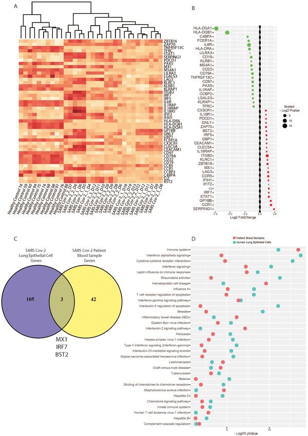
2    Moni et al.

Graphical Abstract

                                                                                                                               Downloaded from https://academic.oup.com/bib/advance-article/doi/10.1093/bib/bbaa376/6041168 by guest on 21 December 2020

GRAPHIC 1: SARS-CoV-2 graphical abstract depicting the study workf low and analysis pipeline. (A) We first conducted
phylogenetic and RNAseq analyses comparing SARS-CoV-2, SARS, MERS, and inf luenza strains, and the overlapping gene
expression effects of infection with these viruses. (B–C) Datasets from a study of transcriptional effects of SARS-CoV-2
infection in human lung epithelial cells (profiled using RNAseq) was analysed to determine differential gene expression. (D)
Gene ontology and curated cell signalling pathway databases were used to screen the differentially expressed gene list for
functional enrichment. (E) SARS-CoV-2 genes were used for drug prediction and survival analyses in disease datasets,
results suggesting pathology associated genes and pathways for subsequent functional analysis. (F) A targeted immune
profile in SARS-CoV-2 infected patient blood samples was analysed to assess system wide immune effects of infection.
Gene expression profiling of SARS-CoV-2 infections        3

Key words: SARS-CoV-2; SARS-CoV; MERS-CoV; coronaviruses; transcriptomics; RNAseq; COVID-19

                                                                      divergence from the influenza viruses examined (Figure 1A).
Introduction
                                                                      RNAseq gene expression profiling of human lung epithelial cells
The current global pandemic of the novel coronavirus SARS-            infected with SARS-CoV-2, SARS-CoV, MERS-CoV and influenza
CoV-2, causing a severe respiratory disease designated as COVID-      strains for 24 h revealed a surprisingly sparse overlapping gene
19, has as of mid-August 2020 infected >21 million people and         expression signature (Figure 1B–C). This is an observation of key

                                                                                                                                             Downloaded from https://academic.oup.com/bib/advance-article/doi/10.1093/bib/bbaa376/6041168 by guest on 21 December 2020
caused >750,000 deaths globally [1]. The highly infectious and        importance; despite their close phylogenetic relationship, the
widespread nature of the disease is attributed in part to its         novel coronavirus SARS-CoV-2 induces a very different tran-
asymptomatic spread, in contrast to earlier pandemic coro-            scriptional response in the host cells compared with SARS-CoV
naviruses SARS-CoV and MERS-CoV, which were more easily               and MERS-CoV, with majority of differentially expressed genes
contained as symptom onset co-occurred with infectivity [2].          (DEGs) distinct to the SARS-CoV-2 set (Figure 1B). There was no
     The development and spread of the novel coronavirus              evident relationship between phylogenetic and gene expression
causing COVID-19 has vastly outpaced the rate of vaccine and          relationships, consistent with the notion that COVID-19 is a quite
therapeutic development. Nevertheless, the global research            different disease entity (at least in its initial stages of devel-
community has rallied; within weeks of the first observations of      opment) to those resulting from the earlier coronaviruses. A
COVID-19, the virus was isolated and characterised. Central           similar conclusion was reached when we compared SARS-CoV-
to effective therapy and vaccine development is an improved           2 and influenza-induced gene expression changes in human
understanding of the cellular pathway and transcriptional             lung epithelial cells (Figure 1C). For this reason, we then focussed
responses to infection in human cells. These are currently very       exclusively on the SARS-CoV-2 transcriptome and pathway data
poorly characterised, both in the early stage of infection, as        to gain insights into early COVID-19 development.
viral load increases before symptom onset; as well as in the              To understand the magnitude of the unique transcriptional
later stage of COVID-19 pneumonia, which is associated with a         effects of SARS-CoV-2 infection, we stringently assessed DEGs
severe cytokine and inflammatory storm in affected patients. It       using a cut-off threshold of absolute log2 fold change (LFC) >1,
remains a mystery why only a small proportion become severely         with an adjusted P value 85% were
affected by SARS-CoV-2 infection, we applied a suite of com-          significantly upregulated compared to MERS-CoV, SARS-CoV and
putational and bioinformatics approaches using SARS-CoV-2             influenza virus infections (Figure 2A). Notably, the top 40 SARS-
genomic and transcriptomic data. We started by comparing              CoV-2 infection response genes show particularly striking upreg-
SARS-CoV-2 with SARS-CoV, MERS-CoV and influenza A strains            ulation compared to the other viruses (Figure 2B). Using a more
H1N1, H3N2 and H5N1 viral genomes, and assessed the effect of         stringent cut-off threshold of absolute log fold change >2, there
infection on transcriptional responses in human lung epithelial       was evident strong upregulation of a unique suite of inflamma-
cells [3–5]. This enabled us to characterise a novel set of SARS-     tory response genes upon SARS-CoV-2 infection; these included
CoV-2 acute response genes. On this set of genes, we performed        interferon response genes IFI44L, IFI27 and IFI6, interleukins IL6
extensive cell signalling pathway and predictive analyses. These      and Interleukin 8 (IL8), and chemokine and complement gene
assessed gene ontologies and cell signalling cascades associated      activation (Figure 2C).
with SARS-CoV-2 infection in human cells, which enabled us to             Collectively, these data further confirm that although
identify clinically approved drugs [6] that affect aspects of the     the novel coronavirus SARS-CoV-2 genome is closely related
early cell response to the viral infection, some of which may be of   to previous pandemic coronaviruses SARS-CoV and MERS-
therapeutic relevance. Having identified the distinct transcrip-      CoV genomes, the transcriptional responses to infection in
tional responses following SARS-CoV-2 infection, we performed         human lung epithelial cells are markedly different. Similarly,
bioinformatics validation studies to determine whether these          comparing the infection responses of pandemic influenza
SARS-CoV-2 response genes and pathways are associated with            strains H1N1, H3N2 and H5N1 with SARS-CoV-2 indicates SARS-
comorbid lung disease, and share concordant signalling path-          CoV-2 infection responses in human lung epithelial cells remain
way responses with the systemic immune response evident in            unique at the gene expression level.
COVID-19 human patient blood samples.

                                                                      Gene ontology and cell signalling pathway analysis
Results
                                                                      finds enriched inflammatory and infection responses
Phylogenetic and RNAseq gene expression analyses                      to SARS-CoV-2 infection
define genomic relationships and infection responses
                                                                      After establishing the unique differential expression profile
between the novel coronavirus SARS-CoV-2, and
                                                                      associated with SARS-CoV-2 infection in human lung epithelial
SARS-CoV, MERS-CoV, and influenza A strains                           cells, we conducted thorough gene ontology and cell signalling
We first compared the viral phylogenetic tree of known pan-           pathway analyses using several curated databases (The Gene
demic coronaviruses (SARS-CoV, MERS-CoV and SARS-CoV-2), as           Ontology, WikiPathways, BioCarta, Reactome and Panther
well as pandemic influenza A strains (H1N1, H3N2 and H5N1).           databases). Assigning significant gene ontologies to SARS-CoV-
We found that SARS-CoV-2 is most closely related to SARS-             2 infection found cytokine and interferon cellular responses
CoV and MERS-CoV, and as expected, these showed significant           dominate the infection profile (Figure 3A). Notably, type 1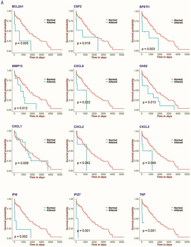
4      Moni et al.

                                                                                                                                                                 Downloaded from https://academic.oup.com/bib/advance-article/doi/10.1093/bib/bbaa376/6041168 by guest on 21 December 2020
Figure 1. Phylogenetic and RNAseq analyses show the genetic relationship between the novel coronavirus SARS-CoV-2, and SARS, MERS and influenza strains. (A)
Phylogenetic viral genome analysis shows SARS-CoV-2 is most closely related to SARS and MERS and is distinct from influenza A strains H1N1, H3N2 and H5N1. (B
and C) Comparison of RNAseq analyses of infected primary human lung epithelial cells with these coronaviruses and influenza A strains indicates the common and
distinct gene sets with upregulated expression in response to infection.

interferon response and cytokine inflammatory terms were the                     Viral infection database screening and drug prediction
most enriched, in line with our differential gene expression                     analyses suggest relevant pathways for subsequent
analysis (Figure 2). Only a small number of viral replication and                study
viral life cycle terms were found significant (Figure 2A and B);                 To aid in contextualising our findings of a unique SARS-CoV-2
this likely reflects the acute nature of the 24 h infection time                 infection gene signature and to provide some external valida-
point used. Cell signalling pathway analyses similarly found                     tion, we conducted an extensive Gene Expression Omnibus (GEO)
striking enrichment of inflammatory interleukin, TNF-alpha                       viral literature screen (Figure 4A). Analysing all the publically
and interferon-mediated signalling effects in host cells, distinct               available viral infection literature in the GEO found infection
from response terms driven by either bacterial or viral infection                with the novel coronavirus SARS-CoV-2 matched most closely
(Figure 3B).                                                                     to studies of SARS-CoV infection, as well as respiration syncytial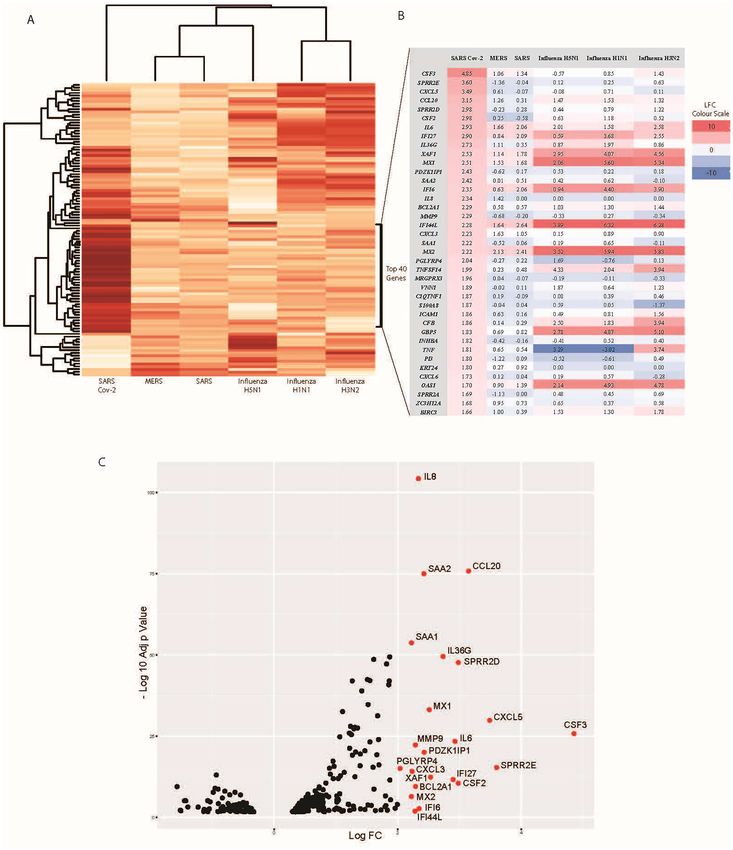
Gene expression profiling of SARS-CoV-2 infections                   5

                                                                                                                                                                            Downloaded from https://academic.oup.com/bib/advance-article/doi/10.1093/bib/bbaa376/6041168 by guest on 21 December 2020
Figure 2. SARS-CoV-2 infection is associated with upregulation of a unique set of genes not seen with SARS, MERS or influenza A infections in human lung epithelial
cells. (A) Heat map depicting the top 108 significant DEGs with infection of the novel coronavirus SARS-CoV-2, compared with top genes related to SARS, MERS and
influenza infections. (B) An expanded view of the top 40 genes with increased expression in SARS-CoV-2 infection, indicating a unique expression profile not seen with
the other viral infections. (C) Volcano plot highlights the most significant SARS-CoV-2 response genes above a log fold-change of 2 and adjusted P value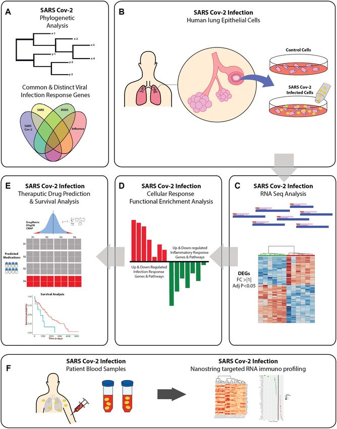
6      Moni et al.

                                                                                                                                                                          Downloaded from https://academic.oup.com/bib/advance-article/doi/10.1093/bib/bbaa376/6041168 by guest on 21 December 2020

Figure 3. Gene ontology and cell signalling pathway analysis finds enriched inflammatory and infection responses to SARS-CoV-2 infection. (A) Gene ontology analysis
finds inflammatory and infection response (bacterial and viral responses pathways combined) significantly enriched with SARS-CoV-2 infection in human lung
epithelial cells. (B) Cell signalling pathway analyses similarly find enrichment, predominantly inflammatory related signalling effects seen with SARS-CoV-2 infection.
Analyses performed using The Gene Ontology, WikiPathways, BioCarta, Reactome and Panther databases.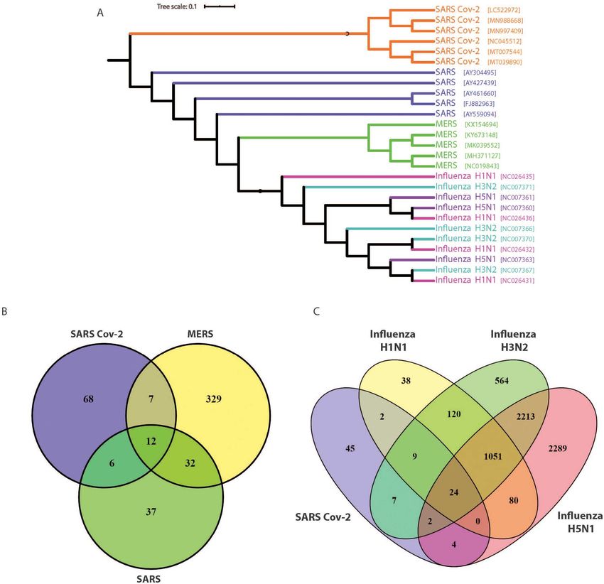
Gene expression profiling of SARS-CoV-2 infections                    7

                                                                                                                                                                               Downloaded from https://academic.oup.com/bib/advance-article/doi/10.1093/bib/bbaa376/6041168 by guest on 21 December 2020

Figure 4. Viral infection literature and drug prediction analysis validates SARS-CoV-2 infection responses and suggests relevant pathways for subsequent study. (A) A
validation GEO viral literature screen was conducted of all publically available literature on viral infections. Analysis finds SARS-CoV-2 infection is most closely related
to published studies of SARS and respiratory viral infections. (B) Predictive drug screening suggests molecules of interest targeting inflammatory, infection, lung fibrotic
and coagulation factors may be of particular relevance to treating early-stage COVID-19 disease.
8 Moni et al.
(P < 0.0001), 1′ -acetoxychavicol acetate (P < 0.0001) and statins find the systemic response is characterised by significant
such as atorvastatin (P < 0.01) (Figure 4B), which are associated perturbation of 140 matched pathways (Supplementary table), of
with increased survival following infections due to anti- which 32 show concordance with those matched in the primary
inflammatory effects [7]. Of additional note are the agents lung cell infection analysis (Figure 6D).
proline dithiocarbamate (P < 0.0001), and dicumarol (P < 0.01); Collectively, these data suggest that while there is a suite
inhibitors having anti-angiogenic and anti-coagulation effects of response genes dysregulated in the acute infection phase,
[8, 9]. These mechanisms may prove relevant to COVID-19 the primary infection in lung cells is characterised by a distinct
pneumonia, with reports emerging of significant cardiovascular altered gene set compared with systemic immune response
involvement in COVID-19 [10], as well as asymptomatic cases genes. Nevertheless, we see common cellular signalling
of acute pulmonary embolism, suggesting significant vascular pathways perturbed in both the primary and systemic acute
Downloaded from https://academic.oup.com/bib/advance-article/doi/10.1093/bib/bbaa376/6041168 by guest on 21 December 2020
effects of currently unknown cause [11] (Figure 4B). It can also be infection responses at the RNA level, and these are dominated
noted that our analysis did not predict the broad spectrum anti- by inflammatory-related cytokine and interferon response
inflammatory hydroxychloroquine, or the antiviral remdesivir pathways.
(agents suggested for the treatment of COVID-19 pneumonia) as
significant targets based on this RNAseq analysis of early acute
Discussion
effects of SARS-CoV-2.
In this study, we have performed phylogenetic viral genome
analyses and extensive transcriptomic characterisations of the
Kaplan–Meier survival estimates using lung
effects of the novel coronavirus SARS-CoV-2, compared with
adenocarcinoma datasets finds a significant
previous human pandemic viruses SARS-CoV, MERS-CoV, and
relationship between SARS-CoV-2 infection response
influenza A strains H1N2, H3N2, and H5N1. To our knowledge,
genes and patient survival this is the largest aggregate comparative genomic and transcrip-
To confirm that there are significant disease associations of the tomic study of the novel coronavirus. We have utilised data
SARS-CoV-2 infection response genes, we performed both uni- from primary human lung epithelial cells, as well as peripheral
variate and multivariate Cox hazard model analysis and Kaplan– blood cells from SARS-CoV-2 infected patients to compare the
Meier survival estimates of lung cancer (LC) adenocarcinoma primary acute lung cell responses with changes in the systemic
patient data (Figure 5 and supplementary table). SARS-CoV-2 immune response. Most notably, despite their close phyloge-
infection response genes are able to stratify LC patients, with netic relationships, SARS-CoV-2 induces a distinct transcrip-
differential expression of the responding gene associated with tional response in infected lung cells compared with other pan-
significantly reduced survival (Figure 5). Notably, we found that demic coronaviruses SARS-CoV and MERS-CoV and is also dis-
inflammatory and chemokine related genes were particularly tinct from influenza A strains. We show that inflammatory
associated with poor prognosis in these patients (P < 0.05). signalling pathways dominate the acute infection response, and
This analysis provides insight into the acute inflammatory while as expected there was little concordance in the pattern of
nature of SARS-CoV-2 infection in the lungs and provides data altered gene expression between infected primary lung cells and
that can be used to examine comorbidity interactions with circulating COVID-19 patient blood cells, there was concordance
COVID-19 early-stage disease. This would support the notion seen in a set of perturbed cell signalling pathways. This might
of giving particular consideration and critical care to patients be expected if the peripheral blood responses are indirect rather
with co-morbidities, particularly with lung related diseases such than due to direct blood cell infection, but these observations
as LC; as survival outcomes in these patients are likely to be may prove therapeutically relevant; it suggests that blocking
significantly impacted by COVID-19 disease. some of these pathways to relieve advanced COVID-19 disease
may also reduce some of pathways particularly active in newly
infected lung epithelial cells. The pathogenic role of these path-
Targeted immune profiling of SARS-CoV-2 infected
ways is not always clear, and while inflammation blockade is
patient blood samples reveals systemic immune
desirable for those seriously ill, it may be less so for the early
responses to infection, and perturbed cell signalling
stages of infection. Nevertheless, further analyses using clinical
pathways similar to that seen in primary lung cell data will be needed to clarify this.
infection As noted above, the human epithelial lung cell transcriptomic
Thus far, we have extensively characterised the acute primary response to SARS-CoV-2 infection after 24 h is surprisingly
human lung epithelial cell response to SARS-CoV-2 infection. different to that of other viruses. Our analyses shows that of
However, in order to understand how this may relate to the the top 108 genes differentially expressed upon SARS-CoV-2,
system-wide immune effects of SARS-CoV-2 infection, we MERS-CoV, SARS-CoV and influenza A viral infections, there is
analysed data from a study of the human immune responses little or no concordance in the responses of infected human lung
in peripheral blood cells of infected individuals. This study used epithelial cells. A notable example is the gene SPRR2E (Small
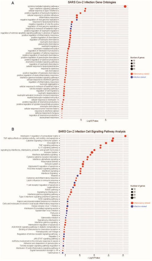
a targeted immune panel on the NanoString platform (Figure 6). proline-rich protein 2E), which encodes small proteins that form
Compared to healthy controls, SARS-CoV-2 infected patient a plasma membrane-associated barrier against extracellular
samples show a distinct systemic infection response at the and environmental factors [12, 13]. SPRR2E is upregulated >3-
RNA level; forming a discrete group by hierarchical clustering fold upon SARS-CoV-2 infection as shown by our analysis, yet
(Figure 6A). A pooled analysis of all patient time-series data is actually downregulated with SARS-CoV and MERS-CoV infec-
found a total of 45 significant DEGs above an absolute log fold- tion, and does not significantly change upon influenza infection
change of 1 (Figure 6B). Notably, compared to our human lung at all (Figure 2). SPRR2E expression has been associated with
epithelial cell analysis, only three genes (MX1, IRF7, BST2) show pulmonary fibrosis [14, 15]; a clinical presentation significant in
concordance between primary lung cell and systemic blood COVID-19 pneumonia [16] and is thus a potentially relevant
cell responses to SARS-CoV-2 infection in the acute phases factor important for further investigation. Additional acute
(Figure 6C). Nevertheless, cell signalling pathway analyses inflammatory responses are also noteworthy. IL8 expressionGene expression profiling of SARS-CoV-2 infections 9
Downloaded from https://academic.oup.com/bib/advance-article/doi/10.1093/bib/bbaa376/6041168 by guest on 21 December 2020
Figure 5. Kaplan–Meier survival estimates using lung adenocarcinoma datasets finds a significant relationship between SARS-CoV-2 infection response genes and
patient survival. Kaplan–Meier estimates for the SARS-CoV-2 infection response genes BCL2A1, CSF2, EPSTI1, MMP13, CXCL6, OAS2, CXCL1, CXCL2, CXCL3, IFI6, IFI27
and TNF. These data indicate a significant relationship with increased mortality in LC patients, suggesting these genes as relevant co-morbidity factors in COVID-19
disease.
is upregulated >2-fold upon SARS-CoV-2 infection in lung will further our abilities to develop targeted therapeutics
cells; however, it is not changed at all with SARS-CoV and the for COVID-19 disease. Indeed, others have also used related
influenza strain infections. IL8 is particularly significant, as transcriptomic study approaches to identified lung epithelium-
it is a chemokine involved in acute inflammatory neutrophil derived factors in COVID-19 but have focussed on (and
infiltrations and tissue damage responses [17]. Understanding confirmed) the interaction with other systems such as blood
mechanisms involved in the distinct SARS-CoV-2 acute response coagulation, a prominent feature of this disease [18].10 Moni et al.
Downloaded from https://academic.oup.com/bib/advance-article/doi/10.1093/bib/bbaa376/6041168 by guest on 21 December 2020
Figure 6. Targeted immune profiling of SARS-CoV-2 infected patient blood samples reveals systemic immune responses to infection, and perturbed cell signalling
pathways that overlap with primary lung infection effects. (A) Heat map of targeted immune RNA profile of 32 patients (n = 10 healthy controls, n = 23 SARS-CoV-
2 infected patients) using a NanoString-targeted immunology panel shows SARS-CoV-2 infected patients have a distinct systemic infection response. (B) A pooled
analysis of time-course data reveals top gene dysregulated in patient blood by SARS-CoV-2 infection. (C) Only three genes are seen to be overlapping between primary
lung cell and blood cell infection responses. (D) Nevertheless, we see concordance at the level of perturbed cell signalling pathways related to immune-inflammatory
cytokine and interferon responses in the host.Gene expression profiling of SARS-CoV-2 infections 11
Beyond the level of expression of individual genes, we sought genes; notably, the human leukocyte antigen genes HLA-DQA1
to investigate ontologies and pathways affected using unbi- and DQB1 are significantly downregulated 5–7 fold compared to
ased database screening approaches. Gene ontology and cell controls (Figure 6B). Such a striking downregulation is notewor-
signalling pathway analyses show a clear acute inflammatory thy; downregulation of the HLAs in circulating immune cells is
response to SARS-CoV-2 infection in lung cells, dominated by also characteristic of HIV [26] and influenza [27, 28] infections,
cytokine and interferon signalling (Figure 3A and B). To contex- as well as seen in a cancer context [17, 29]. This speaks broadly
tualise this finding with prior published literature on viral infec- to the ability of the SARS-CoV-2 virus to induce a response in the
tions, we performed an extensive GEO public literature screen antigen-presenting machinery required for CD4/8 T-cell recogni-
of all available viral infection studies (Figure 4A). We found that tion of viral peptides. While it remains unclear if the peripheral
the SARS-CoV-2 infection transcriptional response in lung cells blood cells themselves become infected with the virus (this may
Downloaded from https://academic.oup.com/bib/advance-article/doi/10.1093/bib/bbaa376/6041168 by guest on 21 December 2020
resembles most that seen in published studies of respiratory be true only of monocytes although still remains unknown [30]),
viral infections SARS-CoV and respiratory syncytial virus, both of our analysis provides initial insight into the systemic responses
which cause respiratory pneumonias in humans. Our predictive in COVID-19 disease that require further validation.
drug target analyses screened hundreds of known agents (most Given SARS-CoV-2 causing COVID-19 disease is associated
with human safety and therapy approvals already) through the with local lung inflammation and damage, and a systemic
Drug Signatures Database screen using DSigDB. Of particular cytokine and inflammatory storm we investigated whether
significance, our analysis matched several broad spectrum anti- any of the DEGs and associated signalling pathways showed
inflammatory drugs (Figure 5B). These data are consistent with concordance between lung cell and peripheral blood cell
the notion that cytokine and inflammatory storms are cause of datasets (Figure 6C and D). We observed little overlap in DEGs;
significant morbidity and mortality in COVID-19 disease [19] and indeed only three genes show concordance (MX1, IRF7 and BST2)
that agents aimed at dampening this overactive inflammatory (Figure 6C). Given the very different nature of the cell types
response may prove efficacious. Antimalarial and antiviral drugs and the infection-reactive nature of the immune responses
have also been examined in COVID-19; notably, Hydroxychloro- evident in the blood cells, this is an expected finding. In contrast,
quine has been subject to much public debate and clinical trials however, we see overlap in terms of significant cell signalling
in COVID-19 patients; although its efficacy is unclear [20]. Our pathways matched to the infection datasets (Figure 6D); this
analysis did not predict hydroxychloroquine as significant in suggests that although different genes are responding, the same
this case, although this may simply reflect the acute nature of cell signalling pathways involved in the two different tissues.
the infection data used in this study rather than the longer term We also note that the peripheral blood cell study employed a
effects of COVID-19. targeted NanoString immune panel and not a complete RNAseq
We gained further insight into the pathogenic nature of genes transcriptome analysis in this case. It is thus possible that
we identified as altered with SARS-CoV-2 infections by conduct- further work using whole transcriptome RNAseq approaches
ing survival analyses using LC patient datasets. We performed would detect larger sets of concordant genes and pathways in
this study to identify factors that have a particularly strong effect affected individual patient tissues. Nevertheless, this analysis
on mortality (and by implication, severe morbidity) in patients provides the first comprehensive comparison between human
with advanced disease that also affects the lung. This work coronaviruses, influenza viruses, as well as data on the immune
revealed that many of these SARS-CoV-2 response genes, espe- and inflammatory responses in lung and peripheral blood cells
cially inflammatory-related genes, are indeed associated with at the level of the transcriptome.
significantly higher mortality in such patients (Figure 5). This
may have important implications for the treatment of COVID-19
patients that also present with co-morbidities, and particularly
Conclusions
so for co-morbid lung conditions that may involve the same We performed phylogenetic and RNAseq analyses to compare
inflammatory gene and pathway sets. It also accords with the the viral genome, and infection responses between SARS-CoV-
general observations of the effectiveness of anti-inflammatory 2, SARS-CoV, MERS-CoV, and pandemic influenza A strains
drugs in reducing disease severity in COVID-19 and in the known H1N1, H3N2, and H5N1. We found that SARS-CoV-2 induces
role of inflammatory factors in the progression of this dis- a unique transcriptional response in human lung epithelial
ease [21–23]. This indicates a strategy to identify those patient cells and that this is dominated by inflammatory cytokine
comorbidities that are at particular risk of having poor COVID-19 and interferon response genes. We provide external validation
outcomes. for these genes and confirmed that although SARS-CoV-2
Thus far our analyses provide novel comparative insight infection is associated with a unique transcriptional response,
into SARS-CoV-2 infection in primary human lung cells. While it resembles most closely those of SARS-CoV and respiratory
our analysis of SARS-CoV-2 infection represents a model of syncytial virus infections in the known literature. COVID-19
the acute primary infection responses in the lungs, the nature patient blood sample analyses also indicate concordance with
of the sustained systemic immune responses is also of great lung cell infection responses at the level of cell signalling
therapeutic importance. There are only limited data available on pathway perturbations.
COVID-19 patient peripheral blood analyses, and at the time of These analyses advance our understating of nature of SARS-
writing only two such datasets are publically available [24, 25]. CoV-2 infection and the cell and immune responses to it and
We analysed the unpublished patient data made available in the show how the infection might be examined for other diseases
preprint by Ong et al. [25] and assessed concordant significant and drug interactions. The pathways we identified can indicate
genes and pathways between peripheral blood and primary a possible role for already approved drugs, although the latter
lung cell responses (Figure 6). Our differential gene expression would only be of utility when if there were further clinical
analyses of healthy control and SARS-CoV-2 infected patient data on these drugs in COVID-19. SARS-CoV-2 is only a very
peripheral blood cells indicate that the latter cluster separately recent discovery, so relatively few studies have been performed
from healthy controls (Figure 6A). A pooled analysis of the sys- on its treatment. Given the global importance of this virus,
temic immune patient time-course data finds a suit of affected more datasets will become available, enabling further work to12 Moni et al.
better uncover mechanisms of the unique SARS-CoV-2 infection Gene ontology and cell signalling pathway analyses
response in humans.
We performed gene set enrichment analysis of Gene Ontology
and cell signalling pathways to evaluate the biological relevance
and functional pathways of the SARS-CoV-2 associated genes. All
Materials and methods
functional analyses were performed using the Enrichr (https://a
Genome information and phylogenetic analysis mp.pharm.mssm.edu/Enrichr/) software tools [31]. For cell sig-
nalling pathway enrichment analyses we employed KEGG [32],
The complete genomes sequences used in this study were col-
WikiPathways [33], BioCarta and Reactome [34] databases. We
lected from the Virus Pathogen Database and Analysis Resource
used GO Biological Process (2018) database for gene ontologi-
(ViPR; https://viprbrc.org/). To determine the relationship among
cal analysis [35]. For this work an adjusted P value ≤0.05 was
Downloaded from https://academic.oup.com/bib/advance-article/doi/10.1093/bib/bbaa376/6041168 by guest on 21 December 2020
all these virus strains, we created a comprehensive phylogram
considered as statistically significant for enrichment analysis.
that includes the complete genomes of 27 human beta-
coronaviruses: SARS-CoV-2: MN988668, MN997409, LC522972,
NC_045512, MT007544 and MT039890; SARS-COV: AY304495, Viral infection database screening
AY427439, AY461660, FJ882963 and AY559094; MERS-COV:
We collected all available virus perturbations datasets in the
NC_019843.3, KX154694, KY673148, MK039552 and MH371127;
NCBI GEO database using Enrichr screening. We identified pub-
H1N1: NC_026435, NC_026436, NC_026432 and NC_026431; H3N2:
lished virus studies that found similar gene expression effects
NC_007371, NC_007366, NC_007370 and NC_007367; and H5N1:
in host cells that we identified in our SARS-CoV-2 infection
NC_007363, NC_007361 and NC_007360. The tree was inferred
analyses.
using the FastME program integrated with a ViPR database using
genomic data with 1000 bootstrap replications. Then, Newick
formatted phylogram was re-designed with the interactive Drug prediction analyses
tree of life online tool (https://itol.embl.de/). Furthermore, the
We have used protein-drug interaction data from the DSigDB,
genomic distance and G + C difference were calculated through
DrugMatrix and CMAP databases to identify potential drugs to
digital DNA–DNA hybridization using Genome-to-Genome
be proposed in the SARS-CoV-2. For each gene set of our interest
Distance Calculator server v2.1 with the default settings. For
from the selected pathways and GO terms, we calculated the
this analysis, we considered reference genomes of SARS-CoV
frequency (f ) of genes in the study set (s) that interact with a
(Refseq: NC_004718.3) and SARS-CoV-2 (Refseq: NC_045512.2);
drug, and the frequency (F) of genes in the population set (S)
and formula-2 derived DDH and GC difference scores as per the
that interact with the same drug. We then performed a test
server’s recommendation.
to determine how likely it would be to obtain at least f genes
interacting with a drug if s genes would be randomly drawn
from the population, given that the frequency F and size S of the
Data pre-processing and identification of DEGs
population. This can be represented mathematically as follows:
We analysed two SARS-CoV-2 related RNAseq transcriptomic
datasets. One was an independent biological triplicate of ! !
cultured primary human lung epithelial cells that received F S−s
either infection with SARS-CoV-2 (GSE147507) or a mock- f F−f
P(f ) = .
treatment [3]. The second dataset was from a study of immune
!
S
responses in healthy controls and COVID-19 cases that employed s
a NanoString Human Immunology Panel to profile collected
peripheral blood cells RNA extracted from whole blood samples
(E-MTAB-8871). We also analysed data from primary epithelial Survival prediction analysis
cells infected with other viruses including SARS-CoV (GSE47963),
To evaluate how LC patient survival is influenced by expression
Mers-CoV (GSE100504) and three different influenza strains
of candidate genes identified as significant in SARS-CoV-2
(H1N1, H3N2 and H5N1) (GSE89008). The SARS-CoV and Mers-
infections we collected clinical and RNAseq data for LC from
CoV data (and respective controls) were generated from human
the TCGA genome data analysis centre (http://gdac.broadinstitu
lung epithelial cells from RNA extracted 24 h post infection. The
te.org). The LC clinical dataset (Lung Adenocarcinoma, TCGA,
influenza virus data are human lung epithelial cell transcrip-
PanCancer Atlas) consists of 566 samples with 81 features,
tome response to infection with 24 h of H1N1, H3N2, and H5N1
and RNAseq gene expression data included 510 cases [36]. We
influenza virus infection. We used the DESeq2 R Bioconductor
matched patient ID in both clinical and RNAseq datasets and
package to analyse all these RNASeq transcriptomic data and
selected patients with data available for both. Among the clinical
the LIMMA (linear models for microarray data) normalization R
variables, we only considered six (ethnicity, anatomical site of
Bioconductor package was used to analyse gene expression data
cancer, the histological grade of cancer, primary tumour site
sets. Note that the dataset we employed was generated within a
and neoplasm status with the tumour) to evaluate survival with
single experimental study, which formed part of the same data
the significant genes. Hence, we considered z-score values to
batch; for this reason, no batch effect correction was required
determine the altered and normal (unaltered) expression of a
in the pre-processing of our data. For each dataset, differential
gene.
gene expression analysis was run between case and control
To determine the patient survival association of the signif-
data, with an adjusted P valueGene expression profiling of SARS-CoV-2 infections 13
Here, h(t|Xi ) is the hazard function at time t on a subject i with Supplementary data
covariate information as the vector Xi , h0 (t), which is the baseline
Supplementary data are available online at Briefings in Bioin-
hazard function and is independent of covariate information.
formatics.
Hence, β indicates a corresponding regression coefficients vector
to the covariates respectively. We have calculated the hazard
ratio (HR) based on the estimated regression coefficients from Code and data availability
the fitted Cox PH model to identify whether a specific covari-
ate affects patient survival. The HR for a covariate xr can be All differential gene expression, drug predictive, and sur-
expressed as exp (β r ). Thus, the HR for any covariate can be vival analyses can be freely accessed through the github
measured by applying an exponential function to the respective repository at: https://github.com/m-moni/COVID-19
Downloaded from https://academic.oup.com/bib/advance-article/doi/10.1093/bib/bbaa376/6041168 by guest on 21 December 2020
(β r ) coefficient.
To determine survival status we used a non-parametric
Funding
method to estimate the survival function. Thus, we used the
product limit estimator known as the Kaplan–Meier estimator The authors have no funding declarations.
for the survival function which is defined as follows [37]:
Y
j
dj
References
Ŝ tj = 1− .
nj 1. Center JHCR. John Hopkins Coronavirus Resource Center.
i=1
2. Wang Y, et al. Unique epidemiological and clinical fea-
Hence Ŝ(tj ) is the estimated survival function at time tj , dj tures of the emerging 2019 novel coronavirus pneumonia
is the number of events occurred at tj and nj is the number of (COVID-19) implicate special control measures. J Med Virol
subjects available at tj . Then two or more groups were compared 2020;92(6):568–76.
using a logrank test. We applied logrank tests to detect the most 3. Blanco-Melo D, et al. Imbalanced host response to SARS-CoV-
significant genes associated with patient survival time, that is, 2 drives development of COVID-19. Cell 2020;181(5):1036, e9–
when comparing groups of patients with altered expression for a 45.
particular gene, compared with groups with normal (unaltered) 4. Heinz S, et al. Transcription elongation can affect genome 3D
expression, the patient survival of these two groups differ. The structure. Cell 2018;174(6):1522, e22–36.
null hypothesis for this test can be formulated as follows: 5. Heller N. Primary human airway epithelial cell transcriptome
response to a wild type Mers-CoV (icMERS-CoV EMC2012). Gene
H0 : Saltered (t) = Snormal (t) Expression Omnibus, 2017.
HA : Saltered (t) 6 = Snormal (t). 6. Yoo M, et al. DSigDB: drug signatures database for gene set
analysis. Bioinformatics 2015;31(18):3069–71.
Here H0 is a survival function that is the same for altered and 7. Parihar SP, Guler R, Brombacher F. Statins: a viable candidate
normal genes and HA indicates survival functions that are not for host-directed therapy against infectious diseases. Nat
the same for these two groups. Rev Immunol 2019;19(2):104–17.
8. Morais C, et al. Anti-angiogenic actions of pyrrolidine dithio-
Key Points carbamate, a nuclear factor kappa B inhibitor. Angiogenesis
• Phylogenetic analysis of the novel 2019 coronavirus 2009;12(4):365–79.
designated as SARS-CoV-2 shows that it is most 9. Timson DJ. Dicoumarol: a drug which hits at least two very
closely related to SARS and MERS at the level of different targets in vitamin K metabolism. Curr Drug Targets
2017;18(5):500–10.
the viral genome. Comparative RNAseq analyses
show SARS-CoV-2 induces a unique transcriptional 10. Sarah Zaman AIM, Jennings GLR, Schlaich M, et al. Car-
response in host human cells compared with SARS, diovascular disease and COVID-19: Australian/New Zealand
consensus statement. Med J Aust 2020.
MERS, influenza H1N1, H3N2 and H5N1.
• The SARS-CoV-2 infection response in human lung 11. Danzi GB, et al. Acute pulmonary embolism and COVID-19
epithelial cells is associated with a unique suite of pneumonia: a random association? Eur Heart J 2020.
inflammatory gene upregulation, and these genes are 12. Nozaki I, et al. Small proline-rich proteins 2 are noncoordi-
enriched in cytokine and interferon-mediated inflam- nately upregulated by IL-6/STAT3 signaling after bile duct
matory processes. ligation. Lab Invest 2005;85(1):109–23.
• Predictive drug screening based on the unique SARS- 13. Oany AR, et al. Design of novel viral attachment inhibitors
CoV-2 signature highlights drugs targeting inflamma- of the spike glycoprotein (S) of severe acute respiratory syn-
drome coronavirus-2 (SARS-CoV-2) through virtual screen-
tory, fibrotic and vascular mechanisms that may prove
ing and dynamics. Int J Antimicrob Agents 2020;106177.
critical in follow up studies on infection disease mech-
14. Kwapiszewska G, et al. Transcriptome profiling reveals the
anisms. And upregulated SARS-CoV-2 genes show sig-
complexity of pirfenidone effects in idiopathic pulmonary
nificant association with increased mortality in an
fibrosis. Eur Respir J 2018;52(5).
independent lung disease dataset; providing insight
15. Taz TA, et al. Network-based identification genetic effect of
into comorbidity associations in COVID-19.
• Analysis of human blood samples from SARS-CoV-2 SARS-CoV-2 infections to idiopathic pulmonary fibrosis (IPF)
patients. Brief Bioinform 2020.
infected people highlights genes altered in the sys-
16. Shi H, et al. Radiological findings from 81 patients with
temic immune response to infection, and some over-
COVID-19 pneumonia in Wuhan, China: a descriptive study.
lapping pathways and ontologies concordant between
Lancet Infect Dis 2020;20(4):425–34.
the primary lung cell response, and the systemic
17. Harada A, et al. Essential involvement of interleukin-8 (IL-8)
responses.
in acute inflammation. J Leukoc Biol 1994;56(5):559–64.14 Moni et al.
18. FitzGerald ES, Jamieson AM. Unique transcriptional changes 1 negative factor (HIV-1 Nef): what might we learn from
in coagulation cascade genes in SARS-CoV-2-infected lung natural sequence variants? Viruses 2012;4(9):1711–30.
epithelial cells: a potential factor in COVID-19 coagu- 27. Koutsakos M, et al. Downregulation of MHC class I expression
lopathies. bioRxiv 2020. by influenza a and B viruses. Front Immunol 1158;10:2019.
19. Mehta P, et al. COVID-19: consider cytokine storm syn- 28. Nain Z, et al. Pathogenetic profiling of COVID-19 and SARS-
dromes and immunosuppression. Lancet 2020;395(10229): like viruses. Brief Bioinform 2020.
1033–4. 29. Hicklin DJ, Marincola FM, Ferrone S. HLA class I antigen
20. Fabio S Taccone JG, Vincent J-L. Hydroxychloroquine in downregulation in human cancers: T-cell immunotherapy
the management of critically ill patients with COVID- revives an old story. Mol Med Today 1999;5(4):178–86.
19: the need for an evidence base. Lancet Respir Med 30. Jafarzadeh A, et al. Contribution of monocytes and
Downloaded from https://academic.oup.com/bib/advance-article/doi/10.1093/bib/bbaa376/6041168 by guest on 21 December 2020
2020. macrophages to the local tissue inflammation and cytokine
21. Conti P, et al. Induction of pro-inflammatory cytokines (IL-1 storm in COVID-19: lessons from SARS and MERS, and
and IL-6) and lung inflammation by Coronavirus-19 (COVI- potential therapeutic interventions. Life Sci 2020;257:118102.
19 or SARS-CoV-2): anti-inflammatory strategies. J Biol Regul 31. Kuleshov MV, et al. Enrichr: a comprehensive gene set
Homeost Agents 2020;34(2):327–31. enrichment analysis web server 2016 update. Nucleic Acids
22. Villar J, et al. Efficacy of dexamethasone treatment for Res 2016;44(W1):W90–7.
patients with the acute respiratory distress syndrome 32. Kanehisa M, Goto S. KEGG: Kyoto encyclopedia of genes and
caused by COVID-19: study protocol for a randomized con- genomes. Nucleic Acids Res 2000;28(1):27–30.
trolled superiority trial. Trials 2020;21(1):717–7. 33. Slenter DN, et al. WikiPathways: a multifaceted pathway
23. Ahamad MM, et al. A machine learning model to iden- database bridging metabolomics to other omics research.
tify early stage symptoms of SARS-Cov-2 infected patients. Nucleic Acids Res 2017;46(D1):D661–7.
Expert Syst Appl 2020;160:113661. 34. Fabregat A, et al. The Reactome pathway knowledgebase.
24. Xiong Y, et al. Transcriptomic characteristics of bron- Nucleic Acids Res 2018;46(D1):D649–d655.
choalveolar lavage fluid and peripheral blood mononuclear 35. The Gene Ontology Resource. 20 years and still GOing strong.
cells in COVID-19 patients. Emerging Microbes & Infections Nucleic Acids Res 2019;47(D1):D330–d338.
2020;9(1):761–70. 36. Hoadley KA, et al. Cell-of-origin patterns dominate the
25. Eugenia Ziying Ong YFZC, Leong WY, Lee NMY, et al. A molecular classification of 10,000 Tumors from 33 types of
dynamic immune response shapes COVID-19 progression. cancer. Cell 2018;173(2):291, e6–304.
Unpublished - preprint. 2020. 37. Rana HK, et al. Machine learning and bioinformatics models
26. Mwimanzi P, et al. Human leukocyte antigen (HLA) class I to identify pathways that mediate influences of welding
down-regulation by human immunodeficiency virus type fumes on cancer progression. Sci Rep 2020;10(1):2795.You can also read