GENERAL ASPECTS ABOUT THE STRUCTURE OF SEVERE ACUTE RESPIRATORY SYNDROME CORONAVIRUS 2 (SARS-COV-2) - SCIELO CUBA
←
→
Page content transcription
If your browser does not render page correctly, please read the page content below

Revista Cubana de Investigaciones Biomédicas. 2020;39(3):e867

                                                                                                                            Artículo de revisión

     General aspects about the structure of Severe Acute Respiratory Syndrome
                                           Coronavirus 2 (SARS-CoV-2)
     Aspectos generales sobre la estructura del coronavirus del síndrome respiratorio
                                            agudo grave 2 (SARS-CoV-2)

Francisco Sotomayor Lugo1 https://orcid.org/0000-0001-9854-8688
José Miguel Corbacho Padilla2 https://orcid.org/0000-0001-6200-1445
Ana Margarita Valiente Linares3 https://orcid.org/0000-0002-2447-6404
Yudelkis Benítez Cordero1 https://orcid.org/0000-0002-9688-7501
Tatiana Viera González4 https://orcid.org/0000-0002-6323-6585

1
    Centro Nacional de Genética Médica. La Habana, Cuba.
2
    Universidad de Ciencias Médicas de la Habana. Cuba.
3
    Hospital Clínico Quirúrgico “Juan Bruno Zayas”. Santiago de Cuba, Cuba.
4
    Centro de Genética Médica de Marianao. La Habana, Cuba.

*
    Autor para la correspondencia: franciscosotomayorl89@gmail.com

ABSTRACT
Introduction: In late 2019, a new coronavirus named Severe Acute Respiratory Syndrome
Coronavirus 2 (SARS-CoV-2) that causes respiratory-related illness was reported in Wuhan,
China. This virus can attack human lung cells causing a disease called coronavirus disease 2019
(COVID-19), which can lead to pneumonia and acute respiratory distress syndrome.
Objective: Describe the structural characteristics of Severe Acute Respiratory Syndrome
Coronavirus 2 (SARS-CoV-2).
Methods: A review was written from 47 bibliographic references. Articles and information from
national and international journals available in the PubMed, Scopus, Medline, SciELO databases

                                                                                                                                              1
              Esta obra se encuentra bajo una licencia Creative Commons CC-By 4.0 https://creativecommons.org/licenses/by-nc/4.0/deed.es_ES
Revista Cubana de Investigaciones Biomédicas. 2020;39(3):e867

were used. The quality, reliability and validity of the selected articles were analyzed to carry out
an adequate review. Analysis-synthesis and logical deduction methods were applied.
Development: An introduction to the general aspects of the structure of SARS-CoV-2 is provided
by stating the characteristics of the structural and non-structural proteins encoded by the viral
genome, which provides the basis for understanding viral entry mechanisms to the host cell, and
may be useful to stimulate the search for novel insights and possible therapeutic targets to fight the
infection.
Conclusions: Knowledge of the structure of the SARS-CoV-2 virus and the characteristics of the
structural and non-structural proteins provides the basis for understanding the viral mechanisms of
infection and the strategies for developing effective therapeutics.
Keywords: Coronavirus Infections; Severe Acute Respiratory Syndrome Coronavirus 2; SARS-
CoV-2; Viral Structure; Viral Genome.

RESUMEN
Introducción: A finales de 2019 se informó el brote de un nuevo coronavirus en Wuhan, China,
llamado Severe Acute Respiratory Syndrome Coronavirus 2 (SARS-CoV-2) que causa alteraciones
en el aparato respiratorio. Este virus puede atacar las células humanas del pulmón causando una
enfermedad denominada enfermedad por coronavirus 2019 (COVID-19), que puede producir
neumonía y un síndrome de dificultad respiratoria aguda.
Objetivo: Describir las características estructurales del virus SARS-CoV-2.
Métodos: Se realizó una revisión bibliográfica a partir de 47 referencias. Se utilizaron artículos e
información de revistas nacionales e internacionales disponibles en las bases de datos PubMed,
Scopus, Medline, SciELO. Para llevar a cabo una revisión adecuada, se analizaron la calidad,
fiabilidad y validez de los artículos seleccionados. Se aplicaron métodos de análisis-síntesis y
deducción lógica.
Desarrollo: Se proporciona una introducción de los aspectos generales de la estructura del SARS-
CoV-2. Se enuncian las características de las proteínas estructurales y no estructurales codificadas
por el genoma viral, lo que provee la base para comprender los mecanismos virales de entrada a la
célula huésped. El artículo resulta de utilidad para estimular la búsqueda de nuevos conocimientos
y posibles objetivos terapéuticos para combatir la infección.

                                                                                                                                              2
              Esta obra se encuentra bajo una licencia Creative Commons CC-By 4.0 https://creativecommons.org/licenses/by-nc/4.0/deed.es_ES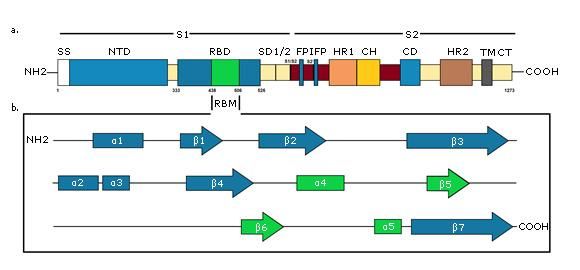
Revista Cubana de Investigaciones Biomédicas. 2020;39(3):e867

Conclusiones: El conocimiento sobre la estructura del virus SARS-CoV-2 y las características de
las proteínas estructurales y no estructurales que lo forman ampara significativamente las bases
para entender los mecanismos virales de la infección y las estrategias para el desarrollo terapéutico
efectivo.
Palabras clave: infecciones por coronavirus; coronavirus del síndrome respiratorio agudo grave
2; SARS-CoV-2; estructuras virales; genoma viral.

Recibido: 28/05/2020
Aceptado: 06/06/2020

                                                         Introduction
Emerging viruses are becoming a great danger to global health and among them, coronaviruses are
a notable example. Very virulent forms have emerged from their natural animal hosts and represent
a threat to human communities. Between 2002-2003, Severe Acute Respiratory Syndrome
Coronavirus (SARS-CoV)(1,2) arose in China from bat populations, passing to civets and finally to
humans. Ten years later, Middle East Coronavirus Respiratory Syndrome Coronavirus (MERS-
CoV)(3) also arose from bats, transferring in the Middle East to dromedary camels and then to
humans. Recently, at the end of 2019, another virus emerged in Wuhan, Hubei Province, China,
which has been recognized as Severe Acute Respiratory Syndrome Coronavirus 2 (SARS-CoV-
2),(4,5) responsible for coronavirus disease 2019 (COVID-19).
Current coronavirus classification recognizes 39 species in 27 subgenus, five genus and two
subfamilies that belong to the Coronaviridae family, Cornidovirineae suborder, Nidovirales order
and Riboviria realm. Within the Coronaviridae family is the Orthocoronavirinae subfamily, which
is made up of four genera, which according to their genetic structure are grouped into taxa called
Alphacoronavirus,           Betacoronavirus,                Gammacoronavirus                     and        Deltacoronavirus.                Both
alphacoronaviruses and betacoronaviruses cause different diseases in different mammalian
species,(6) including respiratory infections and gastroenteritis. SARS-CoV-2 virus belongs to the

                                                                                                                                                3
             Esta obra se encuentra bajo una licencia Creative Commons CC-By 4.0 https://creativecommons.org/licenses/by-nc/4.0/deed.es_ES
Revista Cubana de Investigaciones Biomédicas. 2020;39(3):e867
severe acute respiratory syndrome related coronavirus species among the group of
betacoronaviruses.(7)
Many aspects of the structure and biology of the SARS-CoV-2 virus have not yet been elucidated.
Development of effective preventive and therapeutic strategies may be hindered by the lack (need)
of information related to the structural details of viral proteins, although some crystallographic
structures(8,9) are already available. This review describes the general aspects of the structure of the
SARS-CoV-2 virus, referring to the characteristics of the proteins encoded by the viral genome.
Methods
A review was carried out between March and May 2020. 47 bibliographic references were
analyzed, which correspond to articles from national and international journals available in the
PubMed, Scopus, Medline and SciELO databases. The most recent published literature in relation
to the subject area studied was considered as the bibliography selection criterion. Web pages of the
Ministerio de Salud Pública de Cuba, World Health Organization and Pan-American Health
Organization were explored. For the collection of information, a search strategy was applied using
health science keywords and connectors, and analysis-synthesis and logical deduction methods
were applied to write the article.
General structure of SARS-CoV-2
Coronaviruses are enveloped viruses with a single-stranded, positive-sense RNA molecule.(10)
They belong to a large family of viruses that infect birds and various mammals, including camelids,
bats, civets, rats, mice, dogs, and cats. Viruses in this family are 60-220 nm size and filamentous
forms from 9-13 nm in diameter are usually observed.(11)
SARS-CoV-2 viral genome encodes at least sixteen non-structural proteins (nsps) and four
structural proteins. Structural proteins (Fig. 1) form the viral particle that consists of a
nucleocapsid, formed by the viral genome to which multiple copies of nucleocapsid (N) protein are
attached, which is surrounded by an envelope where proteins are inserted: spike (S), membrane
(M) and envelope (E) proteins are inserted.
4
Esta obra se encuentra bajo una licencia Creative Commons CC-By 4.0 https://creativecommons.org/licenses/by-nc/4.0/deed.es_ESRevista Cubana de Investigaciones Biomédicas. 2020;39(3):e867
Fig. 1 - Schematic diagram of the SARS-CoV-2 virus. The four structural proteins are shown: S, M, N and
E proteins.
SARS-CoV-2 Genome organization
The virus has a positive-polarity single-stranded RNA genome, 26-32 kb long.(12) From this
molecule, proteins that are necessary to finish the complete replication cycle are synthesized. These
proteins include a replicase transcriptase complex that produces more RNA and various structural
proteins that build new virions.
SARS-CoV-2 genome is similar to that of the SARS-CoV and MERS-CoV viruses, with 88 % and
50% of sequence identity,(13) respectively. In 5'-3' sense, the genome is organized into fourteen
open reading frames (ORFs) (Fig. 2a) that encode a variety of structural or non-structural proteins,
according to their functions in the viral particle.
5
Esta obra se encuentra bajo una licencia Creative Commons CC-By 4.0 https://creativecommons.org/licenses/by-nc/4.0/deed.es_ESRevista Cubana de Investigaciones Biomédicas. 2020;39(3):e867
A- Schematic diagram of the sequence of ORFs in viral RNA. B- Schematic diagram of the pp1a and pp1ab polyproteins encoded by the ORF1a
and ORF1b. PLpro, papain-like proteases; TM, transmembrane; Mpro, chymotrypsin-like protease; RdRp, RNA-dependent RNA polymerase;
Hel, helicase; Exo, 3 '→ 5' exonuclease; NU, NendoU: uridylate-specific endoribonuclease; MT, ribose-2′-O-methyltransferase; nsp, non-
structural protein. Created for this article.
Fig. 2 - Structure of the SARS-CoV-2 virus genome.
During the transformation phase, the ORF1a and ORF1b sequences, which correspond to almost
two thirds of the virus RNA, are translated into two large overlapping polyproteins called pp1a and
pp1ab,(14) which are processed into sixteen non-structural proteins (nsps 1-16), many of which form
the replicase transcriptase complex.(15) The nsps 3 and 5 supplement the enzymes papain-like
proteases (PLpro)(16) and chymotrypsin-like protease Mpro (3CLpro),(17) respectively, whom
decides the processing of viral polyproteins. Cleavage occurs between the products of ORF1a and
ORF1b to form pp1a made up of nsps 1–11, while pp1ab will be made up of nsps 1–16 (Fig. 2b).
The other SARS-CoV-2 ORFs that correspond to one third of the viral genome, encode for the four
structural proteins N, S, M and E, as well as other accessory proteins whose function is not yet
known, although it is known that they don’t participate in viral replication in cultured cells.
SARS-CoV-2 non-structural proteins
SARS-CoV-2 non-structural proteins have different functions that decide several processes in the
virus and in the host cell. The non-structural protein (nsp) 1 promote inhibition of type 1 interferon
(IFN) signaling and block the innate immune response in the host cell by degrading the host mRNA,
6
Esta obra se encuentra bajo una licencia Creative Commons CC-By 4.0 https://creativecommons.org/licenses/by-nc/4.0/deed.es_ESRevista Cubana de Investigaciones Biomédicas. 2020;39(3):e867
inhibiting translation and stopping the cell cycle,(18) while nsp2 binds to a proinhibitory protein
whose function is still unknown. In addition to guaranteeing the processing of the pp1a and pp1ab
polyproteins, nsp3 and nsp5 promote the expression of cytokines.(19) The nsps 4 and 6 contribute
to the structure of the double membrane vesicles by determining the transmembrane protein
framework.(20,21) nsp7/8 complex is a hexadecameric complex that support of the replication
enzyme,(22) where nsp8 constitutes a second RNA polymerase that can function as a primase. RNA-
dependent RNA polymerase is represented by nsp12, which together with the helicase RNA
(nsp13) guarantee the assembly of the replicase transcriptase complex.(23) nsp9 constitutes an RNA-
binding protein phosphatase, which like nsp10, is part of the replicase complex. nsp10 establish
also a necessary link for the adequate function of the main viral protease (Mpro).(19) nsp14 is a
bifunctional protein, which exhibits exonuclease activity 3'→5' with a role in maintaining the
fidelity of RNA transcription,(24) and (guanine-N7)-methyl transferase activity, involved in RNA
cap formation.(25) RNA cap formation is also facilitated by nsp16, which is an S-adenosyl-
methionine RNA-dependent with (ribose-2'O)-methyl transferase activity.(26) Finally, nsp15
encode a uridylate-specific endoribonuclease (NendoU)(27) that is crucial for virus replication and
distinguishes nidoviruses from other RNA viruses. These and other functions are summarized in
table.
Table - Functions of the non-structural proteins of the SARS-CoV-2 virus
Non-structural
Function
proteins
nsp1 Inhibition of type 1 IFN signaling; block of the innate immune response in the host cell.
nsp2 Binds to a proinhibitory protein of unknown function.
PLpro activity for processing the pp1a and pp1ab polyproteins; cytokine expression; IFN-β
nsp3
antagonist; deubiquitinase activity.
nsp4 Double membrane vesicles formation.
nsp5 3CLpro activity for processing of pp1a and pp1ab polyproteins; cytokine expression.
nsp6 Double membrane vesicles formation.
nsp7 Hexadecameric complex formation.
nsp8 Primase; hexadecameric complex formation.
nsp9 RNA binding protein phosphatase.
nsp10 Part of the replicase complex.
nsp11 Unknown.
nsp12 RNA-dependent RNA polymerase.
nsp13 RNA Helicase.
7
Esta obra se encuentra bajo una licencia Creative Commons CC-By 4.0 https://creativecommons.org/licenses/by-nc/4.0/deed.es_ESRevista Cubana de Investigaciones Biomédicas. 2020;39(3):e867
nsp14 3'→5' exonuclease activity; N7-MTase activity for the RNA cap formation.
nsp15 Uridylate specific endoribonuclease.
2'O-MTase activity for the RNA cap formation.
nsp16
PLpro, papain-like proteases; IFN, Interferon; 3CLpro, chymotrypsin-like protease Mpro; N7-
MTase, (guanine-N7)-methyltransferase; 2'O-MTase, nucleoside-2'O-methyl transferase.
The three-dimensional structure of the main SARS-CoV-2 protease is very similar to that of SARS-
CoV, sharing a sequence identity of 96 %.(8) This enzyme is essential to translated polyproteins of
viral RNA. Mpro operates at no less than eleven cleavage sites on the ORF1ab polyprotein. The
recognition sequence in most of these sites is Leu-Gln, while the Ser, Ala, Gly residues mark the
cleavage site.(17)
Mpro is a dimer of two identical subunits that together form two active sites,(28) each of which have
three domains identified as I, II and III. This dimerization of the enzyme is necessary for its
catalytic activity. At the catalytic site Cys145 and His41 amino acids carry out the protein cleavage
reaction. In contrast to SARS-CoV virus, SARS-CoV-2 protease presents the substitution of
Ser284, Thr285 and Ile286 by Ala residues which leads to 3.6 times progress of the catalytic
activity of the protease, which go together with a lightly closer packaging of both III domains of
each subunit, leading to greater stability.(8,17)
SARS-CoV-2 structural proteins
M protein is the most abundant structural protein, which is responsible for shaping the virion.
Multiple sequence alignment points show remarkable similarity between the Sars-CoV-2
sequences and those isolated from Pangolin-CoV-MP798 and Bat-CoV-CoVZXC21.(29,30)
However, there is heterogeneity at the N-terminal, where an insert of a Ser residue at position 4 of
SARS-CoV-2 M protein appears to be a unique feature.(31)
M monomer ranges from 25 to 30 kDa, and it is embedded in the envelope through three
transmembrane domains.(32) N-terminal constitutes a small ectodomain, while the C-terminal
endodomain is located on the inner face of the virion membrane contributing to most of the
molecule. The ectodomain can be modified by glycosylation, which influences the tropism of the
8
Esta obra se encuentra bajo una licencia Creative Commons CC-By 4.0 https://creativecommons.org/licenses/by-nc/4.0/deed.es_ESRevista Cubana de Investigaciones Biomédicas. 2020;39(3):e867
organs to be infected. This protein is responsible for the transmembrane transport of nutrients, the
virion release and the envelope formation. Binding with the M protein helps stabilize the N proteins
and promotes the termination of the viral assembly by stabilizing the RNA-N protein complex
within the internal virion.(33)
N protein forms the helicoidal nucleocapsid, joining along the entire viral genome, which assumes
a curl shape. This protein weighs 43-50 KDa and is phosphorylated in a discrete number of serines
and threonines. Although the role of this phosphorylation has not yet been determined, it has been
suggested that it is related to regulatory functions and its ability to bind to the viral genome.(33)
N protein contains two domains that allow it to recognize viral RNA. It is also capable of binding
to nsp3(34) to address the genome to the replicase transcriptase complex and ensure nucleocapsid
packaging. It also works as an IFN-β antagonist,(35) contributing to the mechanisms of viral evasion
to the immune system, by preventing interferon from stopping viral replication in still healthy cells
and the destruction of already infected cells, highlighting the importance of exogenous
administration of interferon in the treatment of disease caused by the virus. N protein also
participates in the repression of host cell interference RNAs that suppress the expression of specific
sequences of the viral genome and that constitute a vital part of the body's immune response to
viruses.(36)
E protein is a small polypeptide that is found in limited amounts in the viral envelope. In particular,
the E protein sequence of SARS-CoV-2 is identical to that isolated from Pangolin-CoV-MP798
and from Bat-CoV-CoVZXC21, CoVZC45 and RaTG13.(32) Two distinguishing features of Sars-
CoV-2 E protein when compared to that of other homologs is the substitution at position 69 where
an Arg replaces Glu, Gln, Asp, and deletion at position 70 corresponding to Gly or Cys in another
E proteins.(37)
During the replication cycle, this protein is abundantly expressed within the infected cell, but only
a small amount is incorporated into the envelope of the virion. Most of it is located in the
intracellular traffic site, such as the Golgi complex, where it participates in the assembly of the
particle and it is very important in the production and maturation of the viral particle.(37)
SARS CoV-2 virus glycoprotein S is a densely glycosylated trimer that projects in the form of
spicules and it is 16-21 nm long.(38) It is a typical class I viral fusion protein that requires the
cleavage of a protease for the activation of its fusion potential, so that during viral infection,(39) it
9
Esta obra se encuentra bajo una licencia Creative Commons CC-By 4.0 https://creativecommons.org/licenses/by-nc/4.0/deed.es_ESRevista Cubana de Investigaciones Biomédicas. 2020;39(3):e867
is cleaved by a furin-like cellular protease into two peptides of the same size, S1 and S2. There is
a cleavage site between the S1 and S2 subunits (amino acids 682-685, RRAR) that creates a
polybasic furin site that has been linked to rapid virus transmission, by facilitating endocytosis
mechanisms to human cells (Fig 3a).(40)
A - Primary structure of S protein. SS, signal sequence; NTD, N-terminal domain; RBD, receptor-binding domain, RBM, receptor-binding motif;
SD1/2, subdomain 1 & 2; S1/S2 and S2', protease cleavage site; FP, fusion peptide; IFP, internal fusion peptide; HR1, heptad repeat 1; CH, central
helix; CD, connector domain; HR2, heptad repeat 2; TM, transmembrane domain; CT, cytoplasmic tail. B - Secondary structure of S protein RBD.
RBM is green colored. Created for this article.
Fig. 3 - Schematic diagram of the SARS-CoV-2 S protein.
S1 subunit is highly variable between different coronaviruses. In the monomeric structure of S
protein, N and C-terminal portions of this subunit fold as two independent domains, the N-terminal
domain (called NTD) and the C-terminal domain (or C domain).(38) Depending on the virus, NTD
or the C domain can serve as the receptor binding domain (RBD). In SARS-CoV-2, the S1 C
domain has the RBD of approximately 21 kDa and 200 amino acid residues.(9) The RBD subdomain
is responsible for S protein organizing in the trimer form and for binding directly to the peptidase
domain (PD) of Angiotensin-Converting Enzyme 2 (ACE2) of human cells.(41,42) In contrast, S2 is
highly conserved and the fusion peptide is located in it, which is responsible for the fusion of the
viral membrane to the host cell membrane, as well as the cytopathic effect that this virus can
produce when it infects cells in vivo.(38)
10
Esta obra se encuentra bajo una licencia Creative Commons CC-By 4.0 https://creativecommons.org/licenses/by-nc/4.0/deed.es_ESRevista Cubana de Investigaciones Biomédicas. 2020;39(3):e867
The trimeric structure of the SARS-CoV-2 S protein has one RBD in one up conformation and two
RBD in down conformations. This process is triggered when the S1 subunit binds to the host cell
receptor.(43) Receptor binding destabilizes the prefusion trimer, resulting in detachment of the S1
subunit and transition of the S2 subunit to a stable post-fusion conformation. To compromise a host
cell receptor, S1 RBD undergoes hinge-like conformational movements that temporarily hide or
expose the determinants of receptor binding.(44) These two states are referred to as down
conformation and up conformation, where the first corresponds to the inaccessible receptor state,
and the second corresponds to the accessible receptor state, which is stable. Observation of this
phenomenon in the SARS-CoV-2 S protein suggests that it shares the same activation mechanism
that is believed to be conserved in the Coronaviridae family, in which binding of the receptor to
exposed RBDs leads to elimination of S1 and the folding of S2.
The final RBD model of the SARS-CoV-2 S protein contains residues from Thr333 to Gly526.
This subdomain has a twisted five-stranded antiparallel β sheet (β1, β2, β3, β4 and β7) with short
helices and connection loops forming the core. Between the β4 and β7 strands in the core, there is
an extended insertion containing short β5 and β6 strands, α4 and α5 helices and loops (Figure 3b).
This extended insertion is the receptor binding motif (RBM) that contains the majority of the
SARS-CoV-2 contact residues for ACE2 binding. There are nine cysteine residues in the RBD,
eight of which form four pairs of disulfide bonds. Among these four pairs, three are in the core
(Cys336-Cys361, Cys379-Cys432, and Cys391-Cys525) to help stabilize the structure of the β
sheet structure, while the remaining (Cys480-Cys488) connects the loops at the distal end of the
RBM.(44,45)
The presence of RBD in the virus S protein determines that it plays the most important roles in
viral binding, fusion and entry into the host cell, making it a promising target for the development
of antibodies, entry inhibitors and vaccines for the prevention and treatment of the disease(46,47).
Conclusions
Knowledge of the structure of the SARS-CoV-2 virus and the characteristics of the structural and
non-structural proteins provides the basis for understanding the viral mechanisms of infection and
the strategies for developing effective therapeutics.
11
Esta obra se encuentra bajo una licencia Creative Commons CC-By 4.0 https://creativecommons.org/licenses/by-nc/4.0/deed.es_ESRevista Cubana de Investigaciones Biomédicas. 2020;39(3):e867
References
1. Zhong NS, Zheng BJ, Li YM, Xie ZH, Chan KH, Li PH, et al. Epidemiology and cause of severe
acute respiratory syndrome (SARS) in Guangdong, People’s Republic of China, in February, 2003.
The Lancet 2003;362(9393):1353-8. DOI: 10.1016/s0140-6736(03)14630-2
2. Kuiken T, Fouchier RA, Schutten M, Rimmelzwaan GF, Van Amerongen G, van Riel D, et al.
Newly discovered coronavirus as the primary cause of severe acute respiratory syndrome. The
Lancet 2003;362(9380):263-70. DOI: 10.1016/S0140-6736(03)13967-0
3. Zaki AM, van Boheemen S, Bestebroer TM, Osterhaus AD, Fouchier RA. Isolation of a novel
coronavirus from a man with pneumonia in Saudi Arabia. New England Journal of Medicine
2012;367(19):1814-20. DOI: 10.1056/NEJMoa1211721
4. Ren LL, Wang YM, Wu ZQ, Xiang ZC, Guo L, Xu T, et al. Identification of a novel coronavirus
causing severe pneumonia in human: a descriptive study. Chinese Medical Journal 2020;133(9):
1015–1024. DOI: 10.1097/CM9.0000000000000722
5. Zhu N, Zhang D, Wang W, Li X, Yang B, Song J, et al. A novel coronavirus from patients with
pneumonia in China, 2019. New England Journal of Medicine 2020;382(8):727-733. Available in:
10.1056/NEJMoa2001017
6. Monchatre-Leroy E, Boué F, Boucher JM, Renault C, Moutou F, Gouilh MA, et al. Identification
of Alpha and Beta Coronavirus in Wildlife Species in France: Bats, Rodents, Rabbits and
Hedgehogs. Viruses 2017;9(12):364. DOI: 10.3390/v9120364
7. of the International CSG. The species Severe acute respiratory syndrome related coronavirus:
classifying 2019-nCoV and naming it SARS-CoV-2. Nature Microbiology 2020;1. DOI:
10.1038/s41564-020-0695-z
8. Zhang L, Lin D, Sun X, Curth U, Drosten C, Sauerhering L, et al. Crystal structure of SARS-
CoV-2 main protease provides a basis for design of improved α-ketoamide inhibitors. Science
2020;368(6489):409-12. DOI: 10.1126/science.abb3405
9. Walls AC, Park YJ, Tortorici MA, Wall A, McGuire AT, Veesler D. Structure, Function, and
Antigenicity of the SARS-CoV-2 Spike Glycoprotein. Cell 2020;180:281-92. DOI:
10.1016/j.cell.2020.02.058
10. Fehr AR, Perlman S. Coronaviruses: an overview of their replication and pathogenesis.
Methods Mol. Biol. 2015;1282:1-23. DOI: 10.1007/978-1-4939-2438-7_1
12
Esta obra se encuentra bajo una licencia Creative Commons CC-By 4.0 https://creativecommons.org/licenses/by-nc/4.0/deed.es_ESRevista Cubana de Investigaciones Biomédicas. 2020;39(3):e867
11. Siddell S, Wege H, Ter Meulen V. The Biology of Coronaviruses. Journal of General Virology
1983;64(4):761-77. DOI: 10.1099/0022-1317-64-4-761
12. Su S, Wong G, Shi W, Liu J, Lai AC, Zhou J, et al. Epidemiology, genetic recombination, and
pathogenesis of coronaviruses. Trends in microbiology 2016;24(6):490-502. DOI:
10.1016/j.tim.2016.03.003
13. Lu R, Zhao X, Li J, Niu P, Yang B, Wu H, et al. Genomic characterization and epidemiology
of 2019 novel coronavirus: implications for virus origins and receptor binding. The Lancet
2020;395(10224):565-74. DOI: 10.1016/S0140-6736(20)30251-8
14. Masters PS. The molecular biology of coronaviruses. Advances in virus research 2006;66:193-
292. DOI: 10.1016/S0065-3527(06)66005-3
15. Chen Y, Liu Q, Guo D. Emerging coronaviruses: genome structure, replication, and
pathogenesis. Journal of Medical Virology 2020;92(4):418-23. DOI: 10.1002/jmv.25681
16. Sun L, Xing Y, Chen X, Zheng Y, Yang Y, Nichols DB, et al. Coronavirus papain-like
proteases negatively regulate antiviral innate immune response through disruption of STING-
mediated signaling. PloS One 2012;7(2). DOI: 10.1371/journal.pone.0030802
17. Anand K, Ziebuhr J, Wadhwani P, Mesters JR, Hilgenfeld R. Coronavirus main proteinase
(3CLpro) structure: Basis for design of anti-SARS drugs. Science 2003;300(5626):1763-7. DOI:
10.1126/science.1085658
18. Lokugamage KG, Narayanan K, Huang C, Makino S. Severe acute respiratory syndrome
coronavirus protein nsp1 is a novel eukaryotic translation inhibitor that represses multiple steps of
translation initiation. Journal of Virology 2012;86(24):13598-608. DOI: 10.1128/JVI.01958-12
19. Astuti I. Severe Acute Respiratory Syndrome Coronavirus 2 (SARSCoV-2): An overview of
viral structure and host response. Diabetes & Metabolic Syndrome: Clinical Research & Reviews
2020;14(4):407–412. DOI: 10.1016/j.dsx.2020.04.020
20. Clementz MA, Kanjanahaluethai A, O’Brien TE, Baker SC. Mutation in murine coronavirus
replication protein nsp4 alters assembly of double membrane vesicles. Virology 2008;375(1):118-
29. DOI: 10.1016/j.virol.2008.01.018
21. Oostra M, Hagemeijer MC, van Gent M, Bekker CP, te Lintelo EG, Rottier PJ, et al. Topology
and membrane anchoring of the coronavirus replication complex: Not all hydrophobic domains of
13
Esta obra se encuentra bajo una licencia Creative Commons CC-By 4.0 https://creativecommons.org/licenses/by-nc/4.0/deed.es_ESRevista Cubana de Investigaciones Biomédicas. 2020;39(3):e867
nsp3 and nsp6 are membrane spanning. Journal of Virology 2008;82(24):12392-405. DOI:
10.1128/JVI.01219-08
22. Deming DJ, Graham RL, Denison MR, Baric RS. Processing of open reading frame 1a replicase
proteins nsp7 to nsp10 in murine hepatitis virus strain A59 replication. Journal of Virology
2007;81(19):10280-91. DOI: 10.1128/JVI.00017-07
23. Perlman S, Netland J. Coronaviruses post-SARS: update on Coronaviruses post-SARS: update
on. Nature Reviews Microbiology 2009;7(6):439-50. DOI: 10.1038/nrmicro2147
24. Kindler E, Thiel V, Weber F. Interaction of SARS and MERS Coronaviruses with the Antiviral
Interferon Response. Advances in Virus Research 2016;96:219-43. DOI:
10.1016/bs.aivir.2016.08.006
25. Chen Y, Cai H, Xiang N, Tien P, Ahola T, Guo D. Functional screen reveals SARS coronavirus
nonstructural protein nsp14 as a novel cap N7 methyltransferase. Proceedings of the National
Academy of Sciences 2009;106(9):3484-9. DOI: 10.1073/pnas.0808790106
26. Menachery VD, Yount BL, Josset L, Gralinski LE, Scobey T, Agnihothram S, et al. Attenuation
and restoration of severe acute respiratory syndrome coronavirus mutant lacking 2′-O-
methyltransferase activity. Journal of Virology 2014;88(8):4251-64. DOI: 10.1128/JVI.03571-13
27. Wen F, Yu H, Guo J, Li Y, Luo K. Identification of the hyper-variable genomic hotspot for the
novel coronavirus SARS-CoV-2. Journal of Infection 2020;11:27. DOI: 10.1016/j.jinf.2020.02.027
28. Tan J, Verschueren KH, Anand K, Shen J, Yang M, Xu Y, et al. Hilgenfeld, pH-dependent
conformational flexibility of the SARS-CoV main proteinase (Mpro) dimer: Molecular dynamics
simulations and multiple X-ray structure analyses. Journal of molecular biology 2005;354(1):25-
40. DOI: 10.1016/j.jmb.2005.09.012
29. Wu F, Zhao S, Yu B, Chen YM, Wang W, Song ZG. A new coronavirus associated with human
respiratory disease in China. Nature 2020;579(7798):265-9. DOI: 10.1038/s41586-020-2008-3
30. Letko M , Marzi A, Munster V. Functional assessment of cell entry and receptor usage for
SARS-CoV-2 and other lineage B betacoronaviruses. Nature microbiology 2020;5(4),562-9. DOI:
10.1038/s41564-020-0688-y
31. Alsaadi EA, Jones IM. Membrane binding proteins of coronaviruses. Future Virology
2019;14(4):275-86. DOI: 10.2217/fvl-2018-0144
14
Esta obra se encuentra bajo una licencia Creative Commons CC-By 4.0 https://creativecommons.org/licenses/by-nc/4.0/deed.es_ESRevista Cubana de Investigaciones Biomédicas. 2020;39(3):e867
32. Bianchi M, Benvenuto D, Giovanetti M, Angeletti S, Ciccozzi M, Pascarella S. Sars-CoV-2
Envelope and Membrane proteins: differences from closely related proteins linked to cross-species
transmission? Preprints 2020;2020040089. DOI: 10.20944/preprints202004.0089.v1
33. Cui J, Li F, Shi ZL. Origin and evolution of pathogenic coronaviruses. Nature reviews
Microbiology 2019;17(3):181-92. DOI: 10.1038/s41579-018-0118-9
34. Srinivasan S, Cui H, Gao Z, Liu M, Lu S, Mkandawire W, et al. Structural Genomics of SARS-
CoV-2 Indicates Evolutionary Conserved Functional Regions of Viral Proteins. Viruses 2020;
12(4):360. DOI: 10.3390/v12040360
35. Lu X, Pan J, Tao J, Guo D. SARS-CoV nucleocapsid protein antagonizes IFN-β response by
targeting initial step of IFN-β induction pathway, and its C-terminal region is critical for the
antagonism. Virus Genes 2011;42(1):37-45. DOI: 10.1007/s11262-010-0544-x
36. Cui L, Wang H, Ji Y, Yang J, Xu S, Huang X, et al. The Nucleocapsid Protein of Coronaviruses
Acts as a Viral Suppressor of RNA Silencing in Mammalian Cells. Journal of Virology
2015;89(17):9029-43. DOI: 10.1128/JVI.01331-15
37. Schoeman D, Fielding BC. Coronavirus envelope protein: current knowledge. Virology Journal
2019;16(1):69. DOI: 10.1186/s12985-019-1182-0
38. Ou X, Liu Y, Lei X, Li P, Mi D, Ren L, et al. Characterization of spike glycoprotein of SARS-
CoV-2 on virus entry and its immune cross-reactivity with SARS-CoV. Nature Communications
2020;11(1):1-12. DOI: 10.1038/s41467-020-15562-9
39. Bosch BJ, van der Zee R, de Haan, CA, Rottier PJ. The coronavirus spike protein is a class I
virus fusion protein: structural and functional characterization of the fusion core complex. Journal
of Virology 2003;77(16):8801-11. DOI: 10.1128/JVI.77.16.8801-8811.2003
40. Wrapp D, Wang N, Corbett KS, Goldsmith JA, Hsieh CL, Abiona O, et al. Cryo-EM structure
of the 2019-nCoV spike in the prefusion conformation. Science 2020;367 (6483):1260-1263. DOI:
http://science.sciencemag.org/content/367/6483/1260
41. Li X, Geng M, Peng Y, Meng L, Lu S. Molecular immune pathogenesis and diagnosis of
COVID-19. Journal of Pharmaceutical Analysis 2020. DOI: 10.1016/j.jpha.2020.03.001.
42. Qiu Y, Zhao YB, Wang Q, Li JY, Zhou ZJ, Liao CH. Predicting the angiotensin converting
enzyme 2 (ACE2) utilizing capability as the receptor of SARS-CoV-2. Microbes and Infection
2020;22(4): 221–225. DOI: 10.1016/j.micinf.2020.03.003
15
Esta obra se encuentra bajo una licencia Creative Commons CC-By 4.0 https://creativecommons.org/licenses/by-nc/4.0/deed.es_ESRevista Cubana de Investigaciones Biomédicas. 2020;39(3):e867
43. Zhang H, Penninger JM, Li Y, Zhong N, Slutsky AS. Angiotensin-converting enzyme 2
(ACE2) as a SARS-CoV-2 receptor: molecular mechanisms and potential therapeutic target.
Intensive Care Med 2020;46:586–590. DOI: 10.1007/s00134-020-05985-9
44. Lan J, Ge J, Yu J, Shan S, Zhou H, Fan S, et al. Structure of the SARS-CoV-2 spike receptor-
binding domain bound to the ACE2 receptor. Nature 2020;581:215–220. DOI: 10.1038/s41586-
020-2180-5
45. Yan R, Zhang Y, Li Y, Xia L, Guo Y, Zhou Q. Structural basis for the recognition of SARS-
CoV-2 by full-length human ACE2. Science 2020; 367(6485):1444-8. Available at:
http://science.sciencemag.org/content/367/6485/1444
46. Tai W, He L, Zhang X, Pu J, Voronin D, Jiang S, et al. Characterization of the receptor-binding
domain (RBD) of protein as a viral attachment inhibitor and vaccine. Cellular & Molecular
Immunology 2020;17:613–620. DOI: 10.1038/s41423-020-0400-4
47. Prompetchara E, Ketloy C, Palaga T. Immune responses in COVID-19 and potential vaccines:
Lessons learned from SARS and MERS epidemic. Asian Pacific Journal of Allergy and
Immunology 2020;38(1):1-9. DOI: 10.12932/AP-200220-0772
Conflict of interests
The authors declare to have not conflicts of interest.
Authors’ contribution
Francisco Sotomayor Lugo: bibliographic review, drafting of the document, design and creation of
figures, final approval of the manuscript.
José Miguel Corbacho Padilla: bibliographic review, drafting of the document, revision of the
manuscript.
Ana Margarita Valiente Linares: drafting of the document, revision and correction of the
manuscript.
Yudelkis Benítez Cordero: bibliographic review, drafting of the document, correction of the
manuscript.
Tatiana Viera González: bibliographic review, design and creation of figures, review and final
approval of the manuscript.
16
Esta obra se encuentra bajo una licencia Creative Commons CC-By 4.0 https://creativecommons.org/licenses/by-nc/4.0/deed.es_ESYou can also read