Genetic Associations between Voltage-Gated Calcium Channels and Psychiatric Disorders - MDPI
←
→
Page content transcription
If your browser does not render page correctly, please read the page content below
International Journal of
            Molecular Sciences

Review
Genetic Associations between Voltage-Gated Calcium
Channels and Psychiatric Disorders
Arturo Andrade * , Ashton Brennecke, Shayna Mallat, Julian Brown, Juan Gomez-Rivadeneira,
Natalie Czepiel and Laura Londrigan
 Department of Biological Sciences, University of New Hampshire, Durham, NH 03824, USA
 * Correspondence: Arturo.Andrade@unh.edu

 Received: 18 June 2019; Accepted: 13 July 2019; Published: 19 July 2019

 Abstract: Psychiatric disorders are mental, behavioral or emotional disorders. These conditions
 are prevalent, one in four adults suffer from any type of psychiatric disorders world-wide. It has
 always been observed that psychiatric disorders have a genetic component, however, new methods
 to sequence full genomes of large cohorts have identified with high precision genetic risk loci for
 these conditions. Psychiatric disorders include, but are not limited to, bipolar disorder, schizophrenia,
 autism spectrum disorder, anxiety disorders, major depressive disorder, and attention-deficit and
 hyperactivity disorder. Several risk loci for psychiatric disorders fall within genes that encode
 for voltage-gated calcium channels (CaV s). Calcium entering through CaV s is crucial for multiple
 neuronal processes. In this review, we will summarize recent findings that link CaV s and their
 auxiliary subunits to psychiatric disorders. First, we will provide a general overview of CaV s structure,
 classification, function, expression and pharmacology. Next, we will summarize tools to study risk
 loci associated with psychiatric disorders. We will examine functional studies of risk variations in CaV
 genes when available. Finally, we will review pharmacological evidence of the use of CaV modulators
 to treat psychiatric disorders. Our review will be of interest for those studying pathophysiological
 aspects of CaV s.

 Keywords: voltage-gated calcium channels; major depressive disorder; autism spectrum disorder;
 schizophrenia; bipolar disorder; attention-deficit and hyperactivity disorder; anxiety; calcium channel
 modulators; psychiatric disorders; auxiliary subunits; genetic risk variations

1. Introduction
     Voltage-gated calcium channels (CaV s) are transmembrane protein activated by depolarization of
membrane potential. The calcium that enters through CaV s is crucial for cellular processes including
gene expression, hormone release, neurotransmitter release, cardiac muscle contraction, and pacemaker
activity [1]. Based on their activation threshold, CaV s are classified as high or low voltage activated
(HVA and LVA). HVA CaV s form multi-protein complexes comprised of the CaV α1 pore-forming and
the auxiliary subunits, CaV α2 δ and CaV β (Table 1). These auxiliary subunits have profound effects on
the biophysical properties and membrane targeting of the CaV α1 subunit [2,3]. Targeted deletions or
disruptive mutations of genes encoding CaV α1 , CaV α2 δ and CaV β subunits result in deleterious effects,
highlighting the importance of these genes [2–8]. Classically, dysfunction of CaV s has been linked
to neurological disorders including Parkinson’s disease, epilepsy, migraine, ataxia and neuropathic
pain [9–15]. More recently, due to the advancement in genetic techniques to sequence and analyze full
human genomes, genes encoding CaV s have been linked to psychiatric disorders [1,14,16]. All of this
combined expands the relevance of CaV s in health and disease.

Int. J. Mol. Sci. 2019, 20, 3537; doi:10.3390/ijms20143537                        www.mdpi.com/journal/ijms
Int. J. Mol. Sci. 2019, 20, 3537 2 of 36
Table 1. Nomenclature and classification of voltage-gated calcium channels CaV α1 subunits based on sequence similarity and biophysical properties.
Protein Name
Expression Profile Subfamily Threshold of Activation
Gene Name Current Type
Cav 1.1
CACNA1S Skeletal muscle (myocytes)
L-type
CaV 1
HVA
Brain, cardiovascular system (smooth muscle of blood vessels, sinoatrial
Cav 1.2 (associated with CaVα2δ and
and atrioventricular nodes, cardiomyocytes), pancreatic islets, adrenal
CACNA1C Cavβ subunits)
medulla (chromaffin cells), intestinal and bladder smooth muscle,
L-type
sympathetic and sensory ganglia, pituitary gland.
Brain, cochlear and vestibular hair cells, retina, heart (sinoatrial and
Cav 1.3
atrioventricular nodes, cardiomyocytes), pancreatic islets, adrenal
CACNA1D
medulla (chromaffin cells) and adrenal cortex, sympathetic and sensory
L-type
ganglia, pituitary gland.
Cav 1.4
CACNA1F Retina (photoreceptors)
L-type
Cav 2.1 Brain (broadly expressed but dominant in cerebellar Purkinje cells and
CACNA1A glutamatergic neurons), spinal cord motor neurons, sympathetic and
P/Q-type sensory ganglia, pancreas and pituitary CaV 2
Cav 2.2 Brain (broadly expressed but dominant in monoaminergic neurons, as
CACNA1B well as cholecystokinin expressing interneurons), sympathetic and
N-type sensory ganglia
Cav 2.3
Brain, heart (atrial myocytes), testis, pituitary, pancreatic islets,
CACNA1E
gastrointestinal system, lungs
R-type
Cav 3.1
Brain, heart (sinoatrial node), aorta, immune system (T-cells), bone, lung,
CACNA1G
glands (pancreas, ovary, testis) LVA
T-type CaV 3
(associated with CACHD1)
Cav 3.2
Brain, heart (sinoatrial node), kidney, liver, adrenal cortex, smooth
CACNA1H
muscle, sensory ganglia (low threshold mechanoreceptors)
T-type
Cav 3.3
CACNA1I Brain, thyroid, spleen, small intestine, adrenal gland
T-typeInt. J. Mol. Sci. 2019, 20, 3537 3 of 36
CaV s are being considered as molecular targets to treat several neurological conditions including
psychiatric disorders [14]. Furthermore, functional studies of CaV gene risk variations identified in
patients with psychiatric disorders are providing mechanistic insights into these conditions. In this
review, we will summarize literature on the structure and function of CaV genes, we will briefly
overview some of the genetic tools that have allowed researchers to establish genetic links between
CaV s and psychiatric disorders, then we will examine studies that have linked CaV genes to several
psychiatric disorders including bipolar disorder (BD), schizophrenia (SCZ), autism spectrum disorders
(ASD), anxiety disorders, major depressive disorder (MDD), and attention-deficit and hyperactivity
disorder (ADHD). If available, we will provide a summary of behavioral studies performed in animal
models where CaV function has been disrupted and a summary of functional studies of risk variations
in CaV genes identified in genetic screenings. Here we will only review phenotypes that are related to
psychiatric disorders; for an in-depth analysis of animal models with targeted disruption of CaV genes
see the following reviews [14,17–25]. Finally, we will summarize literature on therapeutic strategies
that focus on CaV s as pharmacological targets. In this review, we will utilize the gene name for CaV s
when referring to variations in the gene, and the protein name for CaV s when referring to the channel
(Table 1).
2. Structure and Pharmacology of Voltage-Gated Calcium Channels
2.1. CaV α1 Subunits
Ten genes encode the CaV α1 -pore-forming subunit of CaV s (CACNA1). Based on their
pharmacology and sequence similarity, CaV α1 subunits are subdivided in three subfamilies (CaV 1,
CaV 2 and CaV 3) (Table 1). In this review, we will briefly summarize pharmacological aspects of
CaV s, for those interested in a more comprehensive analysis of pharmacological agents that target
CaV s; please see [25–29]. The CaV 1 channel subfamily is comprised of CaV 1.1 (CACNA1S), Cav1.2
(CACNA11C), CaV 1.3 (CACNA1D) and CaV 1.4 (CACNA1F) channels. CaV 1 channels are sensitive to
dihydropyridines (DHPs) and exhibit long-lasting activity relative to the members of CaV 2 and CaV 3,
hence these channels are also known as L-type [28].
The CaV 2 channel subfamily is comprised of CaV 2.1 (CACNA1A), CaV 2.2 (CACNA1B), and CaV 2.3
(CACNA1E). CaV 2.1, CaV 2.2, and CaV 2.3 generate the P/Q-type, N-type and R-type currents respectively.
These channels are generally localized in presynaptic terminals where they control calcium-dependent
transmitter release in central and peripheral synapses, although CaV 2.3 is also present in dendrites and
extra postsynaptic sites [28]. CaV 2 channels are selectively blocked with toxins. CaV 2.1 is sensitive to
ω-agatoxin IVA, CaV 2.2 to ω-conotoxin GVIA, and CaV 2.3 to the SNX-482 peptide toxin [28].
The CaV 3 subfamily is comprised of CaV 3.1 (CACNA1G), CaV 3.2 (CACNA1H), and CaV 3.3
(CACNA1I), which generate T-type currents. CaV 3 channels exhibit small single channel conductance,
and relatively lower threshold of activation compared to all members of the CaV 1 and CaV 2
subfamilies [27]. It is important to note that CaV 1.3 channels exhibit a threshold of activation
that is lower relative to the other members of the CaV 1 subfamily and CaV 2 channels, but slightly
higher than all the CaV 3 subfamily members [30]. All the CACNA1 genes undergo extensive alternative
splicing that produces various splice variants with differences in their tissue expression, pharmacology
and biophysical properties [31]. In some cases, the pharmacological and biophysical properties of a
given splice variant overlap with those ones from members of a different CaV subfamily [32].
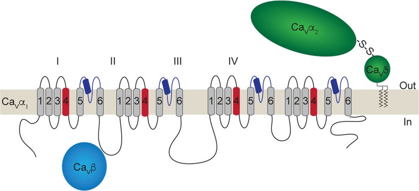
The primary structure of the CaV α1 pore-forming subunit is organized into four homologous
domains (DI-IV). Each domain contains six membrane-spanning segments (S1–S6), with a re-entrant
loop between S5 and S6, which contains negatively charged residues (glutamates and/or aspartates)
that are essential for the selectivity filter. S4 contains positively charged residues (arginines) that
function as voltage-sensors. The amino and carboxyl termini, as well as linker sequences between
the DI-II, DII-III, and DIII-IV are all cytosolic. These sites are important for the interaction of CaV α1properties of a given splice variant overlap with those ones from members of a different CaV
subfamily [32].
The primary structure of the CaVα1 pore-forming subunit is organized into four homologous
domains (DI-IV). Each domain contains six membrane-spanning segments (S1–S6), with a re-entrant
loop between S5 and S6, which contains negatively charged residues (glutamates and/or aspartates)
Int. J. Mol. Sci. 2019, 20, 3537 4 of 36
that are essential for the selectivity filter. S4 contains positively charged residues (arginines) that
function as voltage-sensors. The amino and carboxyl termini, as well as linker sequences between the
DI-II,intracellular
with DII-III, and DIII-IV areasallwell
proteins, cytosolic. These cascades
as signaling sites are important for the
that regulate interaction
calcium of CaVα1 Ca
entry through with
Vs
intracellular
(Figure 1) [33].proteins, as well as signaling cascades that regulate calcium entry through CaVs (Figure
1) [33].
Figure 1. 1. Schematic representation of aa voltage-gated
voltage-gated calcium
calcium channel
channel(Ca(CaVV)) complex.
complex. The
The Ca
CaVVαα11,,
andCa
CaVVαα22δ,δ,and CaVV subunits
ββsubunits areare depicted.
depicted. Transmembrane
Transmembrane segments
segments of the
of the CaVCa α1 subunit
α1Vsubunit (S1–6)
(S1–6) are
are shown
shown arranged
arranged in theinfour
the domains
four domains (DI-IV),
(DI-IV), the voltage
the voltage sensorssensors are indicated
are indicated in red
in red (S4), and(S4),
the
and the reentrant
reentrant loop between
loop between S5 and S5 S6 and S6 (P-loop)
(P-loop) in darkinblue.
darkThe
blue.glycosylphosphatidylinositol
The glycosylphosphatidylinositol (GPI)-
(GPI)-anchored Ca
anchored CaVα2δ isVshown α 2 δ is shown in green, and the cytoplasmic
in green, and the cytoplasmic CaVβ subunitCa V β subunit
in lightin light blue.
blue.
2.2. CaV Auxiliary Subunits, CaV α2 δ and CaV β
Members of the CaV 1 and CaV 2 subfamilies form membrane complexes with the auxiliary
subunits CaV α2 δ and CaV β, influencing several biophysical and pharmacological properties of the
CaV α1 subunit [2,3,22].
2.2.1. CaV α2 δ Subunits
Four genes exist for the CaV α2 δ subunits (CACNA2D1-4), which encode CaV α2 δ-1-4 proteins [22].
Each CaV α2 δ subunit is translated from a single gene, which produces a protein that is cleaved into the
α2 and δ peptides. A disulfide bond links these peptides [34,35]. CaV α2 δ is entirely extracellular, but it
is attached to the cell membrane by a glycosylphosphatidylinositol anchor (GPI) domain (Figure 1) [36].
Interestingly, the α2 peptide contains structural domains such as the von Willebrand factor A domain
(vWA) and two Cache domains [37]. The vWA domain in CaV α2 δ contains a metal-ion-adhesion
site (MIDAS) that is important for membrane trafficking [38]. The functional role of the Cache
domains is poorly understood [18,20]. CaV α2 δ-1 and CaV α2 δ-2 are targets for the gabapentinoid
drugs, gabapentin and pregabalin [39]. Similar to CACNA1 genes, CACNA2D genes are also subject to
extensive alternative splicing that impacts affinity for gabapentinoid drugs and other functions of the
CaV α2 δ subunits [40,41].
2.2.2. CaV β Subunits
Four genes exist for the CaV β subunits (CACNB1-4), which encode CaV β1 –CaV β4 . CaV β subunits
are located in the cytoplasm (Figure 1); however, some splice variants of CaV β2 are attached to
the membrane via a palmitoylation site [42,43], and both CaV β3 and the splice variant CaV β4c can
be mobilized to the nucleus [44–46]. CaV β subunits contain three conserved domains: an inactive
guanylate kinase domain (GK), an src homology domain 3 (SH3), and a HOOK region [47–50]. The
CaV β-GK domain is important for the interaction with the AID domain in the I-II loop of the CaV α1
subunit [50]. The CaV β-SH3 and HOOK domains mediate specific protein–protein interactions of
CaV β subunits, for example, with dynamin [51]. All CACNB genes undergo alternative splicing [52].Int. J. Mol. Sci. 2019, 20, 3537 5 of 36
3. General Function of Voltage-Gated Calcium Channels and Auxiliary Subunits
3.1. CaV α1 Subunits
CaV channels are expressed in a wide variety of tissues where they serve specific functions.
CaV 1.1 is restricted to the skeletal muscle where the movement of the gating mechanisms induced by
depolarization leads to opening of the ryanodine receptors (RYR), a class of calcium channels located
in the sarcoplasmic membrane. The opening of RYR increases intracellular calcium, which results in
activation of calcium-dependent contractile proteins [53]. CaV 1.2 and CaV 1.3 are broadly co-expressed
in various tissues including the brain, heart, smooth muscle, and neurosecretory systems. These
two channels are important for gene expression, calcium transients in dendrites, and the coupling
of electrical signals to hormone secretion [54–58]. CaV 1.2 controls contraction of heart muscle, and
together with CaV 1.3 controls the pacemaking activity of midbrain dopaminergic neurons and adrenal
chromaffin cells [59–62]. CaV 1.3 is key for the pacemaking firing of the sinoatrial node and for
transmitter release from hair cells of the inner ear [63–65]. CaV 1.4 controls glutamate release from
photoreceptors [66,67].
CaV 2.1, CaV 2.2, and CaV 2.3 are involved in the release of neurotransmitters. CaV 2.1 and CaV 2.2
channels have a dominant role in the release of fast transmitters such as GABA, acetylcholine, and
glutamate [68,69]. CaV 2.2 channels are dominant in peripheral terminals that release glutamate and
noradrenaline, as well as in central synapses that release dopamine, serotonin and noradrenaline [70–72].
CaV 2.2 channels are also dominant in interneurons that express the cholecystokinin peptide [73,74].
CaV 2.3 channels are present in the presynaptic terminals and dendrites of certain synapses of the central
nervous system [75]. In the presynaptic terminals, CaV 2.3 channels are localized in the active zones
or in their periphery thereby controlling transmitter release [76]. In the dendrites, CaV 2.3 channels
control calcium-dependent spikes [77]. G-protein coupled receptors for several neurotransmitters
including GABA, endogenous opioids, and endocannabinoids heavily regulate CaV 2 channels [78,79].
This is an important negative feedback mechanism to limit the release of neurotransmitter [78,79].
CaV 2 channels interact with soluble N-ethylmaleimide-sensitive fusion protein receptors (SNAREs),
which promote the fusion of secretory vesicles to the membrane in a calcium-dependent manner. This
calcium generally enters through CaV 2 channels [80].
CaV 3.1, CaV 3.2, and CaV 3.3 channels control repetitive firing and pacemaking activity [27]. CaV 3
channels open at relatively low voltages compared to members of the CaV 1 and CaV 2 subfamilies and
have fast voltage-dependent inactivation. These unique biophysical properties underlie the role of
CaV 3 channels in rhythmic firing of action potentials [25,81]. CaV 3 channels control the pacemaking
activity of the sinoatrial node in the heart [82,83], and the rhythmic bursts of action potential in relay
neurons in the thalamus [84]. CaV 3 channels are not known to be associated with the auxiliary subunits
CaV α2 δ and CaV β; however, recent evidence suggests that CaV 3 channels interact with CACHD1, a
protein closely related to the CaV α2 δ subunits (Table 1) [19,85,86].
3.2. CaV α2 δ Subunits
CaV α2 δ-1 is expressed in skeletal, cardiac and smooth muscle, secretory systems; central and
peripheral neurons [39]. CaV α2 δ-2 is abundantly expressed in the cerebellum, and to a lesser extent
in other areas of the brain [8]. CaV α2 δ-3 is expressed throughout the central and peripheral nervous
systems [21,87]. Finally, CaV α2 δ-4 expression is limited to the retina and endocrine tissue [88].
Expression of CaV α2 δ subunits increases membrane trafficking, stabilizes CaV s complexes in the cell
surface and produces shifts in voltage-dependence of activation as well as inactivation [3,20]. CaV α2 δ
subunits promote synaptogenesis by binding to thrombospondin [89], influence neurotransmission
through interaction with α-neurexins [90,91], and affect synaptic plasticity by interacting with
N-methyl-D-aspartate (NMDA) receptors [92].Int. J. Mol. Sci. 2019, 20, 3537 6 of 36
3.3. CaV β Subunits
CaV β subunits are broadly expressed in several tissues including brain, heart and skeletal muscle.
These proteins promote trafficking of CaV α1 to the cell surface by occluding endoplasmic reticulum
(ER) retention signals present in the linker between DI and DII of CaV α1 [93]. CaV β subunits are key for
modulation of CaV 1 and CaV 2 channels by G-protein coupled receptors and other signaling complexes
including Ras-related GTPases [17,78]. CaV β3 and particularly CaV β4 are thought to induce gene
expression [44,46].
4. Genetic Analysis and Tools to Study Psychiatric Disorders
Few cases exist where the inheritance of a disorder involving CaV genes follows mendelian models.
However, spinal cerebellar ataxia 6 (SCA6) and Timothy syndrome (TS) are two cases that follow
an autosomal dominant pattern. Alterations in the CACNA1A and CACNA1C genes underlie SCA6
and TS, respectively [94,95]. In SCA6, the CACNA1A gene contains between 20 and 33 CAG repeats
that encode glutamines in the C-terminus [96]. Although the molecular mechanism by which these
repeats lead to the disease remains to be fully understood, current evidence suggests transcriptional
dysregulation mediated by a CaV 2.1 C-terminus fragment with the glutamine repeats [97]. In TS,
mutations in CACNA1C (G402S and G406R) produce CaV 1.2 channels with gain of function, and these
mutations are located in the C-terminus of DIS6 [98,99]. TS is a condition that affects the heart and
the nervous system. Patients with Timothy syndrome present several characteristics seen in patients
with ASD [95]. Recent evidence suggests that CaV 1.2 mutations underlying TS produce defects in
neuronal migration during cortical development [100]. In contrast to SCA6 and TS, most psychiatric
disorders are genetically complex conditions that involve the interaction among several genes and
their interactions with the environment [101,102].
4.1. Genetic Strategies to Study Psychiatric Disorders
Several genetic methods have been used to determine the genes or set of genes that are likely to
underlie psychiatric disorders. Historically, linkage and linkage-disequilibrium studies provided the
initial evidence of the genetic origins of psychiatric disorders [103,104]. However, it is now possible
to perform genetic analysis using whole genomes from large populations through genome-wide
association studies (GWAS) to discover new risk variations associated with psychiatric disorders [105].
4.1.1. Linkage Studies
Evidence for linkage is derived from observing the cosegregation of specific genomic regions
with a given disorder. As such, this method is most effective for the study of disorders inherited in a
Mendelian fashion. Pedigrees containing multiple generations of genetic data can be used to elucidate
inheritance patterns and map potential genomic risk loci for a given disorder. The identification of large
families with high prevalence of a given condition often facilitates linkage studies. In these studies, the
inheritance of a genetic loci can be correlated with the presence or absence of the disorder [103].
4.1.2. Linkage-Disequilibrium Studies
In these studies, the aim is to map a nonrandom association of alleles at two or more loci to
discover disease haplotypes. These haplotypes are thought to be inherited from one or a few founding
members of isolated populations [106].
4.1.3. Association Studies
Here the goal is to find risk loci for a specific condition by assessing correlations between disease
status and genetic variation. Of the association studies, GWAS are becoming a popular method to
screen genetic variations of disease across whole genomes of large populations. GWAS have identified
several genetic variations of CaV genes linked to BD, SCZ, ASD, ADHD, and MDD [14,16]. We willInt. J. Mol. Sci. 2019, 20, 3537 7 of 36
review several of these cases below. Thanks to GWAS data, many new risk loci for psychiatric disorders
have been found [107,108].
All the genetic approaches mentioned above have helped to identify associations of several
gene variations to psychiatric disorders. These variations include single nucleotide polymorphisms
(SNPs), small indels, copy number variations (CNVs), de novo variations, and large chromosomal
rearrangements [109].
4.2. Tools to Identify and Analyze Genetic Variations Associated with Psychiatric Disorders
Our understanding of psychiatric disorders is evolving towards a more comprehensive analysis
that includes genetic, genomic, functional and behavioral studies. These are possible thanks to tools
that facilitated the screening of large cohorts of patients (probands) and their corresponding unaffected
relatives. Among these tools are next-generation sequencing, microarrays, endophenotype analysis,
gene network analysis and computational modeling [109].
4.2.1. Next Generation Sequencing (NGS)
These tools include whole exome and genome sequencing (WES and WGS, respectively), as well
as RNA sequencing (RNA-seq). WES detects genetic variations through capture and sequencing of
coding regions within the genomic DNA. Since most of the genome is noncoding, this approach greatly
reduces the amount of sequencing to ~2% of the whole genome [110]. WGS offers an almost complete
sequence coverage (~95%) that includes coding and non-coding regions and is more powerful to detect
exome variations than WES. This increased coverage enables identification of non-coding regions that
include splicing regulatory elements, promoters, enhancers and sites that regulate RNA transport and
stability [111]. WES are more commonly used in genetic screenings for psychiatric disorders because
of their lower cost compared to WGS [112]. Nevertheless, WES studies allow us to focus on regions
where variations can be identified and interpreted faster than in WGS studies [112].
RNA-seq is a common tool used for genetic analysis of psychiatric disorders. This technology
enables quantification of gene expression, detection and quantification of exon splicing, quantification
of rare transcripts and non-coding RNAs, and detection of genome rearrangements. In summary,
RNA-seq provides a whole transcriptome landscape with high signal to noise ratio and with small
amount of RNA input [113].
4.2.2. Microarrays
Studies using microarrays are commonly used to identify genetic risk variations that involve
structural changes >1000 bp [109,114]. Large structural variations detected by microarrays are thought
to increase the risk SCZ, ASD, and ADHD [115–121].
4.2.3. Gene Network Analysis
The discovery of risk variations associated with psychiatric disorders has been a stepping stone to
elucidate the molecular mechanisms that underlie these conditions. Now the challenge is integrating
this information to understand how genetic variations influence complex disorders and traits [122,123].
It is thought that the complex interactions of genes in a network are more likely to explain phenotypes
of psychiatric disorders, rather than the additive effect of those genes. Complex interactions of genes
within networks include transcriptional regulatory, protein–protein interaction, metabolomic networks,
and a hierarchical interaction with other gene networks [124–127]. Furthermore, complex interactions
between gene networks with the environment are becoming increasingly important to fully explain
phenotypes linked to psychiatric disorders [128–133].Int. J. Mol. Sci. 2019, 20, 3537 8 of 36
4.2.4. Endophenotypes
Despite recent advances linking genetic risk variations to psychiatric disorders, the phenotypic
consequences of those variations are poorly understood. However, a combination of molecular
genetics with endophenotypes might represent a promising approach to understand the behavioral
links between risk variations and psychiatric disorders [134]. Endophenotypes are quantitative
neurobehavioral traits that are associated with a disorder, are reasonably heritable, co-segregate with
the disease and are independent of the clinical status of the disorder [135]. Endophenotypes provide
clinical measures of disease diagnosis and progression. Examples of endophenotypes include deficits
in pre-pulse inhibition and sensory gating, decline in working memory, and deficits in face emotion
labeling [134,135]. Interestingly, the latter has been associated with CACNA1C in patients with bipolar
disorder [136].
4.2.5. Computational Psychiatry
Mathematical approaches are being used to integrate findings derived from genetic screenings,
functional studies of gene risk variations, and behavioral phenotypes. Computational psychiatry is
an emerging field that aims to model the compounded effects of individual genes, as well as their
interaction with other genes (gene networks) and with the environment using mathematics [137–140].
Computational approaches have been successfully used to provide insightful mechanisms for disorders
such as SCZ, ASD, and ADHD [141–143].
5. Genetic Associations between CaV Genes and Psychiatric Disorders
Gene network analyses have consistently implicated CaV genes in psychiatric disorders, which
nicely correlates with the role calcium signaling in neuronal function [14,16]. In this section, we will
review several large studies that have provided strong evidence linking CaV genes to psychiatric
disorders and related endophenotypes. We will also briefly describe functional studies, when available,
of risk variations for CaV genes.
5.1. CaV 1.2
CaV 1.2 channels are ion channels that have been extensively studied in the heart. Here, CaV 1.2
channels tightly couple depolarization to muscle contraction through activation of RYR located in
the ER of cardiomyocytes [144]. Additional studies have demonstrated that CaV 1.2 is expressed
in postsynaptic terminals in the brain, and together with CaV 1.3, influences neuronal firing and
couples excitation to gene expression [145]. The activity of neuronal CaV 1.2 and CaV 1.3 channels is
implicated in several processes relevant to psychiatric disorders including learning, memory, and
brain development [146,147]. Although global disruption of CaV 1.2 channels leads to embryonic
lethality in mouse [148], studies of conditional knock out (KO) mice have shown that CaV 1.2 channels
are involved in high-order brain functions such as spatial memory and remote spatial memory
consolidation [149,150]. Furthermore, CaV 1.2 KO heterozygous mice exhibit increased anxiety-like
behavior [151,152]. In line with this, deletion of CaV 1.2 in calcium/calmodulin dependent-protein kinase
IIα expressing cells (mostly forebrain neurons) also leads to anxiety-like behavior [153]. Alterations in
fear is a trait widely observed in patients with anxiety disorders, and conditional CaV 1.2 KO mouse
models have shown alterations in fear responses. Functional ablation of CaV 1.2 in Nestin expressing
cells leads to reduced acquisition of conditioned fear [154], and specific deletion of CaV 1.2 in the
anterior cingulate cortex impairs observational fear [155]. Mice harboring loss of CaV 1.2 channels in
glutamatergic neurons of the forebrain show social behavior deficits associated with the prefrontal
cortex [153]. All this evidence provides strong support for the involvement of CaV 1.2 channels in
psychiatric disorders. Risk variations in the gene that encodes for CaV 1.2, CACNA1C, have been found
in several association studies of BD, and evidence suggest that some of CACNA1C variations are risk
for SCZ, MDD, ADHD, and ASD.Int. J. Mol. Sci. 2019, 20, 3537 9 of 36
Several SNPs in CACNA1C have been linked to psychiatric disorders with most of them being
located in a large intron (~330 kb) between exons 3 and 4 (intron 3). Significant association of the SNP
rs1006737 allele to BD was originally found in a European cohort (>4300 cases and >6000 controls) [156].
Associations of this SNP with BD have been replicated in several other studies; furthermore significant
association of rs100637 with SCZ, ADHD and MDD has also been detected [157–159]. At the molecular
level, rs100637 is correlated with changes in CACNA1C expression, including decreased expression in
the cerebellum [160], but increased expression in the dorsolateral prefrontal cortex and induced human
neurons [136,161]. The latter observation correlates with increased L-type currents seen in induced
human neurons derived from individuals carrying rs1006737 [161]. Furthermore, the minor allele for
rs1006737 (A) is associated with increased methylation of CpG islands located within intron 3 [162].
Imaging studies have shown associations of rs1006737 with changes in structure and activity of brain
regions related to emotion processing, memory formation and cognition, including hippocampus,
inferior occipital fusiform gyrus, prefrontal cortex and amygdala [136,163,164]. For example, carriers
of rs1006737 show greater thickness of the medial orbitofrontal cortex than non-carriers, and the
presence of this SNP correlates with age-related caudal anterior cingulate cortex thickening [164]. In
addition, two independent studies have shown that rs1006737 is associated with increased amygdala
volumes in adults and adolescents [165,166]. Behavioral studies in humans suggest that rs1006737 is
linked to facial emotion recognition in both healthy individuals and patients with BD [167,168]. Some
studies suggest that rs1006737 is also associated with borderline personality disorder in females, but
not males [169,170]. Furthermore, rs1006737 has significant association with reduced baseline affective
startle modulation in healthy males. Alterations in this endophenotype have been observed in severely
depressed and anxious patients, as well as patients with BD in remission [171].
As mentioned above, rs1006737 has also shown strong associations with SCZ. Additive interaction
of the SNP rs1006737 CACNA1C with rs1344706 in the zinc finger protein 804A gene (ZNF804A)
has been linked to defects in white matter microstructure and psychosis [172], although the effect of
rs1344706 is thought to be larger than rs1006737. In MDD, rs1006737 was associated with less baseline
depressive severity [173]. Furthermore, rs1006737 showed biphasic association with antidepressant
treatment in a European population. The A allele was associated with a better outcome of antidepressant
treatment, but it shows the opposite association in a group of individuals with treatment-resistant
depression [174].
The SNP rs2007044 has been associated with SCZ in several studies including Asian, East Asian,
European and Ashkenazi Jew populations [175–179]. This SNP was associated with decreased functional
connectivity between the right dorsolateral prefrontal cortex and right superior occipital gyrus/cuneus,
as well as the anterior cingulate cortex; and at the behavioral level with poor working memory
performance [180]. Also, rs2007044 is associated with increased concentrations of glutamate, glutamine
and glutamate, plus glutamine in subcortical regions such as basal ganglia and thalamus. These
observations have been reported in patients with SCZ, especially in subjects at risk of psychosis [181].
Sleep disturbance is consistently reported in patients with psychiatric disorders including SCZ, BD
and MDD [182–184]. CACNA1C variations in intron 3 have been linked to sleep traits such as narcolepsy
(rs10774044), sleep latency and sleep quality (rs7316184, rs7304986, rs7301906, rs16929275, rs16929276,
rs16929278, rs2051990) [185–187]. The SCZ risk variations in CACNA1C (rs4765913, rs4765914, and
rs2239063) are associated with sleep latency in infants [188]. Allele rs4765914, together with rs7297582,
was identified in two independent studies as genetic risks for BD, MDD, and SCZ [158,189].
Other SNPs in CACNA1C have been linked to several psychiatric conditions. The SNP rs73248708
(intron 3) and rs116625684 (intron 1) were not associated with SCZ or other psychiatric disorders,
but they affect the risk of developing depressive symptoms upon exposure to adult severe trauma
in adulthood [190]. The SNP rs10848635 was identified in a Korean and in a Taiwanese population
as risk factor for SCZ and BD, respectively [191,192]. Associations of rs10848635 with efficacy of
the anti-depressant citalopram were also found [174]. The SNP rs4765913 was identified in two
independent GWAS of European cohorts as genetic risk for BD [193,194]. The alleles rs10848653 andInt. J. Mol. Sci. 2019, 20, 3537 10 of 36
rs2239118 were identified using a family-based association test (parent/affected child trios) and linked
to ASD, this study also identified SNPs in CACNA1G (see below) [193,194].
In addition to variations with mendelian inheritances (TS) and SNPs associated with psychiatric
disorders, two de novo missense variations in CACNA1C were identified in a large whole-exome
sequencing study using massively parallel short-read sequences of more than 2500 patients with SCZ
and more than 2500 control subjects in a Swedish population [195]. One risk variant (G/T) is predicted
to alter a canonical splice donor site for exon 21, which is part of a pair of mutually exclusive exons
(together with exon 22), with exon 21 being dominant in the brain [31]. Exons 21 and 22 encode part
of the extracellular loop between DIIS1 and DIIS2 and a portion of DIIS2 in CaV 1.2. The second risk
variant (C/T) introduces a premature stop codon in the intracellular linker between DIII and DIV [16].
5.2. CaV 1.3
The CACNA1D gene encodes CaV 1.3. This channel contributes to the rhythmic activity of the
sinoatrial node and thereby involved in the regulation of heart rate [145]. As stated above, CaV 1.3
shares some functions with CaV 1.2 in the brain. However, CaV 1.3 is the main contributor to the
pacemaking activity of dopaminergic neurons in the substantia nigra [62]. Mouse genetic models of
CaV 1.3 have provided information on the potential role of CaV 1.3 in psychiatric disorders. CaV 1.3 KO
mice show anxiety-like phenotypes [196], although recent evidence suggests that these phenotypes
are related to hearing deficits [197,198]. However, in an animal model where CaV 1.2 was mutated to
confer resistance to DHPs (CaV 1.2DHP−/− ), thereby allowing specific pharmacological manipulation
of CaV 1.3 channels, activation of these channels led to depressive-like behaviors [198–200]. Finally,
strong evidence suggests that CaV 1.3 channels play a key role in drug seeking behavior, a behavioral
trait linked to addiction [147]. Interestingly, addiction is often found as a comorbidity with psychiatric
disorders including BD and SCZ.
Risk variations in CACNA1D have been associated with BD, SCZ, ADHD, MDD, and ASD. The
non-coding SNP rs893363, located in the 30 UTR of CACNA1D and the putative promoter region of the
choline dehydrogenase gene (CHDH), was found in a genome-wide analysis of five major psychiatric
disorders including BD, SCZ, ADHD, MDD, and ASD [158]. In a study with samples from a cohort of
European-American individuals, 111 non-coding variations in regulatory elements that are predicted to
modify binding of transcription factors to genomic regions of CACNA1D show significant association
with BD [201]. Furthermore, two coding variations in CACNA1D (A1751P and R1771W) located in
the C-terminus segregate with BD type I cases in a large pedigree [202]. Although a study in a Han
Chinese population found no association between CACNA1D SNPs and SCZ [203], more recent studies
that include larger populations of East Asian, Chinese, European and Ashkenazi Jewish individuals
identified the SNP rs2358740 located in a putative promoter region for CACNA1D and the mRNA
decapping enzyme 1A gene (DCP1A) as a risk variant for SCZ [176,204,205].
Several studies point to links between CACNA1D and ASD. Through whole-exome sequencing
three de novo missense variations in the linker between DI and DII in CaV 1.3 (A749G, G407R, and V401L)
were identified as genetic risks for patients with sporadic autism and intellectual disability [206–208].
These genetic risk variations produce a gain of function of CaV 1.3 channels [209–211]. Additional
variations (A59V, S1977L and R2021H) were also identified using WES. The A59V maps to an
N-terminal region of CaV 1.3 that is key for calcium-dependent inactivation. S1977L and R2021H map to
a proline-rich domain of the C-terminus that interacts with SH3 and Multiple Ankyrin Repeat Domain
3 protein (Shank3) [212]. Interestingly, SHANK3 is another gene strongly linked to ASD [213,214]. The
gene CACNA1D is subject to alternative splicing. Interestingly alterations in the relative abundance
of several alternatively spliced exons in CACNA1D have been observed cortical samples of patients
with ASD [215]. Finally, the variation Q567H is located between S1 and S2 of DII in CaV 1.3 and is
linked to moderate hearing impairment and intellectual disability. This variation results in a loss of
function [216].Int. J. Mol. Sci. 2019, 20, 3537 11 of 36
Although risk variations in CACNA1S and CACNA1F encoding CaV 1.1 and CaV 1.4 channels have
been identified in GWAS and WES studies for BD and SCZ, we will not review them here because the
expression of these two genes in the brain is extremely rare relative to the other CaV genes; therefore
the links between their corresponding risk variations and psychiatric disorders are hard to infer [16].
5.3. CaV 2.1
CACNA1A encodes CaV 2.1 channel, which is the most dominant presynaptic calcium channel
in central synapses, particularly those ones from Purkinje cells in the cerebellum and excitatory
synapses of cortex and hippocampus. Although CaV 2.1 KO mice are postnatally lethal [217–219],
forebrain ablation of CaV 2.1 channels results in deficits in spatial learning and memory, and increased
exploratory behavior suggesting a potential role of this channel in psychiatric-related phenotypes [220].
Various mutations in CACNA1A, causing gain or loss of function, have been found in patients with
hemiplegic migraine 1 (FHM-1), Episodic Ataxia 2 (EA-2), SCA-6, and epilepsy [10]. More recently, a
clinical recharacterization of patients with EA-2 and SCA-6 showed that they also present delayed
development, endophenotypes related to learning disabilities, ASD and ADHD [221]. In another
study, some FHM-1 and EA-2 patients also presented SCZ, learning disabilities and ADHD [222].
Furthermore, analysis of the splice isoform landscape across several psychiatric disorders show that
alternative splicing of CACNA1A is altered in ASD [223]. Finally, rs10409541 was among the top 15
most contributory SNPs for ASD diagnosis prediction in a Central European population [194].
5.4. CaV 2.2
CACNA1B encodes CaV 2.2 channels, which are dominant in presynaptic terminals of dorsal root
ganglia and superior cervical ganglia, as well as some interneurons and dopaminergic neurons of
the midbrain. At the behavioral level, ablation of CaV 2.2 channels results in increased locomotion,
exploration, reduced startle [224,225], and reduced ethanol intake [226]. CaV 2.2 KO mice also show
increased aggression and enhanced vigilance state related to disruption in random eye movement
sleep [70]. All this combined suggests a role of CaV 2.2 channels in psychiatric disorders.
Several studies have linked CACNA1B to SCZ, but also some CACNA1B risk variations are
associated with BD and ASD. Purcell et al. identified a de novo variation (G/A) in patients with SCZ
that introduces a premature stop codon in the proximal C-terminus of CaV 2.2 [195]. The intronic SNPs,
rs7036881 and rs78178087, in CACNA1B have been found to be weakly associated with SCZ and the
antipsychotic efficacy of paliperidone palmitate in a study with European patients [227]. In line with
this, another study in a South African population found that the rs2229949 is linked to improved
negative symptomatology during antipsychotic treatment [228]. Deletions in CACNA1B were detected
in 16 patients and duplications of this same gene were detected in 10 patients with SCZ [229].
Several studies have reported that CACNA1B is linked to ASD, MDD, and BD. A monogenic
duplication in CACNA1B has been linked to Asperger Syndrome, a condition that until recently was
considered an ASD [230]. Pathway analysis of variations linked to ASD has shown that CACNA1B,
together with CACNA1C and CACNA1F, converges on MAP kinase/cellular signaling and neuronal
development/axon guidance [231]. CACNA1B, together with CACNA1C and CACNA2D4, has been
also associated with suicide risk in patients with MDD [232]. Finally, WES of 200 individuals from
41 families identified 50 non-coding variations in CACNA1B that increase the risk for BD [201].
5.5. CaV 2.3
The CACNA1E gene encodes the CaV 2.3 channels. These channels are broadly expressed
throughout the nervous system and are located in presynaptic terminals, dendritic spines, and some
extrasynaptic sites [75]. Functional disruption of CaV 2.3 channels leads to increased anxiety-like
behavior and impaired spatial memory [233,234]. CaV 2.3 deficient mice show reduced wake
duration and increased slow-wave sleep, although these results depend on the strategy to knock outInt. J. Mol. Sci. 2019, 20, 3537 12 of 36
CaV 2.3 [235,236]. Nonetheless, this is relevant because alterations in sleep have been observed in
patients with SCZ and BD.
Variations in the CACNA1E gene have been linked to ASD, MDD, SCZ, as well as some
endophenotypes related to these conditions. In a study comprised of 209 families with no previous
history of ASD, parent-child trios with sporadic autism and unaffected siblings were sequenced and a
de novo variant in CACNA1E (G1209S) was identified in one patient [206]. G1209S is located in DIIIS3.
A second de novo synonymous variation in CACNA1E located near a splice site and predicted to affect
an exonic splicing regulator was identified in another patient with ASD [237]. In a genome-wide
meta-analysis study of more than 135,000 cases with more than 340,000 controls, 44 significant risk loci
for MDD were identified, including CACNA1E [238]. The SNP rs4652676 was linked to neuroticism
and subjective well-being, which are endophenotypes associated with MDD [239]. The SNP rs704329
is implicated in the efficacy of serotonin reuptake inhibitors (SSRIs) in a Taiwanese population [240].
Similar to several other CACNA1 genes, CACNA1E has been associated with SCZ as well as working
memory related to cortex and cerebellum [158,241].
5.6. CaV 3.1
CACNA1G encodes the CaV 3.1 channel, a T-type channel member of the CaV 3 subfamily. Specific
ablation of CaV 3.1 channels in the thalamus resulted in frequent and prolonged arousal, which reduced
sleep [242,243]. CaV 3.1 channels also play a key role in prolonged unconsciousness by influencing
thalamocortical rhythmicity [244]. Previous studies have identified risk variations of CACNA1G
as genetic risk for ASD. A linkage study of sibling pairs with only male probands found a strong
association of the chromosomic region 17q11–21, which contains among other genes, CACNA1G [245].
A later study confirmed CACNA1G as a novel candidate gene for ASD by identifying several SNPs
within intron 9 with the strongest association relative to other genes present in the 17q11-21 region [246].
Alleles rs198538 and rs198545, together with some CACNA1C SNPs, were identified as risk variations
for ASD [193]. Furthermore, a de novo synonymous variation in CACNA1G was identified in exome
sequencing of 343 families with one proband and at least one unaffected sibling [207,247]. A de novo
variation screening in childhood-onset cerebellar atrophy identified various disruptive variations in
CACNA1G, some patients with this pathology exhibit autistic traits [248]. However new studies using
transcriptome-wide association, which integrated GWAS with gene expression predictors from several
databases from adult and fetal human brain, found no evidence of association between CACNA1G and
ASD [249].
5.7. CaV 3.2
The CACNA1H gene encodes CaV 3.2 channels, also a T-type channel. This gene is normally
associated with idiopathic epilepsy. However, multiple studies have found associations of CACNA1H
with ASD and SCZ. Mice deficient in CaV 3.2 channels show increased anxiety-like behavior, impaired
memory and reduced sensitivity to psychostimulants such as D-amphetamine and cocaine [250].
CaV 3.2 KO mice exhibit deficits in context-associated memories [251]. CaV 3.2 channels play a minor
role in non-REM sleep [252,253].
Four missense variations (R212C, R902W, W962C and A1874V) were identified in a study of 461
probands with ASD and 480 ethnically matched individuals by targeted sequencing of the CACNA1H
genomic region [254,255]. R212 is located DIS4, R902 in DIIS4, W962 in the P-loop between DIIS5
and DIIS6, and A1874 in the C-terminus. Functional analysis revealed that these variations produce
loss of function of CaV 3.2 by reducing channel conductance, and/or shifting voltage-dependence of
activation in the depolarizing direction [254]. However, these variations have low penetrance, and
some of them were also found in unaffected individuals [254]. In a more recent study using ultra deep
sequencing of 78 ASD candidate genes in the cerebellum and cortical samples of several ASD cases
and neurotypical controls, a synonymous CACNA1H variation was found in the frontal cortex but not
in cerebellum [256]. In this same study, a missense variation in the C-terminus (S1970C) was identifiedInt. J. Mol. Sci. 2019, 20, 3537 13 of 36
in a female diagnosed with ASD [256]. WES from more than 10,000 parents with only one child with
ASD found de novo missense variations in CACNA1H [207,257]. Furthermore, a study of 262 ASD
patients with their unaffected parents from Japan identified a disruptive de novo missense variations in
CACNA1H (R1189C), which is located in the intercellular loop between DII and DII [258]. All of these
studies support that CACNA1H is a susceptibility gene for ASD.
Two rare disruptive variations (7 bp and 2 bp deletions) in the DII-DIII linker in CaV 3.2 that
are predicted to produce a frameshift were found in patients with SCZ [195]. Furthermore, a GWAS
performed in a Swedish population, followed by a meta-analysis with previously identified genes
associated with SCZ, found association of CACNA1H with this condition [259].
5.8. CaV 3.3
CACNA1I encodes CaV 3.3 channels, the third T-type channel member of the CaV 3 subfamily. Of
the CaV 3 members, CaV 3.3 channels have the most depolarized threshold of activation, as well as the
slowest opening and inactivation rate [27]. CaV 3.3 channels regulate sleep spindles. This is supported
with evidence from animal models, mice with functional ablation of CaV 3.3 channels show impairment
in sleep spindle generation [252,253,260,261]. Sleep spindles have been shown to be altered in patients
with SCZ [184]. Not surprisingly, several studies have strongly linked CACNA1I to SCZ and related
endophenotypes. Additional evidence also suggests risk variations of CACNA1I for ADHD and ASD.
Two rare, de novo missense variations of CACNA1I (R1346H and T797M) were identified by
exome sequencing of trio samples that included 105 probands, parents, and unaffected siblings when
available [262]. R797 and R1345 map to the P-loops of DII and DIII of CaV 3.3 respectively. CaV 3.3 was
the only gene with more than one variation [262]. In particular, R1346H impairs N-glycosylation of
CaV 3.3 channels preventing membrane targeting and thereby reducing overall calcium currents [263].
The functional consequences of T797M are unknown, however this mutant produces similar calcium
currents relative to WT [263]. A study by the SCZ working group of the PGC validated CACNA1I as a
risk gene for SCZ [175]. This claim has been supported in other GWAS. The intergenic SNPs between
RPS19BP1 and CACNA1I, rs5757717 and rs9611198, were found in a GWA study of an Ashkenazi Jewish
population and an Irish population respectively [176,264]. The intronic SNP rs3788567 was identified
with very high significance in an Ashkenazi Jewish population [176]. In a study of an Uyghur Chinese
population that comprised 985 patients and 1218 neurotypical controls, six SNPs within CACNA1I were
significantly associated with SCZ (rs132575, rs136805, rs713860, rs738168, rs5757760, rs575087) [265].
Furthermore, rs4522708, rs3788568, rs5750862 were significantly associated with SCZ in a Han Chinese
population [266,267]. Interestingly, rs4522708 was also found in a study of a European population [175].
CACNA1I has been also associated with endophenotypes related to SCZ, such as cognitive ability and
sleep spindle activity. A genome wide meta-analysis study identified an association of CACNA1I with
cognitive ability [268]. The genomic region Chr22: 39975017:40016914, which spans across CACNA1I
was associated with higher amplitude, longer duration and higher intensity of slow spindles in healthy
adolescents [269].
A recent GWAS linked the rs199694726 in CACNA1I to impulsive behavior under extreme negative
emotions [270]. Impulsive traits are a common endophenotype related to psychiatric disorders
including ADHD [271]. Furthermore, a study containing 1013 probands of European descent at the
Children’s Hospital of Philadelphia (CHOP) found a CACNA1I CNV (large deletion) associated with
ADHD [272]. CACNA1I was also identified as a risk gene for ASD in the family-based association
test [273]. The SNP rs5750860 was significantly associated with ASD in an another GWAS [193].
6. Genetic Associations between Auxiliary Subunit Genes CACNA2D (CaV α2 δ) and CACNB
(CaV β) and Psychiatric Disorders
In the previous section we summarized strong evidence linking several genes that encode CaV α1
pore-forming subunits to psychiatric disorders. Given that the auxiliary subunits, CaV α2 δ and CaV β,
heavily influence membrane targeting and overall activity of CaV α1, genes encoding these subunitsInt. J. Mol. Sci. 2019, 20, 3537 14 of 36
are also strongly linked to psychiatric disorders. In this section, we will describe current studies
associating the genes for CaV α2 δ and CaV β with multiple psychiatric disorders.
6.1. CaV α2 δ-1
The CACNA2D1 gene encodes the CaV α2 δ-1 subunit. This subunit is highly expressed in skeletal
muscle, the brain and peripheral nervous system [19], and some studies suggest that it is enriched in
glutamatergic neurons [274]. No brain-related phenotypes have been reported for animal models with
alterations in CaV α2 δ-1 expression (KO, knock in or overexpression) [275–278]. However, compensation
by the CaV α2 δ subunits exists when one of them is disrupted [20].
Various genetic studies have implicated the CACNA2D1 gene in psychiatric disorders including
MDD, BD and SCZ. The genome-wide association metanalysis of MDD that identified CACNA1E,
also found CACNA2D1 as potentially druggable target for this condition [238]. Furthermore, in a
genome-wide association environment study, a suggestive association was found for rs17156280
in CACNA2D1 with an interaction between depressive state and stressful events [279]. A strong
association with depressive traits including subjective well-being and neuroticism was found for the
SNPs in CACNA2D1, rs258668 and rs258677 [239].
A metanalysis of data collected by the Bipolar Disorder Genome Study Consortium identified
rs2367911 as a risk SNP for BD with comorbid binge eating. Indeed, networks/interactomes for
CACNA2D1 and apolipoprotein B gene (APOB) were the top two hits for BD and binge eating in this
study [280]. The same study that identified risk variations for CACNA1C and other CaV genes in a
Swedish population, found a disruptive variation in CACNA2D1 that produces a frameshift associated
with SCZ [195]. A study in a Japanese population a found CNV for CACNA2D1 (a large deletion) in
one patient with SCZ [116].
6.2. CaV α2 δ-2
CACNA2D2 encodes CaV α2 δ-2. Although this protein is broadly expressed in the central nervous
system, there is higher expression in the cerebellum relative to other areas of the brain, particularly
in Purkinje cells [20]. In cortical tissue, some studies suggest that CaV α2 δ-2 is more abundant in
interneurons than in glutamatergic neurons [274]. Deleterious effects from disrupting CaV α2 δ-2 have
been observed in mouse genetic models including ataxia and seizures, however none of them are
directly related to psychiatric disorders [8,281–285]. Purcell et al., found three de novo variations in
CACNA2D2 in patients with SCZ. Two of these three variations introduced premature stop codons,
and the third one is predicted to disrupt a splice donor site [193]. A CACNA2D2 variation (A900T)
scored as a putative second hit in a study of 558 patients with SCZ in a Spanish population [286].
6.3. CaV α2 δ-3
CACNA2D3 encodes CaV α2 δ-3. This protein was initially characterized as a target to treat pain,
however recent studies suggest that the CACNA2D3 is strongly linked to ASD, and to a lesser extent,
SCZ and BD. CaV α2 δ-3 KO mice have alterations in pain processing at the central level [87], as well as
enhanced cross-activation of brain regions involved in processing of auditory, olfactory and visual
sensory information (cross-modal activation) [87,287,288]. Interestingly, patients with ASD and SCZ
often exhibit altered pain perception [289,290] and synesthesia, the latter is a form of cross-modal
activation [291,292].
The same WES that identified variations in CACNA1G, found another variation in CACNA2D3
that is predicted to disrupt a splice junction associated with ASD (A/G) [247]. An inherited variation
with splicing disruption was identified in a study of 2066 unique families with children diagnosed with
ASD, the cohort consisted of 2618 children with ASD (1740 probands and 878 unaffected siblings) [293].
Furthermore, a de novo variation (E508Stop) predicting loss of function of CaV α2 δ-3 was found in
two patients in an exome sequencing study that included 3871 ASD cases and 9937 ancestry controls.
This study also identified several inherited variations in CACNA2D3 which effect is unknown [212].Int. J. Mol. Sci. 2019, 20, 3537 15 of 36
Analysis of CNVs in a study containing samples from 2478 families with children affected with ASD
identified through the Simons Simplex Collection found association to a deletion in CACNA2D3 [294].
In a study where 208 candidate genes were sequenced in 11,730 cases and 2867 controls, two de novo
missense on CACNA2D3 were identified (A773V and A275T) [295]. The SNP rs3773540 was among the
top 15 SNPs contributing to ASD diagnosis as predicted by gene set enrichment analysis [194].
Previous studies have shown that the 3p14 genetic region is associated with SCZ and with an
endophenotype related to the function of the temporal lobe, the antisaccade reflex. Interestingly, this
genomic region contains CACNA2D3 [296,297]. Pathway analysis of SNPs with significant risk for
SCZ suggest association of CACNA2D3 with the response to lurasidone, an antipsychotic used to
treat SCZ [298]. Also, the genomic region 3p21.1_1 is enriched in for both SCZ and BD, this region
contains CACNA1D and CACNA2D3 among six different genes [299]. The SNP rs9849795 located in
CACNA2D3 is associated with functional brain connectivity inferred by functional magnetic resonance,
this trait thought to be compromised in BD and SCZ, this study also identified association with SNPs
in CACNA1C, CACNA2D4 and CACNB2 [300].
6.4. CaV α2 δ-4
CACNA2D4 encodes the CaV α2 δ-4 subunit. This protein is abundantly expressed in the retina,
but it is also found in pituitary and adrenal glands [88,301]. Despite the relatively low expression
of CaV α2 δ-4 in the brain compared with other auxiliary subunits, several studies have identified
the CACNA2D4 as a risk gene for some psychiatric disorders. Although disruptive mutations in
CACNA2D4 in mice cause night blindness, as well as retinal degeneration, phenotypes related to the
brain have not been reported [302–304].
The SNP rs1024582 located between CACNA2D4 and CACNA1C was found highly significant
in a cross-disorder study that included ADHD, BD, ASD, SCZ and MDD [158]. In a later study by
Purcell et al., a de novo variation that produces a frameshift in CACNA2D4 was identified in patients
with SCZ [195]. The SNP rs4765847 was found to associate with DMN, an endophenotype of SCZ [300].
Furthermore, partial deletions of 35.7 kb in CACNA2D4 was found in two unrelated patients with
late onset BD I and one in control individuals [305]. These three deletions eliminate exons 17–26 in
CACNA2D4, which comprise most of the Cache domain [305]. In a linkage disequilibrium study
to detect SNP–SNP interactions that are common in complex diseases a single interaction between
SNPs located near RYR2 and CACNA2D4 was found in samples of the Wellcome Trust Case Control
Consortium (WTCCC) [306].
Genetic associations of CACNA2D4 with MDD and ASD have been also identified. In a WES
study in brain samples of suicide victims suffering from MDD and control subjects with MDD who
died from other causes, a variation in a splice donor (C/A) in CACNA2D4 was identified [232]. For
ASD, a rare homozygous deletion was detected in a male proband that is predicted to affect CACNA1C
and CACNA2D4 (12p13.33) [307].
6.5. CaV β1
CACNB1 encodes CaV β1 . A splice variant of this subunit was originally identified in skeletal
muscle (CaV β1a ) as the only partner of CaV 1.1, later it was demonstrated that splice variations of
CaV β1 are also expressed in the brain (CaV β1b , CaV β1c , and CaV β1d ), particularly in cerebral cortex,
habenula, hippocampus and olfactory bulb [45]. Null mice for CaV β1 subunit show reduced muscle
mass and die of asphyxiation after birth, heterozygous are relatively normal and no phenotypes linked
to higher order brain functions have been reported [308].
Some studies suggest association of the CACNB1 with ASD, BD and SCZ; however, the evidence is
scarce. A metanalysis of five genome-wide linkage scans in 634 affected sibling pairs found a suggestive
association between the chromosome region 17p11.2–q12 and ASD, this region comprises CACNB1,
however this finding requires further replication [309]. For BD, increased CACNB1 expression was
reported in IPSCs derived from patients with BD relative to IPSCs from their unaffected relatives [310].You can also read