Genome-wide mapping of Polycomb target genes unravels their roles in cell fate transitions
←
→
Page content transcription
If your browser does not render page correctly, please read the page content below
Downloaded from genesdev.cshlp.org on March 18, 2015 - Published by Cold Spring Harbor Laboratory Press

Genome-wide mapping of Polycomb
target genes unravels their roles in cell
fate transitions
Adrian P. Bracken,1 Nikolaj Dietrich,1 Diego Pasini,1 Klaus H. Hansen,1 and Kristian Helin1,2,3
1
 Biotech Research and Innovation Centre (BRIC), 2100 Copenhagen Ø, Denmark; 2Faculty of Health Sciences, University of
Copenhagen, 2200 Copenhagen N, Denmark

The Polycomb group (PcG) proteins form chromatin-modifying complexes that are essential for embryonic
development and stem cell renewal and are commonly deregulated in cancer. Here, we identify their target
genes using genome-wide location analysis in human embryonic fibroblasts. We find that
Polycomb-Repressive Complex 1 (PRC1), PRC2, and tri-methylated histone H3K27 co-occupy >1000 silenced
genes with a strong functional bias for embryonic development and cell fate decisions. We functionally
identify 40 genes derepressed in human embryonic fibroblasts depleted of the PRC2 components (EZH2, EED,
SUZ12) and the PRC1 component, BMI-1. Interestingly, several markers of osteogenesis, adipogenesis, and
chrondrogenesis are among these genes, consistent with the mesenchymal origin of fibroblasts. Using a
neuronal model of differentiation, we delineate two different mechanisms for regulating PcG target genes. For
genes activated during differentiation, PcGs are displaced. However, for genes repressed during differentiation,
we paradoxically find that they are already bound by the PcGs in nondifferentiated cells despite being actively
transcribed. Our results are consistent with the hypothesis that PcGs are part of a preprogrammed memory
system established during embryogenesis marking certain key genes for repressive signals during subsequent
developmental and differentiation processes.
[Keywords: Polycomb; chromatin; epigenetics; stem cells; differentiation]
Supplemental material is available at http://www.genesdev.org.
Received January 30, 2006; revised version accepted March 3, 2006.

The human body consists of at least 200 different types                     present gene activity and the physiological status of the
of cells. The response of each of these cells to extra- or                  cell. This code is deciphered by other protein complexes,
intracellular signals depends on their particular lineage                   which specifically recognize and bind the modified his-
“identity.” In other words, despite having identical ge-                    tones and, in turn, elicit the biological effects of the im-
nomes, cells can respond in markedly different ways to                      prints. The Polycomb repressors and Trithorax activa-
the same stimulus. Therefore, a strong “programming”                        tors are thought to be the central players in these epige-
system must function to preserve and specify the iden-                      netic programming events (Orlando 2003; Molofsky et
tity of each cell type. It has become increasingly clear in                 al. 2004; Valk-Lingbeek et al. 2004).
recent years that maintenance of cell identity and, to a                       Polycomb group genes (PcGs) are usually considered as
certain extent, also specification of cell identity are con-                being transcriptional repressors that are required for
trolled by epigenetic events (Fisher 2002; Jaenisch and                     maintaining the correct spatial and temporal expression
Bird 2003; Hsieh and Gage 2004; Valk-Lingbeek et al.                        of homeotic genes during development and were origi-
2004). These epigenetic events, which include DNA                           nally identified based on studies demonstrating that de-
methylation and post-translational modifications of the                     letions of PcG genes lead to homeotic transformations of
histones, control the transcriptional program of each cell                  fruit flies (Orlando 2003; Ringrose and Paro 2004; Pir-
by regulating chromatin structure. The histone modifi-                      rotta and Gross 2005). The vertebrate homeotic HOX
cations are thought to constitute an indexing mecha-                        genes are located in four distinct clusters (A, B, C, D) that
nism, referred to as the “histone code” (Jenuwein and                       are organized into 13 homology (or paralog) groups (Pear-
Allis 2001), containing information such as the past and                    son et al. 2005). The chromosomal organization of the
                                                                            genes in each HOX cluster reflects its anterior–posterior
                                                                            expression in the body plan. The “tail” HOX genes at the
3
 Corresponding author.                                                      5⬘ end of the HOXA locus (i.e., HOXA7–13) are ex-
E-MAIL kristian.helin@bric.dk; FAX 45-3917-9669.
Article published online ahead of print. Article and publication date are
                                                                            pressed predominantly in the posterior of the developing
at http://www.genesdev.org/cgi/doi/10.1101/gad.381706.                      embryo. In PcG knockout mice, the repression of these

         GENES & DEVELOPMENT 20:1123–1136 © 2006 by Cold Spring Harbor Laboratory Press ISSN 0890-9369/06; www.genesdev.org         1123
Downloaded from genesdev.cshlp.org on March 18, 2015 - Published by Cold Spring Harbor Laboratory Press

Bracken et al.

“tail” HOX genes is impaired in the middle and head                  Results
part of the embryo, resulting in posterior-to-anterior
transformation defects (Levine et al. 2004; Lund and van             Identification of gene expression changes in cells
Lohuizen 2004).                                                      depleted of PRC1 and PRC2 members
   Recent biochemical approaches have established that
                                                                     To obtain an understanding of how the PcGs control
the PcG proteins form multiprotein complexes, called
                                                                     development and cell fate decisions, we chose to identify
Polycomb-Repressive Complexes (PRCs). PRC2 contains
                                                                     their target genes. To do this, we wanted to perform
EZH2, EED, SUZ12, and RbAp48, while the PRC1 com-
                                                                     ChIP experiments using specific PcG antibodies fol-
plex consists of >10 subunits including the oncoprotein
                                                                     lowed by probing of microarrays (chips) with the en-
BMI-1 and the HPC proteins (CBX2, CBX4, CBX7,
                                                                     riched material (ChIP-on-chip). As an initial screen, we
CBX8), HPH1-3, RING1-2, and SCML (Levine et al.
                                                                     identified candidate genes by performing expression ar-
2004; N. Dietrich, K. Helin, and K.H. Hansen, unpubl.).
                                                                     ray analysis of PcG-depleted cells. Then subsequently,
Functionally, EZH2 is the catalytically active compo-
                                                                     by generating tiled arrays mapping the complete loci of
nent of PRC2, acting as a histone methyltransferase spe-
                                                                     these potential target genes and probing with the en-
cific for Lys 27 (K27) of histone H3 and K26 of histone H1
                                                                     riched material, we aimed to identify functionally rel-
(Cao and Zhang 2004; Kuzmichev et al. 2004). Interest-
                                                                     evant direct targets.
ingly, the HPC proteins of the PRC1 complex can spe-
                                                                        We transfected proliferating human embryonic diploid
cifically bind to tri-methylated H3K27 (H3K27me3) (Cao
                                                                     fibroblasts (TIG3) with small interfering RNA (siRNA)
and Zhang 2004; Kuzmichev et al. 2004; N. Dietrich, K.
                                                                     oligonucleotides specific for the PRC2 components
Helin, and K.H. Hansen, unpubl.). Since PRC2 is re-
                                                                     (EZH2, EED, and SUZ12) or the PRC1 component BMI-1.
quired for PRC1 binding to chromatin (Rastelli et al.
                                                                     Western blot analysis of lysates prepared 44 h after trans-
1993; Hernandez-Munoz et al. 2005), it has been pro-
                                                                     fection confirmed that the siRNAs efficiently inhibited
posed that this is primarily achieved through binding of
                                                                     the protein synthesis of their specific targets (Fig. 1A). As
HPC proteins to H3K27me3. Recently, the PRC1 com-
                                                                     described previously, the protein levels of PRC2 mem-
plex has been demonstrated to possess a H2A-K119 ubiq-
                                                                     bers are in part dependent on the presence of the other
uitin E3 ligase activity that, like H3K27me3 activity, is
                                                                     partners in the complex (Pasini et al. 2004b). RNA was
associated with the repression of HOX genes (Cao et al.
                                                                     extracted at this point and labeled and hybridized to Af-
2005).
                                                                     fymetrix gene expression arrays (HG-U133). The short
   In addition to being essential regulators of embryonic
                                                                     incubation period with siRNA oligonucleotides gener-
development, the PcGs have also emerged as key players
                                                                     ated small but reproducible expression changes in sev-
in the maintenance of the adult stem cell populations
                                                                     eral hundred genes. We observed a substantial overlap in
(Molofsky et al. 2004; Valk-Lingbeek et al. 2004). For
                                                                     gene expression changes after inhibiting the expression
example, BMI-1 is required for the self-renewal of hema-
                                                                     of BMI-1 and the three proteins of the PRC2 complex
topoietic and neural stem cells (Lessard and Sauvageau
                                                                     (Fig. 1B). In order not to miss any relevant genes for the
2003; Molofsky et al. 2003), while overexpression of
                                                                     subsequent ChIP-on-chip analysis, we selected a total of
EZH2 is capable of blocking the differentiation of muscle
                                                                     341 genes as potential PcG target genes, whose expres-
myoblasts (Caretti et al. 2004) and preventing hemato-
                                                                     sion is significantly changed upon down-regulation of at
poietic stem cell exhaustion (Kamminga et al. 2005).
                                                                     least three out of the four PcG proteins analyzed in our
Consistent with their critical roles in development, dif-
                                                                     experiments (Fig. 1C; Supplementary Table 1). The reli-
ferentiation, and stem cell renewal, several PcGs are on-
                                                                     ability of the expression array data was confirmed by
cogenes, overexpressed in both solid and hematopoietic
                                                                     performing quantitative real-time PCR analysis (qPCR)
cancers (Pasini et al. 2004a; Valk-Lingbeek et al. 2004;
                                                                     of a selection of 18 genes (Fig. 1D; data not shown).
Raaphorst 2005).
   Although substantial progress has been made toward
understanding the biological and biochemical functions
                                                                     PcG binding and overlapping H3K27 tri-methylation
of PcG proteins, we still know little about how precisely
                                                                     of complete gene loci in the HOX gene clusters
they control development and cell fate decisions. Since it
is believed that they primarily function as epigenetic               To identify target genes by ChIP analysis, we first
regulators of transcription, we performed chromatin im-              screened several candidate antibodies specific for PRC1
munoprecipitations (ChIP) and genome-wide screenings                 and PRC2 proteins and the tri-methylated H3K27 for
using tiled arrays to identify PcG target genes in human             their ability to efficiently coimmunoprecipitate histone
cells. Strikingly, we observe a very strong bias for genes           H3 in ChIP conditions (data not shown). From this analy-
controlling development and cell fate decisions. We in-              sis we selected antibodies specific for SUZ12 (PRC2),
vestigate the functional relevance of PcG regulation in              CBX8 (PRC1), and H3K27me3. To validate the specific-
both human embryonic fibroblasts and in a model of                   ity of these antibodies in the ChIP-on-chip analysis, we
neuronal differentiation. Moreover, we show that the                 represented the entire HOXA, HOXB, HOXC, and
PcGs target several tumor suppressor genes and genes                 HOXD loci on tiled chips. We probed these chips with
known to be down-regulated in cancer. Based on these                 DNA amplified by ligation-mediated PCR from ChIPs
data, we propose models for how the PcGs control tran-               performed with antibodies specific for H3K27me3,
scription.                                                           SUZ12, and CBX8 using an antibody for HA as a negative

1124       GENES & DEVELOPMENT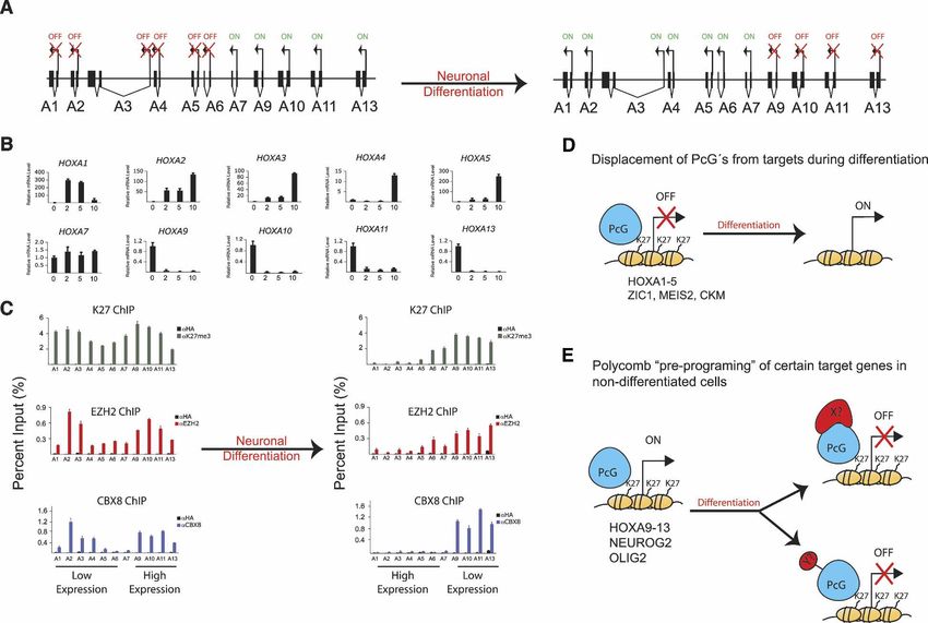
Downloaded from genesdev.cshlp.org on March 18, 2015 - Published by Cold Spring Harbor Laboratory Press

                                                                                                                 Polycomb target genes

Figure 1. Genome-wide expression screen to identify gene changes in Polycomb depleted cells. (A) Western blot analysis of lysates
prepared from TIG3 fibroblasts 44 h after transfection with siRNA oligos designed to inhibit the expression of EZH2, EED, SUZ12, or
BMI-1. Tubulin was used as a loading control. (B) Venn diagram depicting the overlap in genes increased >1.2-fold in PRC2 (EZH2, EED,
and SUZ12 combined) and PRC1 (BMI-1) depletion. (C) Treeview representation of Affymetrix expression data depicting the gene
expression changes in Polycomb-depleted cells. (D) Validation of gene expression changes by qPCR for a selection of genes (indicated
in B). mRNA was prepared from TIG3 cells transfected with Mock (M), EZH2 (Z), EED (E), SUZ12 (S), or BMI-1 (B) siRNAs. The
experiments were performed independently of the gene expression array experiments.

control and an antibody for E2F3 as a positive control for           Identification of target genes whose expression is
the ChIP technique. In Figure 2, A and B, the enrich-                regulated by the PcGs
ments observed across the entire HOXA locus are de-
picted at high resolution in Log2 and normal scale, re-              Next, we represented the 341 gene loci of the genes iden-
spectively. Very strong enrichments of SUZ12, CBX8,                  tified in the expression array analysis starting from 15 kb
and H3K27me3 were observed immediately downstream                    upstream of the transcriptional start site (TSS) and end-
of the HOXA9 gene and extend upstream of the                         ing 5 kb downstream of the 3⬘ end of the last known exon
HOXA13 gene, a stretch of ∼45 kb. Interestingly, the                 (Supplementary Table 1) on custom designed tiled chips.
enrichments observed on the HOXA9–13 genes “blan-                    We hybridized these chips with the DNA used for the
ket” their complete gene loci, and are not restricted to             analysis of the HOX loci. Specific enrichments of CBX8,
their promoter regions. Similar robust enrichments were              SUZ12, and H3K27me3 were detected either at the pro-
observed on the HOXB, HOXC, and HOXD loci (Supple-                   moter or elsewhere within the gene locus of 43 genes,
mentary Fig. S1). As a further confirmation of the anti-             represented as a treeview plot in Figure 3A. A detailed
body specificity, we did not observe any significant en-             description of the exact PcG-binding positions on these
richment of PcGs or H3K27me3 on a large number of                    genes is given in Supplementary Table 2. We observed
control genes, including the E2F target genes CDC6 and               enrichments of two types, either in confined “bell
CCNA2 (Supplementary Fig. S2).                                       curves” that center around a particular point or as the
   To validate the enrichment profiles observed on the               “blanket” type, possibly consisting of multiple peaks
tiled chips, we quantified the immunoprecipitated DNA                fused together. For example, SUZ12, H3K27me3, and
enrichments by qPCR of an independent ChIP experi-                   CBX8 form bell curve enrichments within the TSS of the
ment using primers designed within the promoter re-                  ATF3, BMP2, and DKK2 genes (Fig. 3B; Supplementary
gions of all the HOXA genes (Fig. 2C). This established              Fig. S3), while blanket-type enrichments are observed on
that EZH2 binds along the HOXA locus with an almost                  the OTX2, CCND2, and MT1G genes (Fig. 3B; Supple-
identical profile to SUZ12, CBX8, and H3K27me3. Sig-                 mentary Fig. S3; Supplementary Table 2). The MT1G
nificantly, Polymerase II was only detected at the pro-              gene is part of the metallothionein multigene cluster
moters of the HOXA4 and HOXA5 genes, which are the                   (MT1E, MT1J, MT1A, MT1B, MT1F, MT1G, and MT1H),
only paralogs expressed in embryonic fibroblasts, consis-            which has blanket-type enrichments analogous to the
tent with the fact that these cells are of mesodermal origin.        HOX loci (Supplementary Fig. S3; Supplementary Table 3).

                                                                                                  GENES & DEVELOPMENT            1125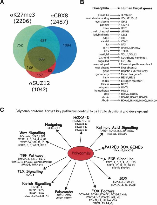
Downloaded from genesdev.cshlp.org on March 18, 2015 - Published by Cold Spring Harbor Laboratory Press

Bracken et al.

Figure 2. The PcG proteins and H3K27me3 are highly enriched on the HOXA gene cluster. (A) ChIP-on-chip tiling array analysis of
the HOXA locus spanning 140 kb of DNA starting at position 26,880,000 on chromosome 7. High-resolution mapping of the 140 kb
was achieved by representing 2840 probes of 50 bp with an average spacing of 3 bp between probes. The Y-axis represents the Log2
signal ratios (bound/input) for the indicated antibodies. The results of the promoter ChIP-on-chip experiment are also shown for the
HOXA promoters. In this analysis, chips were used containing 24,275 human promoters from 1300 bp upstream to 200 bp downstream
of TSS with 15 50-bp probes with an average spacing of 100 bp between probes. (C) Custom ChIP-on-chip data; (P) Promoter ChIP-
on-chip data. (B) XY scatterplot representation of the data depicted in A, created by plotting the average enrichments at 5-kb intervals.
(C) Normal ChIP analysis of the promoters of the HOXA gene cluster. Primers were designed within the promoter regions (indicated
with red bars) of all HOXA genes as indicated at the top of the panel. Enrichment is shown as percentage input.

  Intriguingly, SUZ12 and CBX8 are also found on the                   tified 43 target genes of the PcG proteins, whose expres-
BMI-1 and CBX8 gene loci and are associated with                       sion changes in PcG-depleted cells.
H3K27me3 enrichments (Fig. 3B; Supplementary Fig.                         Perhaps surprisingly, the mRNA transcripts of the pre-
S3). The expression of BMI-1 and CBX8 increases follow-                viously characterized SUZ12 target gene MYT1 as well
ing the siRNA-mediated depletion of other PRC1 or                      the HOXA7–13 genes remained undetectable in PcG-de-
PRC2 members (Figs. 1A, 3A). Therefore, these data sug-                pleted cells (data not shown). This is despite the fact that
gest that PcG proteins autoregulate their own synthesis.               we observe strong PcG enrichments on their promoters
This is conserved during evolution, since PcG proteins                 (Figs. 2C, 3C). Therefore, in order to identify all PcG
bind to the polyhomeotic (Ph) locus in Drosophila (Fau-                target genes in human embryonic fibroblasts, we decided
varque et al. 1995; Bloyer et al. 2003).                               to perform a global unbiased identification of PcG-bound
  Next we validated a selection of these newly identified              promoters independent of their expression.
target genes, for EZH2, SUZ12, CBX8, and H3K27me3
enrichments in an independent experiment (Fig. 3C).
                                                                       Genome-wide identification of human promoters
The previously identified SUZ12 target gene MYT1 (Kir-
                                                                       bound by PcG
mizis et al. 2004) was included as a positive control and
the HOXA1 and CCNA2 genes as negative controls. Fi-                    We interrogated chips containing probes for 24,275 hu-
nally, the expression of several target genes (MT1G,                   man promoters within a defined region spanning 1300
CCND2, SERPINB2, and CYPB1) were tested and shown                      base pairs (bp) upstream to 200 bp downstream of the
to be reduced in BMI-1-overexpressing TIG3 cells (data                 TSS. Significantly, the PcGs bind within this limited
not shown), further validating them as PcG target genes                promoter region in 70% of the target genes described in
in this cellular system. We conclude that we have iden-                Figure 3A (see Supplementary Table 2). As shown in Fig-

1126       GENES & DEVELOPMENT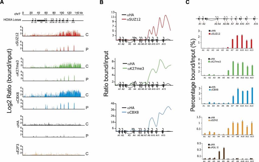
Downloaded from genesdev.cshlp.org on March 18, 2015 - Published by Cold Spring Harbor Laboratory Press

                                                                                                                Polycomb target genes

Figure 3. Identification of 43 PcG target genes whose expression changes in Polycomb-depleted cells. (A) Treeview depiction of the
expression changes in Polycomb-depleted cells of 43 direct PcG target genes. (B) XY scatterplot representations of SUZ12, H3K27me3,
and CBX8 enrichments along the gene loci of BMP2, ATF3, BMI-1, and CCND2. The chromosome number and the region covered are
depicted above each panel. (C) Standard ChIP analysis of newly identified Polycomb target genes using the previously identified MYT1
target gene as a positive control. The CCNA2 and HOXA1 genes are presented as negative controls.

ure 4A and listed in Supplementary Table 3, SUZ12,                   PRC1 and PRC2, which were also tri-methylated on
CBX8, and H3K27me3 are present on a very large num-                  H3K27, consistent with the fact that PRC1 is dependent
ber of promoters. A significant enrichment was found for             on PRC2-mediated K27 tri-methylation for its ability to
CBX8 (PRC1) on 2487 promoters (10.2% of the total), for              bind to chromatin (Hernandez-Munoz et al. 2005; Pir-
H3K27me3 on 2206 promoters (9.0%), and for SUZ12                     rotta and Gross 2005). Several genes with H3K27me3
(PRC2) on 1042 promoters (4.3%). A very significant                  enrichments appeared not to have associated CBX8, sug-
overlap was observed between promoters bound by                      gesting that this mark may execute distinct functions

                                                                                                 GENES & DEVELOPMENT            1127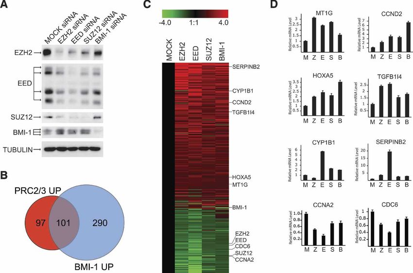
Downloaded from genesdev.cshlp.org on March 18, 2015 - Published by Cold Spring Harbor Laboratory Press

Bracken et al.

Figure 4. Genome-wide mapping of PcG target pro-
moters. (A) Venn diagram depicts significant overlaps
between the presence of SUZ12, CBX8, and H3K27me3
on promoters. (B) A remarkable conservation of Poly-
comb target genes through evolution from Drosophila
to humans. Several confirmed or predicted Drosophila
PcG target genes are shown in the left column. In our
analysis we have identified their human homologs as
being human PcG targets (see Supplementary Table 3).
(C) A selection of PcG target genes identified in this
study, focusing on those known to be involved in key
pathways controlling development, differentiation,
stem cell biology, and cell fate decisions.

independent of PRC1 complexes. Alternatively, PRC1                   locus. As mentioned above, we have found that the hu-
might be bound to these genes through the two chromo-                man PcGs also bind to the BMI-1 and CBX8 gene loci,
domain-containing CBX8 homologs CBX4 or CBX7. The                    and now extend this group to include the CBX4 and
large number of genes identified, which were only sig-               CBX7 loci.
nificantly bound by CBX8, are unlikely to be CBX8 spe-
cific targets. In fact, genes belonging to this category,
                                                                     PcGs target both stem cell- and differentiation-specific
when subsequently tested in quantitative ChIP analysis,
                                                                     genes
were found to also possess significant H3K27me3 and
SUZ12 binding, for example, CYP1B1 and DKK2 (cf. Fig.                We wished to study PcG dynamics on target genes in a
3C and Supplementary Table 3). Therefore the large                   biologically relevant model of differentiation. The global
number of genes bound only by CBX8 is likely a conse-                mapping analysis revealed that the PcGs bind many
quence of the higher affinity of the CBX8 antibody for its           genes that are specifically induced in various types of
epitope rather than a reflection of PRC2-independent re-             differentiation (Table 1). For example, BMP6 (Kugimiya
cruitment. In conclusion, our results using promoter ar-             et al. 2005), the homebox gene PAX4 (Wang et al. 2004),
rays extend the repertoire of PcG target genes to >1000              MYOG (Rohwedel et al. 1994), DMBT1 (Al-Awqati
(Fig. 4A; Supplementary Table 3).                                    2003), and ZIC1 (Sato et al. 2005) are specific differen-
   Next we asked: What is the nature of the target genes             tiation factors induced in osteogenesis, endocrinal differ-
identified? Remarkably, we found that the majority of                entiation, myogenesis, epithelial cell differentiation, and
the genes previously identified or predicted to be PcG               neurogenesis, respectively. Recent results have shown
target genes in Drosophila have human homologs iden-                 that EZH2 represses the muscle creatine kinase gene
tified as PcG targets in this screen (Fig. 4B; Ringrose and          CKM in nondifferentiated myoblasts and is displaced
Paro 2004; Ringrose et al. 2004). In addition to the HOX             from the gene promoter upon muscle differentiation,
genes, we found that Engrailed, Hedgehog, Hairy, and                 when the gene becomes activated (Caretti et al. 2004).
Caudal all have human PcG target homologs. Interest-                 Our results show that the PcGs target a large number of
ingly, another target in Drosophila is the Polyhomeotic              tissue-specific differentiation genes like CKM (see Table

1128       GENES & DEVELOPMENT
Downloaded from genesdev.cshlp.org on March 18, 2015 - Published by Cold Spring Harbor Laboratory Press
Polycomb target genes
Table 1. Examples of differentiation genes identified by PcG ChIP-on-chip
Antibodies used
Gene name Function ␣HA ␣CBX8 ␣SUZ12 ␣K27me3
DLX5 Neuronal differentiation + + +
HEY2 Neuronal differentiation + + +
LHX1 Neuronal differentiation + + +
MASS1 Neuronal differentiation + + +
NEFL Neuronal differentiation + + +
NEUROD1 Neuronal differentiation + + +
NEUROG1 Neuronal differentiation + + +
NEUROG2 Neuronal differentiation + + +
NMU Neuronal differentiation + + +
NXPH2 Neuronal differentiation + + +
OLIG2 Neuronal differentiation + + +
PAX3 Neuronal differentiation + + +
PHOX2A Neuronal differentiation + + +
PHOX2B Neuronal differentiation + + +
POMC Neuronal differentiation + + +
POU4F2 Neuronal differentiation + + +
ROBO3 Neuronal differentiation + + +
SEMA5B Neuronal differentiation + + +
SIM1 Neuronal differentiation + + +
TBR1 Neuronal differentiation + + +
ZIC1 Neuronal differentiation + + +
BMP3 Bone differentiation +
TNFSF11 Bone differentiation + +
CHRDL2 Bone differentiation + + +
BMP6 Bone differentiation +
GDF2 Bone differentiation + +
BMP7 Sex differentiation + +
JAG2 Sex differentiation +
DMRT1 Sex differentiation + + +
DMRT2 Sex differentiation + + +
DMRT3 Sex differentiation + + +
SRY Sex differentiation +
DMRTA1 Sex differentiation + +
BMP8B Sex differentiation + +
FGF9 Sex differentiation +
CD4 T-cell differentiation +
MDFI Chondrogenic differentiation + + +
CSRP3 Muscle development +
MYOG Muscle development + +
SYNE1 Muscle development +
CKM Muscle development + +
MYH11 Muscle development + + +
ACHE Muscle development + + +
TNNT1 Muscle development + + +
HAND2 Cardiogenesis +
DLL1 Cell fate/Notch pathway +
DLL3 Cell fate/Notch pathway + + +
NOTCH4 Cell fate/Notch pathway + +
PAX4 Endocrine differentiation + + +
EDAR Epidermal cell differentiation + +
DMBT1 Epithelial cell differentiation + +
THPO Hematopoiesis + + +
KIT Hematopoiesis + + +
LIF Hematopoiesis +
KDR Hematopoiesis + +
TAL1 Hematopoiesis + + + +
BMP4 Mesoderm cell fate decisions +
SHH Mesoderm cell fate decisions + + +
A selection of enriched target genes identified in the ChIP-on-chip analysis using a 24,275 tiled promoter array.
GENES & DEVELOPMENT 1129Downloaded from genesdev.cshlp.org on March 18, 2015 - Published by Cold Spring Harbor Laboratory Press
Bracken et al.
1), suggesting that the displacement of PcGs from differ- 6A and shown in Figure 6B, the (posterior) 5⬘-end genes
entiation-specific genes during terminal differentiation (HOXA7–13) are expressed in undifferentiated cells but
is a general phenomenon (depicted in Fig. 5A, panel i). are dramatically repressed upon RA treatment. In con-
To test this, we chose to focus on neuronal differen- trast, the genes at the (anterior) 3⬘ end of the locus
tiation for several reasons. A large number of neuronal (HOXA1–5) become strongly activated. We determined
differentiation genes are bound by PcGs (Table 1). The the binding of EZH2 and CBX8 together with H3K27me3
PcGs are required for neuronal development and self- enrichments on the complete HOXA locus both before
renewal of neural stem cells (Leung et al. 2004; Molofsky and after induction to differentiate. Consistent with the
et al. 2004). Additionally, there are several well-estab- genes analyzed in Figure 5, the PcGs are displaced from
lished model systems for neuronal differentiation pro- the activated HOXA1–5 gene loci during differentiation,
cesses. As a model system, we chose the human em- while they are already bound to the HOXA7–13 genes in
bryonic teratocarcinoma cell line NT2/D1, which has undifferentiated NT2/D1 cells (Fig. 6C) and remain
neural progenitor cell properties and irreversibly differ- bound throughout the differentiation process and subse-
entiates along a neural lineage upon treatment with reti- quent decrease in mRNA expression. These results on
noic acid (RA) (Lee and Andrews 1986). the HOXA cluster expand the set of genes, which are
We selected four genes based on their proposed role in transcriptionally active in undifferentiated cells yet
neuronal differentiation: the gene for the zinc-finger do- bound by PcGs prior to their repression in differentiated
main transcription factor ZIC1 required for normal neu- cells.
ronal differentiation (Grinberg and Millen 2005); the ho-
meobox-containing transcription factor MEIS2; the RA
receptor , RARB; and the gene for neurofilament light Discussion
chain, NEFL (Zhu et al. 1997). The expression of these
By genome-wide location analysis, we have identified
genes is induced during differentiation (Zhu et al. 1997;
>1000 putative genes bound by PcG proteins. Strikingly,
Niederreither et al. 2000) and during RA-induced differ-
these genes contain the key members of the Wnt, TGF,
entiation of NT2/D1 cells (Fig. 5B; data not shown). Con-
FGF, Notch, and Hedgehog signaling pathways known to
sistent with the increased expression of these genes dur-
be the regulators of developmental and differentiation
ing differentiation, we observed a progressive decrease in
processes. In addition, target genes are conserved
PcG binding and H3K27me3 enrichments on these genes
throughout evolution from Drosophila to human, sug-
(Fig. 5B; data not shown). These data are consistent with
gesting that this transcriptional regulatory network is
the model depicted in Figure 5A, panel i, and since the
essential for the development and differentiation of all
PcGs associate with a large number of tissue-specific
multicellular organisms.
genes in differentiated cells, this suggests that the differ-
entiation specific signals, which activate tissue-specific
genes, lead to the displacement of PcGs from the genes.
Polycomb target genes
Subsequently we decided to investigate PcG target
genes, which become silenced during differentiation. A By performing gene expression profiling of human em-
potential model for how this would occur is depicted in bryonic fibroblasts in combination with ChIP-on-chip
Figure 5A, panel ii, in which we speculate that PcGs experiments, we identified 43 PcG target genes whose
would be recruited during differentiation. To test this expression is dependent on the PcGs. The large majority
hypothesis, we selected three genes that are highly ex- of these genes become derepressed as a result of specific
pressed in neuronal progenitor cells but silenced during depletion by siRNAs of the PRC2 components (EZH2,
differentiation (Fig. 5C). These included genes for the EED, and SUZ12) or the PRC1 component BMI-1 (39 of
basic helix–loop–helix (bHLH) proneural transcription 43). These target genes include several known markers of
factors OLIG2 (Lee et al. 2005) and NEUROG2 (Ma et al. bone, cartilage, and fat differentiation (Supplementary
1996), and the gene for metallothionein 1G, MT1G. ChIP Table 2). To put this into context, fibroblasts are prolif-
experiments showed, in contrast to our expectations, a erating cells of the mesoderm that compose part of the
strong and significant binding of PcGs together with the connective tissue in almost every tissue and organ. They
presence of H3K27me3 on the promoters of these genes are derived from mesenchymal stem cells, which are
in undifferentiated cells (Fig. 5C), with only slight in- multipotent precursors, capable of differentiating into
creases during differentiation. These results demonstrate osteoblasts, adipocytes, chrondrocytes, endothelial cells,
that binding of PcGs to target genes in undifferentiated and also non-mesoderm-type lineages, such as neuronal-
cells does not strictly correlate with transcriptional si- like cells (Kassem 2004). Interestingly, evidence exists
lencing. that embryonic fibroblasts in tissue culture maintain at
To understand if this intriguing result could be ex- least some of the multipotency of mesenchymal stem
tended to other genes, we analyzed the binding of the cells. For example, mouse embryonic fibroblasts can un-
PcGs to the HOXA locus before and after differentiation dergo chrondrogenic and adipocytic differentiation (Ge
of the NT2/D1 cells. Previous data have shown that the et al. 2002; Lengner et al. 2004). Consistent with this, we
HOX genes display faithful regulation patterns following found that several markers of osteocytic, chrondrocytic,
RA-induced differentiation of NT2/D1 cells (Simeone et adipocytic, and neural differentiation are up-regulated in
al. 1990; Houldsworth et al. 2002). As depicted in Figure Polycomb-depleted cells (Fig. 3A; Supplementary Table
1130 GENES & DEVELOPMENTDownloaded from genesdev.cshlp.org on March 18, 2015 - Published by Cold Spring Harbor Laboratory Press
Polycomb target genes
Figure 5. PcGs bind to genes that are repressed or induced during neuronal differentiation. (A) Models for how PcGs could regulate
gene expression during terminal differentiation. (B) qPCR and ChIP analysis of two PcG target genes (ZIC1 and MEIS2) induced during
neuronal differentiation of NT2/D1 cells treated with 1 µM RA. (C) qPCR and ChIP analysis of two genes (NEUROG2 and OLIG2)
repressed during neuronal differentiation of NT2/D1 cells treated with 1 µM RA.
GENES & DEVELOPMENT 1131Downloaded from genesdev.cshlp.org on March 18, 2015 - Published by Cold Spring Harbor Laboratory Press Bracken et al. Figure 6. Polycombs are present on active HOX genes in nondifferentiated cells. (A) Schematic depiction of the gene expression changes along the HOXA locus upon RA-mediated induction of neuronal differentiation. (B) Quantification of mRNA expression changes of HOXA genes during neuronal differentiation of NT2/D1 cells by qPCR. (C) ChIP analysis of EZH2, H3K27me3, and CBX8 binding to the promoters of the HOXA1 to HOXA13 genes both before and after 10 d of RA treatment. (D) Displacement of PcGs from target genes activated during differentiation, for example, ZIC1, MEIS2, RARB, and HOXA1–5 in neuronal differentiation and CKM in myoblast differentiation. (E) Polycomb “preprogramming” of certain target genes in undifferentiated cells, for example, NEUROG2, OLIG2, and HOXA9–13. These genes are paradoxically expressed while being bound by PcGs in undifferentiated cells. Several hypothetical mechanisms for the triggering of PcG repressive function during differentiation are possible, include post-translational modifications of the PcGs, addition of H1K26me3 or ubiquitinated H2A-K119 marks, recruitment of DNA methyltransferases, the binding of additional transcriptional repressors, or a combination of these mechanisms. 2). For example, both the bone morphogenic protein Strikingly, the expression of only a relatively small BMP2 and the Wnt/-catenin signaling protein DKK2 number of the >1000 identified PcG target genes was function in terminal osteoblast differentiation (Li et al. affected by depleting PcGs in human embryonic fibro- 2005). While HEY1 and TCF7 are transcriptionally acti- blasts, and >90% of the identified target genes are not vated during osteogenesis (de Jong et al. 2004), both detectably expressed. This is consistent with previous CHST11 (Kluppel et al. 2005) and PRKG2 (Chikuda et al. observations by Kirmizis et al. (2004) in SW480 colon 2004) are associated with chrondrocytic differentiation, cancer cells depleted of SUZ12. We hypothesize that the the latter being a molecular switch from proliferation to majority of the 1000 PcG target genes identified here in hypertrophic differentiation of chondrocytes. The G0S2 fibroblasts are permanently silenced. This permanent re- protein is involved in adipocyte differentiation (Zandber- pression could be due to secondary epigenetic modifica- gen et al. 2005), and UCP1 is a specific marker of Brown tions of the target promoters by, for example, the EZH2 adipose tissue. In addition, several other target genes are complex itself or by DNA methylation. Supporting this known regulators of development such as RARB, WT1, notion is the demonstration that EZH2 possesses H1K26 and the homeobox genes SALL1 and HOXA5. In sum- methylation activity (Kuzmichev et al. 2004), and since mary, the combination of ChIP-on-chip and expression H1K26me3 can tether HP1 to chromatin (Daujat et al. array analysis in human embryonic fibroblasts has iden- 2005), this could add an additional layer of transcrip- tified 43 novel PcG target genes with important regula- tional repression on PcG target genes. Additionally, the tory roles in mesenchymal differentiation and develop- expression of EZH2 has recently been shown to be suf- ment. ficient for the recruitment of DNA methyl transferases 1132 GENES & DEVELOPMENT
Downloaded from genesdev.cshlp.org on March 18, 2015 - Published by Cold Spring Harbor Laboratory Press

                                                                                                              Polycomb target genes

to cellular promoters leading to their DNA methylation             model is consistent with previous results, suggesting a
and repression (Vire et al. 2005). It is possible that such        similar role for the PcGs in muscle and germ cell termi-
secondary repressive modifications are not alleviated in           nal differentiation processes (Caretti et al. 2004; Chen et
PcG-depleted cells. As an alternative and complemen-               al. 2005) and predicts that this is a general phenomenon.
tary explanation for the lack of expression of a large                The alternative model describes when PcG target
number of PcG target genes in fibroblasts, we suggest              genes are down-regulated during differentiation (Fig. 6E).
that the fibroblasts may lack the specific transcriptional         This model is based on the surprising observation that
activators required for the expression of the tissue-spe-          the PcGs, in some cases, are bound to their target genes
cific target genes such as those listed in Table 1.                in undifferentiated cells, despite the gene being actively
                                                                   expressed. Based on our results for OLIG2, NEUROG2,
                                                                   and HOXA9–13, we propose that these PcG target genes
Polycomb target genes and cancer                                   are already “preprogrammed” to be repressed upon ap-
                                                                   propriate cell fate signals. Supporting this hypothesis,
The identification of PcG target genes may also provide
                                                                   the Paro laboratory demonstrated that the Drosophila
vital mechanistic insights into the precise nature by
                                                                   PcG and Trithorax proteins bind to Polycomb-Respon-
which PcGs contribute to cancer. It has been proposed
                                                                   sive Elements (PREs) and promoters of HOX genes before
that cancer originates from nondifferentiated or stem
                                                                   their expression levels are set by the early-acting seg-
cell-like cells, referred to as “cancer stem cells” (Reya et
                                                                   mentation factors (Orlando et al. 1998). Further support-
al. 2001). This idea is supported by the recent observa-
                                                                   ing such a concept, we observed H3K27me3 and SUZ12
tion that mammary stem cells are enriched in premalig-
                                                                   enrichments on the entire HoxA locus in mouse embry-
nant breast tissue (Shackleton et al. 2006). The identifi-
                                                                   onic stem cells, resembling the profile observed in un-
cation of a large number of PcG target genes required for
                                                                   differentiated NT/D1 cells (Fig. 6C; Supplementary Fig.
differentiation (Table 1) strongly suggests that the onco-
                                                                   S4; data not shown). This suggests that the transcrip-
genic potential of genes such as BMI-1 and EZH2 can be
                                                                   tional memory system of the HOX genes and other genes
ascribed to their role in stem cell maintenance. Another
                                                                   controlling cell fate are preset or “programmed,” possi-
insight comes from the fact that a significant proportion
                                                                   bly by PcGs in the zygote generated from maternal
of the identified PcG genes are silenced in cancer by
                                                                   mRNA. In this model, specific developmental signals
DNA methylation of their promoter sequences. These
                                                                   such as RA concentrations could trigger the PcG-repres-
include RARB, CCND2, MT1G, KLF4, IGSF4, WT1,
                                                                   sive capacity by as yet unidentified mechanisms. This
NPTX1, HOXA5, BMP2, and G0S2 (Evron et al. 2001;
                                                                   could involve the post-translational modifications of the
Loeb et al. 2001; Fackler et al. 2003; Fukami et al. 2003;
                                                                   PcG proteins, the modification of other proteins/his-
Hagihara et al. 2004; Zhao et al. 2004; Henrique et al.
                                                                   tones by the PcGs, or the specific recruitment/dissocia-
2005; Lewis et al. 2005). Significantly, as pointed out
                                                                   tion of transcriptional regulators from the PcG target
above, EZH2 has recently been shown to act as a plat-
                                                                   genes (Fig. 6E). Therefore, we propose that the PRCs
form for DNA methyltransferases on certain EZH2 tar-
                                                                   function as “platforms” for channeling repressive signals
get genes (Vire et al. 2005). The PRC2 complex could
                                                                   during developmental and differentiation processes.
therefore contribute to cancer development by specifi-
                                                                      In summary, we predict that PcGs form part of an epi-
cally silencing tumor-suppressor genes by DNA methyl-
                                                                   genetic blueprint for development and differentiation es-
ation. Consistent with this, we have found an inverse
                                                                   tablished early in embryogenesis. Future investigations
correlation between the expression levels of the PcGs,
                                                                   may hopefully unravel the precise mechanistic contribu-
EZH2, SUZ12, and BM1–1, and the target genes MT1G,
                                                                   tions of PcG proteins, modifications such as H1K26me3
HOXA5, and RARB in breast cancer (A.P. Bracken, P.
                                                                   and ubiquitinated H2A-K119 and associated proteins,
Cloos, and S. Confalonieri, unpubl.). It will be important
                                                                   such as HP1 and DNA methyltransferases, on the >1000
in future investigations to determine if the silencing of
                                                                   genes identified here during multiple types of cell fate
these genes in cancer contributes to the development of
                                                                   decisions. An additional challenge for the future will be
the disease and if PcGs have a causal role in this.
                                                                   to harness this knowledge such that we can dedifferen-
                                                                   tiate or trans-differentiate cells for therapeutic purposes.
Delineating the mechanisms by which PcGs regulate
cell fate decisions
                                                                   Materials and methods
The identification of genes bound by PcGs has enabled
us to begin to address the mechanisms by which they                Tissue culture
repress transcription. Our results, based on a model of
neuronal differentiation, suggest at least two alternative         The human diploid embryonic lung fibroblast TIG3 cell line and
                                                                   the embryonic carcinoma cell line NTERA2 (NT2/D1) (Lee and
mechanisms (depicted in Fig. 6D,E). The first model is
                                                                   Andrews 1986) were grown in DMEM supplemented with 10%
exemplified by genes such as ZIC1, MEIS2, and HOXA1–               (v/v) FCS. To induce differentiation of NT2/D1 cells, asynchro-
5, which are bound and repressed by PcGs in undifferen-            nously growing cells were seeded at 30% confluency and were
tiated cells. Upon induction to differentiate, PRC1 and            treated 24 h later with 1 µM ATRA (dissolved in DMSO; Sigma).
PRC2 are displaced from these genes by, as yet, uniden-            Cultures treated continuously with RA were collected at 0, 2, 5,
tified mechanism(s), leading to their derepression. This           and 10 d for RNA extraction or harvested for ChIP at 0, 5, and

                                                                                                GENES & DEVELOPMENT           1133
Downloaded from genesdev.cshlp.org on March 18, 2015 - Published by Cold Spring Harbor Laboratory Press
Bracken et al.
10 d. Cultures were reseeded every 3–4 d and collected no less The sequences of the PCR primers are available upon request.
than 24 h after reseeding. Instead for the ChIP-chip analysis, the immunoprecipitated
DNA was amplified by LMPCR as described previously (Ren et
al. 2000) and hybridized to tiled arrays manufactured by
Generation of antibodies
NimbleGen Systems, Inc. (http://www.nimblegen.com).
Two polyclonal CBX8 (hPc3) antibodies “LAST” and “GALD”
were produced in rabbits using synthetic peptides correspond-
ing to amino acids 107–129 (KKPGRSPQDLASTSRAREGLRN
Acknowledgments
MGL) and 295–318 (KKGQGALDPNGTRVRHGSGPPSSGG) of
human CBX8, respectively. These were coupled to Keyhole We thank Simone Minardi at The Affymetrix Microarray Unit
Limpet Hemocyanin (KLH) through the N-terminal lysine resi- of The IFOM-IEO campus (Milan, Italy) for help with the ex-
dues and injected subcutaneously into rabbits according to pression array experiments, Matteo Cesaroni and Rehanah Boup
standard procedures (DAKO). Positive sera produced were affin- for their help with data analysis, and Henrik Winther at DAKO
ity-purified on the respective peptide antigens according to stan- (Denmark) for the production of anti-sera to CBX8. We thank
dard procedures. For ChIP assays, 5 µg of a pool of both affinity- Michael Lees and Claus S. Sørensen for critical reading of the
purified antibodies was used per immunoprecipitation. manuscript and Anders H. Lund, Bruno Amati, Ernesto Gucci-
one, Marco Ciró, and members of the Helin laboratory for dis-
cussions. This work was supported by grants from the Associa-
siRNA interference
tion for International Cancer Research, the Danish Cancer So-
Specific siRNA oligos targeting EZH2, EED, SUZ12, and BMI-1 ciety, the Novo Nordisk Foundation, the Danish Medical
mRNAs were described previously (Bracken et al. 2003; Pasini Research Council, the Danish Natural Science Research Coun-
et al. 2004b). Cells were transfected using Oligofectamine (In- cil, and the European Commission’s 6th Framework Pro-
vitrogen) and harvested 44 h after transfection. gramme.
Quantification of mRNA levels by qPCR
References
cDNA was generated by RT–PCR using the PE Applied Biosys-
tems TaqMan Reverse Transcription Reagents. Reactions were Al-Awqati, Q. 2003. Terminal differentiation of intercalated
determined using the SYBR Green I detection chemistry system cells: The role of hensin. Annu. Rev. Physiol. 65: 567–583.
(Applied Biosystems), using an ABI Prism 7300 Sequence De- Bloyer, S., Cavalli, G., Brock, H.W., and Dura, J.M. 2003. Iden-
tection System. Ubiquitin was used as a control gene for nor- tification and characterization of polyhomeotic PREs and
malization. The sequences of the primers used are available TREs. Dev. Biol. 261: 426–442.
upon request. Bracken, A.P., Pasini, D., Capra, M., Prosperini, E., Colli, E., and
Helin, K. 2003. EZH2 is downstream of the pRB–E2F path-
way, essential for proliferation and amplified in cancer.
Affymetrix expression analysis
EMBO J. 22: 5323–5335.
Total RNA was extracted from mock, EZH2, EED, SUZ12, and Cao, R. and Zhang, Y. 2004. The functions of E(Z)/EZH2-medi-
BMI-1 siRNA-transfected cells. For each treatment, RNA was ated methylation of lysine 27 in histone H3. Curr. Opin.
prepared from six independent experiments and was pooled into Genet. Dev. 14: 155–164.
one sample to reduce the experimental variation. Targets for Cao, R., Tsukada, Y., and Zhang, Y. 2005. Role of Bmi-1 and
microarray hybridization were synthesized accordingly to the Ring1A in H2A ubiquitylation and Hox gene silencing. Mol.
supplier’s instructions (Affymetrix). The human GeneChip ar- Cell 20: 845–854.
ray U133A (Affymetrix), which interrogates ∼33,000 transcripts, Caretti, G., Di Padova, M., Micales, B., Lyons, G.E., and Sar-
was used for gene expression profiling. Fifteen GeneChip arrays torelli, V. 2004. The Polycomb Ezh2 methyltransferase regu-
U133A were used for this analysis; for each different condition, lates muscle gene expression and skeletal muscle differen-
three separate arrays were hybridized with pooled cRNA. Hy- tiation. Genes & Dev. 18: 2627–2638.
bridization, washing, staining, scanning, and data analysis were Chen, X., Hiller, M., Sancak, Y., and Fuller, M.T. 2005. Tissue-
performed at The Affymetrix Microarray Unit at the IFOM-IEO specific TAFs counteract Polycomb to turn on terminal dif-
campus, Milan, Italy, according to the manufacturer’s instruc- ferentiation. Science 310: 869–872.
tions. Expression levels were analyzed using Microarray Analy- Chikuda, H., Kugimiya, F., Hoshi, K., Ikeda, T., Ogasawara, T.,
sis Suite (MAS) 5.0 statistical algorithm software (Affymetrix), Shimoaka, T., Kawano, H., Kamekura, S., Tsuchida, A., Yo-
using the default parameters and scaling (TGT Value) signal koi, N., et al. 2004. Cyclic GMP-dependent protein kinase II
intensities for all the GeneChip arrrays to a value of 500. The is a molecular switch from proliferation to hypertrophic dif-
mock siRNA treatment was used as a baseline condition for ferentiation of chondrocytes. Genes & Dev. 18: 2418–2429.
comparison with the polycomb siRNA-treated samples. Daujat, S., Zeissler, U., Waldmann, T., Happel, N., and
Schneider, R. 2005. HP1 binds specifically to Lys26-methyl-
ated histone H1.4, whereas simultaneous Ser27 phosphory-
ChIP assays
lation blocks HP1 binding. J. Biol. Chem. 280: 38090–38095.
ChIPs were performed and analyzed essentially as described pre- de Jong, D.S., Vaes, B.L., Dechering, K.J., Feijen, A., Hendriks,
viously (Bracken et al. 2003). The antibodies used were rabbit J.M., Wehrens, R., Mummery, C.L., van Zoelen, E.J., Olijve,
anti-E2F3 (sc-878; Santa Cruz), anti-PolII (sc-899; Santa Cruz), W., and Steegenga, W.T. 2004. Identification of novel regu-
anti-H3K27me3 (Upstate), anti-SUZ12 (Upstate), anti-HA (sc- lators associated with early-phase osteoblast differentiation.
805; Santa Cruz), a mouse monoclonal specific for EZH2 (AC22) J. Bone Miner. Res. 19: 947–958.
(Pasini et al. 2004b), and an equal mix of the polyclonal anti- Evron, E., Umbricht, C.B., Korz, D., Raman, V., Loeb, D.M.,
bodies for CBX8, described above. For normal chromatin IPs, the Niranjan, B., Buluwela, L., Weitzman, S.A., Marks, J., and
immunoprecipitated DNA was quantified by real-time qPCR. Sukumar, S. 2001. Loss of cyclin D2 expression in the ma-
1134 GENES & DEVELOPMENTDownloaded from genesdev.cshlp.org on March 18, 2015 - Published by Cold Spring Harbor Laboratory Press
Polycomb target genes
jority of breast cancers is associated with promoter hyper- S., Yano, F., Ogata, N., Katagiri, T., Harada, Y., Azuma, Y., et
methylation. Cancer Res. 61: 2782–2787. al. 2005. Involvement of endogenous bone morphogenetic
Fackler, M.J., McVeigh, M., Evron, E., Garrett, E., Mehrotra, J., protein (BMP) 2 and BMP6 in bone formation. J. Biol. Chem.
Polyak, K., Sukumar, S., and Argani, P. 2003. DNA methyl- 280: 35704–35712.
ation of RASSF1A, HIN-1, RAR-, Cyclin D2 and Twist in in Kuzmichev, A., Jenuwein, T., Tempst, P., and Reinberg, D.
situ and invasive lobular breast carcinoma. Int. J. Cancer 2004. Different EZH2-containing complexes target methyl-
107: 970–975. ation of histone H1 or nucleosomal histone H3. Mol. Cell 14:
Fauvarque, M.O., Zuber, V., and Dura, J.M. 1995. Regulation of 183–193.
polyhomeotic transcription may involve local changes in Lee, V.M. and Andrews, P.W. 1986. Differentiation of NTERA-2
chromatin activity in Drosophila. Mech. Dev. 52: 343–355. clonal human embryonal carcinoma cells into neurons in-
Fisher, A.G. 2002. Cellular identity and lineage choice. Nat. volves the induction of all three neurofilament proteins. J.
Rev. Immunol. 2: 977–982. Neurosci. 6: 514–521.
Fukami, T., Fukuhara, H., Kuramochi, M., Maruyama, T., Iso- Lee, S.K., Lee, B., Ruiz, E.C., and Pfaff, S.L. 2005. Olig2 and
gai, K., Sakamoto, M., Takamoto, S., and Murakami, Y. Ngn2 function in opposition to modulate gene expression in
2003. Promoter methylation of the TSLC1 gene in advanced motor neuron progenitor cells. Genes & Dev. 19: 282–294.
lung tumors and various cancer cell lines. Int. J. Cancer 107: Lengner, C.J., Lepper, C., van Wijnen, A.J., Stein, J.L., Stein,
53–59. G.S., and Lian, J.B. 2004. Primary mouse embryonic fibro-
Ge, K., Guermah, M., Yuan, C.X., Ito, M., Wallberg, A.E., blasts: A model of mesenchymal cartilage formation. J. Cell.
Spiegelman, B.M., and Roeder, R.G. 2002. Transcription co- Physiol. 200: 327–333.
activator TRAP220 is required for PPAR ␥ 2-stimulated adi- Lessard, J. and Sauvageau, G. 2003. Bmi-1 determines the pro-
pogenesis. Nature 417: 563–567. liferative capacity of normal and leukaemic stem cells. Na-
Grinberg, I. and Millen, K.J. 2005. The ZIC gene family in de- ture 423: 255–260.
velopment and disease. Clin. Genet. 67: 290–296. Leung, C., Lingbeek, M., Shakhova, O., Liu, J., Tanger, E., Sare-
Hagihara, A., Miyamoto, K., Furuta, J., Hiraoka, N., Wakazono, maslani, P., Van Lohuizen, M., and Marino, S. 2004. Bmi1 is
K., Seki, S., Fukushima, S., Tsao, M.S., Sugimura, T., and essential for cerebellar development and is overexpressed in
Ushijima, T. 2004. Identification of 27 5⬘ CpG islands aber- human medulloblastomas. Nature 428: 337–341.
rantly methylated and 13 genes silenced in human pancre- Levine, S.S., King, I.F., and Kingston, R.E. 2004. Division of
atic cancers. Oncogene 23: 8705–8710. labor in Polycomb group repression. Trends Biochem. Sci.
Henrique, R., Jeronimo, C., Hoque, M.O., Nomoto, S., Carv- 29: 478–485.
alho, A.L., Costa, V.L., Oliveira, J., Teixeira, M.R., Lopes, C., Lewis, C.M., Cler, L.R., Bu, D.W., Zochbauer-Muller, S., Milch-
and Sidransky, D. 2005. MT1G hypermethylation is associ- grub, S., Naftalis, E.Z., Leitch, A.M., Minna, J.D., and Euhus,
ated with higher tumor stage in prostate cancer. Cancer Epi- D.M. 2005. Promoter hypermethylation in benign breast epi-
demiol. Biomarkers Prev. 14: 1274–1278. thelium in relation to predicted breast cancer risk. Clin.
Hernandez-Munoz, I., Taghavi, P., Kuijl, C., Neefjes, J., and van Cancer Res. 11: 166–172.
Lohuizen, M. 2005. Association of BMI1 with Polycomb Li, X., Liu, P., Liu, W., Maye, P., Zhang, J., Zhang, Y., Hurley,
bodies is dynamic and requires PRC2/EZH2 and the main- M., Guo, C., Boskey, A., Sun, L., et al. 2005. Dkk2 has a role
tenance DNA methyltransferase DNMT1. Mol. Cell. Biol. in terminal osteoblast differentiation and mineralized ma-
25: 11047–11058. trix formation. Nat. Genet. 37: 945–952.
Houldsworth, J., Heath, S.C., Bosl, G.J., Studer, L., and Cha- Loeb, D.M., Evron, E., Patel, C.B., Sharma, P.M., Niranjan, B.,
ganti, R.S. 2002. Expression profiling of lineage differentia- Buluwela, L., Weitzman, S.A., Korz, D., and Sukumar, S.
tion in pluripotential human embryonal carcinoma cells. 2001. Wilms’ tumor suppressor gene (WT1) is expressed in
Cell Growth Differ. 13: 257–264. primary breast tumors despite tumor-specific promoter
Hsieh, J. and Gage, F.H. 2004. Epigenetic control of neural stem methylation. Cancer Res. 61: 921–925.
cell fate. Curr. Opin. Genet. Dev. 14: 461–469. Lund, A.H. and van Lohuizen, M. 2004. Polycomb complexes
Jaenisch, R. and Bird, A. 2003. Epigenetic regulation of gene and silencing mechanisms. Curr. Opin. Cell Biol. 16: 239–
expression: How the genome integrates intrinsic and envi- 246.
ronmental signals. Nat. Genet. 33 (Suppl.): 245–254. Ma, Q., Kintner, C., and Anderson, D.J. 1996. Identification of
Jenuwein, T. and Allis, C.D. 2001. Translating the histone code. neurogenin, a vertebrate neuronal determination gene. Cell
Science 293: 1074–1080. 87: 43–52.
Kamminga, L.M., Bystrykh, L.V., de Boer, A., Houwer, S., Molofsky, A.V., Pardal, R., Iwashita, T., Park, I.K., Clarke, M.F.,
Douma, J., Weersing, E., Dontje, B., and de Haan, G. 2005. and Morrison, S.J. 2003. Bmi-1 dependence distinguishes
The Polycomb group gene Ezh2 prevents hematopoietic neural stem cell self-renewal from progenitor proliferation.
stem cell exhaustion. Blood 107: 2170–2179. Nature 425: 962–967.
Kassem, M. 2004. Mesenchymal stem cells: Biological charac- Molofsky, A.V., Pardal, R., and Morrison, S.J. 2004. Diverse
teristics and potential clinical applications. Cloning Stem mechanisms regulate stem cell self-renewal. Curr. Opin.
Cells 6: 369–374. Cell Biol. 16: 700–707.
Kirmizis, A., Bartley, S.M., Kuzmichev, A., Margueron, R., Niederreither, K., Vermot, J., Schuhbaur, B., Chambon, P., and
Reinberg, D., Green, R., and Farnham, P.J. 2004. Silencing of Dolle, P. 2000. Retinoic acid synthesis and hindbrain pat-
human polycomb target genes is associated with methyl- terning in the mouse embryo. Development 127: 75–85.
ation of histone H3 Lys 27. Genes & Dev. 18: 1592–1605. Orlando, V. 2003. Polycomb, epigenomes, and control of cell
Kluppel, M., Wight, T.N., Chan, C., Hinek, A., and Wrana, J.L. identity. Cell 112: 599–606.
2005. Maintenance of chondroitin sulfation balance by Orlando, V., Jane, E.P., Chinwalla, V., Harte, P.J., and Paro, R.
chondroitin-4-sulfotransferase 1 is required for chondrocyte 1998. Binding of trithorax and Polycomb proteins to the bi-
development and growth factor signaling during cartilage thorax complex: Dynamic changes during early Drosophila
morphogenesis. Development 132: 3989–4003. embryogenesis. EMBO J. 17: 5141–5150.
Kugimiya, F., Kawaguchi, H., Kamekura, S., Chikuda, H., Ohba, Pasini, D., Bracken, A.P., and Helin, K. 2004a. Polycomb group
GENES & DEVELOPMENT 1135Downloaded from genesdev.cshlp.org on March 18, 2015 - Published by Cold Spring Harbor Laboratory Press Bracken et al. proteins in cell cycle progression and cancer. Cell Cycle 3: Lamb, N.E., and Yang, V.W. 2004. Identification of Kruppel- 396–400. like factor 4 as a potential tumor suppressor gene in colorec- Pasini, D., Bracken, A.P., Jensen, M.R., Lazzerini Denchi, E., tal cancer. Oncogene 23: 395–402. and Helin, K. 2004b. Suz12 is essential for mouse develop- Zhu, Q., Couillard-Despres, S., and Julien, J.P. 1997. Delayed ment and for EZH2 histone methyltransferase activity. maturation of regenerating myelinated axons in mice lack- EMBO J. 23: 4061–4071. ing neurofilaments. Exp. Neurol. 148: 299–316. Pearson, J.C., Lemons, D., and McGinnis, W. 2005. Modulating Hox gene functions during animal body patterning. Nat. Rev. Genet. 6: 893–904. Pirrotta, V. and Gross, D.S. 2005. Epigenetic silencing mecha- nisms in budding yeast and fruit fly: Different paths, same destinations. Mol. Cell 18: 395–398. Raaphorst, F.M. 2005. Deregulated expression of Polycomb- group oncogenes in human malignant lymphomas and epi- thelial tumors. Hum. Mol. Genet. 14: R93–R100. Rastelli, L., Chan, C.S., and Pirrotta, V. 1993. Related chromo- some binding sites for zeste, suppressors of zeste and Poly- comb group proteins in Drosophila and their dependence on Enhancer of zeste function. EMBO J. 12: 1513–1522. Ren, B., Robert, F., Wyrick, J.J., Aparicio, O., Jennings, E.G., Simon, I., Zeitlinger, J., Schreiber, J., Hannett, N., Kanin, E., et al. 2000. Genome-wide location and function of DNA binding proteins. Science 290: 2306–2309. Reya, T., Morrison, S.J., Clarke, M.F., and Weissman, I.L. 2001. Stem cells, cancer, and cancer stem cells. Nature 414: 105– 111. Ringrose, L. and Paro, R. 2004. Epigenetic regulation of cellular memory by the Polycomb and Trithorax group proteins. Annu. Rev. Genet. 38: 413–443. Ringrose, L., Ehret, H., and Paro, R. 2004. Distinct contributions of histone H3 lysine 9 and 27 methylation to locus-specific stability of Polycomb complexes. Mol. Cell 16: 641–653. Rohwedel, J., Maltsev, V., Bober, E., Arnold, H.H., Hescheler, J., and Wobus, A.M. 1994. Muscle cell differentiation of em- bryonic stem cells reflects myogenesis in vivo: Developmen- tally regulated expression of myogenic determination genes and functional expression of ionic currents. Dev. Biol. 164: 87–101. Sato, T., Sasai, N., and Sasai, Y. 2005. Neural crest determina- tion by co-activation of Pax3 and Zic1 genes in Xenopus ectoderm. Development 132: 2355–2363. Shackleton, M., Vaillant, F., Simpson, K.J., Stingl, J., Smyth, G.K., Asselin-Labat, M.L., Wu, L., Lindeman, G.J., and Vis- vader, J.E. 2006. Generation of a functional mammary gland from a single stem cell. Nature 439: 84–88. Simeone, A., Acampora, D., Arcioni, L., Andrews, P.W., Bon- cinelli, E., and Mavilio, F. 1990. Sequential activation of HOX2 homeobox genes by retinoic acid in human embryo- nal carcinoma cells. Nature 346: 763–766. Valk-Lingbeek, M.E., Bruggeman, S.W., and van Lohuizen, M. 2004. Stem cells and cancer; the Polycomb connection. Cell 118: 409–418. Vire, E., Brenner, C., Deplus, R., Blanchon, L., Fraga, M., Dide- lot, C., Morey, L., Van Eynde, A., Bernard, D., Vanderwin- den, J.M., et al. 2005. The Polycomb group protein EZH2 directly controls DNA methylation. Nature 439: 871–874. Wang, J., Elghazi, L., Parker, S.E., Kizilocak, H., Asano, M., Sus- sel, L., and Sosa-Pineda, B. 2004. The concerted activities of Pax4 and Nkx2.2 are essential to initiate pancreatic -cell differentiation. Dev. Biol. 266: 178–189. Zandbergen, F., Mandard, S., Escher, P., Tan, N.S., Patsouris, D., Jatkoe, T., Rojas-Caro, S., Madore, S., Wahli, W., Tafuri, S., et al. 2005. The G0/G1 switch gene 2 is a novel PPAR target gene. Biochem. J. 392: 313–324. Zhao, W., Hisamuddin, I.M., Nandan, M.O., Babbin, B.A., 1136 GENES & DEVELOPMENT
You can also read