Gluten sensitivity and epilepsy: a systematic review - Celiac ...
←
→
Page content transcription
If your browser does not render page correctly, please read the page content below
Journal of Neurology
https://doi.org/10.1007/s00415-018-9025-2

    REVIEW

Gluten sensitivity and epilepsy: a systematic review
Thomas Julian1 · Marios Hadjivassiliou2 · Panagiotis Zis2

Received: 9 July 2018 / Revised: 14 August 2018 / Accepted: 17 August 2018
© The Author(s) 2018

Abstract
Objective The aim of this systematic review was to establish the prevalence of epilepsy in patients with coeliac disease
(CD) or gluten sensitivity (GS) and vice versa and to characterise the phenomenology of the epileptic syndromes that these
patients present with.
Methodology A systematic computer-based literature search was conducted on the PubMed database. Information regarding
prevalence, demographics and epilepsy phenomenology was extracted.
Results Epilepsy is 1.8 times more prevalent in patients with CD, compared to the general population. CD is over 2 times
more prevalent in patients with epilepsy compared to the general population. Further studies are necessary to assess the preva-
lence of GS in epilepsy. The data indicate that the prevalence of CD or GS is higher amongst particular epileptic presentations
including in childhood partial epilepsy with occipital paroxysms, in adult patients with fixation off sensitivity (FOS) and in
those with temporal lobe epilepsy (TLE) with hippocampal sclerosis. A particularly interesting presentation of epilepsy in
the context of gluten-related disorders is a syndrome of coeliac disease, epilepsy and cerebral calcification (CEC syndrome)
which is frequently described in the literature. Gluten-free diet (GFD) is effective in the management of epilepsy in 53% of
cases, either reducing seizure frequency, enabling reduced doses of antiepileptic drugs or even stopping antiepileptic drugs.
Conclusion Patients with epilepsy of unknown aetiology should be investigated for serological markers of gluten sensitivity
as such patients may benefit from a GFD.

Keywords CEC · CD · Coeliac · Celiac · Epilepsy · Gluten

Introduction                                                                 protein gliadin [2]. Non-coeliac gluten sensitivity (NCGS)
                                                                             refers to patients with primarily gastrointestinal symptoms
Gluten-related disorders (GRDs) represent a spectrum of                      related to the ingestion of wheat, barley and rye who do
diverse clinical manifestations triggered by the ingestion                   not have enteropathy but who symptomatically benefit from
of gluten. Although the gastrointestinal manifestations of                   a gluten-free diet (GFD) [3]. Amongst patients presenting
gluten sensitivity are the most well studied and popularly                   to neurology, gastrointestinal symptoms are comparatively
recognised, there are a range of debilitating neurological                   rare and thus this definition is unhelpful [4]. Therefore, for
manifestations of gluten sensitivity which are increasingly                  neurological purposes we refer to patients as being gluten
established as the cause of significant disability.                          sensitive (GS), which is defined by positive serology in the
   Coeliac disease (CD) is the best recognised amongst these                 form of anti- gliadin IgG and IgA (AGA), transglutaminase
disorders, affecting around 1% of the population [1]. CD                     (tTG) or endomysial antibodies (EMA) and the presence of a
describes patients with primarily small bowel enteropathy                    range of extra-intestinal symptoms, but without an abnormal
in genetically susceptible individuals after exposure to the                 bowel biopsy which is diagnostic of coeliac disease.
                                                                                Gluten sensitivity is associated with a number of neu-
                                                                             rological conditions including ataxia [5], headaches with
* Thomas Julian                                                              white matter abnormalities on MR imaging [6], peripheral
  thjulian07@gmail.com
                                                                             neuropathy [7] and epilepsy [8]. Perhaps the first description
1
     Sheffield Institute for Translational Neuroscience, University          of an association between gluten sensitivity and epilepsy
     of Sheffield, 385a Glossop Rd, Sheffield S10 2HQ, UK                    was in an article published in 1956 which described a case
2
     Academic Department of Neurosciences, Sheffield Teaching                series of ‘fits’ in children who the author described to be
     Hospitals NHS Trust, Sheffield, UK

                                                                                                                                13
                                                                                                                           Vol.:(0123456789)
Journal of Neurology
afflicted with behavioural and gastrointestinal disturbance 1. Articles which remained unavailable despite attempts to
linked to ingestion of gluten but without diagnostic testing contact the author and requests for access made to the
[9]. Epileptic seizures presenting in the context of gluten British Library.
sensitivity encompass the full spectrum of epilepsy and 2. Studies detailing epilepsy which is secondary to another
include both patients with and without overt brain pathology neurological insult.
who may or may not respond to antiepileptic drugs (AED). 3. Articles detailing gluten sensitivity which was not con-
This spectrum includes a range of interesting pathological firmed using positive serology as a minimum standard.
features including a well-defined syndrome of CD, epilepsy 4. Myoclonus which was reference to anything other than
and cerebral calcifications (CEC) [10]; hippocampal scle- a seizure type in the context of epilepsy.
rosis and temporal lobe epilepsy (TLE) in the context of
gluten sensitivity [11]; and those who apparently display All studies were screened and assessed for eligibility by
no pathological clues to the specific cause of the epilepsy. two authors independently. Details of the inclusion process
The aim of this study was to systematically review the are detailed in Fig. 1.
current literature in order to establish the prevalence of epi-
lepsy in patients with CD and GS, the prevalence of CD Data collection process
and GS in patients with epilepsy and characterise the phe-
nomenology of the epileptic syndromes that these patients Data were extracted from each study in a structured cod-
present with. ing scheme using Google Sheets and included: location of
study; type of study; population size, age and sex; the nature
of gluten sensitivity; the classification of epilepsy; the age
Methodology of onset of epilepsy and gluten sensitivity; imaging results;
serological results; brain and bowel biopsy results; blood test
Protocol results and epilepsy response to treatment including gluten-
free diet, antiepileptic medication and surgery.
This review is not registered on a public database. It is reg- For the purposes of this review, gluten sensitivity was
istered on the database of dissertation projects for the MSc considered to be CD if it was categorised so by satisfying the
in Clinical Neurology at the University of Sheffield. Modified Marsh Criteria (Marsh III) [12]. Gluten sensitivity
was defined as presence of one or more of the following:
Search strategy Positive serum AGA (IgG and/or IgA), anti-tTG or anti-
EMA, without Marsh III on bowel biopsy or in the absence
A systematic PubMed search was performed on the 14th of of biopsy. Consequently, some patients classified with gluten
December 2017. For the search, two medical subject head- sensitivity who had not undergone a bowel biopsy in this
ings (MeSH terms) were used. Term A was “coeliac” or review may have unidentified CD. Epilepsy of unknown
“celiac” or “gluten”. Term B was “epilepsy” or “epileptic” aetiology in patients without a comorbid condition which
or “epilepsia” or “epilepticus” or “myoclonus” or “myo- may result in seizures (e.g. head injury, brain surgery, brain
clonic”. No restrictions were applied in our search strategy. tumour etc) was included.
Inclusion and exclusion criteria Synthesis of results
Articles eligible to be included in the review were required Frequencies and descriptive characteristics extracted were
to meet the following criteria: calculated using Google Sheets. These were calculated as
means and 95% confidence intervals (CI). This study is
1. The study subjects were diagnosed with epilepsy of reported in accordance with the Preferred Reporting Items
unknown aetiology and gluten sensitivity. for Systematic Reviews and Meta-Analysis (PRISMA)
2. The study subjects were human. guidelines.
3. The study contained original data.
4. The study was available as a full text, English language Assessment of bias
article or contained utilisable information in an English
language abstract. None of the studies included in this review are randomised
control trials or interventional studies for which risk of bias
The following were excluded: tools are available. A risk of bias tool was therefore not used.
13Journal of Neurology
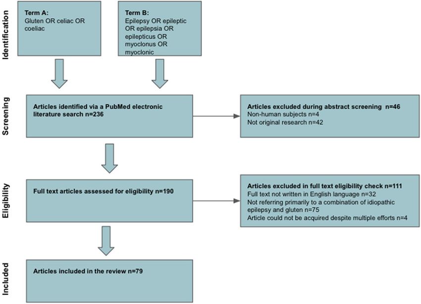
Fig. 1 A PRISMA chart detail-
ing the inclusion process
Compliance with ethical guidelines Epidemiology of gluten sensitivity and CD
associated epilepsy
This article is based upon previously published studies. The
article is in compliance with the journal’s ethical guidelines. Thirteen studies detailed the prevalence of CD amongst
patients with epilepsy of unknown aetiology [16, 21, 33,
37, 43, 45, 48, 51, 59, 63, 67, 69, 74]. The total pooled
prevalence was 2.1% (95% CI 1.64–2.64%, n = 3389).
Results Amongst exclusively paediatric populations the preva-
lence was 1.83% (95% CI 1.26–2.56%, n = 1804) whilst in
Selected studies exclusively adult populations the prevalence was 2.27%
(95% CI 1.52–3.24%, n = 1280). Seven studies detailed
The search strategy identified 236 articles. A total of 46 prevalence of serologically confirmed gluten sensitivity
articles were excluded during the title and abstract screen- amongst patients with epilepsy of unknown aetiology,
ing stage. A total of 111 articles were excluded during the and the pooled prevalence was 3.8% (95% CI 2.65–5.34%,
eligibility assessment. Thus, 79 articles published between n = 861) [28, 37, 41, 45, 48, 63, 68].
1970 and 2017 qualified for inclusion in this review study- Fifteen studies detailed prevalence of epilepsy of
ing a total of 39,579 individuals with either epilepsy, coe- unknown aetiology amongst CD sufferers [8, 13, 19, 20,
liac disease or both [8, 10, 11, 13–88]. Of these, 26 were 25, 38, 42, 54, 56, 57, 61, 65, 74, 86, 88]. The pooled prev-
case reports, 7 were case-control studies and the remain- alence was 1.14% (95% CI 1.03–1.26%, n = 33,217). Two
ing 46 articles were prospective or retrospective case series studies detailed prevalence of epilepsy amongst serologi-
(most of which were consecutively recruited cohort stud- cally confirmed gluten sensitivity. The pooled prevalence
ies). Whilst the studies are published in a range of loca- was 0.93% (95% CI 0.43–1.77%, n = 963). Both of these
tions worldwide, a disproportionate number come from Italy figures are raised relative to a general population point
(n = 24) and Turkey (n = 8). Figure 1 illustrates the study prevalence of 0.64% [89].
selection process. These figures illustrate that there is an increased
prevalence of CD amongst those with epilepsy and vice-
versa. They also demonstrate and increased prevalence
of epilepsy amongst those with GS. A large, population-
based study conducted in Sweden by Ludvigsson et al.
(n = 28,885) demonstrated a bidirectional relationship
13Journal of Neurology
between development of CD and epilepsy which illustrates insult caused by gluten ingestion or be the result of improved
that each population is at increased risk of developing the absorption of AED due to resolution of gastrointestinal dis-
other condition [8]. turbance [69]. Data regarding response to AED alone is only
available for the CEC syndrome subset. These data indicate
Epilepsy classification that GFD can be an effective management of seizures in
patients with gluten sensitivity.
The classification of the epilepsy presentations was reported
in 45 of the articles [8, 10, 11, 13, 15, 16, 19–22, 25–30, 32, CEC syndrome
33, 36, 38, 41–44, 48, 49, 51, 54, 56, 57, 60–69, 74, 75, 77,
81, 86–88,, 60–69, 74, 75, 77, 81, 86–88]. Of these, 25 could Presentation
be used to calculate the prevalence of focal epilepsy amongst
patients with epilepsy [8, 13, 15, 16, 18–21, 28, 33, 36, 41, Of the 79 studies included in this review, 30 detailed cases of
43, 45, 48, 51, 54, 56, 57, 61, 63, 65, 74, 87, 88]. Amongst CEC syndrome making it the best classified presentation of
351 patients, 59% presented with focal seizures. Based on epilepsy in the context of CD and gluten sensitivity [10, 14,
this, it appears that there is a slightly greater tendency for 17, 32, 34, 36, 44, 50, 52, 55, 60, 62, 64, 66, 70–85]. Sixteen
epilepsy in the context of CD/GS to be focal [90]. of these papers are case reports and 14 are retrospective or
prospective case series. These patients generally have focal,
Presentation medically refractory epilepsy and show parieto-occipitally
brain calcifications on CT or MRI. In 82% of cases, cer-
Forty-one studies detailed the age of onset of epilepsy in ebral calcifications were located posteriorly (n = 131). In the
those with CD/GS [11, 14–18, 22, 34–36, 39, 40, 45, 47, remainder, calcifications were either frontal, temporal or in a
52–56, 58, 60–64, 66, 71, 73, 76–80, 82, 83, 87, 70, 71, 73, very small minority in sub-cortical areas. The classification
76–80, 82, 83, 87]. This gave a pooled mean age of onset of of epilepsy was reported in 28 articles, 78% of seizures in
12 (n = 161). Eleven studies (n = 52) provided information CEC syndrome reported in the available literature are focal
which enabled calculation of the total number of patients in nature (n = 121) [14, 17, 32, 34, 36, 50, 52, 55, 60, 62, 64,
suffering CD and epilepsy who suffered gastrointestinal 66, 70–85]. Fourteen studies detailed further localisation of
symptoms [16, 21, 33, 37, 43, 45, 48, 51, 59, 63, 64, 67, the focal epilepsy and 71% of this was occipital in nature
69, 74]. This gave a pooled figure of 40% of patients hav- (n = 85) [14, 17, 36, 50, 52, 55, 60, 64, 71, 74, 76, 81, 83,
ing no gastrointestinal symptoms and who, therefore, only 85]. Our search identified just one study that investigated a
presented with neurological symptoms resulting in the cohort of GFD treated, mostly paediatric CD patients for
diagnosis of CD. This is comparable to other neurological brain lesions independent of neurological signs [65]. The
manifestations of gluten sensitivity in which gastrointestinal authors found no calcifications on CT, but MRI showed
symptoms are rarely present [4]. Nevalainen et al. investi- bilateral or unilateral T2-hyperintensive white-matter lesions
gated the risk of death in patients suffering epilepsy who in 20% of patients (n = 75). These lesions were independent
also had serum markers indicative of CD/gluten sensitivity of specific neurological presentations. This study does not
and found that there was no increased risk of death in this support an increased prevalence of cerebral calcifications
patient cohort relative to those who have epilepsy but no in CD patients but does demonstrate frequent brain imaging
gluten sensitivity [23]. abnormalities in CD. The failure to identify calcifications
is not surprising considering the rarity of CEC syndrome.
Management
Pathology
Thirty-two papers discuss the therapeutic impact of gluten-
free diet [10, 13, 15–18, 22, 31, 34, 36, 39, 45–47, 50, 53, Several studies show that despite improvements in seizure
55, 59–61, 66, 67, 71, 73, 77, 80, 81, 83–85]. For the pur- control with GFD there is no reduction in calcification size
pose of this review, a positive response was defined as either [10, 71, 73, 80]. This has led some researchers to suggest
decreased frequency of seizures with GFD, cessation of sei- that the calcifications may represent an epiphenomenon
zures with GFD, successful reduction of AED with initia- rather than being causative. One speculated that the cause
tion of GFD or cessation of AED following the introduction of the development of calcifications is folate deficiency. Pri-
of GFD. Amongst studies which reported response to GFD marily, this link has been made due to the co-occurrence of
in a consecutively recruited series, 53% of patients were folate deficiency and cerebral calcifications in other situa-
responsive to a GFD and the remaining 47% deemed unre- tions, for instance during methotrexate therapy or in primary
sponsive (n = 34) [13, 15, 16, 36, 49, 61, 67]. The response cerebral folate deficiency [34]. Calvani et al., have previ-
to GFD could reflect resolution/reduction of a neurological ously demonstrated decreased folic acid transport across the
13Journal of Neurology
blood brain barrier as well as decreased intestinal absorption caution as the data include studies using selective popula-
of folic acid in case report and speculate this to be part of the tions (non-consecutive case series and case reports).
pathological process [66]. Whilst there are indeed numerous
examples of co-occurring folate deficiency and CEC syn- Other specific epilepsy syndromes in GS and CD
drome [10, 34, 44, 52, 71, 73, 74, 76–82], it is not a universal
finding and there are numerous examples of patients with A small number of papers have investigated specific epi-
CEC syndrome and folate values within the normal range lepsy syndromes in relation to GS and CD. These studies
[44, 64, 79, 81]. Therefore, the relationship between folate are limited by their small population sizes but demonstrate
and calcifications is inconsistent. a possible interesting link between GS/CD and some specific
A case report by Johnson et al., identified high levels of syndromes.
IgA directed by transglutaminase isoenzyme 6 (TG6) in the
patient’s serum [34]. Raised levels of TG6 have previously Childhood partial epilepsy with occipital paroxysms
been shown to be elevated in other neurological manifesta-
tions of gluten sensitivity and represents a potential marker Three articles investigated the prevalence of CD amongst
of neurological involvement [91]. This study also used indi- those with childhood partial epilepsy with occipital parox-
rect immunofluorescence on monkey brain which identified ysms [21, 26, 64]. According to these, the pooled preva-
cerebral and cerebellar binding of IgA which demonstrates lence of CD amongst such individuals is 10.41% (95% CI
potential autoimmunity to the parenchyma of the brain. 5.11–18.32%, n = 96). This is considerably higher than the
Brain biopsy was conducted in CEC syndrome patients in prevalence CD amongst all epilepsy of unknown aetiology.
two case reports. One case reported a biopsy following right
anterior temporal lobectomy with pathology confirming Children with occipital lobe epilepsy
hippocampal sclerosis with severe cell loss and gliosis in
CA1/CA3/CA4 [17]. The second case report by Bye et al., Dai et al. conducted a case-control study with a cohort of
demonstrated cortical vascular abnormality with patchy pial children with both generalised and focal epilepsy identified
angiomatosis, fibrosed veins and large, jagged microcalci- to be occipital in origin on EEG [27]. This study discovered
fication [76]. 2.22% (n = 90) of the patient cohort also had CD which was
significantly higher than the healthy control group. The two
Management patients with CD were also monotherapy resistant.
The more descriptive nature of the CEC syndrome stud- Adult patients with fixation off sensitivity (FOS)
ies enables insight into the patient’s response to treatment.
Response to AED alone appears to be poor amongst patients Fattouch et al. investigated adult epileptic patients with FOS
with CEC syndrome, with 73% unresponsive to treatment [32]. These patients all presented with simple and complex
(n = 74) [10, 32, 36, 44, 60, 64, 74, 75, 77, 81]. Response focal seizures, which were posterior in onset according to
to GFD appears to be more effective, with 53% of patients EEG. This study showed a prevalence of CD of 20% (n = 15)
managed with GFD demonstrating a good response (n = 47) amongst this patient group.
[10, 36, 60, 77, 81]. Interestingly, three prospective cohort
studies appear to have demonstrated an inverse relationship Progressive myoclonic epilepsy (PME)
between effectiveness of GFD and duration of epilepsy prior
to GFD, perhaps due to increasing neurological damage [36, Franceschetti et al., investigated comorbid conditions
60, 81]. amongst patients with PME of undetermined cause by col-
There are four reported cases of surgical resection of cer- lecting existing medical record data [29]. This study iden-
ebral calcifications with the aim to resolve intractable sei- tified that 0.98% (n = 204) of the patient cohort suffered
zures [17, 36, 52, 77]. Of these, one was resistant to surgery, comorbid CD. This data is limited because as previously
two were responsive to surgery and one responded to a com- established most patients are not symptomatic for CD, and
bination of surgical resection and GFD. Whilst two patients so it is likely checking medical records alone is not suffi-
did respond to surgery, one continued to suffer seizures but cient to rule out CD and GS as the condition will likely be
of lesser severity, and one was commenced gluten-free diet undiagnosed.
immediately following surgery. Consequently, it is feasible
that the improvement is owed to GFD rather than to surgical Temporal lobe epilepsy (TLE) with hippocampal sclerosis
resection. Based on the very limited data available, surgical
resection of calcifications alone does not resolve epilepsy Peltola et al. studied the relationship between temporal lobe
in CEC syndrome. These findings must be interpreted with epilepsy (TLE) with hippocampal sclerosis, and gluten
13Journal of Neurology
sensitivity [11]. Autoimmunity has previously been impli- 6. Patients with epilepsy of unknown aetiology should have
cated in the pathophysiology of hippocampal sclerosis and their serum screened for AGA, anti tTG and EMA. This
thus this study investigated GS antibodies in a prospective is especially important in patients suffering with AED
study of patients with refractory TLE with or without hip- resistant occipital lobe seizures. It is likely that there are
pocampal sclerosis as well as in those with extra-temporal many patients who are being treated with AED poly-
epilepsy. This study identified a significantly raised preva- therapy who can be managed with a GFD alone or with
lence of gluten sensitivity seropositivity in those with TLE GFD and reduced AED.
and hippocampal sclerosis with a figure of 43.75% (n = 16). 7. There is a need to study the prevalence of TG6 anti-
These patients were then tested for CD, with three patients bodies in patients with epilepsy to identify whether
displaying histology for CD (18.75%) and four showing anti-TG6 could be used to identify individuals at risk of
early changes associated with CD. This is the only study epilepsy due to their gluten sensitivity.
linking gluten sensitivity or CD to hippocampal sclerosis
and TLE. This study is limited by its small population. As The results of this review are highly relevant to Dieti-
previously mentioned, hippocampal sclerosis was also pre- cians and Gastroenterologists as well as Neurologists. It is
sent in the biopsy of a patient suffering CEC syndrome in a important that epilepsy is more broadly recognised within
separate study [17]. the spectrum of gluten-related disorders as these patients
can be managed effectively if identified. However, clinicians
Summary must approach these cases with caution so as to not incor-
rectly diagnose epilepsy in those with gluten intolerance
In tandem with studies regarding CEC syndrome, these data who have epilepsy mimics such as syncope, psychogenic
appear to support a hypothesis of a selective vulnerability non-epileptic seizures or migraine, amongst others. Clini-
of specific brain regions to damage in the context of gluten cians must also recognise the limitations to the specificity
sensitivity or coeliac disease. of the GS/CD serum markers and take into account the full
clinical picture when proceeding with diagnosis.
Assessment of bias
Limitations
None of the studies included in this review are randomised
control trials or interventional studies for which risk of bias • There is a significant deal of low quality evidence utilised
tools are available. All articles were reviewed by two authors in this review due to the large number of case reports.
independently and their judgements regarding inclusion Studies which use selected patient groups were utilised
matched in all cases. Case reports are low level evidence to evaluate: Response to treatment, descriptive peculi-
with a high risk of bias. The use of this data is summarised arities where it is explicitly stated that a case report was
in the “Limitations” section of this review. used and for results regarding CEC syndrome. All other
results were calculated using studies with consecutively
Conclusions recruited patient cohorts or entire populations only.
• Not all studies biopsied their GS patients. The patients
This systematic review has identified the following key may therefore have suffered CD. This may cause under-
points: estimation of the link between CD and epilepsy.
• Some studies reviewed medical records rather than pro-
1. There is an increased prevalence of CD amongst patients spectively investigating for CD/GS. Often patients do not
with epilepsy and an increased prevalence of epilepsy suffer classical symptoms of GS/CD and therefore will
amongst those with CD or gluten sensitivity. not be diagnosed. Therefore, the prevalence of CD/GS
2. Patients with CD presenting with neurological symp- amongst epilepsy may be an underestimate.
toms often suffer no gastrointestinal symptoms. • This study addressed the relationship between CD/
3. There appears to be a stronger link between some epilep- GS and epilepsy of unknown aetiology. There were no
tic presentations and GS or CD than others. Future stud- restrictions placed upon date of publication of articles
ies should not treat epilepsy as though it is homogenous included in this review and as such some studies are
when investigating its relationship with GS or CD. dated as far back as 1970. As the understanding of the
4. Gluten-free diet is an effective management of epilepsy causes of epilepsy has developed it is possible that some
in those with epilepsy due to GS/CD. patients included within the studies forming this review
5. CEC syndrome is the best characterised epileptic pres- would now be recognised to have a clear aetiology and
entation linked to CD. therefore will be incorrectly classified.
13Journal of Neurology
• Four articles were excluded because they could not be 14. Bonanni P, Negrin S, Antoniazzi L et al (2017) Clinical implica-
retrieved. tions of interictal epileptiform discharges in cognitive functioning
in CEC syndrome with evolution into epileptic encephalopathy.
• Articles for inclusion in this review were retrieved via a
Neurocase 23:230–238
search in a single electronic database. 15. Sel ÇG, Aksoy E, Aksoy A et al (2017) Neurological manifesta-
tions of atypical celiac disease in childhood. Acta Neurol Belg
117:719–727
Acknowledgements We are sincerely thankful to Dr Stamatina Ili- 16. Bashiri H, Afshari D, Babaei N et al (2016) Celiac Disease and
odromiti for her contribution in the statistical analysis of the data. Dr epilepsy: the effect of gluten-free diet on seizure control. Adv
Zis is sincerely thankful to the Ryder Briggs Fund. Clin Exp Med 25:751–754
This is a summary of independent research carried out at the NIHR 17. Struck AF, Beinlich BR, Rutecki PA (2015) A case of celiac
Sheffield Biomedical Research Centre (Translational Neuroscience). disease, epilepsy, and cerebral calcifications with temporal lobe
The views expressed are those of the authors and not necessarily those epilepsy. WMJ 114:116–117
of the NHS, the NIHR or the Department of Health. 18. Işikay S, Hizli Ş, Kocamaz H (2015) Celiac disease and juvenile
absence epilepsy. Pediatr Emerg Care 31:e19–e20
19. Işikay S, Kocamaz H (2015) The neurological face of celiac
Compliance with ethical standards disease. Arq Gastroenterol 52:167–170
20. Işıkay S, Kocamaz H, Sezer S et al (2015) The frequency of epi-
Conflicts of interest None of the authors have any competing interests. leptiform discharges in celiac disease. Pediatr Neurol 53:78–82
21. Işikay S, Hizli Ş, Yilmaz K (2014) Prevalence of celiac dis-
Open Access This article is distributed under the terms of the Crea- ease in Turkish children with idiopathic epilepsy. Iran J Pediatr
tive Commons Attribution 4.0 International License (http://creativeco 24:280–284
mmons.org/licenses/by/4.0/), which permits unrestricted use, distribu- 22. Casciato S, Morano A, Albini M et al (2015) Cryptogenic focal
tion, and reproduction in any medium, provided you give appropriate epilepsy and “hidden” celiac disease in adulthood: a causal or
credit to the original author(s) and the source, provide a link to the accidental link? Int J Neurosci 125:913–917
Creative Commons license, and indicate if changes were made. 23. Nevalainen O, Auvinen A, Ansakorpi H et al (2014) Autoim-
munity-related immunological serum markers and survival in
a tertiary care cohort of adult patients with epilepsy. Epilepsy
Res 108:1675–1679
References 24. Soós Z, Salamon M, Erdei K et al (2014) LADA type diabetes,
celiac diasease, cerebellar ataxia and stiff person syndrome.
1. Mustalahti K, Catassi C, Reunanen A et al (2010) The prevalence A rare association of autoimmune disorders. Ideggyogy Sz
of celiac disease in Europe: results of a centralized, international 67:205–209
mass screening project. Ann Med 42:587–595 25. Ong MS, Kohane IS, Cai T,, et al (2014) Population-level evi-
2. Fasano A, Catassi C (2001) Current approaches to diagnosis and dence for an autoimmune etiology of epilepsy. JAMA Neurol
treatment of celiac disease: an evolving spectrum. Gastroenterol- 71:569–574
ogy 120:636–651 26. Işıkay S, Kocamaz H (2014) Prevalence of celiac disease in
3. Sapone A, Bai JC, Ciacci C et al (2012) Spectrum of gluten- children with idiopathic epilepsy in southeast Turkey. Pediatr
related disorders: consensus on new nomenclature and classifica- Neurol 50:479–481
tion. BMC Med 10:13 27. Dai AI, Akcali A, Varan C et al (2014) Prevalence of resistant
4. Hadjivassiliou M, Sanders DS, Grünewald RA et al (2010) Gluten occipital lobe epilepsy associated with celiac disease in chil-
sensitivity: from gut to brain. Lancet Neurol 9:318–330 dren. Childs Nerv Syst 30:1091–1098
5. Hadjivassiliou M (2012) Immune mediated acquired ataxias. 28. Vieira C, Jatobá I, Matos M et al (2013) Prevalence of celiac
Handb Clin Neurol 103:189–199 disease in children with epilepsy. Arq Gastroenterol 50:290–296
6. Gabrielli M, Cremonini F, Fiore G et al (2003) Association 29. Franceschetti S, Michelucci R, Canafoglia L et al (2014) Pro-
between migraine and Celiac disease: results from a prelimi- gressive myoclonic epilepsies: definitive and still undetermined
nary case-control and therapeutic study. Am J Gastroenterol causes. Neurology 82:405–411
98:625–629 30. Sarrigiannis PG, Hoggard N, Aeschlimann D et al (2014)
7. Zis P, Rao DG, Sarrigiannis PG et al (2017) Transglutaminase 6 Myoclonus ataxia and refractory coeliac disease. Cerebellum
antibodies in gluten neuropathy. Dig Liver Dis 49:1196–1200 Ataxias 1:11
8. Ludvigsson JF, Zingone F, Tomson T et al (2012) Increased risk 31. Vitelli O, Miano S, Tabarrini A et al (2014) Epilepsy and sleep-
of epilepsy in biopsy-verified celiac disease: a population-based disordered breathing as false friends: a case report. J Child Neu-
cohort study. Neurology 78:1401–1407 rol 29:114–117
9. Guy Daynes (1956) Bread and tears—naughtiness, depression and 32. Fattouch J, Casciato S, Lapenta L et al (2013) The spectrum of
fits due to wheat sensitivity. Proc R Soc Med 49:391–394 epileptic syndromes with fixation off sensitivity persisting in
10. Gobbi G, Ambrosetto P, Zaniboni MG et al (1992) Celiac disease, adult life. Epilepsia 7:59–65
posterior cerebral calcifications and epilepsy. Brain Dev 14:23–29 33. Djurić Z, Nagorni A, Jocić-Jakubi B et al (2012) Celiac dis-
11. Peltola M, Kaukinen K, Dastidar P et al (2009) Hippocampal scle- ease prevalence in epileptic children from Serbia. Turk J Pediatr
rosis in refractory temporal lobe epilepsy is associated with gluten 54:247–250
sensitivity. J Neurol Neurosurg Psychiatry 80:626–630 34. Johnson AM, Dale RC, Wienholt L et al (2013) Coeliac dis-
12. Marsh N, Johnson MW, Rostami M (2015) K. Mucosal histopa- ease, epilepsy, and cerebral calcifications: association with TG6
thology in celiac disease: a rebuttal of Oberhuber’s sub-division autoantibodies. Dev Med Child Neurol 55:90–93
of Marsh III. Gastroenterol Hepatol Bed Bench 8:99–109 35. Javed S, Safdar A, Forster A et al (2012) Refractory coeliac
13. Chapman RW, Laidlow JM, Colin-Jones D et al (1978) Increased disease associated with late onset epilepsy, ataxia, tremor and
prevalence of epilepsy in coeliac disease. Br Med J 2:250–251 progressive myoclonus with giant cortical evoked potentials—a
case report and review of literature. Seizure 21:482–485
13Journal of Neurology
36. Licchetta L, Bisulli F, Di Vito L et al (2011) Epilepsy in coe- 57. Zelnik N, Pacht A, Obeid R et al (2004) Range of neurologic dis-
liac disease: not just a matter of calcifications. Neurol Sci orders in patients with celiac disease. Pediatrics 113:1672–1676
32:1069–1074 58. Pratesi R, Modelli IC, Martins RC et al (2003) Celiac disease and
37. Ertekin V, Selimoğlu MA, Tan H et al (2010) Prevalence of epilepsy: favorable outcome in a child with difficult to control
celiac disease in a sample of Turkish children with epilepsy. seizures. Acta Neurol Scand 108:290–293
Pediatr Neurol 42:380–381 (380; author reply) 59. Essid M, Trabelsi K, Jerbi E et al (2003) Villous atrophy and idi-
38. Bürk K, Farecki ML, Lamprecht G et al (2009) Neurological opathic epilepsy. Tunis Med 81:270–272
symptoms in patients with biopsy proven celiac disease. Mov 60. Arroyo HA, De Rosa S, Ruggieri V et al (2002) Epilepsy, occipital
Disord 24:2358–2362 calcifications, and oligosymptomatic celiac disease in childhood.
39. Sallem FS, Castro LM, Jorge C et al (2009) Gluten sensitivity J Child Neurol 17:800–806
presenting as myoclonic epilepsy with cerebellar syndrome. 61. Volta U, De Giorgio R, Petrolini N et al (2002) Clinical findings
Mov Disord 24:2162–2163 and anti-neuronal antibodies in coeliac disease with neurological
40. Ryan AM, Ryan J, Wan-Ahmed M et al. (2009) Vacuolar leu- disorders. Scand J Gastroenterol 37:1276–1281
coencephalopathy and pulvinar sign in association with coeliac 62. Santos CH, Almeida IL, Gomes MD et al (2002) Bilateral occipi-
disease. BMJ Case Rep tal calcification, epilepsy and coeliac disease: case report. Arq
41. Emami MH, Taheri H, Kohestani S et al (2008) How frequent is Neuropsiquiatr 60:840–843
celiac disease among epileptic patients? J Gastrointestin Liver 63. Luostarinen L, Dastidar P, Collin P et al (2001) Association
Dis 17:379–382 between coeliac disease, epilepsy and brain atrophy. Eur Neurol
42. Briani C, Zara G, Alaedini A et al (2008) Neurological com- 46:187–191
plications of celiac disease and autoimmune mechanisms: a 64. Labate A, Gambardella A, Messina D et al (2001) Silent celiac
prospective study. J Neuroimmunol 195:171–175 disease in patients with childhood localization-related epilepsies.
43. Ruggieri M, Incorpora G, Polizzi A et al (2008) Low prevalence Epilepsia 42:1153–1155
of neurologic and psychiatric manifestations in children with 65. Kieslich M, Errázuriz G, Posselt HG et al (2001) Brain white-
gluten sensitivity. J Pediatr 152:244–249 matter lesions in celiac disease: a prospective study of 75 diet-
44. Della Nave R, Magaudda A, Michelucci R et al (2007) Whole- treated patients. Pediatrics 108:E21
brain histogram and voxel-based analyses of apparent dif- 66. Calvani M Jr, Parisi P, Guaitolini C et al (2001) Latent coeliac dis-
fusion coefficient and magnetization transfer ratio in celiac ease in a child with epilepsy, cerebral calcifications, drug-induced
disease, epilepsy, and cerebral calcifications syndrome. AJNR systemic lupus erythematosus and intestinal folic acid malabsorp-
28:479–485 tion associated with impairment of folic acid transport across the
45. Mavroudi A, Xinias I, Papastavrou T et al (2007) Increased prev- blood-brain barrier. Eur J Pediatr 160:288–292
alence of silent celiac disease among Greek epileptic children. 67. Salur L, Uibo O, Talvik I et al (2000) The high frequency of
Pediatr Neurol 36:165–169 coeliac disease among children with neurological disorders. Eur
46. Harper E, Moses H, Lagrange A (2007) Occult celiac disease pre- J Neurol 7:707–711
senting as epilepsy and MRI changes that responded to gluten-free 68. Lahat E, Broide E, Leshem M et al (2000) Prevalence of celiac
diet. Neurology 68:533–534 antibodies in children with neurologic disorders. Pediatr Neurol
47. Canales P, Mery VP, Larrondo FJ et al (2006) Epilepsy and celiac 22:393–396
disease: favorable outcome with a gluten-free diet in a patient 69. Cronin CC, Jackson LM, Feighery C et al (1998) Coeliac disease
refractory to antiepileptic drugs. Neurologist 12:318–321 and epilepsy. QJM 91:303–308
48. Dalgiç B, Dursun I, Serdaroğlu A et al (2006) Latent and potential 70. Bernasconi A, Bernasconi N, Andermann F et al (1998) Celiac
celiac disease in epileptic Turkish children. J Child Neurol 21:6–7 disease, bilateral occipital calcifications and intractable epilepsy:
49. Mavroudi A, Karatza E, Papastavrou T et al (2005) Successful mechanisms of seizure origin. Epilepsia 39:300–306
treatment of epilepsy and celiac disease with a gluten-free diet. 71. Hernández MA, Colina G, Ortigosa L (1998) Epilepsy, cerebral
Pediatr Neurol 33:292–295 calcifications and clinical or subclinical coeliac disease. Course
50. Díaz RM, González-Rabelino G, Delfino A (2005) Epilepsy, and follow up with gluten-free diet. Seizure 7:49–54
cerebral calcifications and coeliac disease. The importance of an 72. Baquero M, Narciso ML, García M et al (1995) Celiac dis-
early diagnosis. Rev Neurol 40:417–420 ease with occipital calcifications: 2 late cases. Med Clin (Barc)
51. Ranua J, Luoma K, Auvinen A et al (2005) Celiac disease-related 105:781–783
antibodies in an epilepsy cohort and matched reference popula- 73. Lea ME, Harbord M, Sage MR (1995) Bilateral occipital calcifica-
tion. Epilepsy Behav 6:388–392 tion associated with celiac disease, folate deficiency, and epilepsy.
52. Nakken KO, Røste GK, Hauglie-Hanssen E (2005) Coeliac dis- AJNR Am J Neuroradiol 16:1498–1500
ease, unilateral occipital calcifications, and drug-resistant epi- 74. Fois A, Vascotto M, Di Bartolo RM et al (1994) Celiac disease
lepsy: successful lesionectomy. Acta Neurol Scand 111:202–204 and epilepsy in pediatric patients. Childs Nerv Syst 10:450–454
53. Siqueira Neto JI, Costa AC, Magalhães FG et al (2004) Neu- 75. Bardella MT, Molteni N, Prampolini L et al (1994) Need for fol-
rological manifestations of celiac disease. Arq Neuropsiquiatr low up in coeliac disease. Arch Dis Child 70:211–213
62:969–972 76. Bye AM, Andermann F, Robitaille Y et al (1993) Cortical vas-
54. Vaknin A, Eliakim R, Ackerman Z et al (2004) Neurologi- cular abnormalities in the syndrome of celiac disease, epilepsy,
cal abnormalities associated with celiac disease. J Neurol bilateral occipital calcifications, and folate deficiency. Ann Neurol
251:1393–1397 34:399–403
55. Pfaender M, D’Souza WJ, Trost N et al (2004) Visual disturbances 77. Magaudda A, Dalla Bernardina B, De Marco P et al (1993) Bilat-
representing occipital lobe epilepsy in patients with cerebral cal- eral occipital calcification, epilepsy and coeliac disease: clinical
cifications and coeliac disease: a case series. J Neurol Neurosurg and neuroimaging features of a new syndrome. J Neurol Neuro-
Psychiatry 75:1623–1625 surg Psychiatry 56:885–889
56. Pengiran Tengah DS, Holmes GK, Wills AJ (2004) The prev- 78. Piattella L, Zamponi N, Cardinali C et al (1993) Endocranial cal-
alence of epilepsy in patients with celiac disease. Epilepsia cifications, infantile celiac disease, and epilepsy. Childs Nerv Syst
45:1291–1293 9:172–175
13Journal of Neurology
79. Tiacci C, D’Alessandro P, Cantisani TA et al (1993) Epilepsy 86. Hanly JG, Stassen W, Whelton M et al (1982) Epilepsy and coe-
with bilateral occipital calcifications: Sturge-Weber variant or a liac disease. J Neurol Neurosurg Psychiatry 45:729–730
different encephalopathy? Epilepsia 34:528–539 87. Banerji NK, Hurwitz LJ (1971) Neurological manifestations in
80. Fois A, Balestri P, Vascotto M et al (1993) Progressive cerebral adult steatorrhoea (probable Gluten enteropathy). J Neurol Sci
calcifications, epilepsy, and celiac disease. Brain Dev 15:79–82 14:125–141
81. Gobbi G, Bouquet F, Greco L et al (1992) Coeliac disease, epi- 88. Morris JS, Ajdukiewicz AB, Read AE (1970) Neurological dis-
lepsy, and cerebral calcifications. The Italian Working Group on orders and adult coeliac disease. Gut 11:549–554
Coeliac Disease and Epilepsy. Lancet 340:439–443 89. Fiest KM, Sauro KM, Wiebe S et al. Prevalence and incidence of
82. Crosato F, Senter S (1992) Cerebral occipital calcifications in epilepsy: A systematic review and meta-analysis of international
celiac disease. Neuropediatrics 23:214–217 studies Neurology. 2017; 88:296–303
83. Ambrosetto G, Antonini L, Tassinari CA (1992) Occipital lobe 90. Kotsopoulos IA, van Merode T, Kessels FG et al (2002) System-
seizures related to clinically asymptomatic celiac disease in adult- atic review and meta-analysis of incidence studies of epilepsy and
hood. Epilepsia 33:476–481 unprovoked seizures. Epilepsia 43:1402–1409
84. Della Cella G, Beluschi C, Cipollina F (1991) Intracranial calcifi- 91. Hadjivassiliou M, Aeschlimann P, Strigun A et al (2008) Autoan-
cations–seizures–celiac disease: a case presentation. Pediatr Med tibodies in gluten ataxia recognize a novel neuronal transglutami-
Chir 13:427–430 nase. Ann Neurol 64:332–343
85. Ventura A, Bouquet F, Sartorelli C et al (1991) Coeliac disease,
folic acid deficiency and epilepsy with cerebral calcifications.
Acta Paediatr Scand 80:559–562
13You can also read