Guidance For: Tracheostomy Care - The Faculty of Intensive ...
←
→
Page content transcription
If your browser does not render page correctly, please read the page content below
Guidance For: Tracheostomy Care
2 | Guidance For: Tracheostomy Care
Contents

Guidance For: Tracheostomy Care

1.   Executive summary, key standards and recommendations            5
2.   Tracheostomy in light of COVID-19                               8
3.   Introduction                                                    9
4.   Indications for tracheostomy and patient selection              11
5.   Insertion                                                       14
6.   Tracheostomy tube types and choice                              21
7.   Routine care of the established tracheostomy                    23
8.   Vocalisation, communication and oral intake                     30
9.   Emergency management                                            33
10. Decannulation and discharge from ICU                             35
11. Patients with laryngectomies                                     37
12. Key References                                                   39

                                                          Guidance For: Tracheostomy Care | 3
Acknowledgments Guidance For: Tracheostomy Care Neither the Intensive Care Society, Faculty of Intensive Care Medicine nor the authors accept any responsibility for any loss of or damage arising from actions or decisions based on the information contained within this publication. Ultimate responsibility for the treatment of patients and interpretation of the published material lies with the medical practitioner. The opinions expressed are those of the authors and the inclusion in this publication of material relating to a particular product or method does not amount to an endorsement of its value, quality, or the claims made by its manufacturer. Prepared on behalf of the Council of the Intensive Care Society by: The Short-life Standards and Guidelines Working Party of the UK National Tracheostomy Safety Project: Brendan A McGrath (Chair) Sarah Wallace (Speech & Language Therapy) Barbara Bonvento (Physiotherapy) James Lynch (ACCP) Barry Coe (Nursing) Dougal Atkinson (Medical) Reviewed by: • ICS Standards and Guidelines Committee • FICM Professional Affairs & Safety Committee Published date: August 2020 Review date: August 2023 Our patients assume safe care and expect high-quality care. 4 | Guidance For: Tracheostomy Care
1. Executive summary, key standards
                                                                           and recommendations

1. Executive summary, key standards and
recommendations
The patient population in NHS hospitals that are managed with a tracheostomy has evolved
significantly over the last 20 years. The majority are performed percutaneously by intensivists,
in the Intensive Care Unit, on critically ill patients or those recovering from critical illness. Most
tracheostomies are temporary.

The vast majority of patients with ‘surgical’ and ‘non-surgical’ tracheostomies experience critical
care at some stage of their journey. The multidisciplinary nature of tracheostomy care is a familiar
working environment for our speciality, with tracheostomy care being perhaps one of the best
examples truly multidisciplinary care.

There is increasing evidence from national and international quality improvement programs that a
multidisciplinary tracheostomy team that reviews and coordinates the management of tracheostomy
patients can bring benefits for the quality and safety of care, including organisational efficiencies
and significant cost savings.

All patients with tracheostomies admitted to critical care units should expect safe care to be
delivered by appropriately trained, equipped and supported staff. Patient-centred high-quality care
also focusses on communication, vocalisation, mobilisation, information and a prompt return to
oral intake. Improving the quality and safety of patients with tracheostomies and laryngectomies
is a hospital-wide issue, and our speciality is well placed to lead and to contribute to the safe
management of this vulnerable patient group.

International quality improvement efforts should be supported. The key drivers of the Global
Tracheostomy Collaborative (GTC) are examples of the hospital-wide changes that are required,
which will involve and impact the ICU. The GTC key drivers are:

    1. Multidisciplinary team-based care: Multidisciplinary tracheostomy team replacing siloed
       care, meeting face-to-face to coordinate and plan care, overcoming barriers to effective
       communication between providers. Such team-based care has been shown to reduce
       adverse events, length of stay, time to decannulation, increase speaking valve use and
       facilitate patient communication.
    2. Standardisation of care: Standardised care protocols provide consistency in care,
       environment, equipment, and patient and provider expectations. Instituting such pathways
       and procedures promotes coordinated care, increases efficiency, and improves outcomes in
       airway emergencies.
    3. Broad staff education: All patient encounters must involve healthcare staff who have been
       appropriately trained in tracheostomy care. Educational interventions have demonstrated
       marked improvement in objective knowledge and confidence with providing tracheostomy
       care, translating into safer patient care.
    4. Patient and family involvement: Patients, their family and their carers are engaged in QI.
       Prioritising patient-centred care identifies key clinical outcomes, performance measures,
       and improvement areas that may otherwise not have been recognised.
    5. Patient-level data: Hospitals track outcomes using a prospective database with
       detailed patient-level data captured each tracheostomy admission. Analytics allow the
       multidisciplinary team to benchmark over time and anonymously with peers within the
       Collaborative to assess the impact of initiatives.

    This is a standards document. Reference to external resources such as those provided by
    the ICS, FICM, RCoA, NTSP, GTC and individual hospitals (amongst others) are signposted
    throughout. Key evidence-based standards and recommendations are made at the end of each
    chapter. Key standards and recommendations are summarised in the table overleaf.

                                                                          Guidance For: Tracheostomy Care | 5
1. Executive summary, key standards
and recommendations
Table: Key standards in this document linked to key drivers to improve the quality
and safety of tracheostomy care.
Key driver Key standards
Multidisciplinary All in-patients with a tracheostomy are seen at least weekly by a tracheostomy MDT.
(MDT) team-based
care Appointment of an institutional lead (Champion) for tracheostomy care.
All patients considered for tracheostomy as part of their critical illness will have a
multidisciplinary discussion about the benefits and burdens of elective tracheostomy as part
of ICU management.
If a patient is discharged from an ICU with a tracheostomy in situ, the ICU has a responsibility
to ensure that receiving locations are adequately prepared to receive and manage the patient.
Standardisation Institutional multidisciplinary tracheostomy group with oversight of tracheostomy
of care care.
Institutional tracheostomy policy in place.
All patients under the care of an ICU team will have an appropriate consent form completed
prior to elective tracheostomy, whether occurring in the ICU or the operating theatre.
All ICU tracheostomy insertion procedures must include a checklist and LocSSIP.
Appropriate equipment to safely perform tracheostomy will be immediately available on the
ICU. Tube selection should be a multidisciplinary discussion, especially for patients with
abnormal anatomy. Sub-glottic suction tracheostomy tubes should be used as standard for
new tracheostomy.
Weaning from ventilatory support and decannulation should be conducted in a consistent
manner.
Broad staff Bedside staff caring for tracheostomy patients will have received training in
education tracheostomy care as per local policy.
New or existing staff who might be called upon to respond to a tracheostomy emergency
must be appropriately trained, equipped and supported.
All staff involved in insertion of a tracheostomy in ICU or in managing the upper airway
and/or performing bronchoscopy to facilitate tracheostomy will be sufficiently trained and
considered competent.
All patients with a tracheostomy must have communication and swallowing needs assessed
by an SLT with referral when the decision to wean from the ventilator has been made and the
sedation hold started.
Staff must have the knowledge and equipment to facilitate safe vocalisation, through the
use of comprehensive cuff deflation strategies aligned to weaning plans, one-way/speaking
valves, and alternative methods of vocalisation such as ACV.
All tracheostomy and laryngectomee patients must have a bedhead airway sign.
All bedside staff caring for a laryngectomee must understand the airway implications.
6 | Guidance For: Tracheostomy Care1. Executive summary, key standards
and recommendations
Patient and family Institutional Patient Champion engaged in QI program.
involvement
All patients and/or their family/carers will be involved in insertion discussions if appropriate.
Verbal and non-verbal communication methods must be made available to patients with a
tracheostomy. Bedside staff must be familiar with their use.
Information will be made available to patients and/or their family/carers regarding
tracheostomy.
Patient-level data Aggregate patient level data is collected, analysed and reviewed to benchmark and
assess the impact of QI measures.
Guidance For: Tracheostomy Care | 72. Tracheostomy in light of COVID-19 2. Tracheostomy in light of COVID-19 The publication of this document was delayed by the global COVID-19 pandemic. The pandemic resulted in a surge of patients requiring relatively prolonged intubation and ventilation, with around 10% of this patient group requiring a tracheostomy. The highly infectious nature of the SARS-CoV-2 virus meant that usual considerations for tracheostomy and usual care for patients with a tracheostomy were adjusted. As the community learns more about how best to manage this disease, some of the guidance will evolve. Detailed discussion of care of the COVID-19 tracheostomy patient is beyond the scope of this document, but the principles are outlined below, with signposting to appropriate resources. Concerns about the protection of healthcare staff who will be undertaking airway management procedures in patients with confirmed (or suspected) COVID-19 initially led to changes in the timing of tracheostomy for those patients requiring prolonged ventilation. This was primarily to avoid exposing the patient and insertion team to the risks of insertion if a tracheostomy was not going to impact on patient care. A generally more conservative approach to tracheostomy was adopted. Tracheostomy may have some advantages in protecting staff by delivering ventilatory support via a closed system (cuff inflated positive pressure ventilation) in preference to a trial of extubation and possible non-invasive respiratory support and/or re-intubation. However, whilst a closed system protects staff, it likely disadvantages patients, who will not be able to experience cuff deflation strategies early in their tracheostomy journey. International expert consensus statements suggested limiting tracheostomy interventions to the minimum necessary to provide safe care. This meant reductions in the frequency of routine care such as inner cannula changes, stoma care and suction, and limiting endoscopy and FEES. ‘Dry’ circuits using a HME filter instead of active humidification were also recommended. There are limited outcome data emerging that suggest these strategies did not increase harm for patients and they seemed to have protected staff. However, as we learn more about this disease, considering when the risk of infection becomes low enough in a given patient to adopt more ‘usual’ care and interventions will be important in engagement and rehabilitation. At the time of publication, the guidance for tracheostomy management in the COVID-19 era is limited to expert consensus statements and opinion. The changes in practice were supported by an NHS England Safer Tracheostomy Care initiative which will evolve into a comprehensive quality improvement programme via the Adoption & Spread workstream. With further data collection and analysis, best practice recommendations are likely to evolve. Current guidance for the UK and internationally are detailed below. NTSP COVID-19 information http://www.tracheostomy.org.uk/healthcare-staff/improving-tracheostomy-care/covid-19 NHS England Safer Tracheostomy Care Toolkit http://www.tracheostomy.org.uk/storage/files/Safe%20TrachyCareToolkit%20V9b.pdf McGrath et al. Tracheostomy in the COVID-19 era: global and multidisciplinary guidance Lancet Respiratory Medicine (HEALTH-CARE DEVELOPMENT) VOLUME 8, ISSUE 7, P717-725, JULY 01, 2020 https://www.thelancet.com/journals/lanres/article/PIIS2213-2600(20)30230-7/fulltext McGrath et al. Multidisciplinary guidance for safe tracheostomy care during the COVID-19 pandemic: the NHS National Patient Safety Improvement Programme (NatPatSIP). Ananesthesia… https://onlinelibrary.wiley.com/doi/full/10.1111/anae.15120 8 | Guidance For: Tracheostomy Care
3. Introduction

3. Introduction
This is the third revised standards document on tracheostomy care and the first developed by
the joint standards committees of the Intensive Care Society (ICS) and Faculty of Intensive Care
Medicine (FICM).

Since the widespread adoption of bedside percutaneous tracheostomy in the critically ill, there
has been a change in the patient population who are managed with temporary or permanent
tracheostomy in the NHS. Whilst indications for surgical tracheostomies have also evolved,
increasing numbers of complex patients recovering from critical illness have been managed
with tracheostomy, bringing a responsibility for critical care staff, units and their national bodies
to provide guidance and standards for this vulnerable group. Around two-thirds of all new
tracheostomies are now performed in Intensive Care Units (ICUs) on ‘ICU’ patients, who have
predominantly ‘medical’ problems. Alongside this critical care activity, there is also a wider
responsibility to ensure that our tracheostomy patients are discharged from critical care units into
safe locations, with appropriate equipment, training and standards provided in all locations where
patients with tracheostomies are managed.

The first standards document was prompted by an appreciation of a national need to improve
the care of patients with tracheostomies in both critical care units and on general wards. Such
concerns were raised in part by reports to (and from) the NPSA, MHRA, Coroners and other
bodies. The NAP4 audit 2011, NCEPOD study 2014, and other reports highlight continuing
issues: namely obstruction, dislodgement, bleeding, hypoxaemia and cardiac arrest. These
problems are significantly more serious in the critically ill and/or ventilator-dependent patient, with
critical care units consistently highlighted as a high-risk environment for airway management,
including tracheostomy.

There is rightly an increasing awareness and focus on the needs of patients and families who
experience tracheostomy care, with groups such as the National Tracheostomy Safety Project
(NTSP) and the Global Tracheostomy Collaborative (GTC) emerging over the last 10 years. Their
work, amongst others, challenges organisations to provide not just safer care, but the highest
possible quality of care. Recent qualitative research in the UK could be summarised by stating that
our patients assume safe care and expect high quality care. High quality care focusses on the
things that are important to patients; eating, drinking, vocalisation, mobilisation, decannulation and
information for patients and their families before, during and after tracheostomy.

Advances in the non-invasive management of respiratory failure, increased tolerance of trials
of extubation and increased awareness of the potential problems associated with temporary
or permanent tracheostomy have led to a likely stabilisation in the number of new procedures
performed in our ICUs over recent years. However, tracheostomy continues to be an important
procedure available to the multidisciplinary healthcare team and is likely to remain a relevant
intervention as increasingly frail and complex patients are admitted to our units. A typical UK ICU
can expect between 10-20% of their level 3 admissions will undergo tracheostomy, depending
on their case mix, and these patients will spend longer on the ICU and in hospital, occupying
a disproportionately high number of ICU bed days and ventilator days. The NTSP’s Improving
Tracheostomy Care program identified that around 80% of ‘typical’ patients with a new or existing
tracheostomy are admitted to ICU or HDU at some point in their hospital stay where they spend
a median of 23 days, mostly receiving some sort of invasive respiratory support. Tracheostomies
remain in situ for a median of 28 days and hospital stays are typically 50 days, with wide variations
between patients, units and hospitals.

                                                                        Guidance For: Tracheostomy Care | 9
3. Introduction The major indication for tracheostomy remains facilitating anticipated weaning from artificial mechanical ventilation for patients with significant comorbidities, although actual or threatened airway compromise and invasive respiratory physiotherapy are common indications. Many surgical tracheostomies are also admitted to the ICU, with over 90% of all new tracheostomy patients spending at least some time on the ICU. Since the original ICS Standards, there have been a number of detailed national guidelines, which are referred to in this document. This document provides a reasonably concise overview, referring to more detail available elsewhere. The main focus of this document is on the care of adult patients with a temporary tracheostomy in the critical care unit, although many elements are applicable elsewhere. We must ensure that our own medical, nursing and allied health staff are prepared and supported in caring for patients and their families with tracheostomies, but also recognise the role that intensive care medicine can play outside of our units in supporting and troubleshooting problems and in ensuring high quality, safe care is provided. This is increasingly relevant out of regular working hours, where often by default, the multidisciplinary skills of the critical care team are called upon. The multidisciplinary nature of tracheostomy care is a familiar working environment for our speciality, with tracheostomy care being perhaps one of the best examples of bringing expertise from different clinical backgrounds together for patient benefit. There is increasing evidence from national and international quality improvement programs that a multidisciplinary tracheostomy team that reviews and coordinates the management of tracheostomy patients can bring benefits, including reductions in time spent receiving ventilatory support, time in ICU, time with the tracheostomy in situ and time in hospital. As well as improving the quality and safety of care, these tracheostomy teams have also demonstrated a significant impact on the cost of care, mostly through organisational efficiencies and reductions in patient safety incidents. With the vast majority of patients with tracheostomies experiencing critical care at some stage of their journey, our speciality is well placed to lead and to contribute to the safe management of this vulnerable patient group. 10 | Guidance For: Tracheostomy Care
4. Indications for tracheostomy
                                                                              and patient selection

4. Indications for tracheostomy and patient selection
Historical procedures were usually undertaken to provide emergency ‘surgical airways’ to relieve
obstruction to the upper airway, caused by trauma or tumour. Whilst this indication remains, the
advent of modern intensive care has seen a demand for prolonged mechanical ventilation. This is
usually best achieved via a tracheostomy if the patient requires ventilation for more than around
7-10 days, although timing should be considered on an individual basis.

In modern medical practice, the indications have widened both for temporary and permanent
tracheostomy. Indications for tracheostomy can be considered as:

• to facilitate weaning from artificial ventilation in acute respiratory failure and prolonged ventilation
• to enable long-term mechanical ventilation of patients, either in an acute ICU setting or
  sometimes chronically in hospitals or in the community
• to secure and maintain a patent (clear) airway in actual or potential upper airway obstruction
• to secure and maintain a safe airway in patients with injuries to the face, head or neck and
  following certain types of surgery to the head and neck
• to facilitate the removal of bronchial secretions where there is poor cough effort with sputum
  retention: direct suctioning of the trachea can be performed by introducing a catheter via the
  tracheostomy
• in an attempt to protect the airway of patients who are at high risk of aspiration, that is patients
  with incompetent laryngeal, pharyngeal or tongue movements, absent or impaired swallow
  function e.g. neuromuscular disorders, Prolonged Disorders of Consciousness (PDOC),
  unconsciousness, head injuries, stroke, etc. A tube with a cuff can keep some secretions or
  aspirated material out of the airways that would otherwise enter the lungs.

There is no convincing data that can guide clinicians as to the timing of tracheostomy. Prolonged
use of a trans-laryngeal tracheal tube can cause functional problems with the larynx and the upper
airway, and the tube is unpleasant to tolerate, almost always necessitating sedation. Balancing
the risks of managing an airway with prolonged trans-laryngeal tracheal tube, versus the risks of
tracheostomy (procedural and post-placement) is usually difficult. For specific circumstances, such
as extensive elective head and neck surgery or a patient with a clear and prolonged indication for
tracheostomy, the decision can be straightforward.

                                                                        Guidance For: Tracheostomy Care | 11
4. Indications for tracheostomy
and patient selection
Risks of prolonged trans-laryngeal intubation:
• Unpleasant to tolerate
• Prolonged sedation required
• Difficult to re-institute respiratory support without re-intubation
• Upper airway trauma
• Damage to vocal cords
• Breaches larynx, aspiration risks
• Blockage and displacement
• Post-extubation dysphagia
• Post-extubation dysphonia
Risks of tracheostomy:
• Invasive procedure
• Bleeding and airway loss during procedure
• Blockage and displacement risks after procedure
• Stoma infection or breakdown
• Scarring, tracheomalacia, glottic and subglottic stenosis
• Damage to adjacent structures e.g. oesophagus, recurrent laryngeal nerve
12 | Guidance For: Tracheostomy Care4. Indications for tracheostomy
and patient selection
The 2014 NCEPOD report highlighted a significant proportion of critically ill patients underwent
tracheostomy without an attempt at primary extubation. Whilst it may be clear that an individual
patient requires a tracheostomy from early in their journey, advances in sedation and ventilation
strategies in recent years mean that the majority of ventilated patients can undergo a spontaneous
breathing trial. The early use of non-invasive ventilation following extubation may also avoid re-
intubation in some circumstances.
Tracheostomy insertion often commits the patient to prolonged supportive care which can lead to
long-term placement in healthcare or nursing facilities. Whilst this may be entirely appropriate and
desirable by the patient and their family, a permanent or long-term tracheostomy is a significant
lifestyle consideration with burdens for both the patient and their family or carers. All reasonable
efforts to establish the patient’s wishes for long-term support following critical illness should be
explored by the critical care team. This may involve multidisciplinary input or formal best interests
meetings. The indications, burdens and benefits of tracheostomy at the time of insertion should be
clearly documented in the patient’s clinical record by a senior clinician. This information must be
communicated to the patient where possible and appropriate and to their family or carers.
Units are encouraged to offer printed information to patients and their families. Examples can be
obtained and adapted from the NTSP website.
Indications for tracheostomy summary
Standards:
• All patients considered for tracheostomy as part of their critical illness will have a multidisciplinary
discussion about the benefits and burdens of elective tracheostomy as part of ICU management.
• All patients and/or their family/carers will be involved in discussions if appropriate.
• Information will be made available to patients and/or their family/carers regarding tracheostomy.
Recommendations:
• Multidisciplinary discussion within the ICU team is documented.
• Discussion with the patient and/or their family/carers is documented.
• Printed or multimedia information is offered to the family/carers and to the patient when
appropriate.
Information is available from www.tracheostomy.org.uk
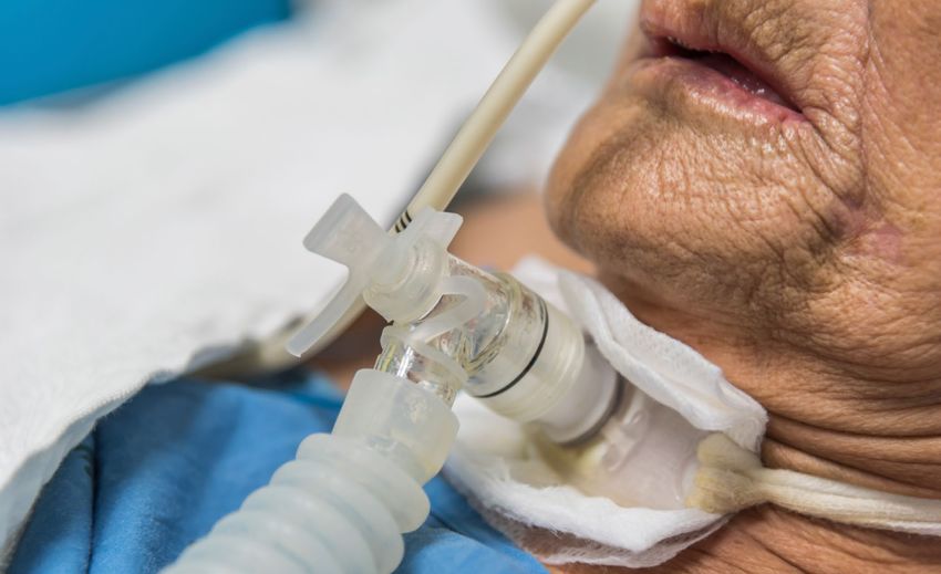
Guidance For: Tracheostomy Care | 135. Insertion 5. Insertion Percutaneous tracheostomy is the highest risk elective procedure that occurs on an ICU. As such, units must prepare, rehearse and train all staff involved to perform the procedure as safely as possible. National Safety Standards for Invasive Procedures (NatSSIP) have been in place since 2015. These detail the standards and considerations for safe performance of procedures, the principles of which can be adapted to Local Safety Standards for Invasive Procedures (LocSSIP). The FICM and ICS have produced WHO-style checklists for percutaneous tracheostomy that can be adapted into LocSSIPs and the NTSP has co-produced a more detailed checklist (see further resources). Around two-thirds of tracheostomy procedures are now performed by intensivists in ICUs rather than by surgeons in operating theatres, with over 90% of ICU tracheostomies performed percutaneously at the bedside. A tracheostomy may be fashioned by an open surgical or percutaneous dilatational technique (PDT), either as an emergency, emergent or elective procedure. Consent and preparation Tracheostomy in the critically ill should almost always be a planned procedure, with the benefits and burdens weighed up by the MDT over a period of days prior to insertion. This should allow ample time to explain the procedure to the patient’s relatives and the patient (if appropriate). Appropriate consent has been considered good practice since the last ICS standards document. In light of the NCEPOD report and several Coronial reports into peri-procedural adverse events, consent must be considered mandatory. It is anticipated that the majority of consent forms will be for patients who lack capacity, necessitating discussion with the patient’s family. Where appropriate, attempts should be made to utilise communication aids and support to determine patient capacity and facilitate consent. Standards and best practices that apply if the patient was transferred to the operating theatre for tracheostomy must apply if the procedure is performed on the ICU. Many of the adverse events reported to NAP4, NCEPOD or incident reporting systems (such as the NRLS or the GTC) around tracheostomy are predictable and can be prevented by appropriate planning and preparation. Appropriate preparation includes that of the equipment, the patient, the team and planning for difficulty. Although perhaps unfamiliar to some staff in ICU, human factors are increasingly acknowledged as perhaps the most important element of managing procedures, procedural complications and emergencies. Human factors include environmental influences, team behaviours and individual performance, each of which have been comprehensively addressed in the DAS guidelines for airway management in the critically ill. Cognitive aids such as checklists and algorithms can improve performance in stressful situations and must be employed prior to undertaking potentially high-risk invasive procedures such as tracheostomy insertion. 14 | Guidance For: Tracheostomy Care
5. Insertion

Equipment preparation

• Airway management kit
• Endoscopic kit for visualising the trachea
• Tracheostomy insertion kit
  • Percutaneous insertion set
    • Limited surgical set if appropriate
• Monitoring
  • End-tidal CO2
  • Visible timer
  • Vital signs, including SpO2 with audible sound

Critically ill patients have a higher incidence of potentially difficult airways than found in routine
anaesthetic practice. This is compounded by the extended neck position and limited access that
can occur during tracheostomy. The person managing the airway must be competent to do so, with
all relevant equipment immediately available. Videolaryngoscopy with a screen visible to all can
make manipulations of trans-laryngeal tracheal tubes easier. Similarly, an endoscope with a screen
visible to all will help teamwork and communication. Supraglottic airways may be used for airway
management and are particularly suited to patients with relatively low airway pressures.

End-tidal CO2 is mandatory for any airway manipulation. Particular care must be given to which
breathing circuit the capnograph is attached to as confusion can occur if, for example, an additional
circuit is used to connect to the new tracheostomy tube whilst the capnograph remains attached to
the ventilator circuit connected to the tracheal tube.

A visible timer is a useful addition and is present on most modern bedside vital signs monitors.

Patient preparation

•   Positioning
•   Ultrasound of the neck to identify vessels and depth from skin to trachea
•   Stopping and aspirating enteral feeding
•   Appropriate ventilation settings
    • 100% oxygen
    • Consider a volume controlled/guaranteed mode
  • Consider a 1:1 I:E ratio
• Identify team member who will monitor vital signs
• Set haemodynamic and oxygen saturation parameters and agree actions
• Check clotting, anticoagulants, antiplatelet agents
• Agree sedation strategy

Patient positioning is important. The neck should be extended by placing a pillow or similar device
under the shoulders. Tilting the bed or torso 30 degrees or so head up will reduce venous pressure
and may reduce bleeding. The patient should be moved laterally towards the side of the bed that
the operator will stand.

Ultrasound is recommended prior to the procedure to identify significant vessels and the depth to
the trachea. The use of peri-procedural ultrasound is limited at present by the footprint of commonly
available probes.

                                                                      Guidance For: Tracheostomy Care | 15
5. Insertion Stopping and starting enteral feed can cause problems with glycaemic control in the critically ill. Prolonged fasting prior to tracheostomy is probably unnecessary, especially as gastric mobility may be impaired. Aspiration of gastric contents prior to airway management is a sensible precaution. Use of an endoscope will impair ventilation and appropriate adjustments to ensure adequate oxygenation should be made prior to the procedure. Patients will often deteriorate during a tracheostomy due to suboptimal positioning for ventilation and interruption/inadequate ventilation. There are no defined parameters that make a tracheostomy safer to perform, but typically a patient should be receiving less than around 50% inspired oxygen and requiring less than around 10cmH2O PEEP prior to tracheostomy. For patients with raised intracranial pressure, the position required for tracheostomy insertion may be a relative contraindication. An identified team member must be responsible for monitoring vital signs and responding to changes with agreed actions. Appropriate sedation and adequate neuromuscular blockade should be used. Airway manipulation can be very stimulating, requiring an increase in opiate and sedative medication. A vasopressor may be required to manage periods when direct airway manipulation is not occurring. Local anaesthesia with an additional vasoconstrictor such as adrenaline may help to reduce bleeding from the superior tissues. Before administering any drugs, allergies or intolerances must be checked. Staff preparation Staff should introduce themselves by name and role, identifying an airway operator, airway assistant, tracheostomy operator and a runner. A team leader should be identified who ideally does not have another role. It is prudent to know how and where additional anaesthetic and surgical assistance can be summoned. For higher risk cases (for example with clotting or anatomical problems) the procedure should be timed following discussion with surgical colleagues so that they are on site and available to assist if required. Prepare for difficulty Critically ill patients may have known or anticipated difficulty in managing the airway, compounded by prolonged oral intubation, laryngeal and oral cavity trauma or secretions, nasogastric tubes, patient positioning and physiological instability. A clear plan must be verbalised for managing the upper airway. Similarly, a clear plan of the key steps of tracheostomy tube insertion must be verbalised, including the number of attempts at tracheal puncture and the planned order of endoscopic confirmation of tracheostomy tube position, suction and then ventilation. It is important to identify who will be summoned to help, what the plan is for unexpected bleeding and what will occur of the tracheostomy tube cannot be inserted. Sign out Clear instructions must be given to bedside staff regarding the care required following tracheostomy insertion. The same standards must be applied as if the patient had left an operating theatre. The position and orientation of the tracheostomy tube must be checked and documented, with the patient in the position that they will be nursed in (rather than the insertion position). This should include the distance from the carina, which is especially important for adjustable flanged tubes. A tube that is considered inadequately positioned must be changed whilst the team and airway equipment are all available. This usually requires a larger or longer tube. 16 | Guidance For: Tracheostomy Care
5. Insertion

The tube must be secured, and the purpose of any sutures identified, with instructions for removal
documented. There is no clear evidence that suturing a tube in place reduces displacement, but
the commonest and most significant problem that occurs with a new percutaneously inserted
tracheostomy tube is displacement, requiring the tube to be secured adequately.

Chest X-rays are not routinely required unless complications are suspected.

The sedation and ventilation plan should be reviewed and confirmed, along with
thromboprophylaxis or anticoagulation plans. Simple analgesia is usually required for patients
whose sedation will be reduced.

A bedhead sign is an effective method of communicating key facts about a tracheostomy in an
emergency and must be completed by the operator as part of the sign out process. The bedhead
sign must include details of any difficulties managing the upper airway. Signs can be downloaded
(and adapted if necessary) from the NTSP website.

Staff competence

The competencies required to participate in tracheostomy insertion are not just limited to technical
skills, but include human factors, situational awareness, teamworking and communication.

Competencies in airway management are defined by the FICM and RCoA. Typically, staff managing
the upper airway will have completed an Initial Assessment of Competency (IAC) in airway
management (or a recognised equivalent level of competence) and have completed a minimum of 3
months of training in Anaesthesia.

Additionally, staff managing the upper airway will be required to use an endoscope during the
procedure, although this could conceivably be performed by an additional person. The FICM
syllabus describes basic competency for endoscopy and bronchoscopy.

Competencies for percutaneous tracheostomy insertion are defined by the FICM and RCoA Step
3 (Advanced) training requirements for ICM. Consultants and senior staff must reflect on their
own competency and frequency of tracheostomy insertion; annual appraisal is an appropriate
opportunity. There are currently no standards around the frequency of training or the frequency of
performing tracheostomy to be considered competent.

For some units, it may be preferable to have a limited group of consultants or senior staff who
are considered competent to insert tracheostomies. This group would be expected to be able to
maintain their competencies by more frequent exposure to the procedure.

Similarly, training opportunities for trainees and skills maintenance opportunities for senior staff
may be limited. It may not always be appropriate to expose non-ICM or junior trainees to the
tracheostomy procedure on the ICU in order to preserve opportunities for regular or senior staff for
whom tracheostomy insertion remains an important skill to develop or maintain.

In considering the required responses to this standards document, it may be useful to work back
from the perspective of considering a death on the ICU due to a major complication of tracheostomy
insertion (e.g. major haemorrhage or loss of the airway). Is the ICU able to defend the environment,
equipment, procedure and competency or level of supervision of those staff involved?

Surgical or percutaneous?

There is no conclusive evidence to justify recommending surgical or percutaneous dilatational
tracheostomy (PDT) over each other. The main advantages of PDT at the bedside of the critically

                                                                    Guidance For: Tracheostomy Care | 17
5. Insertion ill patient are logistical; transfer to the operating theatre is not required and the procedure can be performed in a timely manner, without waiting for an appropriate window in the patient’s ongoing critical care for the procedure to coincide with theatre and surgical availability. The choice will be affected by available expertise, local practices, and individual patient characteristics. Surgical techniques are beyond the scope of this document, but situations in which they may be required are discussed below. There are no comprehensive datasets that can directly compare the risks and benefits of each approach and the guidance below is based on consensus opinion. Analysis of large datasets may help to clarify some of these guidelines in the future. Percutaneous insertion can be considered as a purely ‘needle through skin’ technique, or as a hybrid surgical technique with varying degrees of tracheal exposure prior to puncture of the trachea. Similarly, a surgical technique may involve surgical exposure of the trachea, but entry into the trachea could be achieved with a percutaneous set, or by opening the trachea with a scalpel. As a principle, an operator undertaking any surgical exposure of the trachea or using a percutaneous insertion kit must be appropriately trained and/or supervised. Surgical techniques have the advantage of a wider exposure of the structures of the neck which may allow easier haemostasis, especially if significant blood vessels or the thyroid isthmus overlay the trachea. Surgical techniques also allow the insertion of ‘stay’ or ‘maturation’ sutures, which may be beneficial if tracheal access for later tube changes could be difficult. However, these techniques require additional skills, training and equipment that is not readily available at the bedside. The larger incision and wound associated with a surgical approach may also take longer to heal than a purely percutaneous approach in the critically ill and may not provide such a tight seal against stomal air leaks for patients ventilated with relatively high airway pressures. Surgical tracheostomies may be more suitable for patients with burns, recent neck surgery or cervical spine injuries. Percutaneous procedures involve a much smaller incision and potentially less tissue trauma, with the tamponading effect of the percutaneously inserted tube at least partly effective in reducing post-procedural bleeding. However, significant bleeding can still occur and should be anticipated and planned for. Percutaneous procedures are not necessarily contraindicated in patients receiving anticoagulants or antiplatelet agents and discussion with surgical colleagues is recommended. Factors influencing the risk/benefits include the anatomy, the experience of the operator, the chosen technique, the anticoagulant strategy and clotting results. Percutaneous procedures are safest when the trachea can be identified and easily palpated, with sufficient space between the cricoid cartilage and the sternal notch to puncture the trachea between the second and third tracheal rings. Surgical techniques are usually more appropriate when bleeding is anticipated (especially from an identified vessel) or when anatomical landmarks are difficult to identify. Patients with known or suspected head and neck cancer may benefit from a more precise surgical exploration. A surgical tracheostomy can usually be changed safely after 2 or 3 days, although this depends on the surgical technique, anatomy, stay sutures and often, surgical preference. After 7-10 days, most tracheostomy stomas will have healed adequately to be considered ‘established’ enough that the stoma will remain patent if a tube is temporarily removed. It doesn’t matter too much at this point how a stoma was initially created. This period of time elapsing does not guarantee that tube reinsertion is straightforward, however. If a tube change is required within 7 days of percutaneous tracheostomy, it may be safer to actively manage the upper airway (sedation and re-intubation) and electively re-insert or change the tube. A bedhead sign must be completed whatever the method of insertion so that responders to an airway emergency can clearly identify the nature of the tracheostomy stoma. 18 | Guidance For: Tracheostomy Care
5. Insertion

Surgical tracheostomy

Surgical technique is described comprehensively in surgical texts and although usually confined to
the operating theatre, there may be situations where surgical expertise or assistance is required
at the bedside of a critically ill patient requiring tracheostomy, either electively or in an emergency.
All units should have close links with their head and neck surgical colleagues and agree what
equipment must be immediately available on the ICU for surgical tracheostomy. Discussions
should be held well in advance of any urgent or emergency procedures, and provision of staff and
equipment reviewed following surgical intervention in a tracheostomy procedure on the ICU.

Whilst the precise nature of surgical equipment will be influenced by local considerations, if an ICU
is performing tracheostomies at the bedside, then the location of the following equipment should be
agreed:

• Surgical tracheostomy instrument set, typically containing
  • Tissue tweezers, mosquito forceps, scissors, muscle and skin retractors, and Mayo needle
  		 holder, scalpels, tracheal dilating forceps, tracheal hooks
• Adequate overhead light source
  • Headlight may be preferred by surgical teams
• Diathermy

Elective or emergency tracheostomy?

Almost all tracheostomies performed on the ICU should be planned. There are occasions however
when an emergency tracheostomy could be required. These are discussed in the 2018 Difficult
Airway Society guidelines for the management of intubation in the critically ill.

Following a failed intubation and attempts to oxygenate via the upper airway, emergency airway
access is recommended to occur via surgical approaches to the cricothyroid membrane. This is the
default strategy for ‘Plan D’ in these difficult situations. However, the DAS guidance recognises that
many intensivists are skilled in percutaneous tracheostomy insertion and provision is made for this
in the guidelines. There may be advantages in securing an airway with a tracheostomy, principally
to provide more effective ventilation than with a smaller calibre cricothyroidotomy tube (especially
in acute lung injury) and to facilitate long-term ventilation. However, insertion of a percutaneous or
surgical tracheostomy takes significantly longer than an emergency surgical cricothyroidotomy and
should only be undertaken if the situation demands it and by those trained to do so. A patient who
has had been recently decannulated is an example of a special circumstance where emergency
tracheostomy may be safer and quicker than a cricothyroidotomy.

All units should adopt the DAS guidelines and the principle of providing emergency surgical airway
access via the cricothyroid membrane is commended to staff working in intensive care. Medical
staff providing airway management to critically ill patients must be trained in emergency surgical
cricothyroidotomy and undergo regular rehearsal of airway management scenarios with the
multidisciplinary ICU team. The requirement for emergency tracheostomy should be considered
and planned by the unit management and multidisciplinary team.

                                                                       Guidance For: Tracheostomy Care | 19
5. Insertion Tracheostomy insertion summary Standards: • All patients under the care of an ICU team will have an appropriate consent form completed prior to elective tracheostomy, whether occurring in the ICU or the operating theatre. • All staff involved in managing the upper airway and/or performing bronchoscopy will be sufficiently trained and considered competent. • All staff involved in insertion of a tracheostomy in ICU will be sufficiently trained and considered competent. • Appropriate equipment to safely perform tracheostomy will be immediately available on the ICU. • All ICU tracheostomy insertion procedures must include a checklist and LocSSIP. Standard templates are available from the FICM/ICS and the NTSP (see below). Recommendations: • Medical leadership on the ICU must consider the skill mix of the consultant and senior team with respect to tracheostomy insertion. It may be appropriate to limit the number of staff who insert tracheostomies. • Competency to manage an airway and insert a tracheostomy must be considered for all permanent medical staff. Annual appraisal is a good opportunity to review this. • Competency to manage an airway (and insert a tracheostomy for more senior trainees) must be considered for all new trainees as part of induction medical staff. Further resources www.ficm.ac.uk/safety-and-clinical-quality/safety-checklists-invasive-procedures www.tracheostomy.org.uk 20 | Guidance For: Tracheostomy Care
6. Tracheostomy tube types
                                                                                     and choice

6. Tracheostomy tube types and choice
There are a variety of different tracheostomy tubes available from a number of manufacturers, each
with their own characteristics. It is advisable to use an initial tracheostomy tube that is compatible
with the insertion kit (usually from the same manufacturer), although it is possible to insert different
tubes with different kits. The tube required by an individual patient may change with time and
should be reviewed regularly, ideally by the tracheostomy multidisciplinary team.

There may be advantages in stocking only a limited range of tubes from specific manufacturers.
This approach may aid familiarity for staff and make education easier. This needs to be balanced
against the requirements for the variety of patients managed at the institution. An informed decision
by those involved in tracheostomy care is advised.

Tubes may be classified by material, internal diameter, proximal and distal length, angle, presence
of a cuff, presence of fenestrations, presence of subglottic suction port and presence of an inner
cannula.
The clinical factors to be considered when selecting a tracheostomy tube for a patient are listed
below.

Respiratory function: Most temporary tracheostomies will be inserted whilst a patient is on an
ICU and still requiring some positive pressure ventilation. Typically, this will require the use of a
non-fenestrated cuffed tracheostomy tube. However, it is recognised that long term mechanical
ventilation can be delivered through an uncuffed tube.

Secretion management: Sub-glottic suction is likely to reduce the incidence of ventilator-
associated pneumonia as part of a bundle of interventions. Standard tracheostomy tubes are
available with sub-glottic suction ports. These ports can also be used for above cuff vocalisation
(see later).

Abnormal airway anatomy: Upper airway endoscopy following percutaneous insertion suggests
that a standard tracheostomy tube may be anatomically unsuitable in as many as a third of
adult patients. Obese patients, or those with local neck swelling or oedema, may require a tube
with an extended proximal length, whilst patients with fixed flexion abnormalities may not easily
accommodate tubes with a fixed angulation.

Airway pathology: Localised airway pathology such as tracheomalacia, granuloma formation etc,
may on occasion necessitate the use of a tracheostomy tube that has a longer distal length than
standard.

Weaning: Many patients can be ‘weaned’ from mechanical ventilation to decannulation without any
need to change from the cuffed tracheostomy tube initially inserted. In some cases, it may be useful
to consider options such as downsizing or an uncuffed which promote greater gas flow out via the
upper airways, allowing vocalisation and promoting laryngeal rehabilitation. Fenestrated tubes
may also promote vocalisation, but there are associated risks of surgical emphysema (when used
with positive pressure ventilation) and risks of developing granulation tissue, tracheomalacia and
stenosis. Other options for vocalisation should be explored first.

Clinical environment: Tubes with an inner cannula that is able to be easily removed and cleaned
have a lower incidence of obstruction. For patients who are attached to a warmed and humidified
closed ventilator circuit the benefits of a tube with an inner cannula may not be so marked. For
patients receiving high inspired oxygen concentrations or high airway pressures, the risks and
benefits of circuit disconnection and loss of PEEP associated with cleaning an inner cannula
should be balanced against the potential risks of blockage. For patients on open breathing circuits

                                                                       Guidance For: Tracheostomy Care | 21
6. Tracheostomy tube types and choice or on less respiratory support (typically 50% inspired oxygen concentration) inner cannula are recommended. Inner cannulae will also narrow the effective internal diameter of the tracheostomy tube, potentially increasing airflow resistance and the work of breathing. Patient preference is important for alert patients or those with long term tracheostomies. Some tubes have specialist features such as foam-filled or fluid-filled cuffs, tight-to-shaft cuffs, materials and coatings to reduce biofilm formation or systems to continuously inflate or irrigate the cuff and subglottic space. At present there is no evidence to clearly recommend one tube type over another. Selecting the correct tube is important, especially if the patient has it inserted percutaneously, as it may take 7 days or more for the stoma to mature to more safely allow tube changes. The choice of the size and type of tube for initial insertion may be clinically obvious from the factors detailed above. However, tube size often comes down to experience and the multidisciplinary team can be invaluable. There may be a role for pre-procedural ultrasound or imaging in tube selection, although at present, there are no clear guidelines for tube selection. There is ongoing research into this important area. Videos and information describing tube types in detail are available from the NTSP website and the e-learning in Anaesthesia e-learning modules. Tube types summary Standards: • Tube selection must be a multidisciplinary discussion, especially for patients with abnormal anatomy. • Sub-glottic suction tracheostomy tubes should be used as standard for new tracheostomy. These tubes can reduce the incidence of pneumonia (as part of a bundle of care) and can allow Above Cuff Vocalisation (ACV) without needing to change the tube. Recommendations: • ICUs must consider the range of tubes they stock and use. There is a balance of limiting stock to improve familiarly, but also of providing an adequate range of devices to cater for patient mix. • Staff must be familiar with the variety of tubes available within the ICU. • Consider patient preference for long term tracheostomy patients. 22 | Guidance For: Tracheostomy Care
7. Routine care of the
                                                                        established tracheostomy

7. Routine care of the established tracheostomy
Many potential complications of tracheostomy care can be prevented by basic care, done well. In
the ICU, many of these roles will fall to nursing staff, but all multidisciplinary staff managing patients
with tracheostomies should be aware of the principles of routine care. These should be considered
when reviewing a patient with a tracheostomy.

Cleaning of the tube

Tracheostomy tubes are artificial airways and may become blocked with secretions or blood. To
reduce the chance of blockage, inspired gases must be warmed and humidified where possible and
secretions regularly suctioned.

Inner cannula must be removed and checked at least once per nursing shift (every 8-12 hours),
although the frequency of checking will depend on patient factors. If a patient has a lot of secretions
for example, the frequency should increase. The inner cannula can be cleaned and replaced in
many instances, although some cannulae are disposable. It is advisable to keep a spare inner
cannula at the bedside so that circuit disconnections to change inner cannulae are kept to a
minimum.

The outer part of the tube can be cleaned simply with saline solution. The whole tracheostomy tube
should be changed in line with the manufacturer’s recommendations; typically, every 28 days.

Stoma care

Tracheostomy stomas need to be kept clean and dry. This is usually achieved by applying a specific
absorbent dressing around the tube which will also reduce the chance of direct pressure from the
wings of the tube itself. The stoma should be inspected at least once per day.

If infection is suspected, then the stoma must be swabbed. Barrier creams are sometimes
necessary if there are lots of secretions. Specialist tracheostomy nurses, head and neck surgical
colleagues and tissue viability nurses are a good source of advice if there are problems with the
stoma. Granulation tissue may be treated with topical steroid creams.

The purpose of any sutures in or around the stoma must be clarified at the time of insertion and
documented on the bedhead sign, with a clear plan for removal.

Cuff deflation and cuff management

The tracheal mucosa has a capillary blood supply which will be compromised by the presence
of a tracheostomy tube cuff at high pressure, a problem compounded by critical illness. The cuff
pressure should be maintained as low as possible to prevent air leaks from the upper airways and
ideally below 25cmH2O. Some systems can continually monitor and adjust cuff pressure within set
parameters and these systems likely control cuff pressures more accurately and more safely than
intermittent manual checks.

For manual checks, the cuff pressure should be checked at least once per nursing shift (typically
8-12 hours).

If the ventilator peak inspiratory pressure exceeds the cuff pressure, then leakage of inspired gas
past the cuff can be anticipated. This does not mean that the cuff has failed necessarily. However,
leakage of gas from the upper airways during ventilation, the patient being able to vocalise, the
requirement for high cuff pressures or repeated addition of gas to the cuff may indicate that the

                                                                       Guidance For: Tracheostomy Care | 23
7. Routine care of the established tracheostomy tube is malpositioned or of an inappropriate size, or defective. The patient must be reviewed by a healthcare practitioner competent in tracheostomy care. Cuff deflation should be a goal of routine care of patients with tracheostomies. This can be a complex decision and is best taken following multidisciplinary review by the ICU team and Speech & Language Therapists (SLT) and physiotherapists. The risks of aspiration may be mitigated by a constant upwards flow of gas under positive pressure past the deflated cuff, although an individual assessment is warranted. Fibreoptic Endoscopic Evaluation of Swallow (FEES) by a trained SLT is invaluable in informing a risk assessment for cuff deflation trials and attempts at oral intake. Clearance of subglottic secretions from above the cuff should occur prior to cuff deflation. This is best achieved by using a tube with a subglottic suction port. Oral secretions may need to be addressed prior to deflation (see below). Cuff deflation increases the work of breathing as a variable proportion of inspired gas ‘escapes’ via the upper airways and does not ventilate the lungs. Cuff deflation should normally start as a short trial under direct supervision, extending the frequency and duration of deflation guided by weaning parameters (such as respiratory rate or evidence of fatigue). A ventilator that can tolerate significant leakage of gas needs to be employed for cuff deflation trials. The inspiratory pressure may need to be increased to allow for leakage and reduced ventilatory effectiveness. Patients often require rest after periods of cuff deflation and cuff deflation should form part of an overall weaning strategy, including clear parameters for success and failure, and clear plans for the ventilator setting. Patients should be reassured that any discomfort or unpleasant tastes or smells will usually resolve. Vocalisation is usually possible and is of significant emotional and practical benefit to the patient, their family and to staff. There are likely additional beneficial effects on laryngeal sensory and motor function by promoting trans-laryngeal airflow. If a significant leakage of gas via the upper airways does not occur when the cuff is deflated, then downsizing to a smaller tracheostomy tube should be considered. If problems persist, it may also be prudent to rule out laryngeal pathology such as oedema or vocal cord immobility using visualisation such as laryngoscopy, FEES or laryngeal ultrasound. If cuff deflation becomes established and tolerated, an uncuffed tube should be considered. Secretion management To reduce the chance of blockage, inspired gases must be warmed and humidified where possible. Dehydration will increase sputum thickness. Suction can be ‘open’ (insertion of a separate single-use suction catheter) or ‘closed’ (suction catheter is part of the breathing circuit). A closed system reduces the number of times a breathing circuit is interrupted and may be advantageous if the patient is continually ventilated. Suction catheters are of different lengths and diameters. Specific tracheostomy closed suction catheters are shorter than the endotracheal equivalents. This may not be long enough if a long or adjustable tracheostomy tube is used. Care should be taken to avoid airway trauma when using longer suction catheters. Inability to pass a suction catheter is a ‘red flag’ and may indicate that the tube is badly positioned within the airway. A prompt review by an experienced professional is warranted. Fenestrated inner cannula can cause partial obstruction of the passage of a suction catheter and should be replaced by a non-fenestrated inner cannula prior to suctioning. 24 | Guidance For: Tracheostomy Care
You can also read